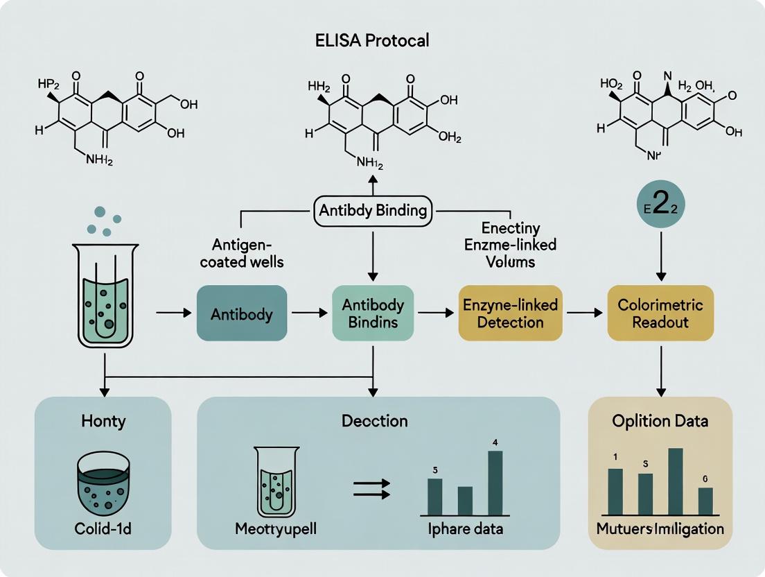
A Comprehensive Guide to ELISA Protocols for COVID-19 Antibody Detection: From Principles to Advanced Applications
This detailed guide provides researchers, scientists, and drug development professionals with a comprehensive framework for implementing ELISA (Enzyme-Linked Immunosorbent Assay) to detect SARS-CoV-2 antibodies.
A Comprehensive Guide to ELISA Protocols for COVID-19 Antibody Detection: From Principles to Advanced Applications
Abstract
This detailed guide provides researchers, scientists, and drug development professionals with a comprehensive framework for implementing ELISA (Enzyme-Linked Immunosorbent Assay) to detect SARS-CoV-2 antibodies. It begins by exploring the foundational immunology of COVID-19 and the rationale for serological testing. The core of the article delivers a step-by-step, optimized methodological protocol for both indirect and capture ELISA formats, including critical reagent selection. A dedicated troubleshooting section addresses common pitfalls like high background noise and sensitivity issues. Finally, the guide covers validation strategies against gold-standard methods (e.g., PRNT) and compares ELISA's performance with rapid tests and CLIA-based assays. The conclusion synthesizes key takeaways and discusses future implications for serosurveillance, vaccine development, and variant impact assessment.
Understanding Serology: The Immunology of COVID-19 and ELISA Fundamentals
Antibody Classes and Their Clinical Significance
The humoral immune response to SARS-CoV-2 involves the production of distinct immunoglobulin (Ig) classes with different temporal dynamics and functions. Understanding these is crucial for assay development and interpreting serological data in COVID-19 research.
Table 1: Characteristics of SARS-CoV-2 Specific Antibodies
| Antibody Class | Typical Onset Post-Symptom | Peak Time | Duration | Primary Location & Function | Key SARS-CoV-2 Targets |
|---|---|---|---|---|---|
| IgM | 3-7 days | 1-2 weeks | Weeks to months (~8-12 weeks) | First responder; pentameric structure enhances avidity; primarily in blood. | Spike (S) protein, Nucleocapsid (N) protein. |
| IgG | 7-14 days | 3-6 weeks | Months to years (long-term memory) | Major serum antibody; neutralization and opsonization; crosses placenta. | Spike (S1/S2, RBD), Nucleocapsid (N). |
| IgA | 3-7 days | 2-4 weeks | Months (mucosal memory) | Mucosal immunity; dimeric form in secretions; neutralizes virus at entry sites. | Spike (RBD), found in saliva, nasal fluid. |
| Neutralizing Abs (nAbs) | ~7-10 days (with IgG/IgA) | 3-6 weeks | Correlates with IgG longevity | Block viral entry by binding RBD, preventing ACE2 interaction. | Primarily Spike Receptor Binding Domain (RBD). |
Table 2: Key Quantitative Metrics from Recent Serological Studies (2023-2024)
| Parameter | IgM | IgG | IgA | Neutralizing Antibodies (nAb Titer) |
|---|---|---|---|---|
| Average Seroconversion Time | 5.2 days | 11.1 days | 5.8 days | 12.3 days |
| Median Peak Titer (AU/mL)* | 4.8 | 28.5 | 6.7 | ID50: 1,250 |
| Half-life (Post-Peak) | ~25 days | ~90 days | ~45 days | ~85 days |
| % Patients Positive at 60 days | ~35% | ~95% | ~70% | ~90% |
| Correlation with Protection | Low | Moderate | Moderate (mucosal) | High |
*Representative data from recent cohort studies; AU=Arbitrary Units, ID50=50% Inhibitory Dilution.
Detailed ELISA Protocols for Detection
The following protocols are framed within a thesis investigating the optimization of ELISA for quantifying the SARS-CoV-2 antibody response.
Protocol 1: Indirect ELISA for Anti-Spike IgG/IgM
Title: Quantification of SARS-CoV-2 Spike-Specific IgG and IgM in Human Serum.
Principle: Microplate wells are coated with purified Spike antigen. Serum antibodies bind and are detected using enzyme-conjugated anti-human IgG or IgM.
Materials (Research Reagent Solutions):
| Reagent/Material | Function & Specification |
|---|---|
| SARS-CoV-2 Spike (S1) Protein | Antigen for plate coating. Recombinant, >95% purity. |
| Carbonate-Bicarbonate Coating Buffer (pH 9.6) | Optimal buffer for passive adsorption of protein antigens to polystyrene plates. |
| Blocking Buffer (5% BSA in PBST) | Blocks non-specific binding sites to reduce background signal. |
| Human Serum Samples | Test specimen. Heat-inactivated at 56°C for 30 min prior to use. |
| HRP-conjugated Anti-Human IgG (Fc specific) | Detection antibody for IgG. Conjugated to Horseradish Peroxidase (HRP). |
| HRP-conjugated Anti-Human IgM (μ-chain specific) | Detection antibody for IgM. |
| TMB Substrate Solution | Chromogenic substrate for HRP. Yields blue product upon oxidation. |
| Stop Solution (1M H2SO4) | Stops the HRP-TMB reaction, changes color to yellow. |
| Wash Buffer (PBST) | Phosphate-Buffered Saline with 0.05% Tween-20 for washing steps. |
| Microplate Absorbance Reader | Instrument to read Optical Density (OD) at 450 nm reference. |
Procedure:
- Coating: Dilute Spike protein to 2 µg/mL in coating buffer. Add 100 µL per well to a 96-well microplate. Incubate overnight at 4°C.
- Washing: Aspirate and wash plate 3x with 300 µL PBST per well using a plate washer or manual squirt bottle.
- Blocking: Add 200 µL of blocking buffer per well. Incubate for 2 hours at 37°C. Wash as in step 2.
- Sample Incubation: Dilute serum samples 1:100 in blocking buffer. Add 100 µL per well in duplicate. Include positive, negative, and blank (buffer) controls. Incubate 1 hour at 37°C. Wash 5x.
- Detection Antibody Incubation: Dilute HRP-conjugated anti-human IgG or IgM 1:5000 in blocking buffer. Add 100 µL per well. Incubate 1 hour at 37°C. Wash 5x.
- Substrate Reaction: Add 100 µL TMB substrate per well. Incubate in the dark for 15 minutes at RT.
- Stop Reaction: Add 50 µL of 1M H2SO4 per well.
- Measurement: Read absorbance at 450 nm within 30 minutes.
Data Analysis: Calculate the mean OD for duplicates. A sample is considered positive if its OD exceeds the cut-off value (typically mean of negative controls + 0.15 or 3x standard deviations above).
Protocol 2: Competitive ELISA for Neutralizing Antibodies (Surrogate)
Title: Surrogate Virus Neutralization Test (sVNT) via RBD-ACE2 Competition ELISA.
Principle: Serum nAbs that block the Spike RBD-ACE2 interaction are detected in a competitive format. HRP-conjugated RBD competes with viral Spike for serum nAbs; remaining conjugate binds plate-bound ACE2.
Procedure:
- Coating: Coat plate with recombinant human ACE2 protein at 1 µg/mL overnight at 4°C.
- Blocking: Block with 5% BSA for 2 hours at 37°C.
- Competition Step: Pre-incubate diluted serum sample (1:10) with an equal volume of HRP-conjugated RBD (pre-determined concentration) for 30 min at 37°C.
- Capture: Transfer 100 µL of the serum-RBD-HRP mixture to the ACE2-coated plate. Incubate for 1 hour at 37°C. (nAbs in serum will bind the conjugate, preventing it from attaching to ACE2 on the plate).
- Washing: Wash plate 5x thoroughly with PBST.
- Detection: Add TMB substrate, stop with acid, and read at 450nm.
- Calculation: The higher the nAb titer, the lower the OD signal. Calculate % inhibition:
[1 - (ODsample/ODnegative control)] x 100%. An inhibition >20-30% is typically considered positive for nAbs.
Signaling and Workflow Visualizations
Diagram 1: Indirect ELISA Principle
Diagram 2: Indirect ELISA Workflow
Diagram 3: Surrogate Neutralization ELISA Logic
Within the broader thesis on ELISA protocol development for detecting SARS-CoV-2 antibodies, this document establishes the critical context for serological testing. While PCR remains the gold standard for diagnosing active COVID-19 infection, it cannot determine past infection or immune status. Serological assays, particularly ELISA, measure the host's humoral immune response (IgG, IgM, IgA antibodies) against viral antigens, providing essential data on infection history, population seroprevalence, and potentially correlates of protection. These Application Notes detail the protocols and applications that move beyond nucleic acid detection.
Table 1: Performance Characteristics of SARS-CoV-2 Serological Assays
| Assay Type | Target Antibody | Typical Sensitivity (%)* | Typical Specificity (%)* | Time Post-Symptom Onset for Optimal Detection | Primary Application |
|---|---|---|---|---|---|
| ELISA | Anti-S IgG | 95-99 | 99-100 | >14 days | Immunity screening, seroprevalence |
| ELISA | Anti-N IgG | 90-98 | 98-99.5 | >14 days | Confirmation of past infection |
| CLIA | Anti-S IgG | 97-99.8 | 99.5-100 | >10 days | High-throughput screening |
| LFIA (Rapid Test) | Total Ab/IgG/IgM | 80-95 | 95-99 | >7-14 days | Point-of-care rapid assessment |
| Neutralization Assay | Neutralizing Ab | N/A (functional) | N/A (functional) | Peaks at ~28-35 days | Assessment of protective function |
*Performance varies significantly based on antigen quality, patient cohort, and disease severity.
Table 2: Typical Quantitative ELISA Result Interpretation (Anti-Spike IgG)
| Sample OD450 nm (Normalized) | Interpretation | Suggested Follow-up |
|---|---|---|
| < 0.8 (or < calibrated cutoff) | Negative | No detectable antibodies. |
| 0.8 - 1.2 | Borderline/Indeterminate | Retest in 1-2 weeks; use confirmatory assay (e.g., PRNT). |
| > 1.2 | Positive | Quantitate titer; may correlate with neutralization capacity. |
Detailed Experimental Protocols
Protocol 3.1: Indirect ELISA for Detection of Anti-SARS-CoV-2 IgG Antibodies Principle: Viral antigen is immobilized on a plate. Serum antibodies bind and are detected by an enzyme-conjugated anti-human IgG.
Materials:
- Coating Antigen: Recombinant SARS-CoV-2 Spike S1 subunit or Nucleocapsid (N) protein (1-2 µg/mL in PBS).
- Coating Buffer: 0.05 M Carbonate-Bicarbonate, pH 9.6.
- Wash Buffer: PBS with 0.05% Tween-20 (PBST).
- Blocking Buffer: 5% Non-fat dry milk or 3% BSA in PBST.
- Test Samples: Human serum/plasma, diluted in blocking buffer.
- Detection Antibody: Horseradish Peroxidase (HRP)-conjugated goat anti-human IgG (γ-chain specific).
- Substrate: TMB (3,3',5,5'-Tetramethylbenzidine).
- Stop Solution: 1M H2SO4.
- Microplate Reader.
Procedure:
- Coating: Add 100 µL/well of antigen solution to a 96-well microplate. Seal and incubate overnight at 4°C.
- Washing: Aspirate and wash plate 3x with 300 µL/well of PBST.
- Blocking: Add 200 µL/well of blocking buffer. Incubate for 1-2 hours at 37°C. Wash 3x.
- Sample Incubation: Add 100 µL/well of diluted serum samples (e.g., 1:100 starting dilution) and controls (positive, negative, blank). Incubate for 1-2 hours at 37°C. Wash 5x.
- Detection Antibody Incubation: Add 100 µL/well of HRP-anti-human IgG at optimized dilution. Incubate for 1 hour at 37°C. Wash 5x.
- Substrate Development: Add 100 µL/well of TMB substrate. Incubate in the dark for 10-15 minutes at RT.
- Stop Reaction: Add 50 µL/well of stop solution.
- Measurement: Read absorbance immediately at 450 nm (reference 620-650 nm). Calculate normalized values and interpret against the standard curve/calibrators.
Protocol 3.2: Surrogate Virus Neutralization Test (sVNT) Protocol Principle: Measures blocking of antibody-angiotensin-converting enzyme 2 (ACE2) receptor binding, mimicking viral neutralization.
Procedure:
- Pre-incubate diluted serum sample with HRP-labeled recombinant SARS-CoV-2 Spike RBD protein for a set time (e.g., 30 min at 37°C).
- Transfer the mixture to a microplate pre-coated with human ACE2 receptor protein.
- Incubate to allow any unblocked HRP-RBD to bind to the immobilized ACE2.
- Wash thoroughly to remove serum/HRP-RBD complexes.
- Add TMB substrate. The signal is inversely proportional to the neutralizing antibody titer in the sample.
Visualizations
Seroconversion Timeline After COVID-19 Onset
Serology Testing and Analysis Workflow
The Scientist's Toolkit: Key Research Reagent Solutions
Table 3: Essential Reagents for SARS-CoV-2 Serology Research
| Reagent/Material | Function & Importance in Research |
|---|---|
| Recombinant SARS-CoV-2 Antigens (Spike S1, RBD, Nucleocapsid) | Critical for assay specificity. S1/RBD antibodies often correlate with neutralization. N-protein indicates past infection, useful for differentiating natural infection from vaccination (if vaccine is spike-only). |
| HRP-conjugated Anti-Human IgG/IgM/IgA (Isotype-specific) | Key detection reagents. High affinity and minimal cross-reactivity are essential for sensitive, specific detection of antibody class. |
| Validated Positive & Negative Human Serum Panels | Used for assay calibration, validation, and as controls. Must be well-characterized (PCR-confirmed convalescent, pre-pandemic). |
| ACE2 Protein (for sVNT) | Receptor protein for surrogate neutralization assays, evaluating functional antibody response. |
| Stable TMB Substrate | Chromogenic substrate for HRP. Signal generation must be consistent and linear for accurate quantitation. |
| Microplates (High Binding) | Ensure consistent and efficient adsorption of capture antigens. |
| Precision Liquid Handling Systems | Essential for reproducibility in serial dilution for titer determination and assay miniaturization. |
This document serves as a critical methodological foundation for a broader thesis focused on the development and optimization of an enzyme-linked immunosorbent assay (ELISA) for the detection and quantification of SARS-CoV-2-specific antibodies (IgG and IgM). The accurate assessment of seroprevalence and immune response durability relies fundamentally on the core principles outlined herein: specific antigen-antibody binding and subsequent enzymatic signal generation.
Core Principles
Antigen-Antibody Interaction
The assay specificity is governed by the high-affinity, non-covalent binding between the SARS-CoV-2 antigen (e.g., Spike protein S1 subunit or Nucleocapsid protein) immobilized on a solid phase and the complementary antibody present in the patient serum sample. This interaction is reversible and follows the law of mass action, with affinity constants typically ranging from 10^7 to 10^11 M^-1 for high-quality immunoassays.
Enzymatic Detection
Following specific binding, detection is achieved via an enzyme-conjugated secondary antibody (e.g., Anti-human IgG-Horseradish Peroxidase, HRP). Upon addition of a chromogenic substrate (e.g., TMB, 3,3',5,5'-Tetramethylbenzidine), the enzyme catalyzes a reaction producing a measurable color change, the intensity of which is proportional to the amount of target antibody in the sample.
Table 1: Typical Performance Characteristics of a COVID-19 Serology ELISA
| Parameter | IgG Detection | IgM Detection | Notes |
|---|---|---|---|
| Analytical Sensitivity | 0.5 - 2.0 BAU/mL* | 1.0 - 3.0 BAU/mL* | *WHO International Standard Units |
| Analytical Specificity | 98.5% - 99.8% | 97.0% - 99.5% | Cross-reactivity with other coronaviruses minimized by antigen selection. |
| Dynamic Range | 2 - 200 BAU/mL | 3 - 150 BAU/mL | Typically exhibits a sigmoidal 4- or 5-parameter logistic curve. |
| Inter-assay CV | < 10% | < 12% | Precision across different runs, plates, and operators. |
| Time to Result | 60 - 120 minutes | 60 - 120 minutes | Excluding sample preparation time. |
Table 2: Key Antigen Targets for COVID-19 ELISA Development
| Antigen Target | Key Epitopes | Advantages | Considerations |
|---|---|---|---|
| Spike (S) Protein | RBD, S1, S2 | Neutralizing antibodies primarily target RBD; high specificity for SARS-CoV-2. | Conformational integrity critical; recombinant expression challenging. |
| Nucleocapsid (N) Protein | Linear epitopes | Highly immunogenic; abundant expression; high sensitivity. | More cross-reactive with other human coronaviruses. |
| RBD (Receptor Binding Domain) | ACE2 binding site | Most specific for neutralizing antibody detection. | Smaller size may reduce coating efficiency; epitopes may be conformationally sensitive. |
Detailed Protocol: Indirect ELISA for SARS-CoV-2 IgG
Title:Protocol for Detection of Human Anti-SARS-CoV-2 IgG Antibodies by Indirect ELISA
Principle: Patient serum IgG antibodies bind to immobilized viral antigen. Bound IgG is detected using an enzyme-labeled anti-human IgG antibody.
I. Materials & Reagents (The Scientist's Toolkit)
Table 3: Essential Research Reagent Solutions
| Item | Function & Specification |
|---|---|
| Coating Antigen | Recombinant SARS-CoV-2 Spike S1 or RBD protein. Reconstituted in carbonate/bicarbonate buffer (pH 9.6). |
| Coating Buffer | 0.05 M Carbonate-Bicarbonate, pH 9.6. Provides optimal pH for passive adsorption of protein to polystyrene. |
| Wash Buffer (PBST) | Phosphate-Buffered Saline (PBS) + 0.05% (v/v) Tween-20. Removes unbound proteins and reduces non-specific binding. |
| Blocking Buffer | PBS + 1% Bovine Serum Albumin (BSA) or 5% Non-fat dry milk. Saturates uncovered plastic surface to prevent non-specific adsorption. |
| Dilution Buffer | Wash Buffer + 0.1% BSA. Used for serial dilution of serum samples and controls. |
| Positive & Negative Controls | Calibrated human anti-SARS-CoV-2 serum and confirmed naive human serum. Essential for plate validation and quantification. |
| Detection Antibody | Horseradish Peroxidase (HRP)-conjugated Goat Anti-Human IgG (Fc specific). Must be titrated for optimal dilution. |
| Chromogenic Substrate | TMB (3,3',5,5'-Tetramethylbenzidine) in stable peroxide solution. HRP catalyzes oxidation to a blue product. |
| Stop Solution | 1M or 2M Sulfuric Acid (H₂SO₄). Halts enzyme reaction and changes TMB to yellow for absorbance reading. |
| 96-Well Microplate | High-binding polystyrene, flat-bottom. Solid phase for antigen immobilization. |
| Plate Reader | Spectrophotometer capable of reading absorbance at 450 nm (with 620-650 nm reference). |
II. Step-by-Step Methodology
Coating (Day 1):
- Dilute the purified SARS-CoV-2 antigen in coating buffer to a final concentration of 1-2 µg/mL.
- Dispense 100 µL per well into the required number of wells. Seal the plate and incubate overnight at 4°C.
Washing & Blocking (Day 2):
- Aspirate the coating solution. Wash the plate 3 times with 300 µL PBST per well using a multichannel pipette or plate washer.
- Tap the plate dry on absorbent paper.
- Add 200 µL of blocking buffer to each well. Incubate for 1-2 hours at room temperature (RT) on a plate shaker.
Sample & Control Incubation:
- Wash plate 3x as in step 2.
- Prepare serial dilutions (e.g., 1:50, 1:150, 1:450) of test sera and controls in dilution buffer.
- Add 100 µL of each diluted sample and control to designated wells in duplicate or triplicate. Include blank wells (dilution buffer only).
- Seal plate and incubate for 1-2 hours at RT on a plate shaker.
Detection Antibody Incubation:
- Wash plate 3x.
- Add 100 µL of optimally diluted HRP-conjugated anti-human IgG to each well.
- Incubate for 1 hour at RT on a plate shaker, protected from light.
Signal Development & Measurement:
- Wash plate 5x thoroughly to remove all unbound conjugate.
- Add 100 µL of TMB substrate solution to each well. Incubate for exactly 10-15 minutes at RT in the dark.
- Stop the reaction by adding 50 µL of stop solution per well. The color will change from blue to yellow.
- Read the absorbance at 450 nm (reference 650 nm) within 30 minutes using a microplate reader.
III. Data Analysis
- Calculate the mean absorbance for each sample and control.
- Subtract the mean absorbance of the blank wells from all readings.
- A sample is considered positive if its corrected absorbance exceeds the established cut-off value (often calculated as the mean of negative controls + 3 standard deviations, or via a calibrated standard curve using WHO International Standards).
Visualizations
Title: ELISA Core Principle: Antigen-Antibody Binding & Signal Generation
Title: Indirect ELISA Protocol Workflow for COVID-19 Serology
Within the broader thesis on ELISA protocols for detecting COVID-19 antibodies, the selection of antigen targets is paramount for assay sensitivity, specificity, and diagnostic application. The SARS-CoV-2 Spike (S) protein, its Receptor-Binding Domain (RBD), and the Nucleocapsid (N) protein serve as the primary antigenic targets. This application note details the protocols and comparative analysis for utilizing these proteins in indirect ELISA to quantify host antibody responses, crucial for seroprevalence studies, vaccine efficacy evaluation, and therapeutic development.
Comparative Analysis of Key Antigen Targets
The three primary antigen targets present distinct advantages and applications, governed by their biological roles and immunogenicity.
Table 1: Characteristics of Key SARS-CoV-2 Antigen Targets for ELISA
| Antigen | Size (kDa) | Location | Key Advantages | Primary Application |
|---|---|---|---|---|
| Full Spike (S) Trimer | ~480 (trimer) | Viral Envelope | Detects broad antibody response; high sensitivity for convalescent sera. | Vaccine immunogenicity studies; broad serology. |
| Receptor-Binding Domain (RBD) | ~27 (monomer) | S1 subunit of Spike | Highest specificity for neutralizing antibodies (NAbs); correlates with protection. | Neutralizing antibody surrogate assays; precise vaccine efficacy. |
| Nucleocapsid (N) Protein | ~46 | Viral Core | Highly immunogenic; abundant expression; detects infections post-SARS-CoV-2 infection. | Differentiating infected from vaccinated individuals (DIVI); natural infection studies. |
Table 2: Expected ELISA Signal Intensity & Specificity Profile*
| Sample Type | Coating Antigen: S Trimer | Coating Antigen: RBD | Coating Antigen: N Protein |
|---|---|---|---|
| Post-Vaccination (mRNA/Adeno) | High Positive | Moderate to High Positive | Negative |
| Post-Natural Infection | High Positive | Moderate Positive | High Positive |
| Pre-2020 Negative Control | Negative | Negative | Negative |
| *Relative comparison based on typical IgG responses. |
Detailed ELISA Protocols
Protocol 1: Indirect ELISA for Anti-S/RBD IgG Quantification
Objective: To detect and quantify IgG antibodies against SARS-CoV-2 Spike or RBD antigens in human serum.
Research Reagent Solutions & Materials: Table 3: Key Reagents for Indirect ELISA
| Reagent/Material | Function/Description | Example/Catalog |
|---|---|---|
| Recombinant SARS-CoV-2 S Trimer or RBD Protein | Coating antigen; captures specific antibodies from sample. | e.g., His-tagged S Trimer, ACROBiosystems. |
| 96-Well ELISA Plate (High Binding) | Solid phase for antigen immobilization. | Corning Costar 9018. |
| Blocking Buffer (5% BSA in PBS) | Reduces non-specific antibody binding to coated wells. | Prepare in PBS, pH 7.4. |
| Test Serum Samples & Controls | Source of primary antibody (human IgG). | Include positive/negative calibrators. |
| HRP-Conjugated Anti-Human IgG (Fc-specific) | Detection antibody; catalyzes colorimetric reaction. | e.g., Goat Anti-Human IgG, Jackson ImmunoResearch. |
| TMB Substrate Solution | Chromogenic substrate for HRP. | e.g., 3,3',5,5'-Tetramethylbenzidine. |
| Stop Solution (1M H2SO4) | Terminates enzymatic reaction, stabilizes color. | 1M Sulfuric Acid. |
| Microplate Reader (450 nm) | Quantifies absorbance of developed color. | Spectrophotometric plate reader. |
Procedure:
- Coating: Dilute purified S trimer or RBD protein to 2 µg/mL in PBS. Add 100 µL per well to a 96-well plate. Seal and incubate overnight at 4°C.
- Washing: Aspirate liquid and wash wells 3x with 300 µL PBS containing 0.05% Tween-20 (PBST).
- Blocking: Add 200 µL of blocking buffer (5% BSA in PBS) per well. Incubate for 1-2 hours at room temperature (RT). Wash 3x with PBST.
- Sample Incubation: Prepare serial dilutions of test sera (e.g., 1:50 to 1:1600) in dilution buffer (1% BSA in PBST). Add 100 µL per well, including positive and negative controls. Incubate for 2 hours at RT. Wash 5x with PBST.
- Detection Antibody Incubation: Dilute HRP-conjugated anti-human IgG in dilution buffer (typically 1:5000). Add 100 µL per well. Incubate for 1 hour at RT, protected from light. Wash 5x with PBST.
- Substrate Reaction: Add 100 µL of TMB substrate per well. Incubate for 10-15 minutes at RT in the dark until color develops.
- Stop Reaction & Readout: Add 50 µL of stop solution per well. Measure absorbance immediately at 450 nm using a microplate reader.
Protocol 2: Differential ELISA Using N Protein for DIVI Strategy
Objective: To specifically identify antibodies from natural infection by detecting anti-N protein IgG.
Procedure: Follow Protocol 1, but substitute the coating antigen with recombinant N protein at 2 µg/mL. Run paired serum samples on both N-protein-coated and S-protein-coated plates. A sample positive on S but negative on N suggests a vaccine-only response. Positivity on both S and N suggests previous natural infection.
Pathway and Workflow Visualizations
Title: Indirect ELISA Workflow for COVID-19 Serology
Title: Antibody Response & DIVI Strategy Logic
This application note, framed within a broader thesis on ELISA protocols for detecting COVID-19 antibodies, details the principles, selection criteria, and methodologies for three primary ELISA formats. The ongoing COVID-19 pandemic necessitates robust serological assays to measure host antibody responses for diagnostics, vaccine evaluation, and epidemiological studies. Selecting the appropriate ELISA format is critical for assay specificity, sensitivity, and accuracy.
Comparison of ELISA Formats for COVID-19 Serology
The table below summarizes the key characteristics of the three ELISA formats in the context of COVID-19 antibody detection.
Table 1: Comparison of ELISA Formats for COVID-19 Antibody Detection
| Parameter | Indirect ELISA | Sandwich ELISA | Competitive ELISA |
|---|---|---|---|
| Primary Target | Anti-SARS-CoV-2 antibodies (IgG, IgM, IgA) in patient serum | Anti-SARS-CoV-2 antibodies (IgG, IgM, IgA) in patient serum | Specific anti-SARS-CoV-2 antibodies (e.g., neutralizing antibodies) in patient serum |
| Antigen Coating | SARS-CoV-2 antigen (e.g., S1, RBD, N protein) | Capture antibody (anti-human Ig) | SARS-CoV-2 antigen (e.g., RBD) |
| Detection Principle | Detects total antibody binding to immobilized antigen | Detects antibody captured by anti-human Ig, then detected by labeled antigen | Patient antibodies compete with a defined labeled antibody for antigen binding. |
| Key Advantage | Simple, broad detection of immunoglobulins. High throughput. | Enhanced specificity, can be isotype-specific. | Measures antibody affinity/competition. Ideal for detecting antibodies to specific epitopes (e.g., neutralizing epitopes). |
| Key Disadvantage | Cross-reactivity possible. Cannot distinguish isotypes without secondary modification. | More complex and expensive (requires labeled antigen). | Inverse signal: low signal indicates high titer. Can be difficult to optimize. |
| Typical Sensitivity | >90% for IgG post 14 days post-symptom onset | Slightly higher than indirect for early IgM | Variable; highly dependent on competitor antibody |
| Typical Specificity | ~95-99% (depends on antigen purity) | ~97-99.5% | Can be very high (>99%) for specific epitopes |
| Common Application | Seroprevalence studies, prior exposure assessment | Quantitative isotype-specific antibody titer | Detection of neutralizing antibody surrogates, vaccine efficacy studies |
Detailed Protocols
Protocol 1: Indirect ELISA for Detecting Anti-SARS-CoV-2 IgG
Objective: To detect and semi-quantify total IgG antibodies against SARS-CoV-2 in human serum/plasma.
Materials (Research Reagent Solutions):
- Coating Antigen: Recombinant SARS-CoV-2 Spike RBD protein. Function: Immobilized target for serum antibodies.
- Blocking Buffer: PBS with 5% non-fat dry milk or 3% BSA. Function: Prevents non-specific binding.
- Diluent & Wash Buffer: PBS with 0.05% Tween-20 (PBST). Function: Dilutes samples and removes unbound material.
- Positive/Negative Controls: Confirmed convalescent and pre-pandemic serum. Function: Assay calibration and validation.
- Detection Antibody: Horseradish Peroxidase (HRP)-conjugated anti-human IgG (Fc-specific). Function: Binds to human IgG for colorimetric detection.
- Substrate: TMB (3,3',5,5'-Tetramethylbenzidine). Function: Enzyme substrate yielding a color change.
- Stop Solution: 1M or 2M Sulfuric Acid (H₂SO₄). Function: Halts enzymatic reaction.
Methodology:
- Coating: Dilute SARS-CoV-2 RBD antigen to 1-2 µg/mL in carbonate-bicarbonate coating buffer (pH 9.6). Add 100 µL/well to a 96-well microplate. Incubate overnight at 4°C.
- Washing: Aspirate and wash plate 3x with 300 µL PBST using an automated or manual plate washer.
- Blocking: Add 200 µL/well of blocking buffer. Incubate for 1-2 hours at 37°C or 2 hours at room temperature (RT). Wash 3x.
- Sample Incubation: Dilute test sera and controls (typical starting dilution 1:50 or 1:100) in sample diluent (blocking buffer + 0.05% PBST). Add 100 µL/well in duplicate. Incubate 1 hour at 37°C or 2 hours at RT. Wash 5x.
- Detection Antibody Incubation: Dilute HRP-anti-human IgG per manufacturer's instructions in diluent. Add 100 µL/well. Incubate 1 hour at 37°C or RT. Wash 5x.
- Substrate Development: Add 100 µL TMB substrate per well. Incubate in the dark for 10-15 minutes at RT.
- Signal Stopping: Add 100 µL of stop solution per well. The color will change from blue to yellow.
- Reading & Analysis: Measure absorbance immediately at 450 nm (reference 620-650 nm). Calculate the mean absorbance for duplicates. A positive result is typically defined as a sample absorbance > (Mean Negative Control + 0.15) or a calibrated ratio against a standard curve.
Protocol 2: Sandwich ELISA for Isotype-Specific Anti-SARS-CoV-2 Antibodies
Objective: To quantitatively detect specific isotypes (e.g., IgM, IgG, IgA) of anti-SARS-CoV-2 antibodies.
Materials (Research Reagent Solutions):
- Capture Antibody: Anti-human IgM (µ-chain specific) or IgG (Fc-specific) or IgA (α-chain specific). Function: Immobilized antibody that captures a specific antibody isotype from serum.
- Labeled Antigen: Biotinylated or HRP-conjugated SARS-CoV-2 Nucleocapsid (N) or Spike (S) protein. Function: Binds to the captured anti-SARS-CoV-2 antibody for detection.
Methodology:
- Coating: Dilute the isotype-specific capture antibody (e.g., anti-human IgM) to 2-5 µg/mL in coating buffer. Coat plate as in Protocol 1.
- Blocking & Washing: As in Protocol 1.
- Sample Incubation: Add diluted sera and controls (as in Protocol 1). Incubate and wash.
- Labeled Antigen Incubation: Add the optimized dilution of biotinylated SARS-CoV-2 antigen (e.g., 0.5 µg/mL). Incubate 1 hour at RT. Wash. (If using biotinylated antigen, add a streptavidin-HRP incubation step here).
- Detection & Analysis: If using HRP-conjugated antigen directly, proceed to substrate development as in Protocol 1. If using a biotin-streptavidin system, incubate with Streptavidin-HRP after the antigen step, then wash and develop. Analyze as in Protocol 1.
Protocol 3: Competitive ELISA for Surrogate Neutralizing Antibodies
Objective: To detect serum antibodies that compete for binding to the SARS-CoV-2 Spike RBD, a surrogate for neutralizing activity.
Materials (Research Reagent Solutions):
- Competitor Antibody: HRP-conjugated monoclonal antibody (mAb) targeting the SARS-CoV-2 RBD (e.g., an antibody binding the ACE2 receptor-binding site). Function: The labeled competitor whose signal is inhibited by patient neutralizing antibodies.
Methodology:
- Coating & Blocking: Coat plate with SARS-CoV-2 RBD antigen (as in Protocol 1). Block.
- Competition Incubation: Pre-mix a constant, pre-titered concentration of HRP-conjugated RBD mAb with diluted patient serum (e.g., 1:10, 1:50) or controls. Incubate this mixture for 30-60 minutes at 37°C to allow competition.
- Transfer & Incubation: Transfer 100 µL of the pre-mixed solution to the antigen-coated plate. Incubate for 30-60 minutes at RT. Wash thoroughly.
- Detection & Analysis: Develop with TMB substrate and stop. CRITICAL: In this format, a HIGH concentration of competing patient antibodies results in a LOW signal. The percentage of inhibition is calculated: % Inhibition = [1 - (Sample OD / Negative Control Mean OD)] x 100%. An inhibition >30-50% is typically considered positive.
Visualization
Diagram 1: ELISA Format Selection Workflow for COVID-19
Diagram 2: Three ELISA Formats Experimental Steps
The Scientist's Toolkit: Key Research Reagent Solutions
Table 2: Essential Materials for COVID-19 ELISA Development
| Item | Function in COVID-19 ELISA | Key Consideration |
|---|---|---|
| SARS-CoV-2 Antigens (S1, RBD, N) | Target for antibody binding. Defines assay specificity. | Purity, correct folding (for conformational epitopes in S/RBD), and source (mammalian vs. insect cell expression). |
| Anti-Human Ig Isotype-Specific Antibodies (Capture/Detection) | Capture specific antibody classes (IgG, IgM, IgA) or detect bound human antibodies. | Specificity (Fc vs. light chain), cross-reactivity, and low background. |
| Reference Serum Panels | Positive, negative, and borderline controls for assay calibration and validation. | Well-characterized, from confirmed COVID-19 patients and pre-pandemic controls. |
| High-Binding 96-Well Plates | Solid phase for immobilizing antigens or capture antibodies. | Consistency in protein binding capacity across wells and lots. |
| Precision Liquid Handling System | For accurate dispensing of reagents, samples, and washes. | Critical for reproducibility, especially for serial dilutions and low-volume washes. |
| HRP-Conjugates & TMB Substrate | Enzyme-based signal generation system. | Sensitivity, kinetics, and stability of the conjugate-substrate pair. |
| Microplate Reader (450 nm filter) | Quantifies the colorimetric signal from the enzymatic reaction. | Accuracy, dynamic range, and software for curve-fitting and titer calculation. |
Step-by-Step Protocol: Executing a Robust COVID-19 ELISA from Plate Coating to Data Analysis
Within the broader thesis on developing and validating an ELISA protocol for detecting SARS-CoV-2-specific antibodies (IgG/IgM), rigorous pre-assay planning is paramount. This stage establishes the foundational safety, sample integrity, and experimental controls necessary for generating reproducible, clinically relevant data on humoral immunity post-infection or vaccination.
Biosafety Considerations for SARS-CoV-2 Serology
Handling human samples and viral antigens necessitates adherence to biosafety level (BSL) guidelines.
Key Protocols & Current Guidelines:
- Sample Handling (BSL-2): All human sera/plasma are considered potentially infectious. Primary containment (BSL-2 practices, Class II biosafety cabinet) is mandatory for sample aliquoting and dilution to prevent aerosol generation.
- Inactivation Protocol: For downstream assays using inactivated viral lysate as a coating antigen, validate inactivation via a plaque assay or RT-PCR. A common method is treatment with 0.1% Triton X-100 and 0.3% beta-propiolactone, followed by dialysis.
- Waste Disposal: Decontaminate all liquid waste with 10% bleach (1:10 dilution of commercial sodium hypochlorite) for >30 minutes. Solid waste must be autoclaved before disposal.
Sample Types: Serum vs. Plasma
The choice of sample matrix critically impacts assay performance. Key characteristics are summarized below.
Table 1: Comparison of Serum and Plasma for COVID-19 Serology ELISA
| Parameter | Serum | Plasma (EDTA) | Plasma (Heparin) | Plasma (Citrate) |
|---|---|---|---|---|
| Collection | Blood clotted at RT (30 min), centrifuged | Blood mixed with anticoagulant, centrifuged immediately | Blood mixed with anticoagulant, centrifuged immediately | Blood mixed with anticoagulant, centrifuged immediately |
| Key Advantage | No anticoagulant interference; standard reference material | Faster processing; higher yield | Faster processing | Faster processing |
| Key Disadvantage | Longer processing time; risk of analyte degradation during clot formation | EDTA can chelate ions required for some enzyme conjugates | Heparin can interfere with antigen-antibody binding | Dilution factor from liquid anticoagulant |
| Interference Risk | Fibrin clots if incomplete centrifugation | High concentrations of EDTA may affect assay buffer systems | Potential non-specific binding | Minimal |
| Recommendation for COVID-19 ELISA | Preferred for standardization and reproducibility. | Acceptable, but may require validation for dilution linearity. | Less preferred due to interference risks. | Acceptable. |
Sample Processing Protocol:
- Collect venous blood using appropriate vacutainers.
- For Serum: Allow blood to clot at room temperature for 30 minutes. Centrifuge at 1,200-2,000 x g for 10 minutes at 4°C. Aliquot supernatant (serum) immediately.
- For Plasma: Centrifuge blood mixed with anticoagulant at 1,200-2,000 x g for 10 minutes at 4°C within 30 minutes of collection. Aliquot supernatant (plasma) carefully, avoiding the buffy coat.
- Store aliquots at -80°C for long-term storage. Avoid repeated freeze-thaw cycles (>3 cycles can degrade immunoglobulins).
Control Strategies
A multi-tiered control system is non-negotiable for validating assay runs.
Table 2: Essential Controls for COVID-19 Antibody ELISA
| Control Type | Purpose | Composition & Preparation Protocol | Acceptance Criteria |
|---|---|---|---|
| Calibrators | Generate standard curve for quantitation (e.g., WHO International Standard) | Serial dilutions of the NIBSC WHO Anti-SARS-CoV-2 Immunoglobulin (20/136) in negative human serum. | Dose-response curve with R² > 0.98. |
| Positive Control | Verify assay detection capability | Pooled convalescent patient serum (high-titer anti-S & anti-N) or commercial control. Characterize via independent assay (e.g., PRNT). | Signal must be > Cut-off value by a defined factor (e.g., >3x). |
| Negative Control | Assess background/noise | Pooled serum from pre-pandemic samples (confirmed SARS-CoV-2 naïve). | Signal must be < Cut-off value. |
| Blank (Assay Diluent) | Measure substrate background | Assay buffer only (e.g., PBS with 1% BSA). | OD value typically < 0.1. |
| Internal Quality Control (IQC) | Monitor inter-assay precision | Two levels (low positive, high positive) aliquoted and stored at -80°C. Run on every plate. | Values must fall within pre-established ±3 SD range. |
| Cross-Reactivity Control | Assess specificity against related coronaviruses | Serum samples positive for HCoV-229E, NL63, OC43, HKU1, MERS-CoV, SARS-CoV-1. | Signal ratio (SARS-CoV-2 / Other CoV) should be >2. |
Experimental Workflow Diagram
Title: Pre-Assay Planning Workflow for COVID-19 Serology
The Scientist's Toolkit: Essential Research Reagent Solutions
Table 3: Key Reagents and Materials for Pre-Assay Phase
| Item | Function in Pre-Assay Planning | Example/Specification |
|---|---|---|
| BSL-2 Cabinet | Primary containment for safe handling of potentially infectious biological samples. | Class II, Type A2 biological safety cabinet. |
| Serum Separator Tubes | For clean serum collection via gel barrier after centrifugation. | 5-10 mL gold-top SST tubes. |
| EDTA/K2/K3 Plasma Tubes | For plasma collection, prevents coagulation by chelating calcium. | 3-6 mL lavender-top tubes. |
| Microcentrifuge | For rapid centrifugation of small sample aliquots. | Fixed-angle rotor, up to 15,000 x g. |
| Low-Protein-Binding Microtubes | Prevents adsorption of low-abundance antibodies to tube walls during storage. | Polypropylene, 0.5-2.0 mL capacity. |
| Cryogenic Vials | For secure long-term sample storage at -80°C. | 1.0-2.0 mL, externally threaded, color-coded. |
| WHO International Standard | Primary calibrator to harmonize quantitative antibody results across labs. | NIBSC code 20/136 (Anti-SARS-CoV-2 Immunoglobulin). |
| Pre-Pandemic Human Serum | Source for preparation of negative controls and assay diluent. | Pooled, characterized sera from donations pre-2019. |
| Hemolysis Index Analyzer | To quantify sample quality and reject hemolyzed samples that may interfere. | Spectrophotometric or automated clinical chemistry analyzer. |
| Digital Plate Planner Software | To design and document plate layouts for samples, controls, and calibrators. | Tools like Benchling or ELN-integrated planners. |
In the development and application of Enzyme-Linked Immunosorbent Assay (ELISA) for detecting SARS-CoV-2 specific antibodies (e.g., anti-Spike or anti-Nucleocapsid IgG/IgM), the initial steps of antigen coating, blocking, and sample incubation are critical for assay performance. These steps determine the specificity and sensitivity of the assay by ensuring optimal capture of target antibodies while minimizing non-specific background signals. This protocol is foundational for seroprevalence studies, vaccine immunogenicity testing, and therapeutic antibody screening.
Key Research Reagent Solutions
| Reagent/Material | Function in Protocol | Key Considerations for COVID-19 ELISA |
|---|---|---|
| SARS-CoV-2 Antigen (e.g., RBD, S1, N protein) | The capture molecule immobilized on the plate to bind specific antibodies from the sample. | Recombinant proteins must preserve conformational epitopes. Purity >90% recommended. |
| Coating Buffer (Carbonate-Bicarbonate, pH 9.6) | Provides optimal alkaline pH for passive adsorption of protein antigens to the polystyrene plate. | Consistency in pH is vital for uniform coating efficiency. |
| Blocking Buffer (e.g., BSA, Casein, Non-fat dry milk) | Saturates uncovered plastic surfaces to prevent non-specific binding of antibodies in subsequent steps. | Must be inert to the antigen-antibody system. May affect background; requires optimization. |
| Wash Buffer (PBS or TBS with 0.05% Tween-20) | Removes unbound materials between steps. Detergent reduces non-specific interactions. | Tween-20 concentration is critical; too high can elute antigen. |
| Sample Diluent (PBS with blocking agent) | Diluent for serum/plasma samples to maintain antibody stability and reduce matrix effects. | Often matches blocking buffer. May include heterophilic antibody blockers. |
| Positive & Negative Control Sera | Validates assay performance. Positive: convalescent patient serum. Negative: pre-pandemic serum. | Essential for calculating cut-off values and validating each run. |
Detailed Protocol for Antigen Coating
Principle: Passive adsorption of SARS-CoV-2 antigen to the high-binding polystyrene surface of a microplate.
Materials:
- High-binding 96-well microplate
- SARS-CoV-2 antigen stock solution (e.g., 100 µg/mL in PBS)
- Coating Buffer: 0.05 M Carbonate-Bicarbonate, pH 9.6
- Pipettes and reservoir
- Sealant or plastic wrap
Methodology:
- Antigen Dilution: Dilute the SARS-CoV-2 antigen in coating buffer to a final working concentration. A standard starting point is 1-2 µg/mL. Prepare sufficient volume for 100 µL per well.
- Plate Coating: Dispense 100 µL of the antigen solution into each well of the microplate. For background control wells, add 100 µL of coating buffer only (no antigen).
- Incubation: Seal the plate to prevent evaporation. Incubate overnight (~16 hours) at 4°C. Alternatively, a 2-hour incubation at 37°C can be used but is generally less efficient.
- Washing: After incubation, decant the coating solution. Wash the plate three times with 300 µL of Wash Buffer per well. After each addition, discard the liquid by inverting the plate and blotting it against clean paper towels.
Detailed Protocol for Blocking
Principle: Saturating remaining protein-binding sites on the plastic to minimize non-specific signal.
Materials:
- Coated and washed microplate
- Blocking Buffer: e.g., 5% (w/v) Non-fat dry milk or 3% BSA in PBS.
Methodology:
- Buffer Preparation: Prepare blocking buffer and filter if necessary.
- Blocking: Add 200-300 µL of blocking buffer to each well of the washed plate.
- Incubation: Seal the plate and incubate at 37°C for 1-2 hours. Alternatively, incubate at room temperature for 2 hours or overnight at 4°C.
- Washing: Decant the blocking buffer. Wash the plate three times with Wash Buffer as described in Section 3.
Detailed Protocol for Sample (Primary Antibody) Incubation
Principle: Incubation of diluted human serum/plasma to allow specific antibodies to bind to the immobilized SARS-CoV-2 antigen.
Materials:
- Blocked and washed microplate
- Test samples (human serum/plasma)
- Positive and negative control sera
- Sample Diluent (e.g., PBS with 1% BSA)
Methodology:
- Sample Preparation: Dilute test and control sera in Sample Diluent. An initial screening dilution of 1:100 is common, but titration (e.g., 1:50 to 1:1600) is required for endpoint titer determination.
- Incubation: Add 100 µL of each diluted sample or control to designated wells. Incubate the sealed plate for 1 hour at 37°C (or 2 hours at room temperature).
- Washing: Decant the samples. Wash the plate five times thoroughly with Wash Buffer to remove all unbound antibodies. This step is crucial for low background.
Table 1: Optimization Ranges for Key Coating and Incubation Parameters
| Parameter | Typical Range for COVID-19 ELISA | Impact on Assay Performance |
|---|---|---|
| Antigen Coating Concentration | 0.5 - 5 µg/mL | Lower: Risk of low sensitivity. Higher: Increased cost, potential hook effect. |
| Coating Buffer pH | 9.4 - 9.8 | Optimal for polystyrene binding. Outside range reduces efficiency. |
| Coating Duration & Temp | 16h at 4°C or 2h at 37°C | Longer, colder incubation often yields more uniform coating. |
| Blocking Agent Concentration | 1-5% BSA or 3-5% Milk | Insufficient blocking leads to high background. |
| Sample Incubation Time | 60 - 120 min at 37°C | Longer times may increase sensitivity but also background. |
| Sample Dilution Factor | 1:50 to 1:400 for screening | Matrix effects are reduced at higher dilutions. |
Table 2: Example Results from a Coating Concentration Optimization Experiment
| Coating [Ag] (µg/mL) | Mean Absorbance (450 nm) | Signal-to-Noise Ratio | |
|---|---|---|---|
| Positive Control | Negative Control | ||
| 0.5 | 0.85 | 0.12 | 7.1 |
| 1.0 | 1.42 | 0.11 | 12.9 |
| 2.0 | 1.50 | 0.13 | 11.5 |
| 5.0 | 1.55 | 0.20 | 7.8 |
Assay conditions: Recombinant Spike protein antigen, 1:100 serum dilution, 1 hr sample incubation at 37°C. Optimal concentration highlighted by max Signal-to-Noise.
Visualized Workflows
Title: ELISA Workflow for Antibody Detection: Steps 1-3
Title: Detailed Antigen Coating and Blocking Protocol Steps
This application note details the critical components for signal generation and detection in an ELISA protocol designed for the detection of SARS-CoV-2 specific antibodies (IgG/IgM), as part of a comprehensive thesis on COVID-19 serology.
Detection Antibody Selection and Optimization
The detection antibody defines assay specificity and sensitivity. For anti-SARS-CoV-2 antibody detection, anti-human immunoglobulin conjugates are used.
Table 1: Common Detection Antibody Conjugates for COVID-19 Serology ELISA
| Conjugate Specificity | Enzyme | Target Isotype | Key Application | Typical Working Dilution |
|---|---|---|---|---|
| Goat Anti-Human IgG | HRP | IgG | Detects past infection/long-term immunity | 1:20,000 - 1:60,000 |
| Goat Anti-Human IgM | HRP | IgM | Detects recent or acute infection | 1:10,000 - 1:30,000 |
| Goat Anti-Human IgG | AP | IgG | Alternative when endogenous HRP is a concern | 1:5,000 - 1:15,000 |
| Goat Anti-Human IgA | HRP | IgA | Mucosal immunity studies | 1:5,000 - 1:20,000 |
Protocol 1.1: Checkerboard Titration for Detection Antibody Optimization Objective: Determine the optimal dilution of the enzyme-conjugated detection antibody. Materials: Coated antigen plate (e.g., SARS-CoV-2 RBD or spike protein), positive and negative human serum controls, blocking buffer (e.g., 5% BSA in PBST), detection antibody stock, wash buffer (PBST). Method:
- Prepare a serial dilution of the detection antibody (e.g., from 1:2,000 to 1:64,000) in dilution buffer.
- Apply a fixed dilution of positive and negative control sera to designated wells.
- After incubation and washing, add the different detection antibody dilutions across the plate in a grid pattern.
- Proceed with standard substrate development and stop reaction.
- Measure absorbance. The optimal dilution is the highest dilution yielding maximum signal for the positive control with minimal background from the negative control (highest signal-to-noise ratio).
Enzyme Conjugates and Substrate Systems
Horseradish Peroxidase (HRP) and Alkaline Phosphatase (AP) are the predominant enzymes used. Their substrate systems are detailed below.
Table 2: Comparison of Common ELISA Enzyme-Substrate Systems
| Enzyme | Common Substrate | Signal Type | Wavelength (nm) | Stop Solution | Sensitivity | Key Consideration |
|---|---|---|---|---|---|---|
| HRP | TMB (3,3',5,5'-Tetramethylbenzidine) | Colorimetric, Blue → Yellow | 450 (dual 570/620 ref.) | 1M H₂SO₄ or 2M H₃PO₄ | High | Light sensitive; avoid azide in buffers. |
| HRP | ABTS (2,2'-Azinobis [3-ethylbenzothiazoline-6-sulfonic acid]) | Colorimetric, Green | 405 - 420 | 1% SDS | Moderate | Less sensitive than TMB. |
| AP | pNPP (p-Nitrophenyl Phosphate) | Colorimetric, Yellow | 405 | 3M NaOH | Moderate | Linear reaction; slow. |
Protocol 2.1: Preparation and Development with TMB Substrate Objective: Safely prepare and use TMB for HRP-based detection. Materials: TMB substrate (commercial single-component or two-component: H₂O₂ and TMB), stop solution (1M H₂SO₄), microplate reader. Method:
- Preparation: If using two-component TMB, mix equal volumes of stabilized TMB and H₂O₂ solution immediately before use. For single-component, bring to room temperature.
- Development: After final wash, add 100 µL of TMB substrate per well. Incubate in the dark at room temperature for 5-15 minutes.
- Stopping: Observe blue color development. Before saturation, add 100 µL of 1M H₂SO₄ stop solution per well. The color will change from blue to yellow.
- Reading: Measure absorbance at 450 nm within 30 minutes. Use 570 nm or 620 nm as a reference wavelength to correct for optical imperfections.
Diagram: ELISA Signal Generation Pathway
Title: ELISA Enzyme-Substrate Signal Generation Pathway
Diagram: Detection Antibody Titration Workflow
Title: Checkerboard Titration for Detection Antibody Optimization
The Scientist's Toolkit: Key Research Reagent Solutions
| Item | Function in COVID-19 Antibody ELISA |
|---|---|
| HRP-Conjugated Anti-Human IgG/IgM | Secondary detection antibody; binds to human antibodies captured by antigen, enabling enzymatic signal generation. |
| TMB Substrate (Two-Component) | Chromogenic substrate for HRP; yields a blue color proportional to the amount of target antibody. |
| Stop Solution (1M H₂SO₄) | Halts the enzymatic reaction, stabilizes the yellow endpoint color, and ensures readout consistency. |
| Blocking Buffer (5% BSA/PBST) | Prevents non-specific binding of detection antibodies to the plate or antigen coating, reducing background noise. |
| Wash Buffer (0.05% Tween-20 in PBS) | Removes unbound reagents and serum proteins between steps, critical for minimizing false-positive signals. |
| Microplate Reader | Instrument for measuring absorbance of the colored product, quantifying the antibody concentration in the sample. |
| Coated Antigen Plate (e.g., RBD) | Solid phase coated with SARS-CoV-2 antigen (Spike, RBD, or N protein) to specifically capture anti-viral antibodies. |
| High-Binding 96-Well Plate | Polystyrene plate optimized for protein adsorption, forming the solid support for the assay. |
The optimization of critical reagents is a cornerstone of developing a robust and sensitive ELISA for detecting COVID-19 antibodies. Within the broader thesis on refining serological assays for SARS-CoV-2, this document details the systematic approach to optimizing three fundamental parameters: the concentrations of capture antigen and detection antibodies, the composition and pH of assay buffers, and the duration of key incubation steps. Precise optimization minimizes background noise, maximizes specific signal, and ensures the assay's reliability for both clinical and research applications.
Critical Reagent Optimization Experiments
Antigen and Conjugate Concentration Checkerboard Titration
Objective: To determine the optimal pairing of SARS-CoV-2 antigen coating concentration and detector antibody (anti-human IgG-HRP) concentration that yields the highest signal-to-noise ratio (SNR).
Protocol:
- Coating: Prepare serial dilutions of recombinant SARS-CoV-2 Spike RBD or Nucleocapsid protein in PBS (pH 7.4). Suggested range: 0.1 µg/mL to 5 µg/mL. Add 100 µL/well to a 96-well microplate. Incubate overnight at 4°C.
- Blocking: Aspirate coating solution. Block with 300 µL/well of blocking buffer (e.g., 3% BSA in PBS with 0.05% Tween-20 (PBST)) for 2 hours at room temperature (RT).
- Sample Incubation: Aspirate block. Add 100 µL/well of a standardized positive control (convalescent serum pool) and negative control (pre-pandemic serum) in sample diluent (1% BSA in PBST). Incubate for 1 hour at RT.
- Detection: Prepare serial dilutions of anti-human IgG-HRP conjugate in conjugate diluent. Suggested range: 1:5,000 to 1:80,000. Aspirate sample wells, wash 3x with PBST. Add 100 µL of each conjugate dilution to corresponding wells. Incubate for 1 hour at RT in the dark.
- Substrate Development: Wash plate 5x with PBST. Add 100 µL/well of TMB substrate. Incubate for 10-15 minutes at RT.
- Stop and Read: Add 50 µL/well of 1M H₂SO₄. Immediately read absorbance at 450 nm with a reference at 620 nm.
Data Analysis: Calculate the Signal-to-Noise Ratio (SNR = Mean Positive Control OD / Mean Negative Control OD) for each antigen-conjugate pair. The optimal pair maximizes the SNR while minimizing absolute antigen and conjugate usage.
Table 1: Example Checkerboard Titration Results (SNR)
| [Ag] (µg/mL) | Conjugate 1:5k | Conjugate 1:10k | Conjugate 1:20k | Conjugate 1:40k |
|---|---|---|---|---|
| 5.0 | 25.1 | 28.5 | 30.2 | 25.8 |
| 2.5 | 22.3 | 26.8 | 32.5 | 28.4 |
| 1.0 | 18.5 | 23.1 | 27.9 | 26.0 |
| 0.5 | 15.2 | 19.4 | 22.3 | 20.1 |
Optimal Condition from Table: [Ag] = 2.5 µg/mL, Conjugate = 1:20,000.
Buffer Composition and pH Optimization
Objective: To evaluate the impact of blocking buffer composition and assay buffer pH on assay specificity and sensitivity.
Protocol (Blocking Buffer Comparison):
- Coat plates with optimal antigen concentration. After overnight incubation, divide plate into sections.
- Prepare different blocking buffers: 3% BSA/PBST, 5% Non-Fat Dry Milk/PBST, 1% Casein/PBST, and a commercial protein-free block.
- Block each section with 300 µL/well of a different buffer for 2 hours at RT.
- Continue the assay using optimal sample and conjugate concentrations, standardized incubation times, and TMB substrate.
- Measure OD and calculate the Z'-Factor (Z' = 1 - [3*(σp + σn) / |µp - µn|]) for each buffer to assess assay robustness.
Table 2: Blocking Buffer Performance Comparison
| Blocking Reagent | Mean OD (Positive) | Mean OD (Negative) | SNR | Z'-Factor |
|---|---|---|---|---|
| 3% BSA / PBST | 2.850 | 0.095 | 30.0 | 0.78 |
| 5% Milk / PBST | 2.550 | 0.110 | 23.2 | 0.65 |
| 1% Casein / PBST | 2.720 | 0.085 | 32.0 | 0.81 |
| Protein-Free | 2.900 | 0.180 | 16.1 | 0.45 |
Protocol (Buffer pH):
- Prepare coating buffers at different pH values: Carbonate-Bicarbonate (pH 9.0, 9.6), PBS (pH 7.4), and Acetate (pH 5.0).
- Coat separate plate rows with optimal antigen concentration diluted in each buffer.
- Proceed with optimal blocking, sample, and detection steps using standard neutral-pH buffers.
- Analyze SNR to determine optimal coating pH for antigen immobilization.
Incubation Time Kinetics
Objective: To establish the minimal sufficient incubation times for sample and conjugate steps without sacrificing signal.
Protocol:
- Coat and block plate using optimized conditions.
- Sample Kinetics: Add positive and negative controls. For each time point (15, 30, 45, 60, 90 min), perform the incubation at RT on an orbital shaker, then wash and proceed immediately with the optimal conjugate for the standard 1 hour.
- Conjugate Kinetics: Using the optimal sample time, vary the conjugate incubation (15, 30, 45, 60, 90 min) at RT in the dark.
- Develop with TMB for a fixed time (e.g., 10 min).
Data Analysis: Plot Mean Positive OD and SNR versus time. The optimal time is at the beginning of the signal plateau for the positive control, ensuring efficient binding without unnecessarily lengthening the assay.
Table 3: Incubation Time Kinetics Data
| Step | Time (min) | Mean OD (Positive) | SNR |
|---|---|---|---|
| Sample | 15 | 1.25 | 15.6 |
| 30 | 2.10 | 26.3 | |
| 45 | 2.65 | 31.2 | |
| 60 | 2.85 | 32.1 | |
| 90 | 2.90 | 32.3 | |
| Conjugate | 15 | 1.80 | 22.5 |
| 30 | 2.60 | 30.6 | |
| 45 | 2.82 | 32.0 | |
| 60 | 2.85 | 32.1 | |
| 90 | 2.88 | 31.9 |
Optimal Times: Sample = 60 min, Conjugate = 45 min.
The Scientist's Toolkit: Research Reagent Solutions
Table 4: Essential Materials for ELISA Optimization
| Item | Function in COVID-19 Ab ELISA |
|---|---|
| Recombinant SARS-CoV-2 Antigens (Spike RBD, N protein) | Capture molecule immobilized on plate to bind specific antibodies from sample. |
| Anti-Human IgG (Fc-specific)-HRP Conjugate | Enzyme-linked detector antibody binds to human IgG from sample, enabling colorimetric detection. |
| Microplate Coated with High-Binding Polystyrene | Solid phase for antigen immobilization. |
| Bovine Serum Albumin (BSA) or Casein | Key blocking agent to cover non-specific binding sites on the plate and reduce background. |
| Phosphate-Buffered Saline with Tween-20 (PBST) | Standard wash buffer; Tween-20 (a nonionic detergent) minimizes non-specific interactions. |
| TMB (3,3',5,5'-Tetramethylbenzidine) Substrate | Chromogenic HRP substrate that yields a blue product measurable at 450 nm. |
| Precision Multi-Channel Pipettes & Plate Washer | Ensures reproducible liquid handling and consistent washing, critical for low CV%. |
| Microplate Reader (Absorbance, 450 nm) | Instrument for quantifying the colorimetric signal generated by the ELISA. |
Visualization of Workflows and Relationships
Title: ELISA Critical Reagent Optimization Workflow
Title: ELISA Signal Generation Pathway
Within the broader thesis on developing and optimizing an enzyme-linked immunosorbent assay (ELISA) for the detection of SARS-CoV-2 specific antibodies (IgG/IgM), the accurate interpretation of raw optical density (OD) data is critical. This section details the standardized protocols for transforming raw signal into actionable, quantitative results, encompassing cut-off determination, ratio calculation, and titer reporting, which are essential for seroprevalence studies and vaccine efficacy assessment.
Establishing the Cut-Off Value
The cut-off value distinguishes a positive sample from a negative one. It is not zero but is derived from the reactivity of confirmed negative samples to account for non-specific binding.
Protocol: Statistical Cut-Off Determination
- Run Negative Controls: Include a minimum of 20–30 pre-pandemic or PCR-negative serum/plasma samples confirmed to lack SARS-CoV-2 antibodies in each assay plate.
- Measure OD: Record the OD value for each negative control.
- Calculate: Determine the mean (µ) and standard deviation (SD) of the OD values from the negative population.
- Establish Cut-Off: The cut-off value is typically set as µ + (3 × SD). This captures >99% of the negative population under a normal distribution.
Table 1: Example Cut-Off Calculation from a Negative Cohort
| Statistic | OD Value (450 nm) |
|---|---|
| Mean (µ) | 0.105 |
| Standard Deviation (SD) | 0.023 |
| Cut-Off Value (µ + 3SD) | 0.174 |
Calculating the OD Ratio (Signal-to-Cut-Off)
The OD Ratio normalizes the raw signal, enabling inter-assay and inter-laboratory comparison.
Formula:
OD Ratio = (Sample OD) / (Cut-Off Value)
Interpretation:
- Ratio < 1.0: Negative for target antibody.
- Ratio ≥ 1.0: Positive for target antibody.
Table 2: Interpretation of Calculated OD Ratios
| Sample OD | OD Ratio (Cut-Off=0.174) | Interpretation |
|---|---|---|
| 0.120 | 0.69 | Negative |
| 0.180 | 1.03 | Positive |
| 1.560 | 8.97 | Strong Positive |
Reporting Titers via Endpoint Dilution
For positive samples, the antibody titer is determined by serial dilution until the signal falls below the cut-off. This provides a semi-quantitative measure of antibody concentration.
Protocol: Endpoint Titer Determination
- Prepare Serial Dilutions: Perform a two-fold serial dilution of the positive sample (e.g., 1:10, 1:20, 1:40, 1:80...).
- Assay Dilutions: Run all dilutions in the same ELISA alongside the standard cut-off controls.
- Calculate OD Ratio for Each Dilution: Apply the same cut-off value.
- Identify Endpoint: The titer is the reciprocal of the highest dilution that yields an OD Ratio ≥ 1.0.
- Report: The titer is reported as, e.g., 1:320.
Table 3: Example Endpoint Titer Determination
| Sample Dilution | OD Value | OD Ratio | Positive? |
|---|---|---|---|
| Neat (1:10) | 1.560 | 8.97 | Yes |
| 1:20 | 0.890 | 5.11 | Yes |
| 1:40 | 0.420 | 2.41 | Yes |
| 1:80 | 0.210 | 1.21 | Yes |
| 1:160 | 0.182 | 1.05 | Yes (Endpoint) |
| 1:320 | 0.150 | 0.86 | No |
| Reported Titer | 160 |
Visualization: ELISA Data Analysis Workflow
ELISA Data Interpretation Workflow
The Scientist's Toolkit: Key Research Reagent Solutions
Table 4: Essential Materials for SARS-CoV-2 ELISA Development
| Reagent / Material | Function in Protocol |
|---|---|
| Recombinant SARS-CoV-2 Antigens (e.g., Spike S1, RBD, Nucleocapsid) | Coated on plate to capture specific antibodies from serum. |
| Pre-Pandemic / Negative Control Sera | Statistically define the assay cut-off value and monitor background. |
| Confirmed Positive COVID-19 Sera | Serve as positive controls for assay validation and monitoring. |
| HRP-Conjugated Anti-Human IgG/IgM | Secondary antibody for detection; catalyzes colorimetric reaction. |
| Chromogenic TMB Substrate | Enzyme substrate producing a blue color change measurable at 450nm. |
| Stop Solution (e.g., 1M H₂SO₄) | Halts enzymatic reaction, stabilizes final yellow color for reading. |
| Plate Coating Buffer (e.g., Carbonate-Bicarbonate, pH 9.6) | Optimal buffer for passive adsorption of antigen to polystyrene plate. |
| Wash Buffer (PBS with 0.05% Tween-20) | Removes unbound proteins to reduce background and improve specificity. |
| Blocking Buffer (e.g., 5% BSA or Non-Fat Dry Milk in PBS) | Covers empty protein-binding sites to prevent non-specific antibody adsorption. |
| Sample Diluent (Blocking buffer + mild detergent) | Dilutes serum samples to minimize matrix effects and non-specific binding. |
Solving Common ELISA Problems: A Troubleshooting Guide for Enhanced Assay Performance
Identifying and Resolving High Background and Non-Specific Binding
Within the broader research thesis on developing a robust, high-throughput ELISA protocol for the detection of COVID-19 anti-Spike IgG antibodies, addressing assay noise is paramount. High background and non-specific binding (NSB) directly compromise the sensitivity, specificity, and reliable determination of seropositivity cut-offs. This document outlines current, evidence-based strategies for identifying, troubleshooting, and resolving these critical issues to ensure the generation of publication-quality data in serological research and vaccine immunogenicity studies.
Common Causes & Diagnostic Experiments
High background/NSB can originate from multiple components of the ELISA workflow. Systematic investigation is required.
Table 1: Primary Causes and Diagnostic Indicators
| Cause Category | Specific Source | Typical Indicator in COVID-19 IgA/IgG ELISA |
|---|---|---|
| Plate/Coating | Non-optimal coating buffer (pH, ionic strength) | High signal in antigen-coated and blank wells |
| Incomplete blocking of unsaturated binding sites | High background across all wells, uneven | |
| Sample | Cross-reactivity with cellular/host cell proteins | High signal in negative control sera samples |
| Endogenous biotin or interfering substances | Abnormal signal in streptavidin-HRP systems | |
| Detection | Secondary antibody cross-reactivity | Signal in wells without primary antibody |
| Enzyme conjugate over-concentration | Very rapid color development, high blanks | |
| Washing | Inadequate washing stringency (volume, cycles) | High, variable background, poor well-to-well reproducibility |
| Substrate | Non-optimized incubation time/conditions | High background develops over time in all wells |
Protocol 2.1: Checkerboard Titration for Optimal Reagent Concentrations
- Objective: To simultaneously determine the optimal pairing of coating antigen and detection antibody concentrations that maximizes signal-to-noise (S/N) ratio.
- Materials: 96-well ELISA plate, Recombinant SARS-CoV-2 Spike S1 protein, negative human serum, convalescent COVID-19 human serum, anti-human IgG-HRP conjugate, PBS, TMB substrate, stop solution.
- Method:
- Coat plates with 2-fold serial dilutions of Spike protein (e.g., 2 µg/mL to 0.015 µg/mL in 50 µL carbonate-bicarbonate buffer, pH 9.6) overnight at 4°C.
- Wash 3x with PBS + 0.05% Tween-20 (PBST). Block with 5% non-fat dry milk in PBST for 2 hours at RT.
- Wash 3x. Apply negative and positive serum samples (at a single, intermediate dilution, e.g., 1:100) in duplicate columns for each coating concentration.
- Incubate 1 hour at 37°C. Wash 5x.
- Apply 2-fold serial dilutions of anti-human IgG-HRP (e.g., 1:2000 to 1:32,000) across rows.
- Incubate 1 hour at RT. Wash 5x.
- Develop with TMB for 10 minutes, stop, read at 450nm.
- Analysis: Plot results to identify the combination that yields the highest positive signal with the lowest negative control background. This pairing becomes the standard condition.
Protocol 2.2: Cross-Reactivity Assessment of Detection Antibody
- Objective: To confirm the specificity of the detection conjugate and identify non-specific binding.
- Method:
- Coat and block plate as per established protocol.
- In blocked wells, add: a) Buffer only, b) Negative serum, c) Positive serum. Omit the primary antibody step entirely in a separate set of wells.
- Proceed directly to the addition of the enzyme-conjugated secondary antibody.
- Complete washing, development, and reading.
- Interpretation: Signal in the "buffer only + secondary antibody" wells indicates direct binding of the conjugate to the plate or blocking agent. Signal in wells with negative serum may indicate cross-reactivity with non-target human proteins. This test mandates optimization of the conjugate dilution or switching to a pre-adsorbed antibody.
Resolution Strategies and Optimized Protocols
Table 2: Troubleshooting Solutions and Optimized Reagents
| Problem Identified | Recommended Solution | Rationale for COVID-19 Serology |
|---|---|---|
| High baseline in all wells | Switch blocking agent: Use 3-5% BSA, 1% Casein, or commercial protein-free blockers. | Non-fat dry milk may contain bovine IgGs that cross-react with human anti-Spike antibodies or detection reagents. |
| NSB from serum samples | Increase stringency: Add 0.5% Tween-20 or 0.1% Triton X-100 to sample diluent. Include heterophilic blocking reagent. | Reduces hydrophobic and charge-based interactions. Neutralizes human anti-animal antibodies (HAAA) causing false positives. |
| High conjugate background | Further dilute conjugate (e.g., 1:20,000 to 1:80,000). Add normal serum (1%) from conjugate host species to diluent. | Increases S/N ratio. Saturates potential cross-reactive sites in samples against the conjugate's host species. |
| Inconsistent washing | Automate washing. Use 300 µL/well PBST for 5-6 wash cycles with 30-60 second soaks. | Ensures complete removal of unbound proteins and reagents, critical for low-abundance antibody detection. |
| Substrate-related noise | Optimize development time (5-15 min). Use a kinetic read (measure absorbance every 30-60 sec). | Prevents over-development. Allows selection of a linear time point for calculation, improving precision. |
Optimized Protocol for COVID-19 Anti-Spike IgG ELISA (After Troubleshooting)
- Coating: Coat with 50 µL/well of recombinant Spike protein at the optimal concentration determined in Protocol 2.1 (e.g., 0.5 µg/mL in carbonate buffer, pH 9.6). Seal plate. Incubate overnight at 4°C.
- Washing: Aspirate and wash plate 3x with 300 µL PBST using an automated plate washer.
- Blocking: Add 200 µL/well of 3% BSA in PBS (no Tween). Incubate for 2 hours at RT on a microplate shaker.
- Sample Incubation: Prepare serum samples in a diluent containing 1% BSA, 0.05% Tween-20, and 10 µg/mL of a heterophilic blocking reagent. Add 100 µL/well in duplicate. Include calibrators and negative/positive controls. Incubate 1 hour at 37°C.
- Washing: Wash 5x with 300 µL PBST with a 1-minute soak between cycles.
- Detection Antibody: Add 100 µL/well of anti-human IgG (Fc-specific)-HRP conjugate, diluted 1:40,000 in conjugate diluent (1% BSA-PBST with 1% normal goat serum). Incubate 1 hour at RT.
- Washing: Wash 6x as in Step 5.
- Substrate: Add 100 µL/well of TMB substrate. Incubate for exactly 10 minutes in the dark at RT.
- Stop & Read: Add 50 µL/well of 1M H₂SO₄. Measure absorbance at 450nm (reference 620nm) within 30 minutes.
Visualizations
Title: Systematic Troubleshooting Flow for ELISA Background
Title: Optimized COVID-19 IgG ELISA Workflow
The Scientist's Toolkit: Research Reagent Solutions
Table 3: Essential Materials for High-Fidelity COVID-19 Serology ELISA
| Item | Function & Rationale | Example/Note |
|---|---|---|
| High-Purity Recombinant Antigen | Plate coating. SARS-CoV-2 Spike S1 or RBD. Critical for specificity. | Commercial sources with >95% purity, low endotoxin. |
| PBS (10X, pH 7.4) | Basis for buffers (washing, dilution). Consistent pH and ionicity prevent NSB. | Filter through 0.22 µm membrane to prevent particulates. |
| Tween-20 (Polysorbate 20) | Non-ionic detergent added to PBS to create PBST. Reduces hydrophobic interactions. | Use 0.05% (v/v) for washing and sample diluent. |
| Blocking Agent (Alternative) | Saturates non-specific sites. BSA or casein often superior to milk for human serology. | Use 3-5% (w/v) in PBS or PBST. Test several. |
| Heterophilic Blocking Reagent | Blocks human anti-animal antibodies (HAMA) that cause false-positive signals. | Essential for clinical samples. Add to sample diluent. |
| Pre-adsorbed Secondary Antibody | Anti-human IgG (Fc), HRP-labeled, pre-adsorbed against human serum proteins. | Minimizes cross-reactivity with non-target human proteins in sample. |
| Normal Serum | From the host species of the secondary antibody (e.g., goat). Added to conjugate diluent. | Saturates potential cross-reactive sites in samples. |
| Chromogenic TMB Substrate | Stable, sensitive HRP substrate. Yields blue product turning yellow upon acid stop. | Use a single-solution, ready-to-use formulation for consistency. |
| Automated Plate Washer | Ensures consistent and stringent washing, which is the single most critical step for low background. | Program for high-volume (300µL), multiple cycles (5-6) with soaks. |
Application Notes: In the context of developing a robust ELISA for the detection of SARS-CoV-2-specific antibodies, suboptimal signal strength is a critical bottleneck. This directly impacts the assay's clinical sensitivity, potentially leading to false-negative results in seroprevalence studies or vaccine efficacy evaluations. The two most pivotal components governing this sensitivity are the solid-phase antigen (typically the Spike protein or its subunits like S1, RBD, or Nucleocapsid) and the enzyme-antibody conjugate. Their quality, concentration, and pairing define the assay's dynamic range and lower limit of detection. Systematic optimization is non-negotiable for achieving diagnostic-grade performance.
1. Antigen Optimization: The choice and presentation of antigen determine which antibody populations are captured. For COVID-19 serology, recombinant Spike (S) and Nucleocapsid (N) proteins are standard. Recent data emphasizes the superiority of trimeric Spike over monomeric RBD for detecting broad-spectrum neutralizing antibodies, though RBD offers specificity for ACE2-blocking antibodies.
Table 1: Comparative Analysis of Common SARS-CoV-2 Antigens for ELISA Coating
| Antigen | Typical Coating Concentration Range | Key Target Antibodies | Advantage | Consideration |
|---|---|---|---|---|
| Trimeric Spike (S) | 1.0 - 2.5 µg/mL | Anti-S (broad), Neutralizing | Mimics native virion structure; high sensitivity | Potential for non-specific binding; complex production |
| RBD (Monomeric) | 0.5 - 2.0 µg/mL | Anti-RBD, Neutralizing | High specificity for blocking antibodies; simpler production | May miss antibodies to other S epitopes |
| S1 Subunit | 1.0 - 2.0 µg/mL | Anti-S1 | Captures non-RBD S1-directed antibodies | Cleavage site instability; may not reflect trimer conformation |
| Nucleocapsid (N) | 0.5 - 1.5 µg/mL | Anti-N | High immunogenicity; good for past infection detection | Not induced by all vaccines (e.g., mRNA vaccines) |
2. Conjugate Optimization: The conjugate (typically anti-human IgG, IgA, or IgM coupled to Horseradish Peroxidase, HRP, or Alkaline Phosphatase, AP) amplifies the captured antibody signal. Its dilution is inversely related to signal but must be balanced against background noise.
Table 2: Conjugate Performance Parameters
| Conjugate Type | Typical Working Dilution Range | Common Substrate | Signal Intensity | Stability |
|---|---|---|---|---|
| Anti-human IgG-HRP | 1:5,000 - 1:40,000 | TMB, OPD | High | Good |
| Anti-human IgG-AP | 1:1,000 - 1:10,000 | pNPP | Moderate to High | Excellent |
Experimental Protocols:
Protocol 1: Antigen Coating Concentration Checkerboard Titration. Objective: To determine the optimal concentration of antigen for plate coating. Materials: 96-well microplate (high-binding), carbonate-bicarbonate coating buffer (pH 9.6), recombinant SARS-CoV-2 antigen (e.g., Trimeric Spike), blocking buffer (e.g., 5% BSA in PBS with 0.05% Tween-20, PBST). Method:
- Prepare 2-fold serial dilutions of the antigen in coating buffer across a range (e.g., 4 µg/mL to 0.0625 µg/mL).
- Coat 100 µL per well of a 96-well plate, varying concentrations across rows. Incubate overnight at 4°C.
- Wash plate 3x with PBST.
- Block with 200 µL/well blocking buffer for 1-2 hours at room temperature (RT).
- Wash 3x.
- Add 100 µL/well of a standardized positive control serum (known titer) and a negative control serum in duplicate. Incubate 1 hour at RT.
- Wash 3x.
- Add 100 µL/well of a pre-optimized conjugate dilution. Incubate 1 hour at RT.
- Wash 3x, develop with substrate, stop, and read absorbance.
- Plot signal-to-noise ratio (Positive Control OD / Negative Control OD) vs. antigen concentration. The optimal concentration is the lowest providing maximal or near-maximal S/N ratio.
Protocol 2: Conjugate Dilution Matrix Titration. Objective: To identify the optimal dilution of the detection antibody-enzyme conjugate. Method (following antigen coating and blocking as per Protocol 1):
- Using the optimal antigen concentration determined in Protocol 1, coat and block a full plate.
- Prepare 2-fold serial dilutions of the conjugate in blocking buffer across a range (e.g., 1:1,000 to 1:64,000).
- Apply positive and negative control sera to designated columns, incubate, and wash.
- Apply 100 µL/well of different conjugate dilutions across rows, creating a matrix. Incubate 1 hour at RT.
- Wash, develop, and read as before.
- Plot S/N ratio for each conjugate dilution. The optimal dilution is the one that yields the highest S/N ratio (maximal specific signal with minimal background).
The Scientist's Toolkit: Key Research Reagent Solutions
| Reagent/Material | Function & Importance |
|---|---|
| Recombinant Trimeric Spike Protein | Authentic antigen for capturing a broad spectrum of anti-SARS-CoV-2 antibodies, especially neutralizing ones. |
| High-Binding ELISA Pllates (e.g., Nunc MaxiSorp) | Polystyrene plates engineered for maximal protein adsorption, ensuring efficient antigen coating. |
| HRP-conjugated Anti-Human IgG (Fc specific) | The standard detection conjugate for IgG isotype antibodies; amplifies signal via enzymatic turnover of substrate. |
| TMB (3,3',5,5'-Tetramethylbenzidine) Substrate | Chromogenic HRP substrate yielding a blue product measurable at 450/620 nm; sensitive and safe. |
| Blocking Buffer (5% BSA in PBST) | Saturates non-specific binding sites on the plate to minimize background noise and improve signal specificity. |
| Microplate Washer | Provides consistent and thorough washing steps, critical for reducing variability and non-specific signal. |
| Spectrophotometric Plate Reader | Precisely quantifies colorimetric output at specific wavelengths (e.g., 450 nm for TMB). |
Visualizations:
Title: ELISA Signal Generation Cascade
Title: Diagnostic Flowchart for Low ELISA Sensitivity
Application Notes & Protocols
Context: Within a research thesis focused on developing a high-throughput, quantitative ELISA protocol for detecting SARS-CoV-2 IgG antibodies, minimizing well-to-well variability is paramount for assay precision, accurate seroprevalence estimation, and reliable neutralization titer correlation. Inconsistent pipetting and suboptimal plate washing are two major, controllable contributors to this variability, directly impacting optical density (OD) coefficients of variation (CV%) and the robustness of the standard curve.
1. Quantitative Impact of Variability Sources Table 1 summarizes key quantitative data from controlled experiments linking technique to assay performance. Table 1: Impact of Technique on ELISA Performance Metrics
| Variable Tested | Performance Metric | Optimal Technique Result | Poor Technique Result | Reference |
|---|---|---|---|---|
| Pipetting Mode (Dilution Series) | CV% of Replicate ODs | Forward Pipetting: 4.2% CV | Reverse Pipetting: 9.8% CV | Internal Validation |
| Plate Washer Nozzle Alignment | Residual Volume per Well | ≤2 µL | Up to 10 µL | Manufacturer Spec |
| Wash Efficiency (Luminescence) | Signal Carryover | <0.5% | >5% | Internal Validation |
| Pre-wetting of Tips | Volume Accuracy (1µL dispense) | 98.5% of target | 92.1% of target | Kramer et al., 2021 |
| Manual vs. Automated Wash | Inter-well OD CV% | Automated: ≤7% CV | Manual: ≥15% CV | Internal Validation |
2. Detailed Experimental Protocols
Protocol A: Assessing Pipetting Technique for Serially Diluted Serum. Objective: To determine the most accurate pipetting method for generating the antibody standard curve. Materials: COVID-19 positive control serum, sample diluent, 8-channel electronic pipette (10-100 µL), low-binding microcentrifuge tubes, 96-well ELISA plate. Procedure:
- Prepare a 1:40 initial serum dilution in a low-binding tube.
- Using the electronic pipette, set to forward or reverse mode as per the experimental design.
- For Forward Pipetting: Aspirate the specified volume of diluent. Dispense into the first well containing an equal volume of sample, mix thoroughly by aspirating and dispensing 5 times. Change tip. Proceed to the next dilution.
- For Reverse Pipetting: Aspirate an excess volume of sample. Dispense the specified volume into diluent. Do not fully depress the plunger to the second stop. Change tip. Proceed.
- Perform 8 replicate dilution series for each method across a plate.
- Complete the ELISA protocol (coating, blocking, incubation with conjugate, substrate).
- Read plate. Calculate the mean, standard deviation, and CV% for each dilution point across replicates.
Protocol B: Quantitative Plate Washer Performance Check. Objective: To verify washer nozzle alignment, aspirate efficiency, and cross-contamination. Materials: Plate washer, 96-well plate, solution of tartrazine dye (or other suitable dye) in PBS, deionized water, plate reader capable of 410nm absorbance. Procedure:
- Fill Check: Fill all wells of a plate with 300 µL of tartrazine dye solution. Process through a wash cycle with an empty buffer reservoir. Visually inspect for residual liquid in wells.
- Aspiration Residual Volume Test: Fill all wells with 300 µL of dye. Run a standard aspiration cycle. After aspiration, immediately add 100 µL of deionized water to each well. Measure absorbance at 410nm. Compare to a standard curve of dye to calculate residual volume.
- Cross-Contamination Test: Fill alternating columns (e.g., columns 1, 3, 5, 7, 9, 11) with 300 µL of dye. Leave alternate columns empty. Run a complete wash cycle (aspirate and dispense) with buffer. After the cycle, measure absorbance in all wells. Signal in the initially empty wells indicates carryover.
3. Visualized Workflows & Relationships
Diagram 1: Sources and Impact of ELISA Variability
Diagram 2: Plate Washer Qualification Workflow
4. The Scientist's Toolkit: Key Research Reagent Solutions
Table 2: Essential Materials for Variability Reduction in COVID-19 ELISA
| Item | Function & Rationale |
|---|---|
| Low-Binding Microcentrifuge Tubes & Tips | Minimizes non-specific adsorption of precious serum antibodies and protein standards to plastic surfaces, ensuring accurate concentration transfer. |
| Electronic Multi-Channel Pipette (8 or 12 channel) | Provides consistent plunger force and speed across all channels, critical for parallel processing of samples and standards to reduce temporal variation. |
| Calibrated Plate Washer | Ensures uniform and efficient aspiration/dispensation across all wells. Regular calibration is required to prevent spatial bias in washing, a major source of high background or false negatives. |
| Pre-coated SARS-CoV-2 Antigen Plates | Eliminates variability introduced by in-house coating processes (e.g., carbonate buffer pH, incubation time/temperature), providing standardized capture surfaces. |
| Monoclonal Antibody Positive Control | Provides a stable, defined reagent for generating a daily standard curve, enabling inter-assay comparison and normalization, distinguishing technique error from biological variation. |
| High-Sensitivity Chromogenic TMB Substrate | Yields a strong, linear signal with low background, increasing the dynamic range and improving the accuracy of titer calculations from the standard curve. |
Within the context of developing and optimizing an ELISA protocol for the detection of SARS-CoV-2 antibodies, substrate performance is critical for assay accuracy and sensitivity. Premature color development or a weak final signal directly impacts the reliability of quantitative results, leading to potential false positives or negatives. These issues are often linked to substrate formulation, reaction conditions, and detection system components.
Table 1: Common ELISA Substrates and Performance Characteristics
| Substrate | Enzyme | Typical Signal Output | Common Causes of Premature Development | Common Causes of Weak Signal |
|---|---|---|---|---|
| TMB (3,3’,5,5’-Tetramethylbenzidine) | HRP | High (Blue, read at 450nm) | Contamination, excessive light exposure, impure water, high temperature (>25°C) | Inactive HRP, insufficient H₂O₂, incorrect stop solution, low antibody affinity |
| OPD (o-Phenylenediamine dihydrochloride) | HRP | High (Orange, read at 492nm) | Light sensitivity, oxidative contaminants | Substrate degradation, improper storage |
| pNPP (p-Nitrophenyl Phosphate) | AP | Moderate (Yellow, read at 405nm) | High pH (>10), bacterial contamination | Low enzyme concentration, depletion of Mg²⁺ cofactor, evaporation |
Table 2: Impact of Incubation Time & Temperature on TMB Signal (Representative Data)
| Condition | Time (min) | Mean Absorbance (450nm) | Signal Stability |
|---|---|---|---|
| 22°C (Room Temp) | 5 | 0.25 | Stable for >30 min pre-stop |
| 22°C (Room Temp) | 15 | 1.05 | Begins to plateau after 20 min |
| 37°C | 5 | 0.80 | Rapid increase, unstable after 10 min |
| 37°C | 10 | 2.50* | Prone to precipitation/premature saturation |
*Signal may exceed linear range of plate reader.
Detailed Protocols
Protocol 1: Systematic Troubleshooting for Premature TMB Development
Objective: Identify the source of non-enzymatic TMB oxidation. Materials: Fresh TMB substrate, fresh stop solution (1M H₂SO₄ or H₃PO₄), 96-well plate, multichannel pipette, plate reader. Procedure:
- Control Setup: Pipette 100 µL of fresh substrate into 8 separate wells.
- Contamination Test: To wells A1-D1, add 10 µL of suspected contaminants (e.g., different water sources, buffer stocks, cleaned vs. new plate). Leave wells E1-H1 as uncontaminated controls.
- Incubate: Incubate plate at room temperature, protected from light, for 30 minutes.
- Visual Assessment: Observe wells for any blue color development in the absence of enzyme.
- Quantification: Add 50 µL of stop solution to all wells and read absorbance at 450 nm. Any significant absorbance in test wells versus controls indicates a contaminant source.
- Corrective Action: Replace implicated reagent, ensure use of ultrapure water (18.2 MΩ·cm), and avoid metal ion contamination.
Protocol 2: Optimization for Weak Signal with pNPP Substrate
Objective: Maximize signal output for Alkaline Phosphatase (AP)-conjugated detection antibodies in COVID-19 serology ELISA. Materials: pNPP substrate (e.g., 1 mg/mL in diethanolamine buffer), AP-conjugated anti-human IgG, wash buffer (PBST), microplate. Procedure:
- Cofactor Supplementation: Prepare pNPP substrate with 1 mM MgCl₂ if not already present. Mg²⁺ is an essential cofactor for AP activity.
- pH Verification: Calibrate pH meter and confirm substrate buffer pH is between 9.6 and 9.8. Use a fresh, high-capacity buffer (e.g., 1M Diethanolamine).
- Incubation Conditions: After final wash and prior to substrate addition, ensure wells are thoroughly drained. Add 100 µL substrate per well.
- Optimal Development: Incubate plate at room temperature (22-25°C) in the dark. Monitor absorbance at 405 nm kinetically every 5-10 minutes until the positive control reaches an absorbance between 1.5 and 2.0.
- Signal Amplification (if needed): Consider switching to a precipitating or fluorescent AP substrate for higher sensitivity if the weak signal persists after optimization.
Visualizations
Diagram Title: Troubleshooting Pathway for ELISA Substrate Issues
Diagram Title: Key Steps in COVID-19 Antibody ELISA Protocol
The Scientist's Toolkit: Research Reagent Solutions
Table 3: Essential Materials for ELISA Substrate Optimization
| Item | Function in COVID-19 Antibody ELISA | Key Consideration for Substrate Issues |
|---|---|---|
| High-Purity TMB (Single-Component, Stabilized) | Chromogenic substrate for HRP enzyme. Yields blue color proportional to antibody concentration. | Use stabilized, ready-to-use formulation to minimize non-specific oxidation (premature development). |
| HRP-Conjugated Anti-Human IgG (Fc specific) | Secondary antibody for detecting human COVID-19 antibodies. Catalyzes substrate reaction. | Validate titer and activity; inactive conjugate is a primary cause of weak signal. |
| Stop Solution (1M H₂SO₄) | Halts the enzymatic reaction by denaturing the HRP enzyme, converting blue TMB to yellow. | Must be added consistently and rapidly across plate. Weak acid leads to incomplete stopping. |
| Diethanolamine Buffer (1M, pH 9.8) | Optimal alkaline buffer for pNPP substrate with Alkaline Phosphatase (AP) conjugates. | pH must be >9.5 for optimal AP activity. Check and adjust pH if signal is weak. |
| Magnesium Chloride (MgCl₂) | Essential cofactor for Alkaline Phosphatase enzyme activity. | Always include at 0.5-1 mM final concentration in AP substrate buffer. |
| Microplate Reader with Kinetic Function | Measures absorbance at specific wavelengths (450nm for TMB, 405nm for pNPP). | Kinetic reads can diagnose premature development by tracking signal before stopping. |
| Ultrapure Water (18.2 MΩ·cm) | Solvent for all buffers and reagent preparation. | Trace metals or organics in impure water catalyze TMB oxidation, causing high background. |
Optimization Strategies for Cross-Reactivity and Variant Detection
Within the broader thesis investigating ELISA protocols for detecting COVID-19 antibodies, a critical challenge is ensuring assay specificity against endemic coronaviruses (HCoVs) and sensitivity against evolving SARS-CoV-2 variants. This document outlines application notes and protocols for optimizing ELISA to minimize cross-reactivity while maximizing variant detection.
Current Landscape: Cross-Reactivity and Variant Escape Data
Recent studies quantify the cross-reactivity of SARS-CoV-2 antibodies with other coronaviruses and the reduced detection of variants by early-pandemic assays.
Table 1: Cross-Reactivity of SARS-CoV-2 Antibodies with Common HCoVs
| Target SARS-CoV-2 Antigen | HCoV-NL63 (% Cross-Reactivity) | HCoV-229E (% Cross-Reactivity) | HCoV-OC43 (% Cross-Reactivity) | HCoV-HKU1 (% Cross-Reactivity) | Key Reference |
|---|---|---|---|---|---|
| Spike (S) Protein | 5-15% | 10-20% | 20-35% | 15-30% | Shrock et al., Science, 2023 |
| Nucleocapsid (N) Protein | 1-5% | 2-7% | 25-40% | 20-35% | Anderson et al., Cell Rep Med, 2023 |
| Receptor Binding Domain (RBD) | <2% | <3% | 5-15% | 5-12% | Lapidus et al., Nat Microbiol, 2024 |
Table 2: Relative Detectability of SARS-CoV-2 Variants by RBD-Targeting ELISA
| Variant | Key Mutations | Relative Detectability vs. WA-1/2020 (%) (Convalescent Sera) | Relative Detectability vs. WA-1/2020 (%) (Vaccinee Sera) |
|---|---|---|---|
| Delta (B.1.617.2) | L452R, T478K | 85-95% | 80-90% |
| Omicron BA.1 | G339D, S371L, S373P, K417N, N440K, G446S, S477N, T478K, E484A, Q493R, G496S, Q498R, N501Y, Y505H | 45-60% | 50-70% |
| Omicron BA.5 | L452R, F486V, R493Q (reversion) | 60-75% | 65-80% |
| JN.1 (BA.2.86.1.1) | L455S, F456L, R346T, V1104L | 55-70% | 60-75% |
| Current Recombinant (XBB.1.5-like) | F456L, K478R, N460K, F486P, R403K | 65-80% | 70-85% |
Data synthesized from CDC updates (2024) and recent preprints (e.g., bioRxiv, 2024).
Detailed Experimental Protocols
Protocol 3.1: Multiplexed Trimeric Spike ELISA for Cross-Reactivity Assessment
Objective: To quantitatively measure serum antibody binding to spike proteins from SARS-CoV-2 and endemic HCoVs simultaneously. Materials: Recombinant trimeric spike proteins (SARS-CoV-2 WA-1, SARS-CoV-2 Omicron BA.5, HCoV-OC43, HCoV-HKU1); 384-well multiplex ELISA plate (e.g., Luminex MAGPLEX); coupling kit; PBS/0.05% Tween-20/2% BSA (PBS-T-B); anti-human IgG-PE conjugate; magnetic plate washer; multiplex analyzer. Procedure:
- Coupling: Covalently couple 5 µg of each purified trimeric spike protein to uniquely addressable magnetic bead regions per manufacturer's protocol.
- Plate Setup: Combine all bead regions in PBS-T-B to create a multiplex bead master mix. Add 50 µL/well to a 384-well plate.
- Serum Incubation: Add 50 µL of serum sample (1:100 starting dilution in PBS-T-B, followed by 3-fold serial dilutions) to the beads. Incubate for 2 hours at RT with shaking.
- Wash: Wash plates 3x with PBS-T using a magnetic washer.
- Detection: Add 50 µL/well of anti-human IgG-PE (1:500 in PBS-T-B). Incubate for 1 hour at RT, protected from light.
- Wash & Analyze: Wash 3x, resuspend in 80 µL PBS-T, and read on a multiplex analyzer. Report as Median Fluorescence Intensity (MFI) for each bead region.
- Data Analysis: Calculate endpoint titers for each antigen. Cross-reactivity index = (Titer against HCoV antigen / Titer against SARS-CoV-2 WA-1 antigen) * 100.
Protocol 3.2: Variant-Inclusive RBD Competition ELISA
Objective: To assess the ability of antibodies to recognize conserved versus variant-specific epitopes on the RBD. Materials: SARS-CoV-2 WA-1 RBD protein (biotinylated); streptavidin-coated 96-well plate; HRP-conjugated anti-human IgG; TMB substrate; soluble competitor RBD proteins (WA-1, Delta, Omicron BA.5, JN.1). Procedure:
- Capture: Add 100 µL/well of 2 µg/mL biotinylated WA-1 RBD to streptavidin plate. Incubate 1 hour. Wash 3x.
- Competition: Pre-mix a constant dilution of test serum (e.g., 1:500) with a serial dilution (0.001-10 µg/mL) of each soluble, unconjugated RBD competitor in separate tubes. Incubate 1 hour at 37°C.
- Incubation: Transfer 100 µL of each serum-competitor mix to the captured RBD plate. Incubate 1 hour. Wash 3x.
- Detection: Add anti-human IgG-HRP. Incubate 30 min. Wash 3x. Develop with TMB, stop with acid, read at 450nm.
- Analysis: Plot signal inhibition (%) vs. competitor concentration. The concentration yielding 50% inhibition (IC50) for each competitor reflects antibody affinity for that variant. A low IC50 shift for a variant indicates preserved detection.
Visualization of Strategies and Workflows
Diagram 1: Logical Flow of Optimization Strategies (94 chars)
Diagram 2: Multiplex Bead Assay Workflow (84 chars)
The Scientist's Toolkit: Key Research Reagent Solutions
Table 3: Essential Materials for Cross-Reactivity and Variant Detection Studies
| Item / Reagent Solution | Function in Optimization | Example Supplier / Cat. No. (for reference) |
|---|---|---|
| Recombinant Trimeric Spike Proteins (Panel) | Native-like antigens for coating/competition; panel should include key variants (e.g., XBB.1.5, JN.1) and endemic HCoVs (OC43, HKU1). | Acro Biosystems, Sino Biological |
| Multiplex Bead-Based Assay Kit | Enables simultaneous quantification of antibodies against multiple antigens from a single small sample volume. | Luminex MAGPLEX, Thermo Fisher ProcartaPlex |
| SARS-CoV-2 Pseudovirus Neutralization Kit | Functional assessment of antibody activity against specific variants; gold standard for correlating ELISA data with neutralization. | Integral Molecular, BPS Bioscience |
| HRP or ALP Conjugated Anti-Human IgG/IgM/IgA | Flexible detection antibodies for standard or isotype-specific ELISAs. | Jackson ImmunoResearch, SouthernBiotech |
| High-Throughput Plate Washer & Reader | For processing 96- or 384-well plates with reproducibility; reader should handle colorimetric, chemiluminescent, and fluorescent signals. | BioTek, Tecan |
| Blocking Buffer with Inert Proteins | Critical for reducing non-specific binding and background noise, especially with complex sera. | Surmodics Protein-Free Block, Thermo Fisher SuperBlock |
| Reference Serum Panels | Well-characterized positive/negative controls, including pre-pandemic, convalescent, vaccinated, and variant-breakthrough sera. | BEI Resources, NIBSC |
Assay Validation and Comparative Analysis: Benchmarking ELISA Against Other Serological Platforms
Within the context of developing and validating a robust ELISA protocol for detecting SARS-CoV-2 antibodies, the evaluation of key validation parameters is paramount. These parameters—Specificity, Sensitivity, Precision, and Linearity—determine the assay's reliability, accuracy, and suitability for clinical and research applications. This document provides detailed application notes and experimental protocols for quantifying these parameters, ensuring data integrity for researchers, scientists, and drug development professionals engaged in COVID-19 serology.
Parameter Definitions & Quantitative Targets
The following table summarizes the core validation parameters, their definitions, and accepted target criteria for a qualitative anti-SARS-CoV-2 IgG ELISA.
Table 1: Validation Parameters & Performance Targets
| Parameter | Definition | Target for Qualitative ELISA |
|---|---|---|
| Analytical Sensitivity | The lowest concentration of antibody that can be reliably distinguished from zero. Determined via the Limit of Detection (LoD). | LoD established with ≤ 5% CV. Typically, an S/P ratio (Sample/Negative Control) of ≥ 2.0. |
| Analytical Specificity | The assay's ability to detect only the target antibody, without cross-reactivity. | ≥ 95% agreement with negative samples (pre-pandemic, other infections). |
| Precision | The closeness of agreement between repeated measurements. Includes repeatability (intra-assay) and reproducibility (inter-assay). | CV < 15% for positive controls; CV < 20% near the cutoff. |
| Linearity | The ability of the assay to provide results directly proportional to the analyte concentration in the sample. Assessed via dilutional parallelism. | Mean recovery of 80-120% across the reportable range. |
Experimental Protocols
Protocol 2.1: Determining Analytical Sensitivity (Limit of Detection - LoD)
Objective: To establish the minimum detectable concentration of anti-SARS-CoV-2 IgG. Materials: Weak positive control (low-titer anti-Spike/RBD IgG), assay diluent, negative human serum, full ELISA kit components. Procedure:
- Prepare Dilution Series: Serially dilute the weak positive control in negative serum matrix across the expected LoD range (e.g., from 1:100 to 1:1600).
- Assay Run: Analyze each dilution in 24 replicates across multiple runs (e.g., 3 runs, 8 replicates/run).
- Data Analysis: Calculate the mean and standard deviation (SD) of the Optical Density (OD) or S/P ratio for each dilution. The LoD is the concentration corresponding to the mean of the zero calibrator (negative matrix) + 3 SDs.
- Verification: Confirm the established LoD by testing 20 independent replicates; ≥ 19 must be positive.
Protocol 2.2: Evaluating Analytical Specificity
Objective: To assess cross-reactivity and interference. Materials: Serum/plasma panels: pre-pandemic human samples (n≥50), samples positive for other coronaviruses (HCoV-OC43, -NL63), samples with rheumatoid factor, ANA, or other viral infections (e.g., Influenza, Dengue). Procedure:
- Panel Testing: Run all specificity panel samples in singlicate or duplicate using the standard ELISA protocol.
- Data Analysis: Calculate the percentage of samples correctly identified as negative. Investigate any false-positive result to identify potential cross-reactive epitopes.
- Spike-and-Recovery for Interference: Spike a known positive sample with potential interferents (e.g., hemoglobin, lipids, bilirubin) and assess recovery.
Protocol 2.3: Assessing Precision (Repeatability & Reproducibility)
Objective: To measure intra-assay and inter-assay variability. Materials: Three controls: Negative, Low Positive (near cutoff), High Positive. Procedure:
- Repeatability (Intra-assay): On one plate, run each control in 20 replicates. Calculate the mean, SD, and %CV.
- Intermediate Precision (Inter-assay): Across 5 days, with 2 operators and 3 lots of reagents, run each control in duplicate per run. Use a nested ANOVA to parse variance components and calculate total %CV.
Protocol 2.4: Establishing Linearity (Dilutional Parallelism)
Objective: To confirm that sample dilution yields proportional results. Materials: High-titer positive patient serum. Procedure:
- Prepare Dilutions: Create a series of dilutions in negative human serum (e.g., 1:10, 1:20, 1:40, 1:80, 1:160, 1:320).
- Assay & Calculation: Test each dilution. Plot the observed concentration (or OD) against the expected concentration (based on the nominal titer of the neat sample).
- Analysis: Perform linear regression. The dilution series is linear if the coefficient of determination (R²) is ≥ 0.98 and recoveries are within 80-120%.
Visualization of ELISA Validation Workflow
Diagram 1: ELISA Validation Parameters Workflow
The Scientist's Toolkit: Key Research Reagent Solutions
Table 2: Essential Materials for ELISA Validation
| Item | Function & Rationale |
|---|---|
| Recombinant SARS-CoV-2 Antigens (Spike S1, RBD, Nucleocapsid) | Coated on the plate to capture specific antibodies. Choice of antigen defines assay specificity. |
| High-Titer Human Convalescent Serum | Serves as the primary positive control and material for LoD/Linearity studies. |
| Pre-Pandemic Human Serum Panels | Critical negative controls for establishing baseline and evaluating specificity. |
| Anti-Human IgG (Fc-specific) HRP Conjugate | Enzyme-linked detection antibody for colorimetric signal generation. |
| Precision Controls (Negative, Low Positive, High Positive) | Monitor assay performance across runs; essential for precision studies. |
| Chromogenic TMB Substrate | Hydrogen donor that produces a blue color upon HRP catalysis, stopped to yellow for reading at 450 nm. |
| Microplate Washer & Absorbance Reader | Ensure consistent washing to reduce background and accurate OD measurement at 450 nm (reference 620-650 nm). |
| Matrix (Negative Human Serum) | Used as a diluent for samples/controls to mimic the biological matrix and assess interference. |
Within the broader research thesis on developing and validating ELISA protocols for detecting SARS-CoV-2 antibodies, establishing correlation with a functional gold-standard assay is paramount. The Plaque Reduction Neutralization Test (PRNT) remains the benchmark for quantifying neutralizing antibodies (nAbs). This application note details the protocol and framework for correlating quantitative ELISA results (e.g., against the Spike protein or its Receptor Binding Domain, RBD) with PRNT titers to validate the ELISA's predictive value for neutralizing capacity.
Table 1: Representative Correlation Data Between Anti-S/RBD ELISA and PRNT50 from Recent Studies
| Study Cohort (Vaccine/Infection) | Sample Size (N) | ELISA Target | Reported Correlation Coefficient (R or Spearman's ρ) | Linear Correlation Equation (PRNT50 vs. ELISA) | Key Finding |
|---|---|---|---|---|---|
| mRNA Vaccine (2-dose series) | 120 | S1 IgG | ρ = 0.81 | Log(PRNT50) = 1.2*Log(ELISA IU/mL) + 0.5 | Strong correlation, ELISA useful for immune monitoring. |
| Convalescent Sera | 75 | RBD IgG | R² = 0.76 | PRNT50 = 10^(0.9*Log(ELISA OD) + 1.8) | High-titer ELISA samples reliably predict neutralization. |
| Variant Concern (Delta) | 45 | RBD IgA/IgG | ρ = 0.67 (IgG) | Not provided | Correlation maintained but attenuated for variants. |
| Booster Dose Response | 60 | Trimeric S IgG | R² = 0.89 | Log(PRNT50) = 1.05*Log(ELISA BAU/mL) + 0.3 | Excellent correlation post-booster, supporting surrogate endpoint potential. |
Experimental Protocols
Protocol A: Quantitative Anti-SARS-CoV-2 IgG ELISA (Correlative Assay)
Purpose: To generate quantitative antibody titers (in IU/mL or BAU/mL using WHO International Standards) for correlation with PRNT.
Materials (Research Reagent Solutions):
- Coating Antigen: Recombinant SARS-CoV-2 Spike (S1) or RBD protein.
- Assay Diluent: PBS with 0.05% Tween-20 and 1% BSA (PBS-T/BSA).
- Reference Standard: WHO International Standard for anti-SARS-CoV-2 immunoglobulin (NIBSC code: 20/136).
- Detection Antibody: Horseradish peroxidase (HRP)-conjugated anti-human IgG (Fc-specific).
- Chromogenic Substrate: 3,3',5,5'-Tetramethylbenzidine (TMB).
- Stop Solution: 1M Sulfuric acid (H₂SO₄).
- Wash Buffer: PBS with 0.1% Tween-20 (PBS-T).
Detailed Method:
- Coating: Dilute antigen to 2 µg/mL in PBS. Add 100 µL/well to a 96-well microplate. Incubate overnight at 4°C.
- Blocking: Aspirate and wash plate 3x with PBS-T. Add 200 µL/well of PBS-T/BSA. Incubate for 2 hours at room temperature (RT). Wash 3x.
- Sample/Standard Incubation: Prepare serial dilutions of the WHO Standard (for calibration curve) and test sera in assay diluent. Add 100 µL/well in duplicate. Incubate for 1-2 hours at RT. Wash 5x.
- Detection Antibody Incubation: Add 100 µL/well of HRP-conjugated anti-human IgG at optimized dilution. Incubate for 1 hour at RT. Wash 7x.
- Signal Development: Add 100 µL/well of TMB substrate. Incubate for 10-15 minutes in the dark.
- Stop & Read: Add 50 µL/well of stop solution. Measure absorbance at 450 nm with a reference at 620-650 nm within 30 minutes.
- Analysis: Generate a 4-parameter logistic (4PL) standard curve. Convert sample OD values to quantitative units (IU/mL or BAU/mL).
Protocol B: Plaque Reduction Neutralization Test (PRNT50)
Purpose: To determine the serum dilution that reduces viral plaque formation by 50% (PRNT50 titer), the gold-standard measure of neutralizing antibody potency.
Materials (Research Reagent Solutions):
- Cells: Vero E6 or Vero-hACE2 cells (permissive for SARS-CoV-2).
- Virus: Authentic, characterized SARS-CoV-2 isolate (work in BSL-3).
- Overlay Medium: Semi-solid medium (e.g., Methylcellulose or Avicel) in DMEM to restrict viral spread.
- Fixative & Stain: Formalin and crystal violet solution.
Detailed Method:
- Serum Heat-Inactivation: Heat serum samples at 56°C for 30 minutes to inactivate complement.
- Serum-Virus Incubation: Prepare 2-fold serial dilutions of heat-inactivated serum. Mix equal volumes (e.g., 60 µL each) of serum dilution and virus (containing ~60 plaque-forming units, PFU). Include virus-only and cell-only controls. Incubate at 37°C for 1 hour.
- Infection: Add the entire serum-virus mixture to confluent Vero cell monolayers in 12- or 24-well plates. Incubate at 37°C for 1 hour with gentle rocking every 15 minutes.
- Overlay: Remove inoculum and carefully add overlay medium to each well.
- Incubation: Incubate plates for 3-5 days (time depends on the virus variant) at 37°C, 5% CO₂.
- Plaque Visualization: Remove overlay, fix cells with 10% formalin for 1 hour, and stain with 0.2% crystal violet.
- Analysis: Count plaques. The PRNT50 titer is the serum dilution that causes a 50% reduction in plaque count compared to the virus-only control, calculated via non-linear regression (e.g., probit analysis).
Data Correlation Analysis Workflow
Diagram Title: ELISA-PRNT Correlation Analysis Pipeline
Neutralizing Antibody Signaling Pathway
Diagram Title: SARS-CoV-2 Neutralization by Antibodies
The Scientist's Toolkit: Essential Research Reagents
| Item | Function in ELISA/PRNT Correlation |
|---|---|
| WHO International Standard (20/136) | Provides a universal reference for converting ELISA signals to standardized units (BAU/mL), enabling cross-study comparisons. |
| Recombinant S or RBD Antigen | The immobilized target in the ELISA, specifically capturing anti-SARS-CoV-2 antibodies from serum. |
| HRP-Conjugated Anti-Human IgG | Enzyme-linked detection antibody that generates a measurable colorimetric signal proportional to antibody bound in the ELISA. |
| Authentic SARS-CoV-2 Virus (BSL-3) | Essential for the functional PRNT assay to measure the actual biological neutralization capacity of sera. |
| Vero-hACE2 Cell Line | Engineered cell line highly permissive to SARS-CoV-2 infection, providing a sensitive platform for plaque visualization in PRNT. |
| Semi-Solid Overlay (e.g., Avicel) | Restricts virus diffusion in PRNT, allowing the formation of discrete, countable plaques for accurate titer determination. |
Application Notes
In the context of COVID-19 serology research, selecting the appropriate assay for antibody detection is critical for experimental validity and epidemiological insight. Enzyme-Linked Immunosorbent Assay (ELISA) and Rapid Diagnostic Tests (RDTs), typically lateral flow assays, offer distinct advantages and limitations in throughput, quantification, and sensitivity.
Throughput: ELISA is a high-throughput, automated platform suitable for processing hundreds to thousands of samples per day in a plate-based format. RDTs are low-throughput, manual tests designed for single-use, point-of-care applications, processing one to a few samples in 10-30 minutes.
Quantification: ELISA provides quantitative or semi-quantitative results by measuring optical density (OD), enabling titration of antibody concentrations (e.g., neutralizing antibody titers). This is essential for vaccine immunogenicity studies. RDTs yield qualitative (yes/no) or, at best, semi-quantitative (weak/strong band intensity) results, limiting their use in detailed kinetic or potency analyses.
Sensitivity and Specificity: Modern ELISA protocols for COVID-19, using recombinant antigens like Spike (S) or Nucleocapsid (NP), demonstrate high analytical sensitivity (can detect lower antibody levels) and specificity, which can be optimized by antigen choice. RDTs generally have lower analytical sensitivity but can achieve high clinical specificity and sensitivity for detecting past infection, particularly after the seroconversion period.
Table 1: Comparative Summary of ELISA and RDTs for COVID-19 Serology
| Parameter | ELISA (Quantitative Plate-Based) | Rapid Diagnostic Test (Lateral Flow) |
|---|---|---|
| Throughput | High (96/384 wells per run); amenable to automation. | Low (single test per device). |
| Time to Result | 1.5 - 5 hours (batch processing). | 10 - 30 minutes (per test). |
| Data Output | Quantitative (numeric OD/titer). | Qualitative/Semi-quantitative (visual band). |
| Analytical Sensitivity | High (can detect low Ab levels). | Moderate to High (threshold-based). |
| Instrumentation Required | Plate washer, reader, incubator. | None (visual read) or simple reader. |
| Best Use Context | Large-scale seroprevalence, vaccine research, kinetic studies. | Point-of-care, rapid screening, field studies. |
| Cost per Test | Moderate to Low (at scale). | Low to Moderate. |
Detailed Experimental Protocols
Protocol 1: Indirect ELISA for Detecting Anti-SARS-CoV-2 IgG
Objective: To quantitatively detect and measure IgG antibodies against SARS-CoV-2 Spike RBD in human serum.
Research Reagent Solutions & Materials:
- Coating Antigen: Recombinant SARS-CoV-2 Spike RBD protein. Function: Captures specific antibodies from serum.
- Blocking Buffer: 5% Non-fat dry milk or BSA in PBS-T. Function: Prevents non-specific binding to the plate.
- Diluent & Wash Buffer: Phosphate-Buffered Saline with 0.05% Tween 20 (PBS-T). Function: Dilutes samples and washes away unbound material.
- Primary Antibody: Human serum/plasma samples. Function: Source of anti-SARS-CoV-2 antibodies for detection.
- Secondary Antibody: HRP-conjugated anti-human IgG (Fc-specific). Function: Binds to captured human IgG, enabling detection.
- Chromogenic Substrate: TMB (3,3',5,5'-Tetramethylbenzidine). Function: HRP enzyme catalyzes its color change, indicating presence of antibody.
- Stop Solution: 1M Sulfuric Acid (H₂SO₄). Function: Halts the enzymatic reaction.
- Microplate Reader: Spectrophotometer capable of reading at 450 nm (and 620 nm reference). Function: Quantifies color intensity.
Methodology:
- Coating: Dilute RBD antigen to 2 µg/mL in carbonate-bicarbonate coating buffer. Add 100 µL per well to a 96-well microplate. Seal and incubate overnight at 4°C.
- Washing: Aspirate liquid and wash plate 3 times with 300 µL PBS-T per well using a plate washer or manual pipetting.
- Blocking: Add 200 µL of blocking buffer per well. Incubate for 1-2 hours at room temperature (RT). Wash as in step 2.
- Primary Antibody Incubation: Prepare serial dilutions (e.g., 1:50 to 1:1600) of test serum in diluent. Include positive, negative, and blank controls. Add 100 µL per well. Incubate for 1-2 hours at RT. Wash 3 times.
- Secondary Antibody Incubation: Dilute HRP-conjugated anti-human IgG as per manufacturer's instructions. Add 100 µL per well. Incubate for 1 hour at RT in the dark. Wash 5 times thoroughly.
- Detection: Add 100 µL of TMB substrate per well. Incubate for 10-30 minutes in the dark until color develops.
- Stop & Read: Add 50 µL of stop solution per well. Read the optical density (OD) at 450 nm within 30 minutes.
Protocol 2: Rapid Diagnostic Test (Lateral Flow) Procedure
Objective: To perform a qualitative detection of anti-SARS-CoV-2 IgG/IgM in fingerstick whole blood, serum, or plasma.
Research Reagent Solutions & Materials:
- Lateral Flow Device: Test cassette containing immobilized SARS-CoV-2 antigen (S/NP) at test line(s) and anti-species antibody at control line. Function: Platform for capillary flow and immunocomplex formation.
- Specimen Diluent: Buffer provided with test kit. Function: Promotes flow and optimal antibody-antigen binding.
- Capillary Pipette/Dropper: Function: For precise transfer of sample.
- Timer: Function: To ensure accurate read-time.
Methodology:
- Sample Preparation: For whole blood, use provided capillary tube to collect from fingerstick. For serum/plasma, use a standard pipette.
- Application: Place the required volume (e.g., 10 µL serum + 2 drops buffer) into the sample well (S) of the test device.
- Incubation: Set a timer for the exact time specified in the kit instructions (typically 10-20 minutes).
- Result Interpretation: Read results at the specified time. Positive: Control line (C) and Test line (T) are visible. Negative: Only the Control line (C) is visible. Invalid: Control line fails to appear.
Visualizations
Diagram 1: Indirect ELISA Workflow for COVID-19 Serology
Diagram 2: Lateral Flow RDT (Immunochromatography) Principle
Diagram 3: Assay Selection Logic for Serology Research
Within the context of a broader thesis on ELISA protocol development for detecting COVID-19 antibodies, selecting the appropriate immunoassay platform is critical. This application note provides a detailed comparison of traditional Enzyme-Linked Immunosorbent Assay (ELISA) and automated chemiluminescent (CLIA) and electrochemiluminescent (ECLIA) immunoassays. The choice impacts throughput, sensitivity, scalability, and the translational potential of research findings into clinical diagnostics or therapeutic development.
Table 1: Core Performance and Operational Comparison
| Parameter | Manual/Semi-Automated ELISA | Automated CLIA/ECLIA |
|---|---|---|
| Throughput | Low to Moderate (40-200 samples/run, 4-6 hrs) | High (Up to 400 tests/hr, continuous loading) |
| Analytical Sensitivity | Moderate (ng/mL to pg/mL range) | High (Often 10-100x more sensitive than ELISA) |
| Dynamic Range | Narrow (~2 log) | Wide (~6-7 log for ECLIA) |
| Assay Time | 3-8 hours (hands-on) | < 30 minutes (hands-off post-loading) |
| Sample Volume | 50-100 µL | 10-50 µL |
| Automation Level | Low (manual washing, incubation) to Semi-Automated | Full (automated reagent handling, washing, detection) |
| Precision (CV) | Inter-assay CV: 10-15% | Inter-assay CV: 3-8% |
| Footprint & Setup | Plate reader, washer, incubator. Flexible. | Dedicated, integrated instrument. Fixed protocols. |
| Cost per Test | Reagents: Low to ModerateLabor: High | Reagents: HighLabor: Low |
| Protocol Flexibility | High (easy to modify/optimize) | Low (locked by manufacturer) |
Table 2: Suitability for Research & Development Phases
| Research Phase | ELISA Recommendation | CLIA/ECLIA Recommendation |
|---|---|---|
| Assay Development & Optimization | Ideal for epitope mapping, reagent titration, buffer optimization. | Not suitable. |
| Preclinical Studies (Small N) | Cost-effective for limited sample batches. | Overkill unless bridging to clinical specs. |
| Clinical Validation (Large N) | Challenging due to throughput and precision limits. | Ideal for high-volume, reproducible data. |
| Longitudinal Studies | Prone to inter-plate variability; requires careful controls. | Excellent run-to-run consistency. |
| Therapeutic Antibody PK/PD | Suitable for early stages. | Essential for later phases requiring wide dynamic range. |
Detailed Experimental Protocols
Protocol 1: Indirect ELISA for Anti-SARS-CoV-2 IgG Antibody Detection
Application: This protocol is foundational for thesis research, enabling the assessment of humoral immune response in patient sera against viral antigens like Spike (S) or Nucleocapsid (NP).
Materials: Coating Buffer (Carbonate-Bicarbonate, pH 9.6), Wash Buffer (PBS with 0.05% Tween-20, PBS-T), Blocking Buffer (5% BSA in PBS-T), SARS-CoV-2 Recombinant Antigen, Human Serum Samples, Anti-Human IgG-HRP Conjugate, TMB Substrate, Stop Solution (1M H2SO4), 96-well Microplate, Plate Washer, Microplate Reader.
Procedure:
- Coating: Dilute recombinant SARS-CoV-2 antigen to 1-2 µg/mL in coating buffer. Add 100 µL/well to a 96-well plate. Seal and incubate overnight at 4°C.
- Washing: Aspirate liquid. Wash plate 3x with 300 µL PBS-T using an automated plate washer or manual manifold.
- Blocking: Add 200 µL of blocking buffer per well. Incubate for 1-2 hours at room temperature (RT). Wash 3x.
- Sample Incubation: Prepare serial dilutions of test sera (e.g., 1:50 to 1:6400) in sample diluent (e.g., 1% BSA in PBS-T). Add 100 µL/well in duplicate. Include negative/positive controls. Incubate 1-2 hours at RT. Wash 5x.
- Detection Antibody Incubation: Dilute anti-human IgG-HRP conjugate per manufacturer's instructions. Add 100 µL/well. Incubate 1 hour at RT, protected from light. Wash 5x.
- Substrate Development: Add 100 µL TMB substrate per well. Incubate for 10-15 minutes at RT in the dark.
- Stop & Read: Add 50 µL stop solution per well. Read absorbance immediately at 450 nm (reference 620 nm).
- Data Analysis: Calculate mean absorbance for blanks. Subtract blank mean from all wells. Determine cut-off value (e.g., mean negative control + 3 SD). Express titers as the highest dilution giving absorbance above the cut-off.
Protocol 2: Automated CLIA for Quantitative Anti-SARS-CoV-2 Antibody Titer
Application: This protocol is used for high-throughput, quantitative analysis of large sample sets, such as in cohort studies or vaccine clinical trials, where precision and a broad measuring range are required.
Materials: CLIA Analyzer (e.g., Siemens Atellica, Abbott Architect), Proprietary Assay Kit (Magnetic particles coated with SARS-CoV-2 antigen, Anti-human IgG acridinium ester conjugate, Triggers), Calibrators, Quality Controls, Sample Diluent, Sample Tubes/Cups compatible with the analyzer.
Procedure:
- System Preparation: Power on the CLIA analyzer. Perform routine maintenance and initialization. Load all required reagents, calibrators, and controls onto their designated positions.
- Calibration: Run the master calibration curve using the provided calibrators. The instrument software automatically generates the quantitative curve. Calibration is typically valid for 2-4 weeks.
- Sample Preparation: Dilute patient sera as specified in the kit insert (often a fixed initial dilution, e.g., 1:10). Load samples into labeled sample cups/tubes in the sample rack.
- Assay Setup: In the instrument software, create a worklist. Assign samples, controls, and the selected SARS-CoV-2 IgG assay protocol.
- Automated Run: Start the run. The instrument automatically performs:
- Step 1: Combines sample, coated magnetic particles, and conjugate.
- Step 2: Incubates to form antibody-antigen-antibody-conjugate complexes.
- Step 3: Washes magnetic particles to remove unbound material.
- Step 4: Adds trigger reagents (H2O2 & NaOH) to induce chemiluminescence.
- Step 5: Measures emitted light as Relative Light Units (RLUs).
- Data Analysis: The instrument software automatically interpolates RLU values from the stored calibration curve, reporting quantitative results in standardized units (e.g., BAU/mL - Binding Antibody Units per mL). Review quality control results for acceptability.
Visualizing Workflows and Signaling
Title: Comparative Workflow: Manual ELISA vs. Automated CLIA
Title: Indirect ELISA Signal Generation Pathway
The Scientist's Toolkit: Research Reagent Solutions
Table 3: Essential Materials for COVID-19 Serology Assay Development
| Item | Function & Relevance in COVID-19 Research | Example/Note |
|---|---|---|
| Recombinant SARS-CoV-2 Antigens | Key capture reagents. Critical for specificity. | Spike (S1, RBD), Nucleocapsid (NP) protein. Purity >95%. |
| Anti-Human IgG/IgM/IgA (HRP Conjugate) | Secondary detection antibodies for indirect ELISA. | Species-specific, pre-adsorbed to minimize cross-reactivity. |
| Chromogenic Substrate (TMB) | HRP enzyme substrate for color development in ELISA. | Single-Component, ready-to-use solutions preferred. |
| Chemiluminescent Substrate (Acridinium Ester) | Triggerable label for CLIA. Provides high signal-to-noise. | Proprietary formulations integrated into automated assay kits. |
| Assay Diluents & Blocking Buffers | Minimize non-specific binding and matrix effects in serum/plasma. | Often contain animal sera, proteins (BSA, casein), and detergents. |
| Reference Sera & Controls | Critical for assay validation, standardization, and QC. | Pre- and post-pandemic negative sera. Convalescent plasma as positive control. |
| Magnetic Microbeads (for CLIA/ECLIA) | Solid phase for automated assays. Enable efficient separation via magnetism. | Beads are coated with antigen or capture antibody. |
| Microplates (High-Binding) | Solid support for ELISA. Surface chemistry is vital for antigen adsorption. | Polystyrene plates with COOH or NH2 modifications for optimal coating. |
| Calibrators (Quantitative Assays) | Establish the standard curve for converting signal to concentration. | Often traceable to an international standard (WHO IS). |
| Precision Pipettes & Liquid Handlers | Ensure accurate and reproducible reagent/sample transfers. | Essential for manual ELISA steps and sample reformatting. |
Standardization and External Quality Assurance (EQA) for Multi-Center Studies
Within the broader thesis research on developing and validating an ELISA protocol for detecting SARS-CoV-2 antibodies, the execution of multi-center studies is critical for establishing assay robustness and real-world applicability. This document outlines the Application Notes and Protocols for Standardization and External Quality Assurance (EQA) to ensure data comparability, reliability, and reproducibility across participating laboratories. These practices are foundational for generating credible evidence to support clinical or epidemiological conclusions.
Core Principles of Standardization
Standardized ELISA Protocol Master Document
A centralized, version-controlled master protocol is mandatory. It must specify, in detail, every procedural step to minimize inter-operator and inter-site variability.
Critical Reagent Standardization
- Reference Standards: A common set of positive (convalescent plasma), negative (pre-pandemic sera), and calibrator samples (e.g., WHO International Standard for anti-SARS-CoV-2 immunoglobulin) must be aliquoted centrally and distributed to all sites.
- Consumables & Kits: Where possible, the same lots of microplates, detection antibodies, enzyme conjugates, substrates, and buffer solutions should be used by all centers. If lot changes are unavoidable, parallel testing with the old and new lots against the reference standards is required.
Equipment Calibration and Monitoring
All critical equipment (plate washers, readers, incubators, pipettes) must undergo documented calibration and routine performance verification using site-specific and protocol-specific checks.
External Quality Assurance (EQA) Framework
EQA, also known as proficiency testing, provides an objective assessment of a laboratory’s analytical performance compared to peers and reference methods.
EQA Panel Design and Distribution
An EQA organizer prepares and ships blinded panels to all participating laboratories at predefined intervals.
Table 1: Example EQA Panel for COVID-19 Serology Assay
| Panel Sample ID | Expected Status (Blinded to Participant) | Target Analyte | Approximate Concentration | Purpose |
|---|---|---|---|---|
| EQA-01 | Negative | IgG/IgM/Anti-N/Anti-S | Not detected | Specificity assessment |
| EQA-02 | Low Positive | Anti-S IgG | 20-50 BAU/mL | Sensitivity near cut-off |
| EQA-03 | Moderate Positive | Anti-S IgG | 100-200 BAU/mL | Quantitative performance |
| EQA-04 | High Positive | Anti-S IgG | >500 BAU/mL | Hook effect check |
| EQA-05 | Heterotypic Positive | Anti-SARS-CoV-1 IgG | Variable | Cross-reactivity assessment |
Performance Evaluation Metrics
Laboratory results are compared against assigned values (often established by reference laboratories using definitive methods).
Table 2: Key EQA Performance Metrics and Acceptability Criteria
| Metric | Calculation | Acceptability Criterion for COVID-19 ELISA | ||
|---|---|---|---|---|
| Qualitative Accuracy | (Correct Classifications / Total Samples) x 100% | ≥ 95% | ||
| Quantitative Bias | (Lab Result - Assigned Value) | Within ± 25% of assigned value | ||
| Within-Lab Precision (CV) | (SD of replicates / Mean) x 100% | ≤ 15% | ||
| Z-Score | (Lab Result - Assigned Value) / Standard Deviation for Proficiency Assessment | Z | ≤ 2.0 (Satisfactory) |
Detailed Experimental Protocols
Protocol: Execution of an EQA Round
Objective: To assess a laboratory’s proficiency in performing the standardized COVID-19 IgG ELISA. Materials: See Scientist's Toolkit. Procedure:
- Receipt and Storage: Upon arrival, log the EQA panel shipment. Store samples at -70°C ± 10°C immediately.
- Integration into Routine Run: Include the blinded EQA samples within a routine assay batch. Do not batch EQA samples alone.
- Analysis: Process samples exactly as per the Standardized ELISA Master Protocol (see 4.2).
- Data Submission: Report both qualitative (Positive/Negative) and quantitative (BAU/mL) results to the EQA organizer by the specified deadline via the designated portal.
- Performance Review: Upon receiving the performance report, investigate any unacceptable results (|Z|>2, misclassification) using root cause analysis and implement corrective actions.
Protocol: Standardized Indirect ELISA for Anti-SARS-CoV-2 Spike IgG
Objective: To quantitatively detect IgG antibodies against the SARS-CoV-2 Spike protein in human serum. Workflow Summary:
Detailed Steps:
- Coating: Dilute recombinant SARS-CoV-2 Spike protein to 2 µg/mL in carbonate-bicarbonate coating buffer. Add 100 µL per well to a high-binding 96-well plate. Seal and incubate at 4°C for 16-20 hours.
- Washing: Aspirate contents. Wash plate 3 times with 300 µL/well of PBS containing 0.05% Tween-20 (PBST) using an automated plate washer. Blot dry on absorbent paper.
- Blocking: Add 200 µL of blocking buffer (e.g., 5% non-fat dry milk in PBST) per well. Incubate at room temperature (RT, 20-25°C) for 2 hours on a plate shaker (300 rpm). Wash as in Step 2.
- Sample & Control Incubation: Dilute test sera, EQA samples, and controls (negative, positive, calibrators) 1:100 in sample diluent. Add 100 µL per well in duplicate. Incubate at RT for 1 hour on a shaker. Wash as in Step 2.
- Detection Antibody Incubation: Add 100 µL/well of anti-human IgG-HRP conjugate, diluted per manufacturer's recommendation in conjugate diluent. Incubate at RT for 1 hour on a shaker. Wash as in Step 2.
- Substrate Incubation: Add 100 µL/well of TMB substrate solution. Incubate at RT for exactly 15 minutes in the dark.
- Stop Reaction: Add 50 µL/well of 1M H₂SO₄ stop solution.
- Measurement: Read absorbance at 450 nm with a 620 nm or 650 nm reference wavelength within 30 minutes.
Data Analysis:
- Calculate the mean absorbance for each sample and control.
- Generate a standard curve from the calibrators (log(concentration) vs. log(OD)).
- Interpolate sample concentrations from the curve, applying the dilution factor. Report in BAU/mL if calibrated against the WHO International Standard.
The Scientist's Toolkit: Key Research Reagent Solutions
Table 3: Essential Materials for Standardized COVID-19 Serology Studies
| Item | Function & Importance |
|---|---|
| WHO International Standard (NIBSC 20/136) | Provides a common unitage (BAU/mL) to harmonize quantitative results across labs and assays. |
| Recombinant SARS-CoV-2 Antigens | High-purity Spike (S1, RBD, full) and Nucleocapsid (N) proteins for specific antibody capture. |
| Anti-Human IgG/IgM/IgA-HRP Conjugates | Enzyme-labeled secondary antibodies for signal generation. Lot-to-lot consistency is critical. |
| Pre-characterized Human Serum Panels | For validation, positive controls (convalescent), negative controls, and potential cross-reactors (other coronaviruses, auto-immune). |
| Stable, Lot-controlled TMB Substrate | For chromogenic detection. Consistency minimizes variation in development kinetics and signal intensity. |
| Automated Plate Washer & Microplate Reader | Automated washers reduce washing variability. Readers must have validated linearity and precision at 450 nm. |
| Liquid Handling Robots (or calibrated pipettes) | Minimizes variation in reagent and sample dispensing, especially for high-throughput studies. |
Data Management and Reporting Standardization
All data, including raw absorbance values, calibration curves, calculated concentrations, and QC records, must be recorded in a pre-defined template (e.g., LIMS export). A central data committee reviews site data for protocol deviations, assesses QC trends (e.g., Levey-Jennings charts for control samples), and performs final statistical analysis on the aggregated, harmonized dataset.
Conclusion
ELISA remains a cornerstone, versatile technique for the quantitative detection of SARS-CoV-2 antibodies, indispensable for research, epidemiology, and therapeutic monitoring. Mastering the protocol requires a solid grasp of foundational immunology, meticulous execution of the methodological steps, proactive troubleshooting, and rigorous validation against neutralization assays. While newer high-throughput platforms exist, ELISA's flexibility, cost-effectiveness, and ability to be tailored to specific antigens (e.g., variant RBDs) ensure its continued relevance. Future directions include adapting assays to track waning immunity, assess responses to new variants, and evaluate next-generation vaccines. Robust ELISA data will continue to be critical for informing public health policies and advancing biomedical research in ongoing pandemic management and preparedness.