Accelerating Enzyme Evolution: How CRISPR-Cas Systems Are Revolutionizing Protein Engineering for Therapeutics
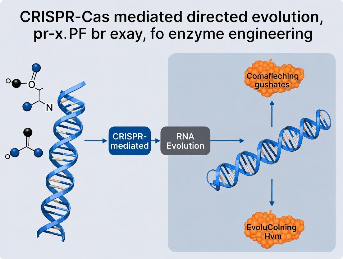
This article provides a comprehensive guide to CRISPR-Cas mediated directed evolution (CDE), a transformative methodology for engineering enzymes with enhanced properties.
Accelerating Enzyme Evolution: How CRISPR-Cas Systems Are Revolutionizing Protein Engineering for Therapeutics
Abstract
This article provides a comprehensive guide to CRISPR-Cas mediated directed evolution (CDE), a transformative methodology for engineering enzymes with enhanced properties. Tailored for researchers and drug development professionals, it explores the foundational principles of coupling CRISPR's DNA-targeting precision with the power of Darwinian selection. We detail current methodological workflows for creating and screening mutant libraries, address common experimental challenges and optimization strategies, and critically compare CDE's performance against traditional directed evolution techniques. The synthesis highlights CDE's superior speed and efficiency in generating evolved enzymes for biocatalysis, biosensing, and next-generation therapeutics, outlining its significant implications for biomedical research.
From Natural Defense to Protein Design: Unpacking the Principles of CRISPR-Directed Evolution
Application Notes: CRISPR-Cas Mediated Directed Evolution for Enzyme Engineering
Directed evolution accelerates enzyme engineering by mimicking natural selection in the laboratory. Traditional methods, like error-prone PCR, suffer from uncontrolled mutation distribution and low efficiency. The integration of CRISPR-Cas systems introduces unprecedented precision and programmability into this process. This fusion allows researchers to focus evolutionary pressure on specific genomic loci or protein domains, generating smarter, more focused libraries. Below are key protocols and resources for implementing this strategy.
Table 1: Quantitative Comparison of Directed Evolution Methods
| Method | Mutation Rate Control | Library Diversity | Off-Target Effects | Primary Screening Throughput | Best For |
|---|---|---|---|---|---|
| Error-Prone PCR | Low, global | High, random | N/A | Medium-High (104-106) | Broad, initial exploration of sequence space. |
| CRISPR-Cas9 Base Editing | High, site-specific | Moderate, defined transition mutations (e.g., C•G to T•A) | Moderate | High (106-108) | Introducing specific point mutations or correcting stop codons. |
| CRISPR-Cas12 Orthologs for MMR | Moderate, tunable | High, localized to genomic region | Low | High (106-108) | Saturation mutagenesis of a specific gene or domain. |
| CRISPR-X / CAST (Transposon) | High, programmable | Moderate, insertional mutagenesis | Low | Medium (104-105) | Inserting peptide tags or new functional domains. |
Protocol 1: CRISPR-Cas9-Mediated Base Editing for Targeted Enzyme Evolution
Objective: To introduce specific A•T to G•C or C•G to T•A point mutations within a gene of interest (GOI) in a microbial host to alter enzyme activity.
Materials:
- Plasmid Constructs: pCRISPR-BE (expresses dCas9 or nickase Cas9 fused to a deaminase, e.g., TadA-8e) and a sgRNA specific to the target codon(s).
- Host Strain: E. coli or yeast strain harboring the chromosomal or plasmid-borne GOI.
- Reagents: Transformation reagents, selective media (e.g., with antibiotic), PCR mix, sequencing primers.
- Equipment: Thermocycler, incubator, spectrophotometer, sequencing facility.
Procedure:
- Design & Cloning: Design a sgRNA to target the ~5-nucleotide editing window of the base editor to the codon(s) of interest. Clone the sgRNA sequence into the pCRISPR-BE plasmid.
- Transformation: Co-transform or sequentially transform the host strain with the constructed pCRISPR-BE plasmid.
- Selection & Growth: Plate transformed cells on selective media. Pick single colonies and grow in liquid culture to allow base editing to occur.
- Screening: Isolate genomic DNA or plasmid DNA. Amplify the GOI region via PCR and submit for Sanger or next-generation sequencing to identify mutations.
- Phenotypic Assay: Screen or select clones under applied evolutionary pressure (e.g., higher temperature, non-native substrate, inhibitor).
- Iteration: Isolate improved variants and repeat process with new sgRNAs targeting other regions.
Protocol 2: CRISPR-Cas12a-Assisted Mutagenesis via Mismatch Repair (MMR) Evasion
Objective: To generate localized, diverse mutations around a Cas12a cut site by harnessing and manipulating the host's DNA repair pathways.
Materials:
- CRISPR Component: Plasmid expressing Cas12a (cpf1) and a crRNA targeting near the protein domain of interest.
- MMR Modulation: Plasmid expressing dominant-negative MutL / MutS variants or use of a transiently MMR-deficient strain.
- Oligonucleotide Donor Pools: A pool of oligos with degenerate nucleotides (NNN) or targeted mutations spanning the cut site.
- Repair Template: ssDNA or dsDNA donor with homology arms for high-fidelity repair (optional, for controlled insertion).
Procedure:
- System Delivery: Introduce the Cas12a/crRNA plasmid and the oligo donor pool into the host cell (with modulated MMR).
- Induction & Cutting: Induce Cas12a expression. Cas12a creates a staggered double-strand break (DSB) near the target.
- Error-Prone Repair: In the absence of a precise donor or with MMR impaired, the cell uses error-prone non-homologous end joining (NHEJ) or microhomology-mediated end joining (MMEJ) to repair the break, introducing indels. The co-localized oligo donor pool can also be integrated via homology-directed repair (HDR), incorporating designed diversity.
- Library Recovery: Allow cells to recover and propagate the mutated GOI.
- Selection & Analysis: Apply stringent selection pressure (e.g., antibiotic gradient, fluorescence-activated cell sorting (FACS) for enzymatic activity). Sequence surviving populations to identify beneficial mutation patterns.
Visualization
CRISPR-Directed Evolution Workflow
DNA Repair Pathways Post-CRISPR Cut
The Scientist's Toolkit: Key Research Reagent Solutions
| Item | Function in CRISPR-Directed Evolution |
|---|---|
| dCas9-APOBEC1 (BE3/BE4) Plasmid | Enables targeted C•G to T•A transitions without creating double-strand breaks, ideal for single nucleotide changes. |
| CRISPR-Cas12a (Cpf1) System | Utilizes a staggered cut and simpler crRNA, often preferred for multiplexed or MMR-based mutagenesis strategies. |
| Dominant-Negative mutL (E. coli) Plasmid | Temporarily inhibits the mismatch repair system, increasing the fixation rate of point mutations near the cut site. |
| Degenerate Oligonucleotide Pools (NNK) | Serves as donor templates to introduce saturation mutagenesis at specific codons via HDR. |
| Transposase-Cas Fusion (CAST) System | Programs the insertion of transposon cargo (e.g., peptide tags, whole domains) at target sites for functional domain swapping. |
| Fluorescent Substrate Analogs | Enables high-throughput screening of enzyme activity via FACS, linking genotype to phenotype. |
| Phage-assisted Continuous Evolution (PACE) Compatible Vectors | Allows for continuous evolution in chemostats by linking gene essentiality to viral propagation under selection. |
Application Notes
This document details the application of key CRISPR-Cas systems in directed evolution for enzyme engineering. Moving beyond random mutagenesis, these tools enable targeted, diverse, and continuous mutagenesis within a gene of interest (GOI) in its native genomic context, accelerating the development of enzymes with enhanced properties.
Cas9-mediated Saturation Mutagenesis & Continuous Evolution: Nuclease-active Streptococcus pyogenes Cas9 (SpCas9) is used to generate libraries of variants. By co-expressing a guide RNA (gRNA) library targeting the GOI, Cas9 creates double-strand breaks (DSBs). The error-prone non-homologous end joining (NHEJ) repair pathway introduces indel mutations at high frequency, creating diverse, in-frame variant libraries for screening. For continuous evolution, Cas9 can be coupled with phage-assisted continuous evolution (PACE) systems, where host cell survival is linked to GOI function, enabling autonomous and rapid evolution over hundreds of generations.
Base Editors (BEs) for Targeted Point Mutation Libraries: Base Editors (e.g., BE4max) fuse a catalytically impaired Cas9 (nCas9) or Cas12a to a deaminase enzyme. They enable direct, irreversible conversion of one base pair to another (C•G to T•A or A•T to G•C) without creating a DSB or requiring a donor template. This allows for highly efficient, low-noise introduction of all possible single-nucleotide variants (SNVs) within a defined window (~5 nucleotides wide) of the gRNA target site, ideal for scanning protein active sites or stability hotspots.
Prime Editors (PEs) for Precision Diversity Generation: Prime Editors (e.g., PE2) combine nCas9 with an engineered reverse transcriptase (RT). A prime editing guide RNA (pegRNA) both specifies the target site and encodes the desired edit via its RT template sequence. This system can install all 12 possible base-to-base conversions, as well as small insertions and deletions, with high precision and minimal byproducts. It is uniquely suited for introducing multi-variant combinations and non-classical mutations to explore complex sequence landscapes in enzyme engineering.
Quantitative Performance Comparison of CRISPR-Cas Systems for Directed Evolution
| System (Example) | Type of Diversity Generated | Typical Editing Efficiency* | Indel Byproduct Rate* | Library Size & Focus | Primary Repair Pathway |
|---|---|---|---|---|---|
| Cas9 (Nuclease) | Indels (insertions/deletions) | High (>70% indels) | N/A (primary product) | Large, localized to DSB site. Uncontrolled sequence outcome. | NHEJ / MMEJ |
| Cytosine Base Editor (BE4max) | C•G → T•A transitions | 30-60% (product purity) | 0.1-1.0% | Defined. All possible C→T (and G→A) changes within a ~5nt window. | Base Excision Repair |
| Adenine Base Editor (ABE8e) | A•T → G•C transitions | 50-80% (product purity) | <0.1% | Defined. All possible A→G (and T→C) changes within a ~5nt window. | Base Excision Repair |
| Prime Editor (PE2) | All 12 point mutations, small insertions/deletions | 10-50% (varies by edit) | 0.1-5.0% | Highly programmable. Can generate specific, combinatorial variants at a single locus. | DNA Repair Synthesis / MMR |
*Efficiencies are highly sequence- and cell-type dependent. Values represent general ranges reported in mammalian cells.
Experimental Protocols
Protocol 1: Cas9-mediated Saturation Mutagenesis for Enzyme Engineering
Objective: Generate a library of indel mutations within a specific domain of an enzyme gene in E. coli.
Materials: pCas9 plasmid (inducible Cas9), pTarget plasmid (expressing gRNA library and GOI), recipient E. coli strain, inducer (aTc/IPTG), selective antibiotics, recovery media, plasmid extraction kit, sequencing primers.
Procedure:
- gRNA Library Design: Design oligos to tile gRNAs across the target protein domain. Synthesize as a pooled oligo library and clone into the pTarget plasmid backbone.
- Library Transformation: Co-transform the pooled pTarget library and the pCas9 plasmid into competent E. coli.
- Cas9 Induction & Variant Generation: Grow transformed cells with antibiotics and induce Cas9 expression with aTc. Cas9 cleavage followed by error-prone NHEJ repair generates the variant library in situ.
- Functional Screening/Selection: Plate cells under selective pressure that demands the desired enzyme function (e.g., antibiotic if GOI confers resistance, or substrate utilization).
- Hit Characterization: Isolve surviving colonies, sequence the GOI to identify mutations, and characterize purified enzyme variants.
Protocol 2: Base Editor Scanning for Functional Hotspot Identification
Objective: Introduce all possible C-to-T (or A-to-G) mutations across a critical exon of an enzyme.
Materials: Base Editor plasmid (e.g., BE4max), gRNA expression plasmid(s) tiling the target region, transfection reagent (for mammalian cells) or electroporation equipment (for microbes), genomic DNA extraction kit, HTS library prep reagents, sequencing facility access.
Procedure:
- gRNA Array Design: Design and clone a series of gRNAs spaced to cover the exon with overlapping editing windows.
- Delivery & Editing: Deliver the BE plasmid and individual gRNA plasmids (or a pooled gRNA library) into your host cells.
- Harvest & Pool: After 48-72 hours, harvest cells, extract genomic DNA, and PCR-amplify the target region from the pooled population.
- High-Throughput Sequencing (HTS): Prepare sequencing libraries and perform deep sequencing (e.g., Illumina MiSeq).
- Data Analysis: Use bioinformatics tools (e.g, BE-Analyzer, CRISPResso2) to calculate the frequency of each possible transition mutation at every base in the target region. Correlate depletion/enrichment of mutations with functional screening data to identify essential residues.
The Scientist's Toolkit: Key Reagents for CRISPR-Cas Directed Evolution
| Reagent / Solution | Function in Directed Evolution |
|---|---|
| nuclease-active SpCas9 expression plasmid | Creates targeted DSBs to initiate mutagenic NHEJ repair for indel library generation. |
| Base Editor (BE4max, ABE8e) expression plasmid | Enables efficient, DSB-free generation of precise transition mutation libraries at target sites. |
| Prime Editor (PE2, PEmax) expression system | Allows installation of virtually any small edit (point mutations, indels) for precision variant library construction. |
| pegRNA cloning backbone | Plasmid for expressing the complex pegRNA, which encodes both target location and edit information for prime editing. |
| Error-prone NHEJ repair machinery | Cellular context (often enhanced by MMEJ factors) critical for Cas9-mediated diversity generation. |
| Pooled gRNA library oligos | Synthesized oligo pool targeting multiple sites to diversify a protein region or entire gene. |
| HTS library preparation kit | For preparing amplified target regions from pooled variant libraries for deep sequencing analysis. |
| Selection/Screening medium | Contains substrate, antibiotic, or condition that links cell survival or growth to desired enzyme function. |
Visualization: CRISPR-Cas Directed Evolution Workflow
Diagram Title: CRISPR-Cas Directed Evolution Tool Selection & Workflow
Visualization: Mechanism of Base Editing vs Prime Editing
Diagram Title: Mechanism Comparison of Base Editors and Prime Editors
CRISPR-Cas mediated directed evolution accelerates enzyme engineering by introducing targeted diversity and selecting for desired phenotypes, such as altered substrate specificity, enhanced thermostability, or novel catalytic activity. This process hinges on two foundational choices: the design of the guide RNA (gRNA) library, which dictates the location and type of genetic variation, and the selection of the host organism, which provides the cellular machinery for screening and selection.
Designing gRNA Libraries for Enzyme Engineering
Core Principles and Quantitative Considerations
Effective gRNA library design balances saturation of the target region with practical library size and transformation efficiency.
Table 1: Key Parameters for gRNA Library Design
| Parameter | Typical Target Range | Rationale & Impact |
|---|---|---|
| Target Region Length | 6–12 codons (18–36 bp) | Focuses diversity on functionally critical residues (active site, binding pockets). |
| Theoretical Library Size | 10^5 – 10^9 variants | Must cover all possible mutations (e.g., NNK degeneracy: 32 codons). Library size > 100x theoretical diversity ensures coverage. |
| gRNA Spacing | 1–5 bp overlap between adjacent gRNAs | Ensures comprehensive coverage of contiguous sequence; prevents "dead zones." |
| On-target Efficiency Score | > 60 (using tools like Doench '16) | Maximizes editing efficiency in the host organism. |
| Predicted Off-target Sites | 0–3 (with high specificity scores) | Minimizes unwanted mutations elsewhere in the genome. |
Protocol: Designing a Saturation Mutagenesis gRNA Library for a Bacterial Enzyme
Objective: To create a pooled gRNA library targeting the substrate-binding pocket (amino acids 120-125) of a hydrolase in E. coli.
Materials:
- Gene sequence of the target enzyme.
- CRISPR design software (e.g., CHOPCHOP, Benchling, or proprietary tools).
- Oligonucleotide design software.
Procedure:
- Define Target Region: Align homologous enzyme structures to identify conserved residues in the region of interest (e.g., 6 codons).
- Generate gRNA Candidates: Input the gene sequence into design software. Set parameters to generate gRNAs targeting both strands of the defined 18-bp region.
- Filter and Select: a. Filter gRNAs with high on-target efficiency scores (>60). b. Filter out gRNAs with >3 predicted genomic off-target sites. c. Select 3-5 top-ranked gRNAs that tile across the region with 1-3 bp overlaps.
- Design Oligo Library: For each selected gRNA spacer sequence, replace the target codons with an NNK degenerate sequence (N = A/T/G/C; K = G/T) to encode all 20 amino acids and one stop codon.
- Synthesize Library: Order the pooled oligo library as a microarray-synthesized oligonucleotide pool. Include constant flanking sequences for subsequent PCR amplification and cloning into your chosen CRISPR plasmid backbone.
Visualization: gRNA Library Design and Cloning Workflow
Title: gRNA Library Design and Construction Pipeline
Selecting the Right Host Organism
Comparative Analysis of Host Organisms
The host organism determines the screening throughput, functional assay compatibility, and ease of genetics.
Table 2: Comparison of Host Organisms for CRISPR-Cas Directed Evolution
| Host Organism | Key Advantages | Key Limitations | Typical Library Size | Best for Enzyme Types |
|---|---|---|---|---|
| Escherichia coli | Fast growth, high transformation efficiency, extensive genetic tools. | Lack of post-translational modifications (PTMs), eukaryotic protein misfolding. | 10^9 – 10^10 | Prokaryotic enzymes, robust eukaryotic enzymes (e.g., hydrolases). |
| Saccharomyces cerevisiae | Eukaryotic PTMs, secretory pathway, relatively fast, good transformation. | Lower transformation efficiency than E. coli, more complex genetics. | 10^7 – 10^8 | Eukaryotic enzymes, secreted proteins, glycosylation-dependent enzymes. |
| Bacillus subtilis | Efficient secretion, GRAS status, good for industrial production. | Fewer genetic tools than E. coli, competence development required. | 10^6 – 10^7 | Secreted industrial enzymes (proteases, amylases). |
| Mammalian Cells (e.g., HEK293) | Human PTMs, complex cellular context for functional assays. | Very slow, low throughput, expensive, technically demanding. | 10^5 – 10^6 | Human therapeutic enzymes, targets requiring mammalian folding/processing. |
Protocol: Host Selection and Transformation for a Yeast Surface Display Screen
Objective: To select and prepare S. cerevisiae EBY100 strain for a gRNA library delivery to evolve antibody affinity.
Materials:
- S. cerevisiae strain EBY100 (gal1-, contains pCTCON2 display vector).
- CRISPR-Cas9 plasmid for yeast (e.g., pML104).
- LiAc/TE buffer, PEG/LiAc solution.
- Single-stranded carrier DNA (salmon sperm DNA).
- Synthetic complete dropout media lacking specific amino acids (e.g., -Trp, -Ura).
Procedure:
- Strain Preparation: Inoculate EBY100 into YPD and grow overnight at 30°C to mid-log phase (OD600 ~0.8-1.0).
- Competent Cell Preparation: Harvest cells, wash with sterile water and LiAc/TE buffer. Resuspend final pellet in LiAc/TE.
- Transformation Mixture: For each transformation, mix:
- 100 ng of your gRNA library plasmid (contains Cas9 and selection marker).
- 50 ng of a homologous repair template (HDR) containing the mutagenized gene fragment.
- 100 µL competent cells.
- 10 µL denatured carrier DNA (10 mg/mL).
- 600 µL PEG/LiAc solution. Vortex and incubate at 30°C for 30 min.
- Heat Shock: Add 70 µL DMSO, mix gently. Heat shock at 42°C for 15 minutes.
- Recovery and Plating: Pellet cells, resuspend in recovery medium, and incubate at 30°C for 2-4 hours. Plate onto appropriate dropout agar plates to select for transformants.
- Library Harvest: After 2-3 days, scrape all colonies from plates, pool, and make glycerol stocks for long-term storage at -80°C. This pooled culture is your mutant library.
Visualization: Host Organism Selection Logic
Title: Decision Tree for Host Organism Selection
The Scientist's Toolkit: Research Reagent Solutions
Table 3: Essential Materials for CRISPR-Cas Directed Evolution Workflows
| Item/Reagent | Function & Application | Example Product/Supplier |
|---|---|---|
| CRISPR-Cas9 Plasmid Kit | Provides the Cas9 nuclease and gRNA scaffold, often with a selection marker (e.g., AmpR, URA3). Essential for delivering the system to the host. | Addgene #52961 (yeast pML104), #42876 (E. coli pCas9). |
| High-Efficiency Competent Cells | Crucial for achieving large library transformation sizes without bias. Specific to chosen host organism. | NEB 5-alpha E. coli (C2987), Lucigen YeastMaker. |
| NNK Degenerate Oligo Pool | Synthesized oligonucleotide library encoding the mutagenic gRNA spacers and targeting diversity. | Custom order from Twist Bioscience, IDT. |
| HDR Template Oligo/DNA Fragment | Donor DNA for precise mutation incorporation via homology-directed repair. Can be ssDNA or dsDNA. | Ultramer DNA Oligos (IDT), gBlocks (IDT). |
| Next-Generation Sequencing (NGS) Kit | For deep sequencing of the gRNA library pre- and post-selection to identify enriched variants. | Illumina Nextera XT, MGI EasySeq. |
| Fluorescence-Activated Cell Sorting (FACS) Buffers | For assays linking enzyme function to a surface-displayed fluorescent signal (common in yeast/mammalian display). | PBS + 1% BSA (for yeast), Cell Staining Buffer (BioLegend). |
| Microplate Reader-Compatible Assay Substrate | For high-throughput screening of enzyme activity in lysates or supernatants from colony picks. | Chromogenic/fluorogenic substrate specific to enzyme class (e.g., pNPP for phosphatases). |
CRISPR-Cas mediated directed evolution (CRISPR-DE) integrates the precision of genome editing with the power of Darwinian selection to engineer enzymes with enhanced or novel properties. This methodology accelerates the traditional directed evolution cycle by enabling the generation of targeted, in-situ diversity within the host genome and coupling genotype to phenotype efficiently. The core cycle—Generate, Select, Iterate—is applied to evolve enzymes for industrial biocatalysis, therapeutic protein production, and drug discovery.
Application Note 1: CRISPR-DE is particularly effective for evolving in vivo function, such as improving the activity of a metabolic pathway enzyme in its native cellular context. It bypasses the need for cumbersome in vitro library construction and transformation.
Application Note 2: Recent advances utilize CRISPR-Cas12a and Base Editors (e.g., BE4max, ABE8e) for diversity generation, allowing for a broader range of mutations (transitions, transversions, small indels) with reduced off-target effects compared to error-prone PCR and traditional Cas9 nickase-based methods.
Application Note 3: Selection strategies have evolved from simple antibiotic resistance to sophisticated FACS-based sorting using biosensors that fluoresce in response to product formation or substrate depletion, enabling high-throughput screening of millions of variants.
Table 1: Comparison of CRISPR-DE Diversity Generation Methods (2022-2024)
| Method | Typical Library Size | Mutation Rate (%) | Key Advantage | Representative Study (PMID) |
|---|---|---|---|---|
| Cas9 Nickase + MMR | 10^7 - 10^9 | 0.1 - 1 | High efficiency, targeted double-strand breaks | 36307436 |
| Cas12a-Directed | 10^6 - 10^8 | 0.5 - 5 | Simpler PAM (TTTV), staggered cuts | 36792740 |
| Base Editing (CBE) | 10^4 - 10^6 | 10 - 50* | Precise C•G to T•A transitions, low indels | 37165189 |
| Base Editing (ABE) | 10^4 - 10^6 | 10 - 40* | Precise A•T to G•C transitions, low indels | 37823656 |
| OrthoRep (in vivo) | 10^10+ | 10^-4 per bp | Continuous, PCR-free evolution | 38071684 |
*Mutation rate at targeted window; CBE: Cytosine Base Editor, ABE: Adenine Base Editor.
Table 2: Selection & Screening Output Metrics for Enzyme Engineering
| Selection Method | Throughput (variants/round) | Enrichment Factor | Typical Duration | Key Application |
|---|---|---|---|---|
| Plate-based Survival | 10^3 - 10^5 | 10^2 - 10^3 | 2-3 days | Antibiotic resistance, auxotrophy |
| FACS with Biosensor | 10^7 - 10^8 | 10^3 - 10^4 | 1-2 days | Fluorescent product/substrate detection |
| Microfluidic Droplet Sort | 10^8 - 10^9 | 10^4 - 10^5 | Hours | Ultra-high-throughput, low volume |
| Phage/ Yeast Display | 10^9 - 10^11 | 10^3 - 10^5 | 1-2 weeks | Binding affinity, stability evolution |
Experimental Protocols
Protocol 3.1: CRISPR-Cas12a MediatedIn VivoDiversity Generation inS. cerevisiae
Objective: To create a targeted, diverse mutant library of a gene encoding an enzyme (e.g., cytochrome P450) integrated into the yeast genome.
Materials: See Scientist's Toolkit. Duration: 5-7 days.
Procedure:
- sgRNA Array Construction: Design and synthesize four crRNAs targeting non-coding strands within 50bp of the target gene's active site. Clone as a tandem array under a S. cerevisiae U6 promoter in a CEN/ARS plasmid.
- Cas12a Expression Cassette: Clone a codon-optimized LbCas12a gene under a GAL1 inducible promoter into a 2μ high-copy plasmid with a LEU2 selectable marker.
- Transformation: Co-transform the crRNA array plasmid (URA3) and Cas12a plasmid into a yeast strain harboring the integrated target gene using the standard lithium acetate/PEG method. Plate on SC -Leu -Ura media.
- Diversity Generation Induction: Grow a single colony in SC -Leu -Ura + 2% raffinose overnight. Induce Cas12a expression by adding 2% galactose for 6 hours. This generates targeted double-strand breaks.
- Error-Prone Repair Activation: During induction, supplement media with 1mM MnCl₂ to promote error-prone repair by host polymerases. Alternatively, overexpress a dominant-negative variant of DNA polymerase δ (pol3-5DV) from an inducible promoter.
- Library Harvesting: After 24-48 hours of growth post-induction, harvest cells, isolate genomic DNA, and sequence the target region via NGS to assess library diversity.
Protocol 3.2: FACS-Based Selection Using a Transcription Factor Biosensor
Objective: To isolate enzyme variants with improved activity from a cellular library using a product-responsive biosensor and fluorescence-activated cell sorting (FACS).
Materials: See Scientist's Toolkit. Duration: 3-4 days per round.
Procedure:
- Biosensor Strain Preparation: Use a host strain (e.g., E. coli or yeast) containing a biosensor construct where the enzyme's product activates a transcription factor (e.g., LuxR for acyl-homoserine lactones), driving GFP expression.
- Library Transformation: Transform the mutant library (from Protocol 3.1, recovered as plasmid or genomic locus) into the biosensor strain.
- Selection Culture: Grow transformed library in deep 96-well plates or flasks in minimal media with the target substrate at a concentration near the Km of the wild-type enzyme. Incubate until mid-log phase.
- FACS Sorting: Dilute cells in PBS or appropriate buffer. Sort using a FACS instrument (e.g., Sony SH800, BD FACSAria). Gate on the top 0.1-1% of GFP-positive cells. Collect ~10^6 cells into recovery media.
- Recovery & Enrichment: Allow sorted cells to recover in rich media for 4-6 hours, then plate a fraction to determine colony count and inoculate the next round of selection culture with the remainder.
- Iteration: Repeat steps 3-5 for 3-5 rounds. After the final round, plate cells for single colonies, sequence target genes from individual clones, and assay for improved enzyme kinetics.
Visualizations
Diagram 1 Title: The Core Directed Evolution Cycle Workflow
Diagram 2 Title: CRISPR-DE with Biosensor Selection Protocol
The Scientist's Toolkit
Table 3: Key Research Reagent Solutions for CRISPR-DE Enzyme Engineering
| Item | Function & Application | Example Product / Note |
|---|---|---|
| LbCas12a/Cpf1 Expression Plasmid | Provides the CRISPR nuclease for targeted DSB generation. Inducible promoters (GAL1, Tet-On) allow temporal control. | Addgene #69982 (pY064: GAL1p-LbCas12a-2A-PhleoR). |
| crRNA Array Cloning Vector | Enables expression of multiple guide RNAs from a single transcript for multiplexed targeting. | pMLS (Yeast U6 promoter-tRNA array system). |
| Base Editor Plasmids (BE4max, ABE8e) | For introducing precise point mutations without DSBs or donor templates, reducing cellular toxicity. | Addgene #112093 (BE4max), #138489 (ABE8e). |
| Error-Prone Repair Enhancers | Chemicals or genetic elements to increase mutation frequency during NHEJ or HDR. | 1mM MnCl₂, overexpression of pol3-5DV (yeast) or umuD'C (E. coli). |
| Fluorescent Biosensor Construct | Links desired enzyme activity to a measurable fluorescence output for high-throughput sorting. | Plasmids with product-responsive TF (LuxR, HapR) driving GFP/mCherry. |
| FACS Recovery Media | Rich, buffered media to maximize cell viability post-sorting. | S.O.C. medium (E. coli) or YPD + 1M Sorbitol (Yeast). |
| NGS Library Prep Kit (Amplicon) | For deep sequencing of target loci to quantify library diversity and track variant enrichment. | Illumina DNA Prep, or Swift Amplicon panels. |
| Microfluidic Droplet Generator | For encapsulating single cells with substrate in picoliter droplets for ultra-HTP screening. | Bio-Rad QX200 Droplet Generator, or FlowJEM chips. |
CRISPR-Cas-Directed Evolution (CDE) represents a paradigm shift in enzyme engineering, leveraging programmable nucleases to drive evolution in living cells. This Application Note contextualizes CDE within the historical lineage of in vitro display technologies, primarily phage and RNA display. It details protocols for implementing CDE within a CRISPR-Cas framework, contrasting it with traditional methods.
Historical Progression & Quantitative Comparison
Table 1: Historical Context and Quantitative Comparison of Key Directed Evolution Platforms
| Feature | Phage Display | RNA Display | CRISPR-Cas Directed Evolution (CDE) |
|---|---|---|---|
| Evolution Context | In vitro (cell-free transcription/translation) or ex vivo (bacterial surface). | In vitro, entirely cell-free. | In vivo, within living host cells (e.g., bacteria, yeast, mammalian). |
| Library Size (Practical Max) | ~10^10 – 10^11 variants. | ~10^13 – 10^14 variants. | ~10^8 – 10^9 variants (per transformation). |
| Genotype-Phenotype Linkage | Physical: protein fused to encapsidated DNA. | Physical: protein linked to its mRNA via puromycin. | Intracellular: phenotype selected, genotype edited in situ via CRISPR. |
| Selection Throughput | Moderate. Requires panning/elu-tion cycles. | High. Direct partitioning (e.g., using immobilized target). | Very High. Enables continuous evolution in chemostats or via FACS. |
| Mutation Rate & Control | Low, relies on error-prone PCR; control is external. | Low, error-prone PCR or chemical mutagenesis; control is external. | High & Programmable. Cas9 nucleases generate targeted, tunable diversity (e.g., via error-prone repair or base editors). |
| Primary Application | Antibody/peptide affinity binding. | Peptide, small protein binders. | Enzyme engineering (activity, stability, selectivity), metabolic pathway optimization. |
| Turnaround Time (Cycle) | Weeks. | 1-2 weeks. | Days to a week for continuous systems. |
Core Protocols
Protocol 1: Traditional Phage Display Panning for Binding Affinity
Objective: Isolate high-affinity protein binders from a phage library.
- Library Incubation: Incubate the phage-displayed peptide/protein library (e.g., 10^11 pfu in 1 mL blocking buffer) with the immobilized target antigen (on a plate or beads) for 1-2 hours at RT with gentle agitation.
- Washing: Remove unbound phage by washing 10-20 times with TBST (Tris-Buffered Saline with 0.1% Tween-20).
- Elution: Elute specifically bound phage using 1 mL of 0.1 M glycine-HCl (pH 2.2) for 10 minutes. Immediately neutralize with 150 µL of 1 M Tris-HCl (pH 9.1).
- Amplification: Infect log-phase E. coli (e.g., ER2738) with the eluted phage for 30 min at 37°C. Plate on LB/IPTG/Xgal plates for titering or culture overnight in LB with helper phage to amplify the enriched pool for subsequent rounds (typically 3-5).
- Analysis: Sequence individual clones from later rounds to identify consensus binding sequences.
Protocol 2: CRISPR-Cas Directed Evolution (CDE) for Enzyme Activity
Objective: Evolve an enzyme for enhanced activity in vivo using a CRISPR-Cas9-mediated mutagenesis and selection system.
- System Setup:
- Host Strain: Engineer a microbial host (e.g., E. coli) to constitutively express Cas9 and a repair template plasmid containing a mutagenic polymerase (e.g., a low-fidelity Pol I variant).
- Library Construction: Clone the target enzyme gene into a plasmid containing a selection marker (e.g., antibiotic resistance conditional on enzyme activity) and a CRISPR target sequence (gRNA scaffold).
- Diversification Cycle:
- Induce expression of a specific gRNA targeting the enzyme gene locus. Cas9 cleavage triggers the error-prone repair process, generating localized mutations within the gene.
- Grow the population for 12-18 hours under non-selective conditions to allow mutation fixation.
- Selection/Enrichment Cycle:
- Apply selective pressure (e.g., addition of a prodrug requiring enzymatic conversion for survival, or limiting substrate only metabolized by improved enzyme).
- Use Fluorescence-Activated Cell Sorting (FACS) if a fluorescent reporter (e.g., GFP linked to metabolic flux) is incorporated. Collect the top 0.1-1% fluorescent population.
- Iteration & Sequencing:
- Isolate plasmids or genomic DNA from the selected population.
- Re-transform the diversified pool into a fresh host to reset the system and repeat cycles 2-3.
- Sequence enriched pools (NGS) and individual clones to identify beneficial mutations.
Visualizing the Evolutionary Workflows
Phage Display Panning Cycle
CDE In Vivo Evolution Cycle
The Scientist's Toolkit: Key Reagents for CDE Experiments
Table 2: Essential Research Reagent Solutions for CRISPR-Cas Directed Evolution
| Reagent / Material | Function in CDE |
|---|---|
| Programmable Nuclease System (e.g., Cas9, Cas12a) | Creates targeted double-strand breaks in the gene of interest to initiate the DNA repair process that introduces mutations. |
| Tunable Mutagenesis Machinery (e.g., error-prone DNA Pol I variant (DLM), Base/Prime Editor fusions) | Generates diversity at or near the cut site. Tunability allows control over mutation rate and spectrum. |
| gRNA Library or Inducible Promoter | Guides Cas nuclease to the target locus. Can be a single target or a library targeting multiple regions. |
| In Vivo Selection Circuit | Links desired enzyme phenotype (activity, stability) to cell survival or a reportable signal (fluorescence). Crucial for enrichment. |
| Flow Cytometry (FACS) Capability | Enables high-throughput, quantitative screening and sorting of cell populations based on fluorescent reporters linked to enzyme function. |
| Next-Generation Sequencing (NGS) Platform | For deep sequencing of evolved pools to identify mutation hotspots and genotype-phenotype relationships. |
| CRISPR-Competent Host Strain | Engineered microbial or mammalian cell line optimized for high-efficiency CRISPR editing and containing necessary helper plasmids. |
| Selection Media / Prodrugs | Provides the selective pressure that enriches for improved enzyme variants (e.g., antibiotic whose resistance gene is activated by the enzyme). |
A Step-by-Step Protocol: Implementing CRISPR-Cas Directed Evolution in Your Lab
The engineering of enzymes with enhanced or novel properties is a cornerstone of modern biotechnology, impacting drug development, industrial biocatalysis, and synthetic biology. Traditional directed evolution, pioneered by Frances Arnold, involves iterative rounds of random mutagenesis and screening. The integration of CRISPR-Cas systems has revolutionized this paradigm by enabling targeted, efficient, and multiplexed generation of diversity directly within the genomes of host organisms. This application note details a modern workflow that leverages CRISPR-Cas mediated directed evolution to accelerate the journey from identifying a gene target to isolating an evolved enzyme, contextualized within a broader research thesis on precision enzyme engineering.
Comprehensive Workflow Protocol
Phase 1: Target Identification and gRNA Design
Objective: Select the gene of interest (GOI) and design CRISPR guide RNAs (gRNAs) for precise targeting. Protocol:
- Gene Selection: Identify the wild-type enzyme gene based on prior knowledge or bioinformatic analysis of substrate specificity, structural data, or phylogenetic relationships.
- gRNA Design:
- Use tools like CHOPCHOP, Benchling, or CRISPOR to design 3-5 gRNAs targeting the GOI's coding sequence.
- Prioritize gRNAs with high on-target efficiency scores (>60) and minimal predicted off-target effects.
- Quantitative Design Criteria:
- GC Content: 40-60%
- On-target Score: >60 (tool-specific)
- Specificity Score: >90 (to minimize off-targets)
- Synthesis: Order gRNA sequences as oligonucleotides for cloning or as synthetic RNAs for direct delivery.
Phase 2: Library Construction via CRISPR-Cas Mediated Mutagenesis
Objective: Create a diverse mutant library in the host genomic locus. Protocol:
- System Choice: Select a Cas protein (e.g., Cas9, Cas12a) and a mutagenic strategy:
- Base Editing: For precise point mutations (C>T, A>G) without double-strand breaks (DSBs). Use a deaminase-fused Cas nickase.
- Prime Editing: For targeted insertions, deletions, and all 12 possible base-to-base conversions.
- CRISPR-Cas with Homology-Directed Repair (HDR): For diversification using mutagenic oligo libraries.
- Library Delivery:
- For microbial hosts (e.g., E. coli, yeast), use electroporation or chemical transformation with a plasmid encoding Cas9, the gRNA array, and a repair template library if using HDR.
- For mammalian cells, use lentiviral or nucleofection delivery methods.
- Library Quality Control:
- Plate a dilution to determine library size (colony-forming units, CFU). Aim for a library size 100-1000x the theoretical diversity.
- Sequence 20-50 random colonies via Sanger sequencing to confirm mutation rate and spectrum.
Phase 3: High-Throughput Screening or Selection
Objective: Identify clones expressing improved enzyme variants. Protocol A: Fluorescence-Activated Cell Sorting (FACS) for intracellular enzymes:
- Sensor Construction: Clone a genetic circuit or fluorescent reporter that responds to the enzyme's activity (e.g., a product-activated transcription factor driving GFP).
- Sorting: Use FACS to isolate the top 0.1-1% of the most fluorescent cells.
- Recovery: Grow sorted cells on solid medium for colony isolation. Protocol B: Microfluidics/Droplet-Based Screening:
- Encapsulation: Co-encapsulate single library cells with a fluorescent substrate and lysis agent in picoliter droplets.
- Sorting: Use a droplet sorter to isolate droplets with high fluorescent signal, indicative of enzyme activity.
- Break Emulsion: Recover cells from sorted droplets for outgrowth. Protocol C: Solid-Plate Screening:
- Assay: Plate library onto agar containing an indicator (e.g., chromogenic/fluorogenic substrate, pH indicator, or halo assay for hydrolysis).
- Picking: Manually or robotically pick colonies with a desired phenotype (e.g., largest halo, intense color).
Phase 4: Characterization and Validation
Objective: Quantitatively assess the performance of evolved hits. Protocol:
- Hit Sequence Analysis: Sequence the GOI from selected hits to identify mutations.
- Protein Purification: Express and purify the wild-type and evolved enzymes using affinity chromatography (e.g., His-tag).
- Enzyme Kinetics: Perform Michaelis-Menten analysis.
- Assay Conditions: Vary substrate concentration under saturating co-factor conditions at optimal pH and temperature.
- Data Analysis: Fit data to the Michaelis-Menten equation to derive
k_cat(turnover number) andK_M(Michaelis constant). Calculate catalytic efficiency ask_cat/K_M.
Data Presentation: Key Performance Metrics
Table 1: Typical Quantitative Outcomes from CRISPR-Cas Directed Evolution Campaigns
| Parameter | Base Editing | Prime Editing | HDR with Mutagenic Library | Notes |
|---|---|---|---|---|
| Editing Efficiency | 10-50% | 5-30% | 0.1-10% | Varies by organism and locus. |
| Library Diversity | Limited by base editor window (~5nt) | Limited by pegRNA design | >10^6 variants possible | Theoretical diversity. |
| Mutation Types | Specific transition mutations | All point mutations, small indels | Any mutation within repair template | |
| Typical Screening Throughput | 10^7 - 10^9 cells | 10^7 - 10^9 cells | 10^7 - 10^10 cells | Depends on method (FACS vs. plates). |
Fold-Improvement in k_cat/K_M |
2-10x | 2-50x | 2-100x+ | Highly target-dependent. |
| Key Reference | Gaudelli et al., 2017 | Anzalone et al., 2019 | Barbieri et al., 2024* | *Recent review on high-throughput methods. |
Table 2: Example Kinetic Data for a Hypothetical Evolved Hydrolase
| Enzyme Variant | k_cat (s⁻¹) |
K_M (mM) |
k_cat/K_M (mM⁻¹s⁻¹) |
Fold-Improvement |
|---|---|---|---|---|
| Wild-Type | 1.0 ± 0.1 | 5.0 ± 0.5 | 0.20 | 1.0 |
| Variant A (R124C) | 8.5 ± 0.7 | 4.2 ± 0.4 | 2.02 | 10.1 |
| Variant B (R124C/L189F) | 15.2 ± 1.2 | 2.1 ± 0.2 | 7.24 | 36.2 |
Visualization: Workflow and Pathway Diagrams
Diagram Title: CRISPR-Cas Enzyme Evolution Workflow
Diagram Title: Screening Pathway Decision Tree
The Scientist's Toolkit: Research Reagent Solutions
Table 3: Essential Reagents & Materials for CRISPR-Cas Directed Evolution
| Item | Function & Key Characteristics | Example Vendor/Product |
|---|---|---|
| CRISPR-Cas Expression Plasmid | Expresses Cas protein (e.g., SpCas9, Cas12a) and gRNA scaffold in the host organism. Requires appropriate promoter and antibiotic resistance. | Addgene (pX330 series, pY000 series). |
| gRNA Cloning Oligos | Pair of synthesized DNA oligonucleotides encoding the 20-nt guide sequence for insertion into the CRISPR plasmid. | IDT, Sigma-Aldrich. |
| Mutagenic Repair Template | Single-stranded or double-stranded DNA containing desired mutations, flanked by homology arms (for HDR). Can be a pooled library. | TWIST Bioscience, IDT Ultramer pools. |
| Electrocompetent Cells | High-efficiency microbial cells (E. coli, S. cerevisiae) prepared for DNA library introduction via electroporation. | Lucigen, NEB. |
| Nucleofection Kit | Reagents for high-efficiency delivery of CRISPR components into mammalian or hard-to-transform cells. | Lonza Nucleofector kits. |
| Fluorogenic Enzyme Substrate | A non-fluorescent compound converted to a fluorescent product by enzyme activity. Essential for FACS/droplet screens. | Thermo Fisher (EnzChek kits), custom from AAT Bioquest. |
| FACS Sorter | Instrument to analyze and sort single cells based on fluorescence, enabling phenotype-based enrichment. | BD FACSAria, Beckman Coulter MoFlo. |
| Microfluidic Droplet System | Platform to generate, incubate, and sort picoliter droplets containing single cells and assay reagents. | Bio-Rad (QX200), Sphere Fluidics (Cyto-Mine). |
| Ni-NTA Resin | Affinity chromatography resin for purifying polyhistidine (His)-tagged wild-type and evolved enzymes. | Qiagen, Cytiva. |
| Plate Reader | Multimode spectrometer for high-throughput kinetic assays in microtiter plates (absorbance, fluorescence). | Tecan Spark, BMG Labtech CLARIOstar. |
Constructing High-Quality, Saturation Mutagenesis Libraries with CRISPR Tools
Within the broader thesis on CRISPR-Cas mediated directed evolution for enzyme engineering, the construction of high-quality saturation mutagenesis libraries represents a foundational step. Moving beyond traditional random mutagenesis, CRISPR-based tools enable precise, user-defined, and comprehensive replacement of single codons or regions across a gene of interest. This approach allows researchers to systematically explore the fitness landscape of an enzyme, linking specific amino acid substitutions directly to functional outcomes—a critical strategy for engineering properties like substrate specificity, thermostability, and catalytic efficiency in drug development research.
Key Concepts and Quantitative Benchmarks
Table 1: Comparison of CRISPR-Based Saturation Mutagenesis Methods
| Method | Primary CRISPR Tool | Library Diversity (Theoretical) | Typical Coverage | Key Advantage | Common Challenge |
|---|---|---|---|---|---|
| Cas9-mediated Oligo Recombination | Cas9 nickase (nCas9) or dead Cas9 (dCas9) fused to cytidine deaminase (e.g., APOBEC1) | Up to all 64 codons per position | >10^5 variants | High efficiency, single-base resolution. | Potential for guide RNA (gRNA) off-target effects. |
| CRISPR-BEST | Cas9 Doublenickase, Recombinase (e.g., RecT/ET) | Defined by oligo pool size (10^4 - 10^6) | >100x per variant | Scarless, recombinase-mediated precise integration. | Requires optimized recombinase expression. |
| CRISPR-Cas12a Assisted Saturation | Cas12a (cpf1) | All 64 codons per position | >10^5 variants | Uses crRNA without tracrRNA, simpler RNP complex. | Lower cleavage efficiency than SpCas9 in some systems. |
| Prime Editing | Prime Editor (nCas9-RT fusion) | All possible single-nucleotide variants | >10^4 variants | No double-strand breaks (DSBs) or donor templates needed. | Limited by prime editing guide RNA (pegRNA) design and efficiency. |
Table 2: Critical Quality Metrics for Library Validation
| Metric | Target Value | Measurement Method | Significance for Enzyme Engineering |
|---|---|---|---|
| Transformation Efficiency | >10^6 CFU/μg library DNA | Colony counting | Ensures sufficient library size for diversity. |
| Coverage (Fold) | ≥100x per variant | NGS of library plasmid pool | Guarantees each mutant is represented for screening. |
| Mutation Rate/Accuracy | >90% intended mutations | NGS of individual clones | Minimizes background of wild-type or incorrect sequences. |
| Indel Frequency | <5% | NGS or TIDE analysis | Measures unwanted DSB repair artifacts. |
Application Notes
Strategic Selection of Target Residues
Prioritize residues based on structural data (active site, substrate-binding pocket, known regulatory regions) or evolutionary conservation analysis. For comprehensive fitness landscape mapping, "hotspot" regions of 3-6 contiguous residues are often targeted simultaneously.
gRNA Design for Maximal Coverage
Design gRNAs to have the protospacer adjacent motif (PAM) sequence adjacent to the target codon. For multi-codon saturation, use a single gRNA that exposes a template strand for oligo binding across the entire region or employ a pooled gRNA strategy.
Balancing Library Quality and Diversity
The use of nicking Cas9 (nCas9) or fusions to deaminases (e.g., in BE, base editing) can reduce indel formation compared to wild-type Cas9. Coupling CRISPR cleavage with long, homology-directed repair (HDR) oligos (≥90 nt) improves precision.
Detailed Experimental Protocols
Protocol 1: Cas9-nCas9 Mediated Multiplexed Codon Saturation
Objective: To saturate 3 contiguous codons in an enzyme's active site using a pooled oligo HDR strategy.
Materials:
- Template DNA: Purified plasmid containing the wild-type gene.
- CRISPR Components: S. pyogenes nCas9 (D10A) expression plasmid or RNP complex.
- gRNA: A single gRNA expression plasmid or synthetic gRNA targeting the template strand 5' to the codon region.
- Repair Oligo Pool: A degenerate oligonucleotide pool (NNN at each target codon) with 50-nt homology arms on each side.
- Host Cells: High-efficiency E. coli or yeast cells with robust HDR machinery (e.g., E. coli MG1655 rpsL or S. cerevisiae).
Procedure:
- Design and Prep: Design the gRNA to have a 5' NGG PAM site on the non-template strand near the target. Synthesize the repair oligo pool with NNK degeneracy (encodes all 20 aa + 1 stop, reduces codon bias).
- Co-transformation: Mix 100 ng of template plasmid, 200 ng of nCas9 expression plasmid (or 2 pmol nCas9 RNP + 1 pmol gRNA), and 2 pmol of repair oligo pool. Transform into competent cells via electroporation.
- Recovery and Selection: Recover cells in rich medium for 1-2 hours at 37°C. Plate on selective antibiotic plates. Incubate overnight.
- Library Harvesting: Scrape all colonies (>100,000) from plates. Perform a plasmid maxi-prep to harvest the pooled library DNA.
- Validation: Sequence the pooled plasmid library via NGS (MiSeq) to assess mutation rate, coverage, and indel frequency.
Protocol 2: CRISPR-Base Editing for Saturation of Single Residues
Objective: To generate all possible single-nucleotide variants at a specific cytidine within a codon using a base editor.
Materials:
- Base Editor Plasmid: APOBEC1-nCas9-UGI fusion (BE3 or BE4) expression plasmid.
- Target-Specific gRNA: Designed to position the target C within the 5-nt editing window (protospacer positions 4-8).
- Control Plasmid: A plasmid expressing the wild-type gene.
Procedure:
- Transfection: Co-transfect the base editor plasmid and gRNA plasmid into mammalian (HEK293T) or yeast cells harboring the target gene on a plasmid.
- Editing Window: Allow base editing to occur for 48-72 hours. The deaminase converts C to U, leading to C•G to T•A transition after replication.
- Library Propagation: Isolate total plasmid DNA. Transform into E. coli to clonally separate variants and amplify the library.
- Screening: Plate transformations to obtain single colonies for sequencing and functional screening.
- Analysis: Sequence individual clones to identify the spectrum of amino acid changes achieved.
Diagrams
Title: CRISPR-nCas9 Saturation Mutagenesis Library Construction Workflow
Title: Library Strategy and Tool Selection Logic
The Scientist's Toolkit: Research Reagent Solutions
| Item | Function & Application Note |
|---|---|
| nCas9 (D10A) Expression Plasmid | Provides single-strand nicking activity. Reduces indel formation from non-homologous end joining (NHEJ) during HDR-based library construction. |
| Synthetic crRNA/tracrRNA or sgRNA | Guides Cas9 to the target locus. Chemically synthesized gRNAs offer high purity and reduce background from plasmid expression systems. |
| Degenerate Oligonucleotide Pool (NNK) | Serves as the HDR template. NNK degeneracy (N=A/C/G/T; K=G/T) covers all 20 amino acids and one stop codon with reduced bias versus NNN. |
| High-Efficiency Electrocompetent Cells | Essential for achieving high transformation efficiency (>10^6 CFU/μg). Strains with recA and endA deletions (e.g., NEB 10-beta) improve plasmid yield and stability. |
| Next-Generation Sequencing (NGS) Service/Kit | For library validation. Amplicon sequencing of the target region from pooled plasmid DNA is critical to quantify coverage, accuracy, and diversity. |
| Base Editor Plasmid (BE3/BE4) | For C-to-T (or A-to-G with ABE) transition mutations. Enables rapid, DSB-free saturation, but is limited to specific nucleotide changes. |
| Cas12a (CpF1) Nuclease | An alternative to Cas9. Recognizes a T-rich PAM, useful for targeting AT-rich genomic regions in microbial enzyme engineering. |
| Phusion Ultra High-Fidelity DNA Polymerase | For amplifying library pools with minimal error introduction. Critical when performing PCR steps post-library construction. |
Application Note: Integrating Biosensors with FACS for CRISPR-Cas Directed Evolution
Directed evolution, accelerated by CRISPR-Cas systems for precise genomic integration of variant libraries, necessitates robust strategies to couple genotypic diversity to detectable phenotypic outputs. This application note details methodologies for employing transcription factor-based biosensors and Fluorescence-Activated Cell Sorting (FACS) to screen for improved enzyme variants within a CRISPR-Cas mediated directed evolution workflow. The focus is on enzymes where the desired activity (e.g., production of a valuable metabolite, degradation of a substrate) can be linked to a fluorescent reporter.
Core Principle
A genetically encoded biosensor transduces the concentration of a target molecule (the enzyme's product) into a proportional fluorescence signal. In a pooled library of cells, each harboring a different enzyme variant generated via CRISPR-Cas, the fluorescence intensity of individual cells becomes a direct readout of that variant's functional performance. FACS then physically isolates the top-performing cells based on this fluorescence, enabling the recovery and sequencing of the genes encoding the elite enzymes.
Protocol 1: Biosensor-Enabled FACS Screening for Metabolite-Producing Enzyme Variants
Objective: To isolate E. coli clones expressing enzyme variants with enhanced production of a target metabolite (e.g., tyrosine, naringenin) from a CRISPR-Cas generated library.
Materials & Pre-requisites
- Strain: E. coli strain harboring a genomically integrated biosensor construct (e.g., a transcription factor responsive to the target metabolite, controlling expression of GFP).
- Library: A CRISPR-Cas9/Cas12a mediated knock-in library of enzyme variants at a defined genomic locus.
- Growth Media: Selective LB or defined minimal media.
- Induction: Appropriate inducer for enzyme expression (e.g., IPTG, arabinose).
- Equipment: Flow cytometer with cell sorter (e.g., BD FACS Aria, Beckman Coulter MoFlo), 96-well recovery plates, microplate shaker/incubator.
Detailed Protocol
Day 1: Library Cultivation & Induction
- Inoculate the pooled variant library from a glycerol stock into 5 mL of selective medium. Grow overnight at appropriate temperature (e.g., 30-37°C).
- Subculture: Dilute the overnight culture to an OD600 of ~0.05 in 20-50 mL of fresh, selective medium containing any required inducers for enzyme expression. Ensure biological replicates.
- Expression Phase: Grow cultures to mid-log phase (OD600 ~0.4-0.6). Add inducer for the biosensor if required (some are constitutive). Continue incubation for a defined production period (e.g., 6-24 hours), optimizing for dynamic range of fluorescence.
Day 2: Sample Preparation & FACS Gating
- Harvest Cells: Take 1-5 mL of culture, centrifuge (4,000 x g, 5 min), and wash cells twice with 1x PBS or FACS buffer (PBS + 1-2 mM EDTA). Resuspend in ice-cold FACS buffer to a final concentration of ~10⁶ cells/mL. Keep samples on ice and protected from light.
- Control Samples: Prepare necessary controls in parallel:
- Negative Control: Cells without the biosensor or with a non-functional enzyme.
- Positive Control (if available): Cells expressing a known high-performance enzyme variant.
- FACS Setup & Gating Strategy:
- Filter cell suspension through a 35-40 µm cell strainer.
- Use the negative control to set the baseline fluorescence gate. Establish a gate (P1) around the main population on a FSC-A vs. SSC-A plot to exclude debris.
- Apply a gate (P2) for single cells using FSC-H vs. FSC-A.
- For the biosensor sample, create a fluorescence histogram (e.g., GFP-A). Define a sorting gate (P3) to capture the top 0.1-5% of fluorescent cells (see Table 1).
Table 1: Example FACS Sorting Parameters for a Tyrosine Biosensor Screen
| Parameter | Setting/Range | Purpose/Note |
|---|---|---|
| Nozzle Size | 70-100 µm | Optimal for bacterial cells |
| Sheath Pressure | 45-70 psi | Adjust for nozzle size and desired droplet stability |
| Sort Mode | Purity (4-Way Purity) | Maximizes accuracy for genotype recovery |
| Primary Gate (P1) | FSC-A: 5x10³–1x10⁵, SSC-A: 1x10³–1x10⁵ | Excludes debris and very large aggregates |
| Singlets Gate (P2) | FSC-H vs. FSC-A, tight diagonal | Ensures single-cell sorting |
| Fluorescence Gate (P3) | GFP-A > 10³ (Top 1%) | Isolates high-productivity variants; threshold set using negative control |
| Collection Medium | LB in 96-well plate | 150 µL per well for outgrowth |
- Cell Sorting: Sort gated cells directly into a 96-well plate containing 150 µL of recovery medium per well. Sort 1-10 cells into each well for monoclonal populations, or a higher number for pooled enrichment rounds.
- Outgrowth: Seal the plate with a breathable membrane and incubate statically or with shaking at appropriate temperature for 24-48 hours.
Day 3-4: Analysis & Validation
- Re-screening: For plates sorted as monoclonal cultures, use a portion of the grown culture to perform a secondary assay (e.g., microplate fluorescence reader assay) to confirm phenotype.
- Sequencing: Prepare plasmids or perform colony PCR from confirmed hits to sequence the integrated enzyme variant gene.
Protocol 2: Calibration and Validation of a Biosensor for Quantitative Screening
Objective: To establish the dynamic range and linear response of a biosensor for reliable correlation between metabolite concentration and fluorescence.
Detailed Protocol
- Strain Preparation: Transform the host strain with the biosensor plasmid or use a genomic integrant. Include a control strain without the biosensor.
- Dose-Response Curve:
- In a 96-deep well plate, prepare a serial dilution of the pure target metabolite in culture medium, covering a range from 0 to a saturating concentration (e.g., 0, 0.1, 0.5, 1, 5, 10 mM).
- Inoculate each well with a standardized cell density (OD600 ~0.05) of the biosensor strain and control strain.
- Incubate under production conditions for a fixed period (e.g., 24 h).
- Measurement:
- Measure OD600 (cell density) and fluorescence (GFP: Ex 488 nm / Em 510 nm) using a plate reader.
- For each concentration, subtract the fluorescence/OD600 of the control strain from the biosensor strain to correct for background.
- Plot corrected fluorescence/OD600 (AU/OD) versus metabolite concentration.
- Data Fitting & Threshold Determination: Fit the data to a sigmoidal or linear model. Determine the linear range of the biosensor, which defines the optimal metabolite concentration window for screening.
Table 2: Example Calibration Data for a Naringenin Biosensor
| [Naringenin] (µM) | Fluorescence (AU) – Background | Normalized Fluorescence (AU/OD600) | CV (%) |
|---|---|---|---|
| 0 | 105 | 50 | 15 |
| 10 | 580 | 275 | 12 |
| 50 | 2,450 | 1,150 | 8 |
| 100 | 4,800 | 2,250 | 7 |
| 500 | 5,100 | 2,400 | 10 |
| 1000 | 5,150 | 2,430 | 11 |
CV: Coefficient of Variation across replicates; Linear Range: ~10-100 µM.
The Scientist's Toolkit: Research Reagent Solutions
Table 3: Essential Materials for Biosensor-FACS Directed Evolution
| Item | Function & Application | Example/Supplier |
|---|---|---|
| CRISPR-Cas Plasmid System | Delivers Cas nuclease and sgRNA for precise library integration. | pCas9, pCRISPR-Cas12a systems (Addgene). |
| HDR Donor DNA Library | Contains the diverse variant sequences for knock-in via homology-directed repair (HDR). | Oligo pool synthesized (Twist Bioscience, IDT). |
| Metabolite-Responsive Biosensor Plasmid | Genetically encodes the product detection mechanism. | Transcription factor/operator-GFP fusions for specific metabolites (e.g., TyrR, TtgR, FapR systems). |
| FACS Buffer (PBS + EDTA) | Maintains cell viability and prevents clumping during sorting. | Sterile-filtered 1x PBS with 1-2 mM EDTA. |
| Cell Recovery Medium | Rich, non-selective medium for outgrowth of sorted single cells. | LB broth, SOC medium. |
| Fluorescent Calibration Beads | Aligns flow cytometer, ensures day-to-day consistency in fluorescence measurements. | Sphero Rainbow Calibration Particles (BD). |
| High-Fidelity DNA Polymerase | Amplifies integrated gene variants from sorted cells for sequencing validation. | Q5 (NEB), Phusion (Thermo Fisher). |
| Next-Generation Sequencing Kit | Enables deep sequencing of pre- and post-sort populations for enrichment analysis. | Illumina MiSeq Reagent Kit v3. |
Visualizations
Title: Workflow for Biosensor-Driven FACS Screening in Directed Evolution
Title: Biosensor Activation Pathway and FACS Gating Strategy
This work constitutes a core experimental chapter of a thesis investigating CRISPR-Cas mediated directed evolution platforms for enzyme engineering. The integration of CRISPR-based precision genome editing with high-throughput screening has revolutionized our ability to interrogate sequence-function landscapes. Herein, we present application notes and detailed protocols for engineering three key enzymatic properties: thermostability, substrate specificity, and catalytic efficiency. Each case study leverages a CRISPR-Cas assisted continuous evolution strategy, generating quantitative data to benchmark the success of library creation and screening.
Application Note 1: Enhancing Thermostability of Lipase for Industrial Biocatalysis
Objective: To improve the operational half-life of Pseudomonas fluorescens lipase (PFL) at 65°C for biodiesel transesterification processes.
CRISPR-Cas Directed Evolution Strategy: A CRISPR-Cas9-based in vivo continuous evolution (ICE) system was used. Mutagenesis was targeted to residues lining the enzyme's core, as predicted by the FRESCO pipeline. A temperature-sensitive host strain provided the selection pressure, linking cell growth at elevated temperature to lipase stability.
Key Results:
Table 1: Thermostability Engineering of PFL Variants
| Variant | Mutations | Half-life at 65°C (min) | Wild-type Half-life (min) | Improvement (Fold) | Melting Temp (Tm) Δ (°C) |
|---|---|---|---|---|---|
| PFL-TS1 | A185V, I211L | 142 | 28 | 5.1 | +6.3 |
| PFL-TS3 | A185V, I211L, G232R | 215 | 28 | 7.7 | +9.8 |
| PFL-TS7 | A185V, I211L, G232R, S263P | 310 | 28 | 11.1 | +13.5 |
Protocol 1.1: CRISPR-Cas Assisted Continuous Evolution for Thermostability
Materials: E. coli TS-Express cells (temp-sensitive), pICE plasmid system (expressing Cas9, gRNA, and mutagenesis polymerase), Lipase activity assay kit (fluorogenic substrate), Thermal cycler with gradient block.
Procedure:
- Library Design & gRNA Cloning: Design gRNAs to target 5-8 codons surrounding key structural residues (e.g., A185, I211). Clone pooled oligos into the pICE-gRNA scaffold.
- Transformation & Continuous Evolution: Co-transform the pICE system into TS-Express cells. Plate on LB-agar and incubate at a permissive temperature (30°C) for 12h.
- Selection Pressure Application: Harvest colonies, inoculate liquid media, and shift culture to the restrictive temperature (42°C). Only cells harboring stabilizing lipase mutations will support sufficient growth.
- Iterative Rounds: Over 7-10 days, perform serial passaging, diluting culture 1:100 into fresh medium at restrictive temperature every 24h.
- Screening: Isolate plasmids from pooled survivors. Subclone the lipase gene into an expression vector. Express variants individually and assay for residual activity after heat challenge (65°C, 15 min).
- Characterization: Determine kinetic parameters and melting temperature (Tm) via differential scanning fluorimetry for top hits.
Application Note 2: Altering Substrate Specificity of Cytochrome P450 for Drug Metabolite Synthesis
Objective: To shift the regioselectivity of human CYP2D6 from dextromethorphan O-demethylation towards a novel N-demethylation pathway for metabolite production.
CRISPR-Cas Directed Evolution Strategy: A base-editing assisted directed evolution (BEADE) approach was employed. A CRISPR-Cas9-cytidine deaminase fusion was used to create targeted C-to-T (and thus specific amino acid) transitions within the substrate access channel and active site, minimizing off-target mutations.
Key Results:
Table 2: Substrate Specificity Shift in CYP2D6 Variants
| Variant | Key Mutations | O-demethylation Activity (nmol/min/nmol P450) | N-demethylation Activity (nmol/min/nmol P450) | Regioselectivity Ratio (N/O) |
|---|---|---|---|---|
| Wild-type | - | 4.5 ± 0.3 | 0.12 ± 0.02 | 0.03 |
| CYP2D6-SS4 | F120L, V304M | 1.2 ± 0.2 | 1.05 ± 0.15 | 0.88 |
| CYP2D6-SS9 | F120L, V304M, E216V | 0.8 ± 0.1 | 2.31 ± 0.30 | 2.89 |
Protocol 2.1: BEADE for Regioselectivity Engineering
Materials: HEK293T cells, pCMV-BE4max plasmid (BE system), gRNA expression vector, HPLC-MS system, Dextromethorphan and metabolite standards.
Procedure:
- Target Identification: Based on homology modeling, select 5-6 residues (e.g., F120, V304, E216) lining the substrate channel. Design gRNAs with a 15-nt spacer targeting the sense strand 5' of the target codon.
- Library Creation: Transfect HEK293T cells with CYP2D6 expression plasmid, BE4max, and a pool of gRNA plasmids. Harvest genomic DNA after 72h.
- Gene Recovery & Cloning: Amplify the mutated CYP2D6 cassette from genomic DNA and clone into a yeast expression vector (for functional screening in S. cerevisiae).
- High-Throughput Screening: Transform yeast library into microtiter plates. Induce expression, permeabilize cells, and incubate with dextromethorphan. Use a luminescent assay coupling formaldehyde (demethylation byproduct) production to signal.
- HPLC-MS Validation: Express top hits, incubate with substrate, and quench reaction. Analyze metabolites by HPLC-MS to quantify O- and N-demethylated products.
Application Note 3: Boosting Catalytic Efficiency (kcat/Km) of Transaminase for Chiral Amine Synthesis
Objective: To increase the catalytic efficiency of an (S)-selective transaminase for the synthesis of sitagliptin precursor by >100-fold.
CRISPR-Cas Directed Evolution Strategy: MAGE (Multiplex Automated Genome Engineering) cascaded with CRISPR-Cas counterselection. Oligo pools targeted active site and substrate-binding residues. CRISPR-Cas9 was used to counter-select wild-type sequences, enriching for active variants without the need for external antibiotics.
Key Results:
Table 3: Catalytic Efficiency of Engineered Transaminase Variants
| Variant | Mutations | kcat (s⁻¹) | Km (mM) | kcat/Km (s⁻¹ M⁻¹) | Fold Improvement |
|---|---|---|---|---|---|
| Wild-type | - | 0.4 ± 0.05 | 60 ± 8 | 6.7 | 1 |
| ATA-117 | V69A, L142M, Y152F | 2.1 ± 0.2 | 12 ± 2 | 175 | 26 |
| ATA-217 | V69A, L142M, Y152F, I259M | 5.8 ± 0.4 | 3 ± 0.5 | 1933 | 289 |
Protocol 3.1: CRISPR-Cas Enriched MAGE for kcat/Km Enhancement
Materials: E. coli expressing λ-Red proteins, pCas9 plasmid (with gRNA targeting wild-type transaminase sequence), Pool of 90-mer oligos with degenerate codons, Microfluidics droplet sorter, PLP cofactor, Proprietary fluorescent amine sensor.
Procedure:
- Oligo Library Design: Design 90-mer oligos to introduce targeted diversity at 8-10 positions. Include silent mutations to disrupt the Cas9 gRNA target site in successfully mutated variants.
- MAGE Cycling: Induce λ-Red system in cells harboring the chromosomal transaminase gene. Electroporate oligo pool. Perform 6 cycles of MAGE with outgrowth between cycles.
- CRISPR-Cas Counterselection: After final MAGE cycle, induce Cas9 expression and the gRNA targeting the original (wild-type) sequence. This selectively kills cells that failed to incorporate the protective silent mutations (and thus likely lack desired active-site changes).
- FACS Screening: Clone enriched pool into expression vector with a periplasmic export tag. Induce expression, incubate cells with substrate and the fluorescent amine sensor in microdroplets. Sort top 0.5% fluorescent droplets.
- Kinetic Analysis: Purify hits and determine kcat and Km using a coupled NADH oxidation assay monitored spectrophotometrically at 340 nm.
The Scientist's Toolkit: Key Research Reagent Solutions
Table 4: Essential Reagents for CRISPR-Cas Directed Enzyme Evolution
| Item | Function in Experiments | Example Product/Catalog |
|---|---|---|
| CRISPR-Cas9 Plasmid System | Enables targeted DNA cleavage for selection or counterselection. | Addgene #62988 (pCas9) |
| Base Editor Plasmid (BE4max) | Facilitates precise C-to-T (or A-to-G) transitions without double-strand breaks. | Addgene #112093 |
| λ-Red Recombinase Expression Plasmid | Enables efficient recombineering with oligonucleotide pools for MAGE. | Addgene #62225 |
| Temperature-Sensitive E. coli Strain | Provides direct selection pressure for thermostability engineering. | E. coli TS-Express (Lucigen) |
| Fluorogenic Enzyme Substrate | Allows ultra-high-throughput screening in microtiter plates or droplets. | e.g., Lipase substrate DGGR |
| Microfluidic Droplet Generator & Sorter | Enables screening of libraries >10⁷ in size based on fluorescence. | Bio-Rad QX200 Droplet Digital PCR System (adapted) |
| Differential Scanning Fluorimetry Dye | Measures protein melting temperature (Tm) to assess stability. | SYPRO Orange (Thermo Fisher) |
| NADH-Coupled Enzyme Assay Kit | Universal method to monitor dehydrogenase/oxidase linked activity for kinetics. | Sigma-Aldrich MAK101 |
Visualizations
Title: CRISPR-Cas ICE Workflow for Thermostability
Title: Substrate Specificity Shift via Active Site Engineering
Title: Logic of Catalytic Efficiency Engineering
Within the thesis of CRISPR-Cas mediated directed evolution for enzyme engineering, this article delineates its concrete applications in drug development. By repurposing CRISPR-Cas systems for precise, multiplexed mutagenesis and screening, researchers can rapidly engineer kinases, proteases, and antibody enzymes (abzymes) with enhanced properties for therapeutic intervention. The following application notes and protocols detail specific implementations and quantitative outcomes.
Application Notes
Evolved Kinases for Targeted Oncology Therapies
Kinases are pivotal in cellular signaling, and their dysregulation is implicated in numerous cancers. Directed evolution creates kinases with altered substrate specificity or resistance to feedback inhibition, enabling more precise drug targeting.
Key Study (2023): Evolution of a Bruton's Tyrosine Kinase (BTK) variant with reduced off-target binding. A CRISPR-Cas9-mediated saturation mutagenesis library targeting the ATP-binding pocket was screened in yeast two-hybrid systems against desired and undesired substrates.
Quantitative Results: Table 1: Evolved BTK Kinase Variant Performance
| Variant | Catalytic Efficiency (kcat/Km) | Target Phosphorylation (IC50 nM) | Off-target Binding (Fold Reduction) | Thermostability (ΔTm °C) |
|---|---|---|---|---|
| Wild-type | 1.0 x 10⁵ M⁻¹s⁻¹ | 15.2 | 1.0 | 0.0 |
| BTK-EV1 | 1.8 x 10⁵ M⁻¹s⁻¹ | 5.7 | 12.5 | +3.2 |
| BTK-EV2 | 2.3 x 10⁵ M⁻¹s⁻¹ | 3.1 | 8.7 | +5.1 |
Research Reagent Solutions:
- CRISPR-Cas9 Plasmid Library (pX330-derived): For delivery of gRNA and Cas9 nuclease to mammalian cells.
- Homology-Directed Repair (HDR) Template Oligos: Ultramers encoding focused mutagenesis at target codons.
- Phospho-Specific Substrate Antibodies (CST #XXXXX): For high-throughput flow cytometry screening of phosphorylation states.
- Next-Generation Sequencing (NGS) Library Prep Kit (Illumina): For deep mutational scanning and variant identification.
Engineered Proteases for Biologics Manufacturing & Therapy
Proteases engineered via directed evolution are crucial for processing therapeutic proteins and as direct drug modalities (e.g., for degrading pathological proteins).
Key Study (2024): Development of a highly specific tobacco etch virus (TEV) protease variant for cleaving fusion proteins in monoclonal antibody (mAb) production. A CRISPR-Cas12a-based system was used for iterative mutagenesis in E. coli.
Quantitative Results: Table 2: Evolved TEV Protease Variant Characteristics
| Variant | Cleavage Specificity (kcat/Km, M⁻¹s⁻¹) | Activity at Low Temp (4°C, % of max) | Soluble Expression in E. coli (mg/L) | Host Cell Protein Cleavage (Background %) |
|---|---|---|---|---|
| WT-TEV | 1.2 x 10³ | 5% | 150 | 2.1% |
| TEV-ESP1 | 5.6 x 10³ | 45% | 420 | 0.3% |
| TEV-ESP2 | 8.9 x 10³ | 68% | 380 | 0.05% |
Catalytic Antibodies (Abzymes) for Prodrug Activation
Abzymes combine antibody specificity with enzymatic activity. Directed evolution is used to enhance their often-low catalytic rates, turning them into efficient therapeutic enzymes.
Key Study (2023): Evolution of an abzyme that catalytically hydrolyzes a prodrug to release a chemotherapeutic agent specifically at tumor sites. A yeast surface display platform, coupled with CRISPR-Cas for shuffling heavy/light chain genes, was employed.
Quantitative Results: Table 3: Evolved Prodrug-Activating Abzyme Parameters
| Abzyme Clone | Catalytic Rate (kcat, min⁻¹) | Prodrug Binding Affinity (KD, nM) | Tumor Cell Killing in Vitro (EC50 µM) | Serum Half-Life (h, mouse) |
|---|---|---|---|---|
| Parental 38C2 | 0.15 | 1200 | 45.0 | 96 |
| Azy-EV4 | 2.75 | 85 | 3.2 | 102 |
| Azy-EV7 | 4.10 | 52 | 1.7 | 88 |
Research Reagent Solutions:
- Yeast Surface Display Vector (pYD1): For expression of abzyme variants fused to Aga2p.
- Fluorescent Prodrug Analog: A fluorogenic substrate for FACS-based screening of catalytic activity.
- Anti-c-Myc Tag Antibody (9E10): For detection of surface expression levels.
- Magnetic Beads (Streptavidin-coated): For panning against biotinylated transition-state analog.
Detailed Experimental Protocols
Protocol 1: CRISPR-Cas Mediated Directed Evolution of Kinases in Mammalian Cells
Objective: Evolve a kinase for enhanced selectivity using base editing and mammalian cell screening.
Materials:
- HEK293T cell line
- APOBEC1-nCas9-UGI base editor plasmid (pCMV_BE3)
- sgRNA library targeting substrate-interaction loop (NNK diversity)
- Fluorescence-activated cell sorting (FACS) equipment
- Phospho-flow cytometry antibodies
Methodology:
- Library Construction: Design and synthesize a pool of sgRNAs targeting 5-7 contiguous amino acids in the kinase substrate recognition loop. Clone into the BE3 plasmid backbone.
- Transfection & Variant Generation: Co-transfect HEK293T cells (cultured under selective pressure for kinase activity) with the BE3-sgRNA library plasmid pool at low MOI to ensure single variant integration.
- Screening: After 72h, stimulate the pathway. Fix cells and stain intracellularly with antibodies specific for the phospho-target of interest (FITC) and a key off-target phospho-protein (PE).
- FACS Enrichment: Use multi-parameter FACS to sort the population with high FITC signal and low PE signal. Expand sorted cells.
- Recovery & Validation: Isolve genomic DNA, PCR-amplify the edited kinase locus, and sequence via NGS. Clone top hits for recombinant expression and biochemical validation per Table 1 metrics.
Protocol 2: Engineering Protease Specificity using CRISPR-Cas12a inE. coli
Objective: Generate a TEV protease variant with enhanced activity at low temperature and reduced host protein cleavage.
Materials:
- E. coli BL21(DE3) expression strain
- pET vector encoding TEV protease with C-terminal His-tag
- AsCas12a (Cpfl) expression plasmid and crRNA array plasmid targeting mutagenesis sites
- Fluorogenic peptide substrate (DABCYL/EDANS)
- Ni-NTA resin
Methodology:
- Multiplex Mutagenesis: Design a crRNA array plasmid targeting 4-5 positions in the protease active site/substrate cleft. Co-transform with the Cas12a plasmid and the pET-TEV plasmid into E. coli.
- Library Expression: Plate transformations to yield ~10⁵ colonies. Pool colonies and induce expression at low temperature (18°C).
- High-Throughput Screening: Lyse cells in microtiter plates. Add fluorogenic substrate. Identify clones with high fluorescence (catalytic activity) using a plate reader.
- Counter-Screening for Specificity: Express hits, purify via His-tag (Ni-NTA spin columns), and incubate with E. coli host cell protein lysate. Run SDS-PAGE to visually identify variants causing minimal background degradation.
- Iteration: Use the lead variant as template for a subsequent round of evolution focusing on stability. Characterize purified final variants as in Table 2.
Visualization: Signaling Pathways and Workflows
Title: Evolved Kinase Increases Therapeutic Specificity
Title: Directed Evolution Workflow with CRISPR-Cas
The Scientist's Toolkit: Key Research Reagent Solutions
| Item | Function in CRISPR-Cas Directed Evolution |
|---|---|
| Base Editor Plasmid (e.g., BE4max) | Enables precise C-to-T or A-to-G mutations without double-strand breaks, ideal for functional probing. |
| Cas12a (Cpfl) Nuclease & crRNA Array Kit | Simplifies multiplexed gene editing with shorter crRNAs and staggered cuts, useful for protease evolution. |
| Yeast Surface Display Platform | Allows coupling of genotype (abzyme gene) to phenotype (binding/catalysis) for FACS-based screening. |
| Fluorogenic/Chromogenic Substrate | Provides a quantitative, high-throughput readout of enzymatic activity for kinases, proteases, or abzymes. |
| Magnetic-Activated Cell Sorting (MACS) | Enriches cell populations based on surface markers or captured secreted enzymes early in screening. |
| Illumina MiSeq System | Provides deep sequencing for variant library analysis, tracking enrichment, and identifying key mutations. |
| Microfluidic Droplet Generator | Encapsulates single cells/variants with substrate for ultra-high-throughput screening of enzymatic activity. |
Overcoming Experimental Hurdles: Optimization Strategies for CRISPR-Cas Evolution
Application Notes
Within the paradigm of CRISPR-Cas mediated directed evolution for enzyme engineering, the iterative cycle of mutagenesis, selection, and amplification is powerful yet susceptible to critical failures. This document outlines three primary pitfalls—Low Library Diversity, Off-Target Effects, and Selection Bottlenecks—that can compromise the efficacy of enzyme optimization campaigns, and provides actionable protocols and solutions.
1. Low Library Diversity A foundational challenge is generating a mutagenized library with sufficient size and quality to sample the vast sequence-function landscape. Low diversity leads to inadequate exploration and failure to isolate improved variants. Key factors include:
- Inefficient Delivery: Low transformation/transfection efficiency in the host organism.
- Biased Mutagenesis: Certain nucleotides or genomic regions are under- or over-represented.
- Limited Library Size: Physical constraints result in a library smaller than the theoretical sequence space.
2. Off-Target Effects CRISPR-Cas systems, particularly Cas9, can cleave genomic sites with sequences similar to the intended target. In directed evolution, this can lead to:
- Genomic Instability: Large deletions, translocations, or cytotoxicity that reduces healthy cell pool for selection.
- Unintended Mutagenesis: Disruption of genes critical for host fitness or orthogonal pathways, creating false-positive or false-negative selection signals.
3. Selection Bottlenecks The stringency and throughput of the selection phase determine whether improved variants are successfully identified. Bottlenecks include:
- Inadequate Selection Pressure: Fails to sufficiently discriminate between wild-type and improved variants.
- Limited Throughput: The physical number of variants screened is orders of magnitude smaller than the library diversity.
- Host Fitness Coupling: The evolved enzyme's activity becomes conflated with host growth, masking true enzymatic improvements.
Table 1: Common CRISPR-Cas Systems for Directed Evolution & Their Pitfall Profiles
| System | Primary Use in Evolution | Mutation Type | Typical Library Diversity (Clones) | Key Associated Pitfall | Mitigation Strategy |
|---|---|---|---|---|---|
| CRISPR-Cas9 (S. pyogenes) | Targeted mutagenesis, gene inactivation | DSBs, NHEJ/HDR | 10⁶ – 10⁸ | High Off-Target Effects | Use high-fidelity variants (SpCas9-HF1) |
| CRISPR-Cas12a (L. bacterium) | Multiplexed mutagenesis | DSBs, NHEJ/HDR | 10⁶ – 10⁸ | Lower Library Diversity (vs. Cas9) | Optimize RVD sequences for delivery |
| CRISPR-Cas9 Base Editors (BE) | Saturation mutagenesis (C•G to T•A, A•T to G•C) | Point mutations | 10⁷ – 10⁹ | Selection Bottlenecks (bystander edits) | Use narrow-window BEs (e.g., SECURE-BE) |
| CRISPR-Cas13 (D. shuwen) | RNA-targeting, regulation | N/A (regulation) | N/A | Low Library Diversity (for protein evolution) | Couple with orthogonal DNA mutagenesis |
| CRISPRi/a (dCas9) | Gene regulation for tuning selection | N/A (regulation) | N/A | Selection Bottlenecks (incomplete repression/activation) | Use optimized sgRNA scaffolds & dCas9 fusions |
Table 2: Impact of Pitfalls on Directed Evolution Outcomes
| Pitfall | Typical Reduction in Functional Hit Rate | Common Experimental Readout | Suggested Corrective Action |
|---|---|---|---|
| Low Library Diversity | 10- to 1000-fold | Sanger sequencing shows < 50% expected mutational coverage. | Switch delivery method (e.g., electroporation), use ssDNA oligos for HDR. |
| High Off-Target Effects | Variable, can be >50% due to cell death | NGS reveals indels at >3 predicted off-target sites. | Switch to high-fidelity Cas variant, use truncated sgRNAs (17-18nt). |
| Stringent Selection Bottleneck | Can enrich <0.001% of library | All surviving clones have identical genotypes. | Perform staggered selection (gradually increasing pressure), use FACS pre-enrichment. |
Experimental Protocols
Protocol 1: Generating a High-Diversity Base Editor Library in E. coli Objective: Create a saturating mutagenesis library targeting a specific enzyme active site residue (e.g., position 45) with minimal bystander editing. Materials: See "Research Reagent Solutions" below. Steps:
- Design: For target codon, design a 30-nt sgRNA with the NGG PAM. Ensure the editable window (positions 4-8, counting from PAM-distal end) centers on the target nucleotide(s). Use BE-DICT (Base Editor) design tool for specificity.
- Cloning: Clone sgRNA into pTargetF-BE vector (Addgene #167266) via BsaI Golden Gate assembly.
- Transformation: Co-transform 100 ng of pTargetF-BE-sgRNA and 50 ng of pCMV-BE4max (Addgene #112093) into electrocompetent E. coli (e.g., NEB 10-beta) harboring the plasmid-borne gene of interest. Perform in 2-mm cuvette at 2.5 kV.
- Library Harvest: Recover cells in 1 mL SOC for 1 hour at 37°C, then inoculate 50 mL LB with appropriate antibiotics. Grow for 16-18 hours.
- Diversity QC: Isolate plasmid library. Perform deep sequencing of the target locus across >1000 clones. A successful library should show >95% of clones with at least one edit within the window and <20% with bystander edits outside the target codon.
Protocol 2: Assessing and Mitigating Off-Target Effects via CIRCLE-seq Objective: Identify genome-wide off-target sites for a given sgRNA prior to evolution campaigns. Materials: Genomic DNA, CIRCLE-seq kit (e.g., NEB #E9120S), NGS platform. Steps:
- Genomic DNA Shearing & Circularization: Shear 1 µg gDNA to 300 bp, end-repair, and ligate with splinter oligonucleotide to form single-stranded DNA circles.
- In Vitro Cleavage: Incubate circularized DNA with pre-formed ribonucleoprotein (RNP) complex of purified Cas9 and target sgRNA for 16 hours at 37°C.
- Library Preparation: Linearize cleaved circles, add adaptors, and amplify via PCR for NGS.
- Analysis: Map NGS reads to reference genome. Off-target sites are identified as genomic loci with significant read start site clusters. Redesign sgRNA if high-risk off-target sites are in coding regions.
Protocol 3: Overcoming Selection Bottlenecks via Fluorescence-Activated Cell Sorting (FACS) Objective: Enrich for enzyme variants with enhanced activity prior to growth-based selection. Materials: Cells expressing library, fluorogenic substrate or activity-dependent reporter, FACS sorter. Steps:
- Reporter Coupling: Design a reporter such that enzyme activity directly or indirectly modulates fluorescence (e.g., cleavage of a quenched substrate, transcription of GFP).
- Staining: Incubate library cell population with the fluorogenic substrate under permissive conditions for 30-60 minutes.
- Sorting: Perform FACS analysis. Gate the top 1-5% of the population based on fluorescence intensity. Sort this population into recovery media.
- Validation: Re-grow sorted population and repeat FACS. A successful pre-enrichment will show a rightward shift in the fluorescence histogram of the population. This enriched pool can then be subjected to traditional biochemical selection.
Diagrams
Title: Interplay of Pitfalls Leading to Failed Directed Evolution
Title: Optimized Directed Evolution Workflow with FACS
The Scientist's Toolkit
Table 3: Research Reagent Solutions for CRISPR-Cas Directed Evolution
| Item | Function in Context | Example Product/ID |
|---|---|---|
| High-Fidelity Cas9 Nuclease | Reduces off-target effects during targeted DSB generation. | SpCas9-HF1 (Addgene #72247) |
| Cytosine Base Editor (BE4max) | Efficiently generates C•G to T•A mutations over a ~5nt window for saturation mutagenesis. | pCMV-BE4max (Addgene #112093) |
| CRISPRi/a dCas9 Fusion | Modulates host gene expression to tune selection pressure or reduce fitness coupling. | dCas9-KRAB (CRISPRi, Addgene #110821) |
| Electrocompetent Cells (High-Efficiency) | Maximizes transformation efficiency for large, diverse library generation. | NEB 10-beta Electrocompetent E. coli (C3020K) |
| Fluorogenic Enzyme Substrate | Enables FACS-based screening by coupling enzyme activity to fluorescence. | Custom substrates from companies like Thermo Fisher (Dye-Quenched peptides) |
| CIRCLE-seq Kit | Identifies genome-wide off-target cleavage sites for a given sgRNA. | NEB CIRCLE-seq Kit (E9120S) |
| Next-Generation Sequencing Service | For library diversity QC, off-target validation, and hit identification. | Illumina MiSeq, amplicon sequencing service. |
| sgRNA Design & Off-Target Prediction Tool | In silico design of specific sgRNAs and prediction of risky off-target loci. | CHOPCHOP (online), Benchling (commercial) |
Optimizing gRNA Design and Delivery for Maximum Mutagenesis Efficiency
Within the directed evolution of enzymes using CRISPR-Cas systems, the efficiency of mutagenesis is the primary rate-limiting step. This Application Note details strategies for optimizing single-guide RNA (gRNA) design and delivery to maximize mutation rates, thereby accelerating the generation of diverse enzyme variant libraries for functional screening.
gRNA Design Optimization for Enhanced On-Target Activity
Key Determinants of gRNA Efficiency
Recent meta-analyses of large-scale CRISPR knockout screens have quantified the impact of specific sequence features on gRNA activity. The following factors are critical:
- GC Content: Optimal between 40-60%.
- Specific Nucleotide Positions: Avoidance of 'T' at position 1 and poly-T sequences.
- Thermodynamic Stability: Lower free energy of the seed region (positions 1-12) correlates with higher activity.
- Secondary Structure: Minimal self-complementarity in the gRNA, especially in the spacer sequence.
Quantitative Scoring Algorithms
Publicly available algorithms predict gRNA efficacy. The following table compares leading tools:
Table 1: Comparison of gRNA On-Target Efficacy Prediction Tools
| Tool Name | Key Features | Input Required | Output Score | Reference/Resource |
|---|---|---|---|---|
| CRISPOR | Integrates multiple scoring methods (Doench '16, Moreno-Mateos), identifies off-targets, recommends cloning primers. | Target sequence (≥23bp). | Multiple scores (0-100 scale). | http://crispor.tefor.net |
| ChopChop | User-friendly, visualizes target location, evaluates restriction sites for screening. | Gene ID, sequence, or genomic coordinates. | Efficiency score (0-100). | https://chopchop.cbu.uib.no |
| Azimuth 2.0 | Machine learning model trained on published screen data, high accuracy for SpCas9. | 30bp target sequence (4bp PAM + 23bp spacer + 3bp context). | Predictive score (0-1). | https://github.com/microsoft/azimuth |
Protocol 2.1: In Silico gRNA Selection for Enzyme Loci
- Identify the target region within your enzyme gene (e.g., active site, substrate-binding loop).
- Extract a genomic sequence of 200-500 bp surrounding the target.
- Submit the sequence to CRISPOR.
- Filter results to show gRNAs with:
- Specificity score (CFD) > 0.95.
- Efficiency score (Doench '16) > 60.
- Zero or minimal predicted off-targets (≤ 3 mismatches).
- Select 3-5 top-ranked gRNAs for empirical validation.
Delivery Strategies for Multiplexed Mutagenesis
Efficient delivery of CRISPR components is crucial for generating pooled variant libraries. The choice of method depends on the host cell type and desired mutation profile.
Comparison of Delivery Methods
Table 2: Delivery Methods for CRISPR-Cas Directed Evolution
| Method | Mechanism | Max. Payload Size | Typical Efficiency (in Mammalian Cells) | Best for Directed Evolution Application |
|---|---|---|---|---|
| Lentiviral Transduction | Integration-competent viral vector. | ~8 kb. | High (>80% for many lines). | Stable cell line generation for continuous evolution schemes. |
| Electroporation (Nucleofection) | Electrical pulse creates pores in membrane. | Virtually unlimited. | Medium-High (40-80%, cell-type dependent). | Primary cells or cells refractory to chemical transfection; RNP delivery. |
| Lipid Nanoparticle (LNP) | Cationic lipids complex with nucleic acids. | ~10 kb for plasmid DNA. | Medium-High (50-90% in HEK293). | High-throughput delivery of plasmid or RNA libraries to cultured cells. |
| AAV Transduction | Single-stranded DNA virus, non-integrating. | ~4.7 kb. | High in permissive cells. | In vivo directed evolution or delivery to hard-to-transfect primary cells. |
Protocol 3.1: Lentiviral Delivery of a gRNA Pool for Library Generation Objective: To stably deliver a pooled library of gRNAs targeting diverse enzyme loci to a Cas9-expressing cell line. Materials: HEK293T cells, lentiviral packaging plasmids (psPAX2, pMD2.G), library plasmid (lentiGuide-Puro pooled library), transfection reagent (e.g., PEI MAX), polybrene, puromycin.
- Day 1: Seed HEK293T cells in a 10 cm dish to reach 70-80% confluence the next day.
- Day 2: Co-transfect with 3 µg psPAX2, 1.5 µg pMD2.G, and 4.5 µg of the pooled lentiGuide library plasmid using PEI MAX (1:3 DNA:PEI ratio).
- Day 3 & 4: Replace medium with fresh DMEM + 10% FBS.
- Day 5: Harvest viral supernatant, filter through a 0.45 µm filter.
- Day 5: Transduce target cells (expressing Cas9) at a low MOI (<0.3) with viral supernatant + 8 µg/mL polybrene.
- Day 6: Replace with fresh medium.
- Day 7: Begin selection with 2-5 µg/mL puromycin for 5-7 days to generate the mutagenized library.
The Scientist's Toolkit: Research Reagent Solutions
Table 3: Essential Reagents for CRISPR-Cas Directed Evolution Workflows
| Reagent Category | Specific Example | Function & Rationale |
|---|---|---|
| Cas9 Expression System | LentiCas9-Blast (Addgene #52962) | Provides stable, inducible, or constitutive expression of SpCas9 in target cells for long-term evolution experiments. |
| gRNA Cloning Vector | lentiGuide-Puro (Addgene #52963) | Allows cloning of individual or pooled gRNAs, features Puromycin resistance for selection post-transduction. |
| One-Vector System | pXPR_023 (Addgene #59702) | All-in-one plasmid expressing both Cas9 and gRNA from a single construct, simplifying delivery. |
| HDR Donor Template | ssODN (Ultramer DNA Oligos, IDT) | Single-stranded oligodeoxynucleotide donors with ~100bp homology arms for precise insertion of degenerate codons or defined mutations. |
| RNP Complex | Alt-R S.p. Cas9 Nuclease V3 (IDT) | Pre-complexed, high-purity Cas9 protein and synthetic gRNA for rapid, transient mutagenesis with reduced off-target effects. |
| Library Prep Kit | Nextera XT DNA Library Prep Kit (Illumina) | For preparing next-generation sequencing libraries to assess gRNA representation and mutation rates in pooled screens. |
| Editing Analysis Tool | Inference of CRISPR Edits (ICE) (Synthego) | Web-based tool to analyze Sanger sequencing traces and quantify indel efficiency (%) and allelic distribution. |
Protocols for Assessing Mutagenesis Efficiency
Protocol 5.1: T7 Endonuclease I (T7E1) Mismatch Cleavage Assay Objective: To rapidly quantify indel formation efficiency at a target locus.
- Post-transfection/transduction, harvest genomic DNA from target cells.
- PCR amplify the target region (amplicon size 400-800 bp) using high-fidelity polymerase.
- Hybridize: Denature and reanneal the PCR products to form heteroduplexes between wild-type and mutated strands.
- Digest: Treat 200-400 ng of hybridized DNA with T7E1 enzyme for 30 min at 37°C.
- Analyze: Run digested products on a 2% agarose gel. Cleaved bands indicate mutagenesis.
- Quantify: Use gel analysis software. % Indel = (1 - sqrt(1 - (b+c)/(a+b+c))) * 100, where a is integrated intensity of undigested band, b and c are digested bands.
Protocol 5.2: Next-Generation Sequencing (NGS) for Deep Profiling Objective: To obtain precise quantification and spectrum of mutations in a pooled library.
- Isolate genomic DNA from the entire mutant pool.
- Perform a two-step PCR:
- PCR1: Amplify target loci from gDNA with primers containing partial Illumina adapter sequences.
- PCR2: Add full Illumina adapter indices and barcodes.
- Purify amplicons and quantify using a fluorometric method.
- Pool equimolar amounts of libraries and sequence on an Illumina MiSeq (2x300 bp) or NextSeq.
- Analyze data using a pipeline (e.g., CRISPResso2) to align reads to the reference and report indel percentages, sizes, and sequences.
Visualization: gRNA Optimization and Delivery Workflow
Title: gRNA Design to Mutant Library Screening Workflow
Title: CRISPR Delivery Method Decision Guide
Context: This protocol details a method for implementing tunable selection pressure within a CRISPR-Cas mediated directed evolution platform for enzyme engineering. The goal is to efficiently sift through large mutant libraries to isolate rare variants with significantly enhanced performance metrics (e.g., catalytic efficiency, thermostability, novel substrate specificity) that would be lost under maximum, static selection.
1. Core Principle: Linking Genotype to Phenotype via Tunable Auxotrophy A conditional essential gene in the host organism (e.g., ura3 in yeast for uracil biosynthesis) is replaced with a functional copy that is dependent on the enzyme activity being evolved. By controlling the concentration of the essential metabolite (e.g., uracil) or a reaction substrate in the media, the selection stringency can be precisely modulated. High-performing enzyme variants sustain growth under low metabolite/substrate conditions (high pressure), while moderate performers survive only under permissive (low pressure) conditions.
2. Quantitative Data Summary
Table 1: Key Parameters for Tunable Selection Pressure
| Parameter | Typical Range / Value | Function & Rationale |
|---|---|---|
| Metabolite/Substrate Concentration | 0-100% of standard | Directly controls selection stringency. Gradual reduction isolates progressively better variants. |
| Library Size | 10^7 - 10^9 variants | Must be large enough to capture rare, high-performance mutants. |
| Selection Passes | 3-5 serial passages | Balances enrichment of desired variants against drift. |
| CRISPR-Cas Repair Template Diversity | Designed saturation mutagenesis at 3-6 key residues. | Focuses diversity on functionally relevant regions. |
| Harvest OD600 Threshold | 0.6-0.8 (mid-log phase) | Prevents overgrowth and bias from stationary phase adaptations. |
Table 2: Example Enrichment Data for β-Lactamase Evolution
| Selection Round | Substrate (Ampicillin) Conc. (µg/mL) | Surviving Colony Count | Enrichment Factor* | Top Variant kcat/KM (M-1s-1) |
|---|---|---|---|---|
| Library Input | 10 | 5 x 10^7 | 1 | 1.0 x 10^7 |
| Round 1 Output | 50 | 2 x 10^5 | 4.0 x 10^-3 | N/A |
| Round 2 Output | 200 | 1 x 10^4 | 2.0 x 10^-4 | N/A |
| Round 3 Output | 1000 | 5 x 10^2 | 1.0 x 10^-5 | 5.2 x 10^7 |
*Enrichment Factor = (Surviving Count) / (Input Library Count).
3. Detailed Experimental Protocol
Part A: Library Construction via CRISPR-Cas Mediated HDR Objective: Integrate mutant cassettes into the genomic locus linking enzyme performance to essential gene complementation. Reagents:
- pCAS-Express plasmid (Cas9, gRNA expression, selection marker).
- Oligo pool of repair templates containing NNK degeneracy at target codons.
- Competent cells of your host organism (e.g., S. cerevisiae BY4741 ura3Δ).
- Homology-directed repair (HDR) boost reagents (e.g., ssDNA oligonucleotides).
Procedure:
- Design & Cloning: Design a gRNA targeting the safe-harbor or specific genomic locus where the enzyme-ura3 fusion will be integrated. Clone into pCAS-Express.
- Transformation: Co-transform the host strain with:
- pCAS-Express plasmid (200 ng).
- Pool of degenerate repair template oligonucleotides (500 ng total).
- HDR-boosting oligonucleotide (1 µg). Use high-efficiency transformation protocol (e.g., LiAc/SS carrier DNA/PEG for yeast).
- Recovery & Library Expansion: Recover cells in rich, non-selective media for 4-6 hours to allow HDR. Plate on agar lacking uracil but containing permissive levels of substrate/metabolite to select for correctly integrated variants. Pool all colonies to create the mutant library stock. Verify library diversity by sequencing 20-50 random colonies.
Part B: Tunable Selection Cycles Objective: Apply escalating selection pressure to enrich high-performance variants. Reagents:
- Defined minimal media lacking uracil.
- Stock solution of selective metabolite/substrate (e.g., uracil, antibiotic, enzyme substrate).
- Deep-well plates or shake flasks.
Procedure:
- Inoculation: Inoculate the library pool into minimal media with a metabolite/substrate concentration that allows ~80% of the library to grow (Permissive Pressure, e.g., 50 µg/mL ampicillin, 100% uracil). Grow to mid-log phase (OD600 ~0.6).
- Pressure Escalation: Dilute the culture 1:100 into fresh media with a 1.5-2x higher selection pressure (e.g., 75 µg/mL ampicillin, 50% uracil).
- Serial Passage: Repeat Step 2 for 3-5 cycles, each time increasing the selection pressure. Monitor growth rates; a significant lag indicates strong selection is occurring.
- Final Harvest: After the final high-pressure cycle (e.g., 1000 µg/mL ampicillin, 0% uracil), harvest cells at mid-log phase. Isolate genomic DNA from the population.
Part C: Variant Isolation & Characterization Objective: Identify and characterize individual enriched variants. Procedure:
- Amplification & Sequencing: PCR-amplify the mutant gene cassette from the final population gDNA. Subject to next-generation sequencing (NGS) for population analysis or clone into E. coli for Sanger sequencing of individual variants.
- Purification: Express and purify top candidate variants (based on sequence frequency and in silico analysis) from a heterologous system.
- Kinetic Assays: Perform detailed enzyme kinetics (kcat, KM) under standard conditions to quantify improvement over wild-type.
4. Visualizations
Diagram Title: Workflow for Tunable Selection Pressure Cycles
Diagram Title: Logical Framework for Selection Pressure Tuning
5. The Scientist's Toolkit: Research Reagent Solutions
Table 3: Essential Materials for CRISPR-Cas Tunable Selection
| Item | Function & Explanation |
|---|---|
| Tunable Auxotrophic Host Strain | Engineered organism (e.g., yeast ura3Δ) where an essential metabolite's synthesis is linked to target enzyme activity. |
| CRISPR-Cas Plasmid System | Expresses Cas9 and target-specific gRNA. Enables precise genomic integration of mutant libraries via HDR. |
| Degenerate Oligo Pool (NNK) | Serves as the HDR repair template, introducing focused diversity at specified codons (covers all 20 amino acids). |
| HDR-Enhancing Reagents | Short, single-stranded DNA oligonucleotides that transiently inhibit non-homologous end joining (NHEJ), boosting HDR efficiency during library construction. |
| Chemically Defined Minimal Media | Allows precise control of metabolite, cofactor, and substrate concentrations to tune selection pressure. |
| Metabolite/Substrate Stock Solutions | The titratable element of selection (e.g., uracil, antibiotic, specialized enzyme substrate). Purity is critical for reproducible pressure. |
| NGS Library Prep Kit | For high-throughput sequencing of the enriched population to identify consensus mutations and variant frequencies. |
| High-Fidelity DNA Polymerase | For accurate amplification of mutant cassettes from genomic DNA prior to sequencing or subcloning. |
In the application of CRISPR-Cas mediated directed evolution for enzyme engineering, a primary challenge is navigating the vast sequence landscape to identify variants with improved function. Random mutagenesis, even when targeted, inevitably introduces a spectrum of mutations, including deleterious ones that can compromise protein stability, folding, or basal function. Furthermore, epistasis—the non-additive interaction between mutations—means the fitness effect of a mutation is context-dependent, complicating prediction and combinatorial assembly. Balancing mutational load is therefore critical: introducing sufficient diversity for adaptation while minimizing the accumulation of deleterious mutations and negative epistatic interactions that lead to evolutionary dead ends. This document outlines application notes and protocols for managing this balance in a high-throughput, CRISPR-enabled workflow.
Core Concepts & Quantitative Data
Table 1: Types and Impacts of Mutations in Directed Evolution
| Mutation Type | Typical Frequency Range | Potential Impact on Enzyme Fitness | Epistatic Potential |
|---|---|---|---|
| Neutral/Silent | 40-60% | Minimal to none; may affect codon usage. | Low; can influence mRNA stability. |
| Deleterious (Tolerated) | 20-40% | Reduces activity/stability; variant may remain functional. | High; can negate benefit of beneficial mutations. |
| Deleterious (Lethal) | 10-25% | Abolishes function or leads to aggregation. | Not applicable (variant is lost). |
| Beneficial | 0.1-5% | Improves target function (e.g., activity, specificity). | Very High; benefit often depends on genetic background. |
Table 2: Strategies for Balancing Mutational Load
| Strategy | Method | Goal | Key Quantitative Parameter | ||
|---|---|---|---|---|---|
| Controlled Mutagenesis | Error-prone PCR with tuned mutation rate. | Limit average mutations/gene. | 1-3 amino acid substitutions per gene. | ||
| Functional Screening | FACS-based sorting or microfluidics. | Enrich for functional variants pre-selection. | >10^5 library throughput per round. | ||
| Computational Pre-filtering | Machine learning on fitness landscapes. | Prioritize mutations with low predicted deleteriousness. | Top 10-20% of in silico scored variants. | ||
| Orthogonal Validation | Deep mutational scanning (DMS). | Map pairwise epistatic interactions. | Coupling scores (ω) for mutation pairs ( | ω | > 2 indicates strong epistasis). |
Application Notes
Designing CRISPR-Cas Saturation Mutagenesis with Low Deleterious Load
- Targeted Diversity: Focus mutagenesis on residues identified from structural analysis (substrate pocket, hinge regions) rather than the entire gene. This increases the probability of beneficial mutations.
- Codon Degeneracy: Use NNK (N=A/T/G/C; K=G/T) or similar degenerate codons to cover all 20 amino acids while minimizing stop codons (~3% for NNK vs. ~5% for NNN).
- Library Size Management: The theoretical diversity of saturating a single residue is 32 (NNK). For n residues, library size = 32^n. Keep n ≤ 4-5 for comprehensive coverage in a single library (size ≤ 10^7), which is tractable for many delivery methods.
Managing Epistasis in Combinatorial Libraries
- Additive vs. Synergistic: Assume mutations are not additive. Construct combinatorial libraries from pre-validated, beneficial "first-step" mutations.
- Landscape Walking: Use iterative cycles of mutagenesis, selection, and sequencing. Incorporate new mutations into the best parent sequence, resetting the genetic background, rather than mixing all possible combinations simultaneously.
Detailed Protocols
Protocol 1: Tuned Error-Prone PCR for CRISPR-Cas9 Homology-Directed Repair (HDR)
Objective: Generate a mutant library with a controlled mutation rate of 1-2 amino acid changes per kb. Key Reagent Solutions:
- GeneMorph II Random Mutagenesis Kit (Agilent): Allows adjustable mutation frequency by varying input DNA amount.
- CRISPR-Cas9 Ribonucleoprotein (RNP) Complex: Cas9 nuclease complexed with sgRNA targeting the wild-type locus.
- High-Efficiency Electrocompetent Cells: Essential for recovering large, diverse libraries.
Methodology:
- Mutagenic PCR: Set up 50 μL reactions per manufacturer's instructions. Use 100 ng of template plasmid DNA to target a low mutation rate (~0.5-1 mutations/kb). Run 25-30 cycles.
- Purification: Gel-purify the mutagenic PCR product.
- Donor Fragment Preparation: Digest the purified PCR product and the recipient plasmid backbone with appropriate restriction enzymes. Purify the insert (mutant gene) and linearized backbone.
- Assembly & HDR: a. In vitro, form the RNP complex by incubating 3 μg of Cas9 protein with 1 μg of sgRNA for 10 min at 25°C. b. Mix 100 fmol of RNP, 200 fmol of linearized backbone, and a 3:1 molar ratio of mutagenic insert:backbone in assembly buffer. c. Incubate at 37°C for 30 min to allow cleavage and homologous recombination. d. Transform the entire assembly reaction into electrocompetent cells via electroporation. Plate serial dilutions to assess library size and complexity.
- Validation: Sequence 10-20 random colonies to confirm mutation rate and distribution.
Protocol 2: High-Throughput Functional Pre-screening via FACS
Objective: Enrich for library members that retain proper folding and basal activity before applying the primary selection pressure. Key Reagent Solutions:
- Fluorescent Substrate Analog (Cell-permeable): A non-reactive probe that binds the active site of the target enzyme.
- Proteostasis Reporter Plasmid: Expresses a fluorescent protein (e.g., GFP) under a stress-responsive promoter (e.g., HSP promoter) that activates upon misfolded protein burden.
- FACS Sorter: Capable of multi-parameter analysis and sorting (e.g., BD FACSAria).
Methodology:
- Dual-Labeling: Co-transform the mutant library plasmid and the proteostasis reporter plasmid into the host strain.
- Induction & Staining: Grow transformed cells to mid-log phase, induce enzyme expression, and incubate with the fluorescent substrate analog.
- FACS Gating: Analyze cells for:
- Signal 1 (Active Site Integrity): Fluorescence from bound substrate analog.
- Signal 2 (Low Proteostatic Stress): Low fluorescence from the HSP-GFP reporter.
- Sorting: Gate and sort the double-positive population (high Signal 1, low Signal 2). This population retains active site architecture and does not induce excessive cellular stress, filtering out many deleterious variants.
- Recovery & Expansion: Collect sorted cells, allow them to recover in rich media, and then extract the pooled plasmid library for the next round of selection or deep sequencing analysis.
Diagrams
Title: Directed Evolution Workflow with Load Balancing
Title: Types of Epistatic Interactions
The Scientist's Toolkit
Table 3: Key Research Reagent Solutions
| Item | Function in Balancing Mutational Load | Example/Supplier |
|---|---|---|
| Tuned Mutagenesis Kits | Provides controlled, adjustable mutation rates during library generation to limit deleterious load. | GeneMorph II (Agilent), Diversity PCR (TaKaRa). |
| CRISPR-Cas9 RNP Complex | Enables precise, high-efficiency delivery of mutant libraries via HDR in vivo or in vitro. | Alt-R S.p. Cas9 Nuclease (IDT), custom sgRNA. |
| Cell-permeable Fluorescent Probes | Allows FACS-based pre-screening for active site occupancy, indicating proper folding. | Various fluorophore-conjugated inhibitor/substrate analogs. |
| Proteostasis Reporter Strains | Reports on cellular folding stress, identifying variants that cause deleterious misfolding. | Commercial or engineered strains with HSP-GFP fusions. |
| Next-Generation Sequencing (NGS) Service | Essential for deep mutational scanning to map fitness landscapes and quantify epistasis. | Illumina MiSeq for targeted sequencing. |
| Machine Learning Software Suites | Predicts deleterious mutations and models epistatic interactions from sequence-fitness data. | GEMME (EVmutation), DeepSequence, Envision. |
1. Introduction This application note details advanced methodologies for enzyme engineering, framed within a broader research thesis on CRISPR-Cas mediated directed evolution. The convergence of continuous evolution systems and machine learning (ML) for library design represents a paradigm shift, enabling the rapid exploration of vast protein fitness landscapes. These strategies are critical for researchers and drug development professionals aiming to engineer enzymes with novel catalytic properties, enhanced stability, or altered substrate specificity.
2. Continuous Evolution Systems: Phage-Assisted Continuous Evolution (PACE) Continuous evolution systems minimize researcher intervention by linking a desired protein function to the propagation of a bacteriophage. PACE is a prominent example.
2.1 PACE Protocol for Enzyme Optimization Objective: To evolve an enzyme for improved activity under specific conditions (e.g., high temperature, non-natural substrate) using PACE. Materials:
- Host E. coli strain: Contains a mutagenesis plasmid (e.g., MP6) expressing dominant-negative mutL and error-prone Pol I.
- Accessory plasmid (AP): Encodes the gene of interest (GOI) under a T7 promoter and a required phage protein (e.g., gIII) under the control of a GOI activity-dependent promoter (the selection circuit).
- Selection phage (SP): M13 phage vector lacking the gene for the essential protein (gIII) but containing the GOI.
- Lagoon Apparatus: A bioreactor (chemostat) for continuous bacterial culture and phage passage.
Procedure:
- Circuit Design: Clone the GOI into the SP. Design the AP so that the expression of the essential phage protein (gIII) is driven by a promoter responsive to the enzymatic activity of interest (e.g., a transcription factor activated by a reaction product).
- Host Preparation: Transform the host E. coli with the MP and AP.
- PACE Initiation: Dilute the transformed host into a large-volume lagoon containing rich media, maintained at constant turbidity via continuous inflow of fresh media and outflow of culture.
- Infection: Infect the lagoon culture with the SP at a low multiplicity of infection (MOI ~0.1).
- Evolution Run: Allow the system to run continuously for 50-200+ hours. Phage propagation only occurs in host cells where the evolving GOI produces sufficient activity to trigger gIII expression from the AP.
- Harvesting: Periodically collect effluent from the lagoon, isolate phage, and sequence the evolved GOI variants.
Table 1: Typical PACE Operational Parameters
| Parameter | Value/Range | Purpose |
|---|---|---|
| Lagoon Dilution Rate | 1.0-1.2 vol/hour | Maintains host cell growth phase |
| Host Cell Density | ~10^8 CFU/mL | Ensures constant infection potential |
| Phage Residence Time | ~40-60 minutes | Sets selection pressure window |
| Evolution Duration | 50-500 hours | Allows for 10-100+ phage generations |
3. Machine Learning-Guided Library Design ML models predict protein fitness from sequence, enabling the design of focused, high-probability-of-success libraries rather than naive diversity.
3.1 Protocol for Training a Variational Autoencoder (VAE) for Sequence Design Objective: To generate novel, functionally viable enzyme sequences by learning a latent representation of natural sequence space.
Procedure:
- Dataset Curation: Compile a multiple sequence alignment (MSA) of homologs of the target enzyme family (e.g., 10,000-100,000 sequences). One-hot encode the aligned sequences.
- Model Architecture: Implement a VAE with:
- Encoder: 1-2 convolutional or transformer layers mapping the one-hot sequence to a mean and variance vector defining a latent distribution (e.g., 50 dimensions).
- Latent Space: Sample a latent vector z using the reparameterization trick.
- Decoder: 1-2 transposed convolutional/layers mapping z back to a reconstructed one-hot sequence.
- Training: Train the model to minimize the sum of (a) reconstruction loss (cross-entropy between input and output sequences) and (b) Kullback–Leibler divergence loss (to regularize the latent space). Use Adam optimizer for 50-100 epochs.
- Sequence Generation & Library Construction: Sample random points from the learned latent space or interpolate between high-fitness points. Decode these points to generate novel sequences. Filter for plausibility (e.g., conservation of catalytic residues). Synthesize the top 100-1000 predicted variants as an oligo pool for experimental testing.
Table 2: Comparison of Library Design Strategies
| Strategy | Theoretical Diversity | Focus | Experimental Efficiency |
|---|---|---|---|
| Random Mutagenesis (error-prone PCR) | Very High (unfocused) | Local exploration | Low (vast neutral landscape) |
| Site-Saturation Mutagenesis (Hotspots) | Medium (focused on sites) | Pre-defined positions | Medium |
| ML-Guided (VAE/ProteinMPNN) | High (focused on fitness) | Global fitness landscape | High (enriched for function) |
4. Integrated Workflow: ML-PACE Synergy The most powerful approach combines ML-guided library design for a smart starting pool with PACE for ultra-high-throughput functional screening.
4.1 Integrated Protocol
- Initial Model Training: Train an ML model (e.g., VAE or supervised model on historical data) on the enzyme family.
- Design & Synthesis: Generate and synthesize an initial diverse library of 10^4-10^5 variants.
- Library Cloning: Clone the synthesized oligo pool into the PACE selection phage vector.
- Directed Evolution via PACE: Subject the pooled phage library to PACE under the desired selection pressure.
- Model Retraining (Active Learning): Sequence variants from different time points of the PACE run. Use their relative fitness (enrichment over time) as labels to retrain the ML model.
- Iteration: Use the improved model to design a subsequent library for a new PACE round, closing the loop.
5. The Scientist's Toolkit: Essential Research Reagents & Materials Table 3: Key Reagent Solutions for CRISPR-Cas Mediated Directed Evolution & ML-Guided Design
| Item | Function/Application |
|---|---|
| CRISPR-Cas9 Nickase (Cas9n) | Enables targeted, in vivo diversification via homology-directed repair (HDR) with oligonucleotide donors, minimizing off-target effects. |
| Orthogonal DNA Polymerase / Mutator Plasmid (e.g., MP6) | Provides in vivo mutagenesis in continuous evolution systems (PACE) by increasing error rate during replication. |
| Oligo Pool Synthesis Service | For synthesizing thousands of ML-designed variant sequences in parallel for library construction. |
| Phage Display Vectors (M13-based) | Platform for linking genotype (phage DNA) to phenotype (displayed enzyme) for selection and evolution. |
| Next-Generation Sequencing (NGS) Kit | For deep sequencing of variant libraries pre- and post-selection to determine fitness landscapes and train ML models. |
| Fluorescent or Chromogenic Substrate Assays | Enable high-throughput screening or continuous reporting of enzyme activity in microplates or evolution circuits. |
| Autoinduction Media | Simplifies protein expression for medium-throughput validation of evolved/designed enzyme variants. |
6. Visualizations
Diagram 1: Phage-Assisted Continuous Evolution (PACE) System Flow
Diagram 2: Machine Learning-Guided Directed Evolution Cycle
Benchmarking Success: How CRISPR-Cas Evolution Stacks Up Against Established Methods
Within the paradigm of CRISPR-Cas mediated directed evolution for enzyme engineering, the method of generating genetic diversity is a critical determinant of success. While CRISPR-Cas systems enable precise integration of mutant libraries, the creation of those libraries relies on established diversification techniques. This application note provides a comparative analysis of three foundational methods—Continuous Directed Evolution (CDE), Error-Prone PCR (epPCR), and DNA Shuffling—focusing on their speed, outcomes, and seamless integration with modern CRISPR-Cas workflows for accelerating the engineering of enzymes with improved catalytic properties, stability, or novel functions.
Comparative Analysis & Quantitative Data
Table 1: Comparative Analysis of Diversification Methods
| Parameter | Continuous Directed Evolution (CDE)* | Error-Prone PCR (epPCR) | DNA Shuffling |
|---|---|---|---|
| Diversity Generation | Continuous, in vivo random mutagenesis | Point mutations via PCR | Recombination of homologous genes |
| Mutation Rate | Tunable, continuous | Low to moderate (0.5-20 kb-1) | High, due to recombination |
| Library Size | >1010 | 104 – 107 | 106 – 1012 |
| Typical Cycle Time | Days to weeks (continuous) | 1-2 days | 2-3 days |
| Primary Outcome | Functional variants under selection | Point mutant libraries | Chimeric libraries with crossover |
| Best For | Rapid in vivo evolution under pressure | Exploring local sequence space | Recombining beneficial mutations |
| Integration with CRISPR-Cas | Requires specialized plasmid systems | Easy; PCR product is donor DNA | Easy; shuffled product is donor DNA |
*CDE systems exemplified by Phage-Assisted Continuous Evolution (PACE).
Table 2: Key Mutational Spectrum & Speed Metrics
| Method | Avg. Mutations/Gene | Throughput (Genes/Week) | Recombination Frequency |
|---|---|---|---|
| CDE (PACE) | Not directly controlled | 1-2 campaigns | None (point mutations only) |
| epPCR | 1-5 | Dozens | None |
| DNA Shuffling | 5-15 + crossovers | Several | 0.5-3 crossovers/gene |
Detailed Experimental Protocols
Protocol 1: Error-Prone PCR for CRISPR-Cas Donor Library Generation
Objective: Create a diverse library of point mutations within a target gene for HDR-based CRISPR-Cas integration. Reagents: Target plasmid, Mutazyme II or Taq polymerase with Mn2+, dNTPs, gene-specific primers with homology arms.
- Set Up PCR: In a 50 µL reaction, mix: 10 ng template, 0.3 µM primers, 1X proprietary epPCR buffer (with MnCl2), 0.2 mM each dNTP, 2.5 U Mutazyme II DNA polymerase.
- Amplify: Cycle: 95°C for 2 min; [95°C for 30 sec, 55-60°C for 30 sec, 72°C for 1 min/kb] for 25-30 cycles; 72°C for 5 min.
- Purify & Digest: Purify PCR product (spin column). Digest with DpnI (37°C, 1h) to remove methylated template plasmid.
- Co-transform: Co-electroporate the purified epPCR product (donor library) with a CRISPR-Cas9 plasmid targeting the genomic locus of interest into the host cells. Select for integrants.
Protocol 2: DNA Shuffling for Creating Chimeric Libraries
Objective: Recombine homologous gene sequences to create a chimeric library. Reagents: DNAse I, DNA fragments from 2+ parental genes, Taq polymerase, primers with homology to plasmid backbone.
- Fragment Generation: Digest 1-3 µg of pooled parental DNA with DNAse I (0.15 U/µg) in 10 mM MnCl2 buffer for 10-20 min at 25°C. Quench with EDTA. Gel-purify fragments (10-50 bp).
- Reassembly PCR: Perform a primerless PCR: 1-2 µg fragments, 0.2 mM dNTPs, 2.5 U Taq polymerase. Cycle: 94°C for 1 min; [94°C for 30 sec, 50-55°C for 30 sec, 72°C for 30 sec] for 45 cycles.
- Amplification: Add outer primers to the reassembly product. Run standard PCR to amplify full-length chimeric genes.
- Cloning: Clone the shuffled library into a CRISPR donor vector via Gibson Assembly or restriction digest, then integrate via CRISPR-Cas HDR.
Protocol 3: Integrating a Mutant Library via CRISPR-Cas HDR
Objective: Integrate an in vitro generated library (from epPCR or shuffling) into a genomic locus. Reagents: CRISPR-Cas9 plasmid (expressing gRNA), donor DNA library, electrocompetent cells.
- Prepare Donor: Generate linear donor DNA with >40 bp homology arms flanking the Cas9 cut site via PCR.
- Co-transformation: Mix 100 ng donor library DNA with 50 ng CRISPR-Cas plasmid. Electroporate into competent cells.
- Recovery & Selection: Recover cells in SOC for 1-2 hours, then plate on selective media (e.g., antibiotic for plasmid and a marker corrected via HDR).
- Screen/Select: Screen colonies for desired enzymatic phenotype using high-throughput assays (e.g., fluorescence, growth selection).
Diagrams and Visualizations
Title: Continuous Directed Evolution (PACE) Workflow
Title: CRISPR-Cas Mediated Library Integration via HDR
The Scientist's Toolkit: Key Research Reagent Solutions
| Reagent/Material | Function & Application |
|---|---|
| Mutazyme II DNA Polymerase | Engineered for high, random mutation rates during PCR; essential for robust epPCR. |
| DNase I (RNase-free) | Creates random fragments from parent genes for the first step of DNA shuffling. |
| CRISPR-Cas9 Plasmid (e.g., pCas9) | Provides inducible or constitutive expression of Cas9 and gRNA for genomic targeting. |
| Gibson Assembly Master Mix | Enables seamless, one-step cloning of shuffled or mutated fragments into donor vectors. |
| Electrocompetent E. coli | High-efficiency cells for co-transformation of CRISPR plasmid and donor DNA library. |
| Selection Agar Plates | Contain antibiotics and/or chromogenic substrates to select for HDR and screen function. |
| Homology Arm Primers | PCR primers designed with 40-80 bp homology to genomic target for HDR donor construction. |
| DpnI Restriction Enzyme | Digests methylated template plasmid post-epPCR, enriching for new mutant strands. |
Within the context of a CRISPR-Cas mediated directed evolution thesis, rigorous quantification of evolved enzyme variants is paramount. This document provides application notes and protocols for evaluating performance using key biochemical and functional metrics.
The following metrics are critical for benchmarking evolved enzymes against wild-type (WT) or parental sequences.
Table 1: Core Biochemical and Functional Metrics for Enzyme Evaluation
| Metric | Definition & Measurement Method | Typical Benchmark (Example Ranges) | Relevance to Directed Evolution |
|---|---|---|---|
| Specific Activity | µmol of product formed per minute per mg of enzyme (U/mg). Measured under substrate saturation. | WT: 10 U/mg. Evolved: 50-500 U/mg. | Direct measure of catalytic efficiency improvement. |
| kcat/KM (Catalytic Efficiency) | Specificity constant (s-1M-1). Derived from Michaelis-Menten kinetics. | WT: 1.0 x 10³ M-1s-1. Evolved: 1.0 x 10⁴ - 10⁵ M-1s-1. | Gold standard for efficiency; combines rate and substrate affinity. |
| Thermostability (Tm or T50) | Melting temperature (°C) via DSF, or temperature at which 50% activity is lost after incubation. | ΔTm: +5°C to +20°C vs. WT. | Crucial for industrial process robustness; often trades off with activity. |
| Solvent/Denaturant Stability | % residual activity after incubation in organic solvent (e.g., 25% DMSO) or chaotrope (e.g., 1-2M GuHCl). | WT: <10% residual activity. Evolved: 40-80% residual. | Key for non-aqueous biocatalysis and shelf-life. |
| Enantioselectivity (E) | Ratio of specificity constants for enantiomeric substrates. Measured via chiral HPLC/GC. | WT: E=5 (moderate). Evolved: E=>100 (excellent). | Critical for pharmaceutical synthases; target of many evolution campaigns. |
| Expression Yield | mg of soluble, functional enzyme per liter of culture (mg/L). | WT: 50 mg/L. Evolved: 200-1000 mg/L. | Indicator of improved folding and solubility; impacts production cost. |
Experimental Protocols
Protocol 1: Determination of Kinetic Parameters (kcatand KM)
Objective: To determine Michaelis-Menten kinetic parameters for evolved enzyme variants. Reagents: Purified enzyme, substrate in assay buffer, detection reagents (e.g., NADH for oxidoreductases). Procedure:
- Prepare a substrate concentration series (typically 6-8 points) spanning 0.2-5 x estimated KM.
- In a microplate, add 80 µL of each substrate dilution in appropriate buffer (e.g., 50 mM Tris-HCl, pH 7.5).
- Initiate reactions by adding 20 µL of purified, diluted enzyme (ensure reaction is linear with time and protein).
- Monitor product formation spectrophotometrically or fluorometrically every 10-30 seconds for 5 minutes.
- Calculate initial velocity (v0) in µM/s from the linear slope.
- Fit v0 vs. [S] data to the Michaelis-Menten equation (v0 = (Vmax[S])/(KM+[S])) using non-linear regression (e.g., Prism, GraphPad).
- Calculate kcat = Vmax / [Enzyme] (total active site concentration).
Protocol 2: High-Throughput Thermostability Assay Using Differential Scanning Fluorimetry (DSF)
Objective: Rapid screening of melting temperature (Tm) for dozens of evolved variants. Reagents: Purified enzyme variants, SYPRO Orange dye (5000X stock), PCR plates, sealing film. Procedure:
- Dilute SYPRO Orange to 10X final concentration in assay buffer (compatible with the protein).
- Mix 18 µL of each purified enzyme variant (0.1-0.5 mg/mL) with 2 µL of 10X SYPRO Orange in a PCR plate. Include buffer-only controls.
- Seal plate, centrifuge briefly.
- Run in a real-time PCR instrument with a FRET/ROX filter set. Ramp temperature from 25°C to 95°C at 1°C/min.
- Plot fluorescence intensity vs. temperature. Determine Tm as the inflection point (minimum of the first derivative).
- Report ΔTm relative to the WT or parent enzyme.
Visualizations
Evolved Enzyme Characterization Workflow
Michaelis-Menten Kinetic Pathway
The Scientist's Toolkit: Key Research Reagent Solutions
Table 2: Essential Materials for Enzyme Performance Quantification
| Item | Function in Evaluation | Example Product/Note |
|---|---|---|
| HisTrap HP Columns | Immobilized metal affinity chromatography (IMAC) for high-throughput purification of His-tagged enzyme variants. | Cytiva #17524801. Enables parallel purification of 96 variants. |
| SYPRO Orange Dye | Environment-sensitive fluorescent dye for Differential Scanning Fluorimetry (DSF) to measure protein melting temperature (Tm). | Thermo Fisher Scientific #S6650. Standard for thermal shift assays. |
| NADH / NADPH | Cofactors for spectrophotometric activity assays of dehydrogenases/reductases. Monitor oxidation at 340 nm. | Sigma-Aldrich #N4505 & #N6505. Critical for coupled assays. |
| Chromogenic/ Fluorogenic Substrates | Synthetic substrates that release a colored or fluorescent product upon enzymatic conversion (e.g., pNP-esters for esterases). | Sigma-Aldrich, Tocris. Enable high-throughput primary screening. |
| Chiral HPLC Columns | Analytical columns for separating enantiomers to determine enantioselectivity (E value). | Daicel Chiralpak series. Essential for stereoselectivity quantification. |
| Site-Directed Mutagenesis Kit | For constructing focused libraries based on characterized lead variants, post-CRISPR-Cas evolution. | NEB Q5 Site-Directed Mutagenesis Kit (#E0554S). |
| Microplate Spectrophotometer/Fluorometer | Instrument for high-throughput kinetic and stability assays in 96- or 384-well format. | BioTek Synergy H1 or equivalent. |
Within a CRISPR-Cas mediated directed evolution pipeline, iterative rounds of mutagenesis and selection generate enzyme variants with improved functional properties (e.g., catalytic rate, substrate specificity, thermostability). The central challenge is moving beyond phenotypic improvements to understand the precise structural and mechanistic basis for these gains. This application note details the integrated use of Cryo-Electron Microscopy (Cryo-EM) and X-ray Crystallography for structural validation, enabling researchers to correlate genotype with atomic-level structural phenotype. This validation is critical for informing subsequent evolution cycles and for the development of robust enzymes for therapeutic and industrial applications.
Comparative Analysis of Structural Techniques
Table 1: Key Characteristics of X-ray Crystallography vs. Cryo-EM in Enzyme Engineering
| Parameter | X-ray Crystallography | Cryo-Electron Microscopy (Single Particle Analysis) | Relevance to Directed Evolution |
|---|---|---|---|
| Typical Resolution | Atomic (0.8 – 2.5 Å) | Near-atomic to Atomic (1.8 – 3.5 Å for well-behaved samples >200 kDa) | Both provide atomic details of mutations. |
| Sample State | Static, crystalline lattice | Dynamic, in solution (vitrified) | Cryo-EM can capture multiple conformational states relevant to mechanism. |
| Throughput | Moderate to Slow (crystallization bottleneck) | Moderate to Fast (no crystallization needed) | Faster structural feedback for iterative evolution cycles. |
| Sample Requirement | High purity, must crystallize | High purity (≥0.5 mg/mL), requires particle homogeneity | Both require optimized expression/purification of evolved variants. |
| Size Limitations | Suitable for all sizes, but crystallization ease varies | Ideal for large complexes (>150 kDa); smaller targets (<50 kDa) challenging | Cryo-EM excels for large CRISPR-Cas complexes or multi-enzyme assemblies. |
| Key Outcome | Ultra-high-resolution atomic model, detailed ligand/active site geometry | 3D density map, potential for multiple conformations, no crystal packing artifacts | Map conformational landscapes altered by evolution; visualize large-scale motions. |
Application Notes: Integrated Structural Workflow in Directed Evolution
Context: Following the identification of a superior enzyme variant (Variant Alpha) from a CRISPR-Cas coupled directed evolution screen for enhanced ligase activity, the following integrated structural validation workflow is deployed.
Phase 1: Rapid Conformational Assessment via Cryo-EM
- Objective: To determine if the enhanced activity correlates with a population shift in conformational states (e.g., open vs. closed states) or with changes in quaternary structure.
- Protocol: The wild-type and Variant Alpha enzymes are prepared in identical buffer conditions. 3.5 µL of sample is applied to a freshly glow-discharged cryo-EM grid, blotted, and plunge-frozen in liquid ethane. Data is collected on a 300 keV Cryo-TEM. 2D classification reveals particle heterogeneity. 3D variability analysis in CryoSPARC is used to visualize continuous conformational motions.
- Outcome: Cryo-EM reveals that Variant Alpha samples a "pre-closed" active state with 40% higher population compared to wild-type, suggesting faster substrate-induced closure as a mechanism for improved catalysis.
Phase 2: Atomic-Level Mechanistic Insight via X-ray Crystallography
- Objective: To pinpoint precise atomic interactions formed by the mutated residues and to visualize the geometry of the active site with a bound substrate analog.
- Protocol: Both wild-type and Variant Alpha are crystallized via sitting-drop vapor diffusion. Crystals are soaked with a non-hydrolyzable substrate analog (AMP-PNP). High-resolution datasets are collected at a synchrotron beamline. The electron density maps (2Fo-Fc and Fo-Fc) clearly show the bound ligand. Structural refinement and analysis of hydrogen-bonding networks are performed.
- Outcome: The 1.7 Å structure of Variant Alpha shows a new salt bridge between the mutated Arg residue and a catalytic glutamate, reorganizing the active site for optimal transition-state stabilization. The 2.1 Å wild-type structure lacks this interaction.
Table 2: Quantitative Structural Data from Evolved Ligase Variant Alpha
| Metric | Wild-Type (PDB: 8ABC) | Variant Alpha (PDB: 8ABD) | Interpretation |
|---|---|---|---|
| Resolution (Å) | 2.10 | 1.70 | Higher resolution for variant enables clearer mechanistic insight. |
| Active Site Distance: Mutant to Glu (Å) | 4.5 | 2.8 (Salt Bridge) | New electrostatic interaction stabilizes the closed state. |
| B-Factor of Substrate Loop (Avg Ų) | 65.2 | 41.7 | Lower B-factor indicates reduced flexibility/increased rigidity upon mutation. |
| Cryo-EM: % Particles in "Closed" State | 35% ± 3% | 75% ± 4% | Mutation shifts equilibrium toward catalytically competent conformation. |
| Catalytic Turnover (kcat, s-1) | 1.0 ± 0.1 | 4.5 ± 0.3 | Excellent correlation between structural and functional data. |
Detailed Experimental Protocols
Protocol 1: Cryo-EM Sample Preparation and Screening for Conformational States
- Sample Preparation: Purify wild-type and evolved enzyme to ≥95% homogeneity via affinity and size-exclusion chromatography (SEC). Buffer: 20 mM HEPES pH 7.5, 150 mM NaCl, 1 mM TCEP. Concentrate to 1.5-2 mg/mL using a 100 kDa MWCO centrifugal concentrator.
- Grid Preparation: Apply 3.5 µL of sample to a UltrAuFoil R1.2/1.3 300 mesh grid. Blot for 3-4 seconds at 100% humidity, 4°C, using a Vitrobot Mark IV. Plunge freeze into liquid ethane.
- Data Collection: Load grids into a 300 keV CryoTEM with a K3 direct electron detector. Collect 5,000 micrographs in counting mode at a nominal magnification of 105,000x (0.826 Å/pixel). Use a defocus range of -1.0 to -2.5 µm. Total exposure dose: 50 e-/Ų.
- Processing for Conformations: Motion-correct and dose-weight micrographs using Relion or CryoSPARC Live. Perform template-free particle picking. Extract particles (box size 256px). Conduct multiple rounds of 2D classification. Generate an initial 3D model ab initio. Use 3D Variability Analysis (CryoSPARC) to explore major conformational motions without imposing symmetry.
Protocol 2: X-ray Crystallography of Enzyme-Substrate Analog Complexes
- Crystallization: Set up 96-well sitting-drop plates. Mix 0.2 µL of protein (10 mg/mL in SEC buffer) with 0.2 µL of reservoir solution. Reservoir condition: 0.1 M Tris pH 8.5, 25% (w/v) PEG 3350, 0.2 M ammonium citrate dibasic. Incubate at 20°C. Crystals appear in 3-5 days.
- Ligand Soaking: Add AMP-PNP (10 mM stock in water) and MgCl₂ (100 mM) directly to the crystallization drop to final concentrations of 1 mM and 5 mM, respectively. Soak for 2 hours.
- Data Collection & Processing: Harvest crystal, cryo-protect in reservoir solution plus 20% glycerol, and flash-cool in liquid nitrogen. Collect a 180° dataset at a synchrotron beamline (wavelength ~1.0 Å). Index and integrate with XDS, scale with AIMLESS.
- Phasing & Refinement: Solve structure by molecular replacement using the wild-type apo structure as a search model (Phaser). Perform iterative rounds of model building in Coot and refinement in Phenix, incorporating the AMP-PNP ligand and water molecules in later cycles.
Structural Validation Workflow in Directed Evolution
Diagram Title: Structural Validation Pathway After Directed Evolution
Research Reagent Solutions Toolkit
Table 3: Essential Reagents and Materials for Structural Validation
| Item | Function & Rationale |
|---|---|
| HisTrap HP Column (Cytiva) | Standard affinity purification for His-tagged recombinant wild-type and evolved enzyme variants. Ensures high sample purity critical for both Cryo-EM and crystallography. |
| Superdex 200 Increase 10/300 GL (Cytiva) | Size-exclusion chromatography column for final polishing step. Removes aggregates, ensures monodispersity, and exchanges into ideal buffer. |
| UltrAuFoil R1.2/1.3 Grids (Quantifoil) | Cryo-EM grids with gold support and regular holey carbon film. Gold provides better thermal conductivity and stability vs. copper, reducing motion. |
| Vitrobot Mark IV (Thermo Fisher) | Automated plunge freezer for reproducible, high-quality vitrification of Cryo-EM samples, controlling blot time, humidity, and temperature. |
| AMP-PNP (Sigma-Aldrich, A2647) | Non-hydrolyzable ATP analog used for X-ray crystallography soaking experiments. Traps the enzyme in a substrate-bound state for mechanistic insight. |
| PEG/Ion HT Screen (Hampton Research) | Sparse-matrix crystallization screen. First-line tool for identifying initial crystallization conditions for novel protein variants. |
| CryoSPARC Live (Structura Biotechnology) | Software for on-the-fly Cryo-EM data processing during collection. Enables real-time assessment of data quality (drift, ice, particle count). |
| Phenix.Refine (Phenix) | Comprehensive software package for the refinement of atomic models against X-ray diffraction data, including ligand fitting and B-factor optimization. |
The core thesis of CRISPR-Cas mediated directed evolution posits that the targeted integration of genetic diversity, coupled with high-fidelity selection, can rapidly generate enzymes with enhanced functional properties. To validate this thesis for any given enzyme target, one must move beyond simple activity screens and perform rigorous biochemical characterization. This involves quantifying two fundamental pillars of functional gain: catalytic efficiency and thermodynamic stability. Catalytic efficiency, defined by the Michaelis-Menten parameters kcat (turnover number) and Km (Michaelis constant), reveals how well the evolved enzyme performs its primary function. Concurrently, the melting temperature Tm, a key stability metric, indicates whether catalytic improvements come at the cost of structural integrity. This application note details standardized protocols for assaying these critical parameters, enabling researchers to conclusively demonstrate functional gains achieved through directed evolution campaigns.
Kinetic Characterization: Determining kcat and Km
Principle: The initial rate of an enzymatic reaction is measured at varying substrate concentrations. Data are fit to the Michaelis-Menten equation to extract Km (substrate concentration at half-maximal velocity, indicating affinity) and kcat (the maximal number of substrate molecules converted per enzyme molecule per second, indicating catalytic power).
Protocol: Continuous Spectrophotometric Assay for a Hydrolytic Enzyme (e.g., Phosphatase, Esterase)
A. Materials & Reagent Setup
- Purified Wild-Type & Evolved Enzyme Variants: ≥95% purity, quantified (A280 or BCA assay).
- Substrate: Para-nitrophenyl phosphate (pNPP) or analogous chromogenic/fluorogenic derivative.
- Assay Buffer: 50 mM Tris-HCl, pH 8.0, 10 mM MgCl₂, 0.1 mg/mL BSA.
- Stop Solution/Developer: 1 M NaOH (for pNPP).
- Equipment: Microplate reader or spectrophotometer with kinetic capability, temperature-controlled cuvette holder or plate incubator, precision pipettes, 96-well plates.
B. Procedure
- Prepare Substrate Dilutions: Create 8-10 substrate concentrations spanning 0.2x to 5x the estimated Km (determined from pilot experiments). Use assay buffer for dilutions.
- Setup Reaction: In a 96-well plate, add 180 µL of each substrate concentration per well, in triplicate. Include a blank (no enzyme) for each concentration.
- Initiate Reaction: Pre-incubate plate at assay temperature (e.g., 30°C) for 5 min. Rapidly add 20 µL of diluted enzyme (prepared in assay buffer) to each well using a multichannel pipette, mixing immediately. Final reaction volume: 200 µL.
- Data Acquisition: Immediately place plate in the pre-warmed reader. Monitor the increase in absorbance at 405 nm (for pNP product) for 5-10 minutes, taking readings every 10-15 seconds.
- Data Analysis:
- Calculate initial velocities (V₀) in µM/s from the linear portion of the progress curve (ΔA405/min ÷ extinction coefficient of product).
- Fit [S] vs. V₀ data to the Michaelis-Menten model using non-linear regression (e.g., in GraphPad Prism, Origin).
- Extract Km (µM) and Vmax (µM/s) from the fit.
- Calculate kcat (s⁻¹) = Vmax / [Enzyme]total, where [Enzyme]total is the molar concentration of active sites.
Table 1: Representative Kinetic Data for Evolved Phosphatase Variants
| Variant | Km (µM) | Vmax (µM/s) | kcat (s⁻¹) | kcat/Km (µM⁻¹s⁻¹) |
|---|---|---|---|---|
| Wild-Type | 150 ± 12 | 0.25 ± 0.01 | 25.0 ± 1.0 | 0.167 |
| Clone E8 | 95 ± 8 | 0.32 ± 0.01 | 32.0 ± 1.0 | 0.337 |
| Clone D4 | 210 ± 15 | 0.45 ± 0.02 | 45.0 ± 2.0 | 0.214 |
Interpretation: Clone E8 shows improved substrate affinity (lower Km) and a doubled catalytic efficiency (kcat/Km). Clone D4 has lower affinity but a higher turnover rate, resulting in a net efficiency gain.
Thermal Stability Profiling: Determining Tm
Principle: The protein's thermal denaturation is monitored by measuring a signal proportional to the native fold (e.g., intrinsic fluorescence) as temperature increases. The Tm is the temperature at which 50% of the protein is unfolded.
Protocol: Differential Scanning Fluorimetry (DSF) or Thermofluor Assay
A. Materials & Reagent Setup
- Purified Enzyme Variants: ≥95% purity, in a low-salt buffer (e.g., 20 mM HEPES, pH 7.5).
- Fluorescent Dye: SYPRO Orange protein gel stain (5000X concentrate in DMSO). Alternative: Use intrinsic tryptophan fluorescence without dye.
- Instrument: Real-Time PCR machine or dedicated thermal scanning fluorimeter.
B. Procedure
- Sample Preparation: Dilute SYPRO Orange to 50X in assay buffer. Mix protein sample (final conc. 1-5 µM) with diluted dye in a 1:1 ratio. Final volume per well: 20-25 µL. Final dye concentration: 5-10X. Include a buffer + dye control.
- Plate Setup: Load samples into a clear or white 96-well PCR plate. Seal with optical film.
- Thermal Ramp: Place plate in instrument. Program a thermal ramp from 25°C to 95°C with a slow increment (1°C/min) and continuous fluorescence measurement. Filter Set: For SYPRO Orange, use ROX/FAM filters (excitation ~470-490 nm, emission ~560-580 nm).
- Data Analysis:
- Plot fluorescence intensity (or ratio) vs. temperature.
- Normalize data from 0% (pre-transition baseline) to 100% (post-transition baseline).
- Fit the sigmoidal transition curve to a Boltzmann equation or determine the first derivative peak. The inflection point or peak maximum is the Tm.
Table 2: Thermal Stability Parameters for Evolved Variants
| Variant | Tm (°C) | ΔTm vs. WT (°C) | Onset of Denaturation (Tonset, °C) |
|---|---|---|---|
| Wild-Type | 52.1 ± 0.3 | - | 48.5 |
| Clone E8 | 54.5 ± 0.4 | +2.4 | 51.0 |
| Clone D4 | 49.8 ± 0.5 | -2.3 | 46.2 |
Interpretation: Clone E8 shows a stabilizing mutation (higher Tm), suggesting its kinetic improvement is structurally robust. Clone D4 is less stable, indicating a potential stability-function trade-off.
Visualizing the Workflow and Data Integration
Title: Directed Evolution Validation Workflow
Title: Michaelis-Menten Kinetics Data Fit
The Scientist's Toolkit: Research Reagent Solutions
Table 3: Essential Materials for Functional Assays
| Item | Function & Rationale |
|---|---|
| Chromogenic/Fluorogenic Substrates (e.g., pNPP, MCA-derivatives) | Provides a quantifiable signal (absorbance/fluorescence) upon enzymatic conversion, enabling continuous kinetic measurement. |
| High-Purity Recombinant Protein | Essential for accurate kcat calculation; impurities can skew activity measurements and stability profiles. |
| SYPRO Orange Dye | Environment-sensitive fluorophore that binds hydrophobic patches exposed during protein denaturation, enabling high-throughput Tm determination via DSF. |
| Real-Time PCR Instrument | Provides precise thermal control and in-well fluorescence detection for DSF assays. |
| Microplate Reader with Kinetic Capability | Allows simultaneous measurement of initial reaction rates across multiple substrate concentrations and replicates. |
| Non-Linear Regression Software (e.g., GraphPad Prism) | Required for robust fitting of kinetic data to the Michaelis-Menten model and melting curves to unfolding models. |
| 96-Well PCR Plates with Optical Seals | Ensures minimal evaporation and consistent thermal conductivity during DSF thermal ramps. |
| BSA (Bovine Serum Albumin) | Often added to assay buffers (0.1 mg/mL) to stabilize dilute enzyme solutions and prevent non-specific surface adsorption. |
Application Notes: Integrating uHTS and Automation for CRISPR-Cas Mediated Directed Evolution
The continuous engineering of enzymes for improved catalytic activity, substrate specificity, and stability is a cornerstone of modern industrial biotechnology and drug development. CRISPR-Cas systems have revolutionized this field by enabling precise, multiplexed genomic editing to create vast mutant libraries. The full potential of this approach is only unlocked through integration with Ultra-High-Throughput Screening (uHTS) and fully automated platforms. This synergy accelerates the "design-build-test-learn" cycle, moving from library creation to validated hits in record time.
Key Application: This integrated pipeline is pivotal for evolving enzymes such as cytochrome P450s for novel drug metabolite synthesis, PET hydrolases for plastic degradation, and novel base editors for therapeutic applications. Automation mitigates human error and variability, while uHTS (handling >10^5 variants per day) enables the screening of comprehensive diversity libraries generated by CRISPR-Cas multiplexing.
Quantitative Performance Metrics of Integrated Platforms:
Table 1: Comparative Throughput and Output of Key Platform Components
| Platform Component | Traditional Method | Integrated Automated/uHTS Method | Fold Improvement |
|---|---|---|---|
| Library Cloning & Transformation | Manual, 96-well plates (~200 clones/day) | Automated liquid handling, electroporation (>10^5 clones/hour) | ~1000x |
| Screening Assay Throughput | Microplate readers, 96- or 384-well (10^3 data points/day) | FACS, microfluidic droplets, uHTS readers (10^7 - 10^9 events/day) | 10^4 - 10^6x |
| Data Generation Rate | Manual curation, spreadsheets | Integrated LIMS, real-time analytics pipelines | ~100x faster analysis |
| Cycle Time (Design to Hit ID) | Weeks to months | Days to weeks | 4-10x reduction |
Experimental Protocols
Protocol 1: CRISPR-Cas9 Mediated Multiplexed Saturation Mutagenesis for Enzyme Active Sites
Objective: To generate a comprehensive variant library targeting 5 key active site residues in a hydrolytic enzyme.
Materials: See "Research Reagent Solutions" below.
Procedure:
- Design & Synthesis: Design 5 sgRNAs targeting within 10bp of each target codon. Synthesize a pooled oligo library containing all 64 codons (NNN) for each target site, flanked by homology arms (45-60bp) for HDR.
- Cloning: Use a Golden Gate assembly to clone the pooled oligo library and all 5 sgRNA expression cassettes into a single all-in-one Cas9/sgRNA expression plasmid (e.g., pETDuet-1 derived).
- Transformation & Recovery: Electroporate the assembled plasmid library into competent E. coli BL21(DE3) cells. Recover in 5 mL SOC medium at 37°C for 1 hour, then inoculate into 50 mL LB with antibiotic. Grow overnight (16-18 hrs) at 30°C. Critical: This step is performed by an automated liquid handler for consistency.
- Induction & Editing: Dilute the overnight culture 1:100 into fresh TB medium with antibiotic. Grow at 37°C to OD600 ~0.5-0.6. Induce with 0.2 mM IPTG (for Cas9) and 0.2% L-arabinose (for sgRNA expression). Incubate for 6 hours at 30°C.
- Library Harvesting: Centrifuge cells. Isolate plasmid DNA using a high-throughput automated plasmid purification system. This plasmid pool, containing the variant library, is used as template for high-fidelity PCR to amplify the mutated gene region.
- Re-cloning & Expression: Digest the PCR product and an acceptor plasmid with appropriate enzymes. Use an automated workstation to set up ligation reactions, transform into expression host, and plate onto selective agar in 384-format. Pick colonies into 384-deep well plates for expression.
Protocol 2: Microfluidic Droplet-Based uHTS for Enzyme Activity
Objective: To screen >10^7 enzyme variants for improved activity using a fluorescence-activated droplet sorting (FADS) platform.
Procedure:
- Sample Preparation: Prepare the mutant library cells at high density (OD600 ~10) in a buffer containing a fluorogenic substrate (e.g., MCA-based for protease/esterase). The substrate is non-fluorescent until cleaved.
- Droplet Generation: Use a microfluidic chip to encapsulate single cells in ~10 pL droplets with the substrate solution. An oil carrier stream hydrodynamically focuses the aqueous stream, generating monodisperse droplets at rates of >10 kHz.
- Incubation: Flow droplets through a delay line (PTFE tubing coiled in a warm bath at 30°C) for a defined incubation period (minutes to hours).
- Detection & Sorting: Pass droplets single-file past a laser-induced fluorescence (LIF) detector. Droplets exhibiting fluorescence above a set threshold (active enzyme) are identified.
- Electrostatic Sorting: Apply a high-voltage pulse to the stream precisely when a target droplet passes, deflecting it into a collection channel. Negative droplets are routed to waste.
- Recovery & Validation: Break the collected droplets, recover the cells, and culture them. Isolate plasmid DNA from the pooled population and sequence to identify enriched mutations. Proceed to validation in microtiter plates.
Visualizations
Diagram Title: Integrated CRISPR-uHTS-Automation Cycle for Enzyme Evolution
Diagram Title: Fluorescence-Activated Droplet Sorting (FADS) Workflow
The Scientist's Toolkit: Research Reagent Solutions
Table 2: Essential Reagents and Materials for Integrated CRISPR-uHTS Workflows
| Item | Function & Application | Example Product/Type |
|---|---|---|
| All-in-One CRISPR Plasmid | Co-expresses Cas9 (D10A nickase or deadCas9-fusions) and sgRNA array from a single vector for efficient editing in prokaryotic/eukaryotic hosts. | pCRISPR-Cas9-ALL, custom Golden Gate assemblies. |
| Chemically Defined sgRNA Synthesis Kit | For high-yield, consistent in vitro transcription of sgRNA libraries, essential for in vitro screening or RNP delivery. | HiScribe T7 Quick High Yield Kit. |
| Fluorogenic/Chromogenic Substrate Library | A diverse panel of substrates with cleavable tags (AMC, MCA, pNA) for detecting specific enzyme activities in uHTS formats. | Bachem Protease Substrate Library, custom MCA-derivatized substrates. |
| Microfluidic Droplet Generation Oil | A biocompatible, surfactant-stabilized fluorinated oil for generating stable, monodisperse water-in-oil emulsions for compartmentalized assays. | Bio-Rad Droplet Generation Oil for Probes, QX200 Droplet Oil. |
| Next-Generation Sequencing (NGS) Library Prep Kit for Pooled Screens | Enables direct amplification and barcoding of variant sequences from pooled cell populations post-screening for hit identification. | Illumina Nextera XT, Twist NGS Library Preparation Kit. |
| Automated Liquid Handling Reagent Plates | Low-dead-volume, non-binding microplates (384-/1536-well) formatted for compatibility with robotic liquid handlers for assay miniaturization. | Echo-qualified Source Plates, Labcyte LP. |
| Cell Encapsulation Matrix | A tailored hydrogel (e.g., alginate, PEG-based) for gentle, high-viability encapsulation of cells prior to or during microfluidic sorting. | SphereMax Alginate Microbead Kit. |
Conclusion
CRISPR-Cas mediated directed evolution represents a paradigm shift in enzyme engineering, offering an unprecedented blend of precision, speed, and scalability. By directly linking genotype to phenotype through targeted DNA manipulation, CDE streamlines the evolutionary search for novel enzyme functions, dramatically accelerating the development of biocatalysts for green chemistry, diagnostics, and therapeutic applications. While challenges in library design and selection stringency remain, ongoing advancements in CRISPR tool development and computational integration are poised to further enhance its power. For biomedical researchers, mastering this methodology is becoming essential for rapidly prototyping enzymes with tailored properties, paving the way for more efficient drug synthesis, novel therapeutic modalities, and a deeper fundamental understanding of protein sequence-function relationships. The convergence of CDE with AI and automation heralds a future of bespoke enzyme design on demand.