Advanced Strategies for Optimizing Enzyme Assay Buffer Conditions: A Guide for Robust and Reproduc Research
This article provides a comprehensive guide for researchers and drug development professionals on optimizing enzyme assay buffer conditions.
Advanced Strategies for Optimizing Enzyme Assay Buffer Conditions: A Guide for Robust and Reproduc Research
Abstract
This article provides a comprehensive guide for researchers and drug development professionals on optimizing enzyme assay buffer conditions. It covers the foundational principles of enzyme kinetics and assay components, explores advanced methodological approaches including Design of Experiments (DoE) and progress curve analysis, details systematic troubleshooting for common issues like high background and weak signal, and outlines rigorous method validation protocols. By integrating modern optimization techniques with traditional knowledge, this guide aims to enable the development of highly reliable, sensitive, and reproducible enzymatic assays critical for both basic research and therapeutic development.
Core Principles and Components of a Robust Enzyme Assay Buffer
Core Concepts and Definitions
What are the fundamental parameters of Michaelis-Menten kinetics, and what do they represent?
The Michaelis-Menten model describes enzyme-catalyzed reactions where a single substrate is transformed into a single product. The key equation is:
$$v = \frac{dp}{dt} = \frac{V{\max} a}{Km + a}$$
where:
- (v) is the initial reaction rate
- (a) is the substrate concentration
- (p) is the product concentration
- (V_{\max}) is the maximum reaction rate achieved at saturating substrate concentrations
- (Km) is the Michaelis constant, defined as the substrate concentration at which the reaction rate is half of (V{\max}) [1].
Biological Significance:
- (V{\max} = k{cat} E0), where (k{cat}) (the catalytic constant) represents the turnover number of the enzyme—the number of substrate molecules converted to product per enzyme molecule per second [1].
- (Km) reflects the enzyme's affinity for its substrate; a lower (Km) value generally indicates higher affinity [1].
- The specificity constant, (k{cat}/Km), measures catalytic efficiency, combining both substrate binding and turnover rate [1].
How does the reaction progress curve analysis differ from initial velocity studies?
- Initial Velocity Assay: Measures initial rates of reaction for a range of substrate concentrations. These initial rates are then fitted using linear transformations (e.g., Lineweaver-Burk plots) to estimate (Km) and (V{\max}) [2].
- Progress Curve Assay: The entire time course of the reaction (the progress curve) is fitted to the solution of a differential equation or an integrated rate equation. This approach uses data more efficiently and can require fewer experiments to estimate kinetic parameters [2] [3].
Advantages of Progress Curve Analysis:
- Significantly lower experimental effort in terms of time and costs [3].
- More efficient use of data from a single experiment [2].
Frequently Asked Questions (FAQs)
My progress curves show significant curvature at low substrate concentrations. How can I obtain accurate initial rates?
Significant curvature at low substrate concentrations is a common challenge, as the early linear portion of the curve can be very short. To address this:
- Use Integrated Rate Equations: Employ software tools that fit the entire progress curve to the integrated form of the Michaelis-Menten equation or its approximations. The ICEKAT tool offers a "logarithmic mode" for this purpose, which is particularly useful when the initial linear phase is difficult to measure [4].
- Avoid Manual Linear Fits: Manual selection of a linear range is time-consuming and prone to user bias, especially with curved traces [4]. Semi-automated tools like ICEKAT provide more objective and reproducible fits.
When is the standard Michaelis-Menten equation not valid for progress curve analysis?
The standard Michaelis-Menten equation, based on the standard quasi-steady-state approximation (sQ model), is invalid when the enzyme concentration is not much lower than the substrate concentration or the Michaelis constant (K_m) [2]. This condition is often violated in in vivo systems where enzyme concentrations can be high.
Solution:
- Use the total QSSA (tQ) model, which remains accurate for a wider range of enzyme and substrate concentrations [2]. The tQ model is described by a more complex equation but provides unbiased parameter estimates regardless of the enzyme concentration [2].
What is the optimal experimental design for estimating (Km) and (k{cat}) accurately?
A key challenge is that parameter identifiability often requires prior knowledge of (K_m), creating a circular problem. A modern approach involves:
- Pool Data from Different Conditions: Conduct progress curve experiments under both low and high enzyme concentrations. Using the tQ model, these datasets can be pooled to improve the accuracy and precision of the estimates [2].
- Use Bayesian Inference: Bayesian methods, combined with the tQ model, allow for the design of optimal experiments that ensure parameters can be identified with minimal data. This approach can accurately estimate kinetic parameters for enzymes with disparate catalytic efficiencies [2].
How can I quickly optimize my enzyme assay conditions?
Instead of the traditional "one-factor-at-a-time" (OFAT) approach, which can take over 12 weeks, use Design of Experiments (DoE) [5].
- Fractional Factorial Design: Efficiently screens multiple factors (e.g., buffer pH, ionic strength, substrate concentration, enzyme concentration) to identify which ones significantly affect enzyme activity [5].
- Response Surface Methodology: Follows the initial screening to model the relationship between the significant factors and the response (e.g., reaction rate) and find the optimal condition values [5].
- This structured approach can reduce the optimization process to less than three days [5].
Troubleshooting Common Experimental Issues
| Problem | Potential Cause | Solution |
|---|---|---|
| Low signal-to-background ratio | Suboptimal reagent concentrations | Titrate enzyme and substrate levels; use universal assays (e.g., Transcreener) for robust detection [6]. |
| High data variability | Unoptimized buffer composition | Systematically optimize pH, ionic strength, and cofactors using DoE [5]. |
| Parameter unidentifiability | Poor experimental design | Use Bayesian optimal design; pool data from low/high [E] with tQ model [2]. |
| Non-Michaelis-Menten behavior | Substrate inhibition/activation | Use software (e.g., ENZO) to test complex models [7]. |
| Inaccurate initial rates | High [E] or manual fitting errors | Use tQ model; apply semi-automated tools (ICEKAT) [2] [4]. |
Essential Software Tools for Kinetic Analysis
The following table summarizes key software tools available for enzyme kinetics analysis.
| Software Tool | Key Features | Availability | Best For |
|---|---|---|---|
| ICEKAT [4] | Interactive initial rate fitting; works directly in a web browser; Michaelis-Menten/IC50/EC50 analysis. | Free web app | Semi-automated, user-friendly initial rate determination. |
| ENZO [7] | Web tool; automatically generates models from drawn reaction schemes; real-time curve fitting. | Free web app | Testing and evaluating complex kinetic models easily. |
| KinTek Explorer [8] | Simulation & data fitting; visual parameter scrolling; robust error analysis. | Free full-featured version (education/research) | Advanced research; complex mechanism simulation. |
| OriginLab Enzyme Kinetics App [9] | Fits and ranks multiple models (e.g., inhibition, activation, two-substrate). | Free app (requires OriginPro) | Comparing different kinetic models. |
Research Reagent Solutions
| Essential Material | Function in Enzyme Kinetics | Application Notes |
|---|---|---|
| Universal Assay Kits (e.g., Transcreener ADP2, AptaFluor SAH) [6] | Detect common enzymatic products (e.g., ADP, SAH) via fluorescence. | Simplifies assay development for multiple targets within an enzyme family (kinases, methyltransferases). |
| Coupled Enzyme Systems | Use a secondary enzyme to convert product into a detectable signal (e.g., luminescence). | Provides signal amplification; requires validation to avoid interference [6]. |
| Homogeneous "Mix-and-Read" Assays [6] | Directly detect product without separation steps (e.g., using FP, TR-FRET). | Reduces variability, increases throughput, ideal for HTS. |
Experimental Protocols & Workflows
Workflow for Robust Progress Curve Analysis
The following diagram illustrates a recommended workflow for conducting progress curve analysis, integrating best practices for experimental design and data fitting.
Protocol: Estimating (Km) and (k{cat}) via Progress Curve Analysis
Objective: To accurately determine the Michaelis constant ((Km)) and the catalytic constant ((k{cat})) using progress curve data.
Materials:
- Purified enzyme
- Substrate
- Appropriate assay buffer (optimized via DoE) [5]
- Detection system (e.g., spectrophotometer, fluorometer)
- Kinetic analysis software (e.g., ICEKAT, ENZO, or KinTek Explorer) [7] [4] [8]
Procedure:
- Prepare Reaction Mixtures: Create a series of reactions with a fixed, known concentration of enzyme and varying concentrations of substrate. It is recommended to include substrate concentrations both above and below the suspected (Km) value. If the (Km) is unknown, a wide range should be explored. For enhanced parameter identifiability, consider performing the experiment at two different enzyme concentrations [2].
- Initiate Reactions and Monitor: Start the reaction, typically by adding the enzyme, and immediately begin recording the formation of product or the depletion of substrate over time. Data points should be collected at regular intervals until the reaction approaches completion or the rate becomes very slow.
- Data Fitting and Parameter Estimation:
- Option A (Using Integrated Equation): Fit the progress curve data for each substrate concentration to the integrated form of the Michaelis-Menten equation or the more robust total QSSA (tQ) model using appropriate software [2] [4]. This directly yields estimates for (V{max}) (and thus (k{cat})) and (K_m).
- Option B (Using Initial Rates): Obtain initial rates ((v)) from the early, linear part of each progress curve. Use a tool like ICEKAT to ensure consistent and unbiased fitting, especially for traces with significant curvature [4]. Then, plot the initial rate ((v)) against the substrate concentration (([S])) and fit the data to the Michaelis-Menten equation to determine (V{max}) and (Km).
Calculation:
- (k{cat}) is calculated from the estimated (V{max}) using the formula: (k{cat} = V{max} / E0) where (E0) is the total molar concentration of enzyme active sites used in the reaction [1].
Model Selection and Advanced Applications
Choosing Between Kinetic Models
The decision to use the standard model (sQ) or the total QSSA model (tQ) is critical for accurate parameter estimation. The following diagram outlines the decision process.
Representative Kinetic Parameters for Various Enzymes
The table below shows experimentally determined kinetic parameters for a selection of enzymes, illustrating the diversity of catalytic efficiencies found in nature [1].
| Enzyme | (K_m) (M) | (k_{cat}) (s⁻¹) | (k{cat}/Km) (M⁻¹s⁻¹) |
|---|---|---|---|
| Chymotrypsin | (1.5 \times 10^{-2}) | 0.14 | 9.3 |
| Pepsin | (3.0 \times 10^{-4}) | 0.50 | (1.7 \times 10^{3}) |
| tRNA synthetase | (9.0 \times 10^{-4}) | 7.6 | (8.4 \times 10^{3}) |
| Ribonuclease | (7.9 \times 10^{-3}) | (7.9 \times 10^{2}) | (1.0 \times 10^{5}) |
| Carbonic anhydrase | (2.6 \times 10^{-2}) | (4.0 \times 10^{5}) | (1.5 \times 10^{7}) |
| Fumarase | (5.0 \times 10^{-6}) | (8.0 \times 10^{2}) | (1.6 \times 10^{8}) |
Biochemical buffers are fundamental components in enzyme assays, playing a crucial role in maintaining enzyme stability and functionality. Their primary function is to regulate pH levels, ensuring enzymes operate under optimal conditions for catalysis. Enzyme activity is highly dependent on pH, as it influences the ionization state of amino acid residues in enzyme active sites. A slight deviation in pH can lead to changes in enzyme structure and function, affecting the reaction rate. Beyond pH stabilization, buffers also influence the ionic strength of the solution, which impacts enzyme-substrate interactions, binding affinity, and turnover rate. Furthermore, buffers can serve as a medium for essential assay components like cofactors and metal ions. The careful selection and optimization of buffer components are therefore critical for achieving reliable, reproducible, and accurate results in enzymology and drug discovery [10] [11].
Frequently Asked Questions (FAQs)
Q1: Why is the precise preparation of a buffer so critical for my enzyme assay? Accurate buffer preparation is essential for obtaining reproducible and consistent results. A buffer described simply as "25 mM phosphate pH 7.0" is ambiguous and can be prepared in multiple ways, each resulting in different ionic strengths, buffering capacities, and electroosmotic flow rates. This lack of specificity makes it impossible to reproduce the work. For consistency, the exact procedure must be defined, including the specific salt forms used and the precise pH adjustment procedure [12].
Q2: What are the key characteristics of a "Good" buffer? In 1966, Norman Good and colleagues defined several criteria for optimal biochemical buffers:
- A pKa between 6 and 8.
- High solubility in water.
- Exclusion by biological membranes.
- Minimal salt effects.
- Minimal effects on dissociation from changes in temperature and concentration.
- Minimal interactions between buffer components and critical reaction components.
- Chemical stability.
- No absorption of UV or visible light used for detection.
- Ease of use [13].
Q3: A common practice is to dilute a concentrated stock pH-adjusted buffer. Is this acceptable? Diluting a pH-adjusted stock buffer is not considered good working practice. For example, diluting a 2 M sodium borate stock solution (pH 9.4) to 500 mM resulted in a pH of 9.33. Similarly, diluting a 1 M sodium di-hydrogen orthophosphate stock (pH 2.50) to 500 mM resulted in a pH of 2.58. The recommended practice is to prepare the buffer at its final working concentration and pH [12].
Q4: My enzyme reaction is not proceeding as expected. Could a buffer component be interfering? Yes, buffers are not inert and can interact with assay components. For instance:
- Phosphate buffers can cause calcium to precipitate as calcium phosphate.
- Tris buffers contain a reactive amine group that can react with diethylpyrocarbonate (DEPC) and are unsuitable for RNase-free work.
- Citrate buffers act as calcium chelators and should be avoided where calcium concentration is critical. Always select a buffer that does not chelate or interact with essential metal ions or cofactors in your assay [13].
Troubleshooting Guide
This section addresses common issues encountered during enzyme assays related to buffer components.
Incomplete or No Enzyme Activity
| Problem | Potential Cause | Solution |
|---|---|---|
| Low or no activity | Incorrect buffer pH far from enzyme optimum. | Determine the enzyme's pH optimum and prepare the buffer accordingly. |
| Inhibition by buffer counter-ion. | Switch to a different buffer with the same pKa but a different ionic composition. | |
| Co-factor depletion or absence. | Ensure required co-factors (e.g., Mg²⁺, NAD(P)H, ATP) are included at optimal concentrations [10]. | |
| Chelation of essential metal ions by the buffer. | Avoid buffers like citrate that chelate metal ions; use alternative buffers for metal-dependent enzymes [13]. |
Poor Reproducibility Between Experiments
| Problem | Potential Cause | Solution |
|---|---|---|
| Irreproducible results | Inconsistent buffer preparation. | Document the buffer preparation protocol in exquisite detail, including the salt form, acid/base used for pH adjustment, and temperature of measurement [12]. |
| pH measurement performed at the wrong temperature. | Prepare and adjust the pH of the buffer at the temperature at which the assay will be performed, as pH is temperature-dependent [13]. | |
| Changes in buffer pH after addition of other components. | Measure the final pH of the assay mixture after all components (e.g., substrates, organic solvents) are added [12]. |
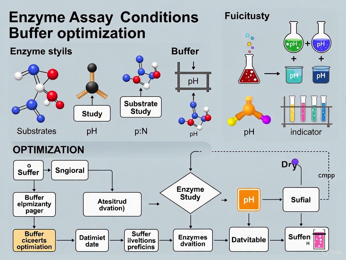
Optimizing Buffer Conditions: A Systematic Methodology
The traditional "one-factor-at-a-time" (OFAT) approach to assay optimization is inefficient and often fails to detect critical interactions between variables. In contrast, Design of Experiments (DoE) is a statistical methodology that allows for the simultaneous variation of multiple factors, enabling researchers to identify optimal conditions and understand complex interactions with fewer experiments [5] [14].
For instance, using a DoE approach, the process of identifying significant factors and optimal assay conditions for an enzyme like human rhinovirus-3C protease can be reduced to less than three days, compared to over 12 weeks with the OFAT approach [5]. The following workflow outlines a typical DoE process for buffer optimization.
Experimental Workflow for DoE-based Buffer Optimization
Key Buffer Factors to Optimize
When designing your experiment, consider these critical buffer-related factors and their interactions:
| Factor | Description | Optimization Consideration |
|---|---|---|
| Buffer Type & pKa | The chemical system (e.g., Phosphate, Tris, HEPES). | Select a buffer with a pKa within ±1 unit of your target pH [13] [10]. |
| pH | The specific hydrogen ion concentration. | Center your experimental range around the suspected enzyme optimum. |
| Ionic Strength | The concentration of ions in solution. | Optimize to balance enzyme-substrate interactions and avoid excessive current/heating [12] [10]. |
| Cofactor Concentration | Essential ions or molecules (e.g., Mg²⁺, NADH). | Determine the concentration required for maximal activity without inhibition. |
| Additives | Components like detergents or reducing agents. | Test for their necessity in stabilizing enzyme activity. |
The Scientist's Toolkit: Essential Reagents and Materials
| Item | Function | Example/Note |
|---|---|---|
| Universal Assay Kits | Homogeneous, mix-and-read assays for common products (e.g., ADP, SAH). | Simplifies HTS; platforms like Transcreener can be used for multiple targets [11]. |
| High-Fidelity (HF) Restriction Enzymes | Engineered enzymes with reduced star activity. | Provides more specific cleavage, reducing unwanted side reactions [15]. |
| BSA-free / rAlbumin Buffers | Reaction buffers without bovine serum albumin, using recombinant albumin. | Reduces variability and potential contaminants in enzymatic reactions [15]. |
| Zwitterionic Buffers | Buffers like MOPS and HEPES with positive and negative charges. | Do not pass through biological membranes; often preferred for in vitro assays [13]. |
| Spin Columns for DNA Cleanup | Kits for purifying DNA prior to enzymatic digestion. | Removes contaminants like salts and inhibitors that can affect enzyme efficiency [15]. |
Advanced Theoretical Framework: The Sabatier Principle in Biocatalysis
Recent research on self-sufficient heterogeneous biocatalysts (ssHBs) reveals that enzyme performance can be governed by the Sabatier principle, a concept well-known in heterogeneous catalysis. This principle states that maximum catalytic efficiency is achieved when the binding strength between a substrate (or cofactor) and a catalyst is neither too strong nor too weak. In ssHBs, where enzymes and cofactors are co-immobilized on a support, the binding thermodynamics between the cofactor and the support material create a volcano-shaped activity plot. The activity increases with binding strength until an optimum is reached, after which stronger binding decreases activity by making the cofactor less accessible to the enzyme [16].
This principle highlights the critical role of buffer components like pH and ionic strength, as they directly modulate the electrostatic interactions between cofactors and charged surfaces or polymers within the immobilized system. Optimizing these parameters is therefore not just about maintaining enzyme structure, but also about fine-tuning the thermodynamic availability of reactants [16]. The following diagram illustrates this relationship.
Best Practices for Buffer Preparation and Handling
- pH Meter Care: The pH meter is often the most neglected piece of equipment. Electrodes must be clean, properly filled, and calibrated with fresh buffers that span the pH range of interest. Temperature should be set to ambient during measurement [13].
- Avoiding Overshooting: When adjusting pH, avoid adding an excessive amount of acid or base, which alters the ionic strength. If you overshoot, it is better to discard the solution and start over rather than titrating back and forth [12].
- Documentation: A complete method should specify the electrolyte salt form used, the exact pH adjustment procedure (including the nature and concentration of the acid/base used), and at what point in the preparation the pH was measured (e.g., before or after adding an organic solvent) [12].
Troubleshooting Guide: FAQs on Enzyme Assay Additives
Q1: My enzyme assay shows low or no activity. Could detergent inhibition be the cause, and how can I resolve this?
A: Yes, detergents are a common cause of enzyme inhibition. This can occur if the detergent forms micelles that disrupt the enzyme's structure or binds non-specifically to the active site.
- Solution:
- Clean Up Your DNA/Protein: Salt or other contaminants from purification can inhibit enzymes. Clean up your sample using spin columns to remove inhibitors [17].
- Verify Detergent Concentration: Ensure the detergent concentration is appropriate. Concentrations near or above the critical micellar concentration (CMC) are more likely to cause issues. Reduce the number of detergent units if necessary [17].
- Check for Salt Inhibition: Some enzymes are sensitive to salt. If your detergent preparation or buffer has high salt, dilute the sample or reduce its volume in the reaction (the DNA solution should be no more than 25% of the total reaction volume) [17].
- Try a Different Detergent: Standard detergents like DDM or Foscholine 12 may not be optimal for all proteins. Consider specialized detergents, such as calix[4]arene-based ones, which are designed to stabilize membrane proteins by structuring their membrane domains [18].
Q2: I am observing unexpected bands or smears in my gel after a restriction digest. What additive-related issues could be responsible?
A: This is a common problem often linked to the behavior of enzymes and detergents in the reaction mix.
- Solution:
- Reduce Enzyme Units: A high concentration of enzyme can lead to "star activity" (cleavage at non-specific sites) or cause the enzyme to bind to the DNA, creating a smear. Lower the number of enzyme units in your reaction [17].
- Add SDS to Loading Buffer: If the enzyme is bound to the DNA, adding SDS (0.1–0.5%) to the gel loading buffer can dissociate the enzyme from the DNA, eliminating the smear [17].
- Avoid Excessive Glycerol: If the enzyme storage buffer contains glycerol, ensure its final concentration in the reaction does not exceed 5% v/v. High glycerol can promote star activity [17].
- Use High-Fidelity (HF) Enzymes: Where possible, use engineered HF restriction enzymes that are designed to eliminate star activity [17].
Q3: How can I stabilize my enzyme to maintain its activity during storage and the assay?
A: Enzyme instability can result from denaturation, aggregation, or proteolytic degradation.
- Solution:
- Add Stabilizers: Incorporate stabilizers like calcium ions (Ca²⁺), polyols (e.g., glycerol, sorbitol), and boric acid into your storage and assay buffers. These help maintain the enzyme's native structure [19] [20].
- Use Reducing Agents: For enzymes with critical cysteine residues, adding reducing agents like Dithiothreitol (DTT) or β-mercaptoethanol (BME) can prevent the formation of incorrect disulfide bonds, maintaining activity [18].
- Optimize Buffer Conditions: pH and ionic strength dramatically affect stability. Systematically optimize the buffer composition using approaches like Design of Experiments (DoE) to find conditions that maximize stability [5].
- Consider Encapsulation: In detergent formulations, enzymes are sometimes encapsulated to protect them from harsh conditions until the assay begins. This strategy can be adapted for lab assays to improve shelf-life [20].
Q4: My enzymatic reaction progress curve is not linear. How can additives help achieve initial velocity conditions?
A: Non-linear progress curves often mean the reaction is not in the initial velocity phase, where less than 10% of the substrate has been consumed. This can be due to enzyme instability, product inhibition, or substrate depletion [21].
- Solution:
- Include Stabilizers: As in Q3, stabilizers like polyols and specific ions can prevent the time-dependent inactivation of your enzyme, extending the linear phase of the reaction [19].
- Reduce Enzyme Concentration: Lowering the amount of enzyme is one of the most effective ways to extend the period during which initial velocity conditions are met, preventing rapid substrate depletion [21].
- Ensure Cofactor Availability: For enzymes requiring cofactors (e.g., Mg²⁺ for kinases), ensure the buffer contains an adequate and stable supply. Some cofactors may require stabilizers themselves.
Experimental Protocols for Additive Evaluation
Protocol 1: Systematic Optimization of Additive Concentrations Using DoE
This protocol uses a Design of Experiments (DoE) approach to efficiently optimize multiple additives simultaneously, significantly speeding up the process compared to traditional one-factor-at-a-time methods [5].
- Define Objective: Identify the primary response to optimize (e.g., enzyme activity, signal-to-background ratio, Z'-factor for assay robustness).
- Select Factors: Choose the additives and conditions to test (e.g., detergent concentration (0.01-0.1%), stabilizing agent (e.g., glycerol, 1-5%), reducing agent (DTT, 0.1-1 mM)).
- Choose Experimental Design: A fractional factorial design is suitable for initial screening to identify the most influential factors.
- Prepare and Run Experiments: Use a liquid handler to prepare assay plates according to the DoE matrix. Run the enzymatic assay under standard initial velocity conditions [21].
- Analyze Data: Fit the data to a model to determine the effect of each factor and their interactions. The model will predict the optimal concentrations of each additive.
- Validate: Run a confirmation experiment using the predicted optimal conditions to verify the improvement.
Protocol 2: Testing Detergent Efficacy for Membrane Protein Extraction and Stabilization
This protocol is adapted from methods used to evaluate novel calix[4]arene-based detergents [18].
- Membrane Preparation: Prepare membrane fractions containing the target protein.
- Solubilization: Incubate membranes (at 2 mg/ml protein concentration) with the test detergent (at a detergent:protein ratio of 5:1 w/w) for 2 hours at 4°C.
- Separation: Centrifuge at 100,000 x g for 1 hour to separate solubilized proteins (supernatant) from insoluble material (pellet).
- Analysis:
- Extraction Efficiency: Analyze the supernatant and pellet fractions by SDS-PAGE and Western blot to determine the amount of target protein extracted.
- Functional Assay: Measure the activity of the solubilized protein. For example, for a transporter like BmrA, measure ATPase activity in a reaction containing 50 mM Tris-HCl pH 8.0, 50 mM NaCl, 5 mM ATP, and 5 mM MgCl₂, incubated for 20 minutes at 37°C [18]. Compare the activity retained after extraction with different detergents.
Data Presentation: Additive Properties and Functions
Table 1: Common Stabilizers and Reducing Agents in Enzyme Assays
| Additive | Typical Concentration Range | Primary Function | Key Considerations |
|---|---|---|---|
| Glycerol | 5-25% (v/v) | Stabilizer: Prevents denaturation by reducing molecular motion and forming protective hydrogen bonds [19]. | High viscosity may affect pipetting accuracy and reaction kinetics. |
| BSA/rAlbumin | 0.1-1.0 mg/mL | Stabilizer: Binds to surfaces to prevent enzyme adsorption; can scavenge contaminants. | May interfere with some detection methods. Note: some vendors are switching to recombinant albumin (rAlbumin) [17]. |
| DTT (Dithiothreitol) | 0.5-1.0 mM | Reducing Agent: Maintains cysteine residues in a reduced state, preventing incorrect disulfide bond formation [18]. | Unstable in aqueous solution over time; make fresh solutions frequently. |
| β-Mercaptoethanol | 1-10 mM | Reducing Agent: Alternative to DTT for keeping sulfhydryl groups reduced. | Less efficient and more volatile than DTT. |
Table 2: Types of Detergents and Their Applications
| Detergent Type | Examples | Common Applications | Mechanism & Notes |
|---|---|---|---|
| Ionic(Anionic/Cationic) | SDS, Foscholine 12 (FC12) | Strong denaturation; solubilizing insoluble proteins. | Can disrupt protein structure and cause inactivation. Not suitable for functional assays [18]. |
| Non-Ionic | Dodecyl Maltoside (DDM), Triton X-100 | Solubilizing and stabilizing membrane proteins for functional studies [18]. | Gentler; can preserve native protein structure and activity. |
| Zwitterionic | CHAPS | Solubilizing membrane proteins while maintaining a mild environment. | Contains both positive and negative charges; useful for isoelectric focusing. |
| Specialty | Calix[4]arene-based (C4Cn) | Extracting and stabilizing difficult membrane proteins (e.g., ABC transporters) [18]. | Designed to structure membrane domains via hydrophobic interactions and salt bridges. |
The Scientist's Toolkit: Research Reagent Solutions
Table 3: Essential Reagents for Additive Optimization
| Reagent | Function | Example in Context |
|---|---|---|
| Design of Experiments (DoE) Software | Enables rapid, multivariate optimization of assay conditions (e.g., buffer, additives) by exploring complex parameter interactions, significantly speeding up the process [5]. | Identifying the optimal combination of detergent, glycerol, and pH in less than 3 days, versus 12 weeks with traditional methods [5]. |
| Universal Assay Platforms (e.g., Transcreener) | Homogeneous, "mix-and-read" assays that directly detect universal enzymatic products (e.g., ADP, SAH). They simplify workflow and are highly amenable to automation and HTS [19]. | A single assay platform can be used for multiple targets within an enzyme family (e.g., kinases), simplifying additive optimization across projects. |
| High-Fidelity (HF) Restriction Enzymes | Engineered enzymes that have been modified to eliminate star activity (cleavage at non-canonical sites), providing more reliable and specific digestion [17]. | Essential for achieving clean, specific digests without the need for extensive buffer and additive troubleshooting. |
| Self-Driving Lab Platforms | Integrated systems that use machine learning and automation to autonomously plan and execute experiments, rapidly navigating high-dimensional parameter spaces [22]. | A platform can autonomously determine optimal reaction conditions (pH, T, [cofactors]) for enzymatic catalysis with minimal human intervention [22]. |
Workflow and Decision Diagrams
Additive Selection Decision Guide
Frequently Asked Questions (FAQs)
1. What is the Z'-factor and why is it a critical assay metric? The Z'-factor (Z-prime factor) is a statistical parameter used to assess the quality and robustness of an assay, particularly in high-throughput screening (HTS). It is calculated using only positive and negative control data, providing a quality metric before testing actual samples. A high Z'-factor indicates a robust assay with a good separation band between controls, which is crucial for reliable hit identification in drug discovery [23] [24].
2. How do I calculate the Z'-factor for my enzyme assay? The Z'-factor is defined by the following equation: Z' = 1 - [3(σp + σn) / |μp - μn|] Where:
- μp and μn are the means of the positive and negative controls.
- σp and σn are the standard deviations of the positive and negative controls [24]. This equation evaluates the dynamic range between the controls and the data variation associated with them. You can compute the means and standard deviations from your control data using standard statistical software and then apply this formula [25].
3. My assay's Z'-factor is below 0.5. What should I do? A Z'-factor below 0.5 indicates a marginal or potentially unusable assay for screening purposes. The following table outlines the standard interpretations for Z'-factor values [24]:
| Z'-factor Value | Interpretation |
|---|---|
| 1.0 | Ideal assay (theoretical maximum) |
| 0.5 to 1.0 | Excellent assay |
| 0 to 0.5 | Marginal or "yes/no" type assay |
| < 0 | Assay is not suitable for screening |
If your value is low, you should optimize your assay conditions. Key areas to investigate include:
- Buffer Composition: The choice of buffer can significantly impact enzyme activity, especially for metalloenzymes. For example, Tris-HCl can chelate metal ions, reducing activity, while HEPES often has a lower metal-binding constant and may be preferable [26].
- Reagent Concentrations: Optimize the concentrations of enzyme, substrate, and co-factors.
- Assay Protocol & Technology: Ensure consistency in procedures and check the performance of your detection instrument [23]. Employing systematic optimization methods like Design of Experiments (DoE) can efficiently identify optimal conditions [5].
4. How does buffer choice specifically impact my assay metrics? The buffer is not an inert component and can profoundly affect enzyme activity, thereby influencing key metrics like specific activity and the Z'-factor. This is particularly critical for metalloenzymes, which require metal ion cofactors. Different buffers can chelate metal ions to varying degrees, altering the free metal ion concentration available to the enzyme [26].
- Experimental Evidence: A 2023 study characterized a Mn2+-dependent dioxygenase (BLC23O) in three different buffers (HEPES, Tris-HCl, and Sodium Phosphate). The results showed that the observed catalytic efficiency (kcat/Km) and metal ion dissociation constant (Kd) varied significantly depending on the buffer used. HEPES buffer yielded the greatest catalytic efficiency for this enzyme [26].
- Non-Metalloenzymes: The same study found that the activity of trypsin, a nonmetalloenzyme, was not significantly affected by the different buffers [26]. This highlights the need for buffer optimization for any new enzyme system.
5. What is a "Robust Z'-factor" and when should I use it? The standard Z'-factor can be sensitive to outliers in the control data because it uses the mean and standard deviation. A Robust Z'-factor substitutes the median for the mean and the median absolute deviation (MAD) for the standard deviation [27]. This approach is highly recommended for complex cell-based assays or any data where outliers are a concern, as it provides a more reliable quality assessment that is less sensitive to extreme values [27].
6. What is the difference between the Z-factor and the Z'-factor? These two related statistics are often confused. The key distinction lies in the data used for their calculation, as summarized below [23]:
| Parameter | Data Used | Situation | Evaluates |
|---|---|---|---|
| Z'-factor | Positive and negative controls only | Assay development and validation, before sample testing | The inherent quality and potential of the assay format |
| Z-factor | Includes test samples and a control | During or after a screening run | The actual performance of the assay with test compounds |
Troubleshooting Guides
Problem: Low or Negative Z'-factor
Potential Causes and Solutions:
Cause: High variability in positive or negative control signals.
- Solution:
- Ensure reagents are fresh and prepared consistently.
- Check pipette calibration and technique for accuracy.
- Verify that your detection instrument (e.g., microplate reader) is functioning properly with low noise and consistent performance across wells [23].
- Use a robust Z'-factor (based on median and MAD) if outliers are skewing your results [27].
- Solution:
Cause: Insufficient dynamic range (small difference between positive and negative control means).
- Solution:
- Re-optimize the concentration of your enzyme to increase the signal from the positive control.
- Ensure your negative control fully inhibits the enzyme reaction or provides a genuine background signal.
- Investigate if the buffer system is inhibiting enzyme activity, particularly for metalloenzymes. Switch from a chelating buffer like Tris-HCl to a low metal-binding buffer like HEPES and re-test [26].
- Solution:
Cause: Suboptimal assay conditions.
- Solution: Systematically optimize all assay components. Instead of the traditional, inefficient "one-factor-at-a-time" (OFAT) approach, use a Design of Experiments (DoE) methodology. DoE allows you to efficiently identify critical factors (like buffer type, pH, substrate concentration) and their interactions, leading to a robust assay with a high Z'-factor in less time [5] [14].
Problem: Inconsistent Specific Activity Measurements
Potential Causes and Solutions:
Cause: Uncontrolled or fluctuating buffer pH and composition.
- Solution:
- Always accurately prepare buffers and measure the pH at the temperature your assay will be run.
- Understand the properties of your buffer. For metalloenzymes, avoid strong metal-chelating buffers unless the metal-chelate complex is the intended substrate.
- Refer to the table below for common buffer properties.
- Solution:
Cause: Inaccurate determination of enzyme concentration.
- Solution: Use a validated protein quantification method (e.g., Bradford, BCA assay) and ensure the enzyme preparation is pure and stable.
Research Reagent Solutions
The following table lists key reagents and their critical functions in enzyme assay development and optimization.
| Reagent | Function in Assay |
|---|---|
| HEPES Buffer | A zwitterionic buffer with a physiological pKa and, crucially, a low constant for metal ion binding. It is often the preferred choice for assays involving metalloenzymes to avoid chelation of essential cofactors [26]. |
| Tris-HCl Buffer | A common primary amine buffer with a physiological buffering range. Its amino group can chelate metal ions, which may inhibit metalloenzyme activity. Its pH is also sensitive to temperature [26]. |
| Sodium Phosphate Buffer | An inorganic buffer that mimics extracellular environments. It can interact with and precipitate certain di- and trivalent metal ions (e.g., Ca²⁺), potentially interfering with metalloenzyme function [26]. |
| Positive Control Compound | A compound known to elicit a maximum response in the assay (e.g., a potent inhibitor for an inhibition assay). It defines the upper or lower bound of your assay window for Z'-factor calculation [23] [24]. |
| Negative Control Compound | A compound known to elicit a minimum response (e.g., a vehicle or blank solution). It defines the opposite bound of your assay window for Z'-factor calculation [23] [24]. |
Experimental Protocols
Detailed Methodology: Investigating Buffer Impact on a Metalloenzyme
This protocol is adapted from a 2023 study investigating buffer effects on metal-dependent enzymes [26].
1. Principle: To determine whether and how different buffer systems influence the kinetic parameters and metal binding of a metalloenzyme, using a catechol dioxygenase (BLC23O) as an example.
2. Reagents:
- Purified metalloenzyme (e.g., BLC23O)
- Substrate (e.g., 3-methylcatechol)
- Metal salt (e.g., MnCl₂ for BLC23O)
- Buffer salts: HEPES, Tris-HCl, Sodium Phosphate
- Equipment for UV-Vis spectroscopy or other suitable detection methods
3. Procedure:
- Step 1: Prepare Assay Buffers. Prepare three separate assay buffers (e.g., HEPES, Tris-HCl, Sodium Phosphate) at the same pH and ionic strength.
- Step 2: Determine Optimal pH and Temperature. For a new enzyme, first identify the optimal pH and temperature in one buffer system.
- Step 3: Determine Metal Ion Kd. In each buffer, prepare a series of reactions containing the enzyme and a range of metal ion concentrations. Measure the initial reaction rates and plot them against metal concentration to determine the dissociation constant (Kd) for each buffer.
- Step 4: Determine Kinetic Parameters. In each buffer, with saturating metal ions, perform Michaelis-Menten kinetics experiments. Measure initial velocities at various substrate concentrations.
- Step 5: Data Analysis. Fit the kinetic data to non-linear regression models to determine the Km (Michaelis constant) and kcat (turnover number) for the enzyme in each buffer system. Compare the catalytic efficiency (kcat/Km) across buffers.
4. Key Experimental Parameters from Literature: The table below summarizes the different kinetic parameters obtained for the Mn2+-dependent enzyme BLC23O in three buffer systems, demonstrating the practical impact of buffer choice [26].
| Buffer | kcat (s⁻¹) | Km (mM) | kcat/Km (mM⁻¹ s⁻¹) | Kd (Mn²⁺) |
|---|---|---|---|---|
| HEPES | 0.45 ± 0.01 | 0.54 ± 0.02 | 0.84 ± 0.02 | 1.49 ± 0.05 µM |
| Tris-HCl | Data not fully reported in excerpt, but stated as less efficient than HEPES. | |||
| Sodium Phosphate | Data not fully reported in excerpt, but stated as less efficient than HEPES. |
Diagnostic Diagrams
Z Prime Factor Concept
Assay Optimization Workflow
Modern Methodologies for Efficient Assay Development and Optimization
Implementing Design of Experiments (DoE) for Multi-Factor Optimization
Troubleshooting Common DoE Implementation Issues
FAQ: My initial experimental runs show high variability, making it difficult to identify significant factors. What should I do?
High variability in initial runs often stems from poorly controlled factor levels or inadequate understanding of the system. First, conduct a robustness test by running the same experimental condition multiple times to quantify inherent process variability. Use this information to determine if your measurement system is sufficiently precise. Second, ensure you are controlling for environmental factors like temperature fluctuations or reagent lot variations that can introduce noise. Consider applying a screening design like a fractional factorial approach to first identify the most influential factors from a larger set before proceeding to full optimization. This method allowed researchers to identify factors significantly affecting human rhinovirus-3C protease activity in less than three days, dramatically speeding up the optimization process [5].
FAQ: How do I handle multiple responses with conflicting optimal conditions?
This common challenge requires multi-objective optimization. For enzyme assays, you may need to maximize activity while minimizing cost or substrate consumption. The most effective approach involves using Response Surface Methodology (RSM) to model each response, then applying a desirability function or optimization algorithm to find a compromise. In other fields, such as 3D printing, researchers have successfully used the Non-dominated Sorting Genetic Algorithm II (NSGA-II) to handle conflicting objectives like simultaneously maximizing tensile strength and elastic modulus [28]. For enzymatic cascade reactions where different enzymes require distinct pH conditions, innovative solutions like biomolecular condensates that create localized pH environments have shown promise [29].
FAQ: My model shows significant lack-of-fit. What steps should I take?
Significant lack-of-fit indicates your model doesn't adequately represent the underlying process. First, verify you haven't omitted important factors or interactions. Second, consider whether transformation of your response variable might improve fit. Third, assess if adding higher-order terms (quadratic or cubic) would better capture curvature in your response surface. If these steps don't resolve the issue, you may need to augment your design with additional experimental runs. Machine learning approaches like Random Forest regression have demonstrated 40% better predictive capability (R²) on test data compared to traditional RSM when dealing with complex, non-linear responses [28].
FAQ: How can I accelerate the traditionally time-consuming process of enzyme assay optimization?
Traditional one-factor-at-a-time optimization can take more than 12 weeks for enzyme assays [5]. To accelerate this process, consider these approaches:
- Adopt fractional factorial designs to reduce the number of initial experimental runs while still capturing main effects and important interactions [5].
- Implement machine learning-driven platforms that can autonomously determine optimal reaction conditions with minimal experimental effort. One study demonstrated optimization in a five-dimensional design space across multiple enzyme-substrate pairings through over 10,000 simulated optimization campaigns [22].
- Utilize universal assay platforms with mix-and-read formats that simplify automation and produce robust results, reducing development time for new targets [30].
Table: Comparison of Traditional vs. Advanced DoE Approaches for Enzyme Assay Optimization
| Approach | Time Requirement | Factors Typically Optimized | Key Advantages |
|---|---|---|---|
| Traditional One-Factor-at-a-Time | >12 weeks [5] | pH, temperature, substrate concentration | Simple to implement, intuitive |
| Fractional Factorial with RSM | ~3 days for initial optimization [5] | Buffer composition, enzyme concentration, substrate concentration | Captures interactions, efficient |
| Machine Learning-Driven Self-Driving Labs | Significantly accelerated [22] | pH, temperature, cosubstrate concentration, ionic strength | Autonomous, handles high-dimensional spaces |
| Universal Assay Platforms | Reduced development time [30] | Multiple enzyme targets with same detection method | Broad applicability, simplified workflows |
Experimental Design and Optimization Workflows
The following workflow diagrams illustrate proven methodologies for implementing DoE in enzyme assay optimization.
Screening and Optimization Workflow
Machine Learning-Enhanced DoE Workflow
Detailed Experimental Protocols
Protocol 1: Fractional Factorial Screening Design for Enzyme Assay Optimization
Purpose: To efficiently identify the most significant factors affecting enzyme activity from a larger set of potential variables.
Materials:
- Purified enzyme (e.g., human rhinovirus-3C protease) [5]
- Substrate solution
- Assay buffer components
- Plate reader capable of measuring enzyme activity
Procedure:
- Factor Selection: Identify 5-7 potential factors that may influence enzyme activity (e.g., buffer pH, ionic strength, enzyme concentration, substrate concentration, temperature, cofactors).
- Experimental Design: Create a Resolution IV fractional factorial design that maintains the ability to detect main effects and two-factor interactions. For 6 factors, this typically requires 16-20 experimental runs instead of the 64 required for a full factorial design.
- Randomization: Randomize the run order to minimize confounding from systematic errors.
- Execution: Perform experiments according to the design matrix, measuring initial reaction rates as the response variable.
- Statistical Analysis: Conduct ANOVA to identify significant factors (p < 0.05). Use Pareto charts to visualize factor importance.
- Follow-up: Select the 3-4 most significant factors for further optimization using Response Surface Methodology.
Troubleshooting Note: If no factors show statistical significance, ensure your factor levels are sufficiently spaced (e.g., pH 6 vs 8 rather than 7.0 vs 7.2) to detect effects above background noise [5].
Protocol 2: Response Surface Methodology for Buffer Condition Optimization
Purpose: To model the relationship between key factors and enzyme activity, and identify optimal conditions.
Materials:
- Significant factors identified from screening design
- Central Composite Design (CCD) or Box-Behnken design template
- Statistical software for RSM analysis
Procedure:
- Design Selection: For 3 factors, select a Box-Behnken design (15 runs) or Central Composite Design (20 runs) depending on your need for predicting response at extremes.
- Experimental Runs: Execute the design in randomized order, measuring enzyme activity as the response.
- Model Fitting: Fit a quadratic model to the data: Y = β₀ + ΣβᵢXᵢ + ΣβᵢᵢXᵢ² + ΣβᵢⱼXᵢXⱼ
- Model Validation: Check for lack-of-fit and R² values. The model should explain at least 80% of variability (R² > 0.8).
- Optimization: Use the fitted model to locate optimal conditions through canonical analysis or desirability functions.
- Verification: Conduct confirmation experiments at the predicted optimum to validate model accuracy.
Technical Note: Researchers have successfully applied this approach to reduce enzyme assay optimization time from >12 weeks to under 3 days while providing more comprehensive factor interaction information [5].
Research Reagent Solutions for DoE Implementation
Table: Essential Reagents and Materials for Enzyme Assay DoE Studies
| Reagent/Material | Function in DoE | Application Notes |
|---|---|---|
| Universal Assay Platforms (e.g., Transcreener) [30] | Detects common enzymatic products (e.g., ADP) | Enables study of multiple targets within enzyme families with same detection method |
| Environmentally-Sensitive Dyes (e.g., PRODAN) [29] | Measures local environment properties within experimental systems | Confirmed condensates are less polar than water (comparable to isopropanol) |
| Biomolecular Condensate Forming Constructs (e.g., Laf1-BTL2-Laf1) [29] | Creates localized reaction environments with distinct properties | Increases enzymatic activity 3-fold by stabilizing open, active conformation |
| RGG Intrinsically Disordered Region [29] | Drives phase separation in chimeric enzyme constructs | Enables formation of enzymatic condensates with concentration factors up to 73,000X |
| Plate Readers with Multiple Detection Modes [30] | Measures various assay outputs (fluorescence intensity, polarization, TR-FRET) | Supports multiple detection methods for assay development flexibility |
Advanced Applications and Integration Strategies
FAQ: How can I implement DoE with limited experimental resources?
With limited resources, focus on definitive screening designs that can evaluate 6-12 factors with just 13-25 runs. These designs efficiently separate main effects from two-factor interactions while requiring fewer runs than traditional fractional factorial designs. Additionally, leverage universal assay platforms that can be applied across multiple enzyme targets with minimal re-optimization, significantly reducing development time for new targets [30].
FAQ: What emerging technologies can enhance traditional DoE approaches?
Machine learning-powered self-driving laboratories represent the cutting edge in experimental optimization. These systems integrate automated liquid handling, real-time analytics, and AI-driven experimental planning to rapidly navigate complex parameter spaces. One platform demonstrated accelerated optimization of enzymatic reaction conditions in a five-dimensional design space across multiple enzyme-substrate pairings [22]. These systems can autonomously determine optimal reaction conditions with minimal experimental effort and without human intervention.
Biomolecular condensates offer another innovative approach, creating localized environments that can optimize enzymatic reactions. Research has shown these condensates can generate distinct pH environments compared to the surrounding solution, maintaining high enzymatic activity even in suboptimal bulk solution conditions. This capability enables cascade reactions involving multiple enzymes with different optimal pH requirements [29].
In the field of enzyme assay development, optimizing buffer conditions is a critical but time-consuming process that can dictate the success of downstream drug discovery efforts. Traditional One-Factor-at-a-Time (OFAT) approaches, while straightforward, often require months of iterative experimentation and can miss critical interaction effects between variables. In contrast, statistical Design of Experiments (DoE) methodologies can compress this timeline to days by systematically exploring multiple factors simultaneously. This technical support article demonstrates how implementing DoE can dramatically accelerate optimization while providing more robust, reproducible assay conditions suitable for high-throughput screening (HTS) environments.
For researchers facing pressure to accelerate preclinical timelines, this paradigm shift from OFAT to DoE represents more than just a technical improvement—it enables faster candidate progression while ensuring data quality. The following sections provide practical guidance, troubleshooting advice, and illustrative case studies to facilitate adoption of DoE methodologies in your enzymatic assay workflow.
Understanding the Fundamental Differences: OFAT vs. DoE
What are the core methodological differences between OFAT and DoE approaches?
OFAT (One-Factor-at-a-Time) methodology involves varying a single factor while keeping all other parameters constant. This sequential approach tests factors in isolation, requiring numerous experimental cycles. A typical OFAT optimization of five factors at three levels each would necessitate 3⁵ = 243 experiments, consuming significant time and resources [31].
DoE (Design of Experiments) employs statistical principles to vary multiple factors simultaneously according to a predefined experimental matrix. This approach directly captures factor interactions—how the effect of one factor depends on the level of another—with dramatically fewer experiments. A screening DoE for five factors might require only 16-20 experiments to identify critical factors [14].
Table: Fundamental Methodological Comparison
| Characteristic | OFAT Approach | DoE Approach |
|---|---|---|
| Experimental Strategy | Sequential variation of single factors | Simultaneous variation of multiple factors |
| Factor Interactions | Cannot detect or quantify | Explicitly models and quantifies |
| Experimental Efficiency | Low (exponential growth with factors) | High (polynomial growth with factors) |
| Time Requirement | Typically months for complex systems | Often days to weeks |
| Statistical Rigor | Limited, prone to local optima | High, with defined confidence intervals |
| Resource Consumption | High (reagents, personnel time) | Optimized for minimal resource use |
Why does DoE typically identify different optimal conditions than OFAT?
DoE captures interaction effects that OFAT inherently misses. In enzymatic systems, factors like pH, ionic strength, and cofactor concentrations frequently interact. For example, the optimal pH for enzyme activity may shift at different magnesium concentrations. OFAT would fix magnesium at one level while optimizing pH, potentially identifying a local optimum that isn't robust across the full operational range. DoE directly models these interactions, leading to more robust and often different optimal conditions [14].
Case Study: Quantitative Comparison in Cellulase Production Optimization
What tangible benefits has DoE demonstrated in real enzyme optimization projects?
A 2025 study on cellulase production from Enterococcus faecium and Stutzerimonas stutzeri provides compelling quantitative evidence of DoE's advantages. Researchers compared OFAT and Response Surface Methodology (RSM, a DoE technique) for optimizing carboxymethyl cellulase (CMCase) production [32].
Table: Optimization Results Comparison for Cellulase Production
| Optimization Method | E. faecium CMCase Activity (U/mL) | Improvement Factor | S. stutzeri CMCase Activity (U/mL) | Improvement Factor |
|---|---|---|---|---|
| Unoptimized Conditions | 8.22 | 1.00x | 11.05 | 1.00x |
| After OFAT Optimization | 14.92 | 1.81x | 19.64 | 1.78x |
| After RSM DoE Optimization | 20.40 | 2.43x | 24.08 | 2.18x |
The DoE approach not only achieved higher final enzyme activity but also identified critical interaction effects between factors like incubation temperature and pH that OFAT had missed. This resulted in a 2.43-fold improvement for E. faecium compared to the 1.81-fold improvement with OFAT alone [32].
Experimental Protocols and Implementation Guide
What is a standard workflow for implementing DoE in enzyme assay development?
How do I select the appropriate DoE design for my enzyme assay?
The choice of DoE design depends on your specific optimization goals and the number of factors being investigated:
- Screening Designs (Plackett-Burman): Ideal for initial phase when investigating 5+ factors to identify the most influential ones with minimal experiments [31].
- Response Surface Designs (Central Composite, Box-Behnken): Used for optimization after screening, typically with 2-4 critical factors, to model curvature and locate optima [33] [14].
- D-Optimal Designs: Computer-generated for constrained design spaces or when traditional designs are inefficient [14].
For most enzyme assay optimizations involving 3-5 factors, a Central Composite Design (CCD) provides excellent balance between efficiency and information gain. A typical CCD for 4 factors requires 25-30 experiments versus 3⁴ = 81 for full factorial [14].
What are common pitfalls when transitioning from OFAT to DoE?
- Insufficient factor range definition: Testing factors over too narrow a range prevents finding the true optimum. Solution: Conduct preliminary range-finding experiments.
- Ignoring randomization: Running experiments in systematic order introduces bias from temporal drift. Solution: Always randomize run order [14].
- Overlooking model diagnostics: Relying solely on R² without checking residuals or prediction statistics. Solution: Use multiple model quality metrics including Q² [14].
- Inadequate replication: Without center point replicates, you cannot distinguish signal from noise. Solution: Include 3-5 center point replicates to estimate pure error [14].
Advanced Methodologies: Machine Learning and Autonomous Optimization
How are emerging technologies further accelerating optimization?
Machine learning (ML) and autonomous laboratories represent the next frontier in optimization technology. Recent advances include:
ML-Driven Self-Driving Labs: A 2025 study demonstrated a platform that autonomously optimized enzymatic reaction conditions in a 5-dimensional parameter space using Bayesian Optimization. This approach conducted over 10,000 simulated optimization campaigns to identify optimal algorithms, then executed real experiments with minimal human intervention [22].
Deep Learning for Kinetic Prediction: The CataPro model uses deep learning to predict enzyme kinetic parameters (kcat, Km) from sequence and substrate information, enabling in silico pre-screening of promising enzyme variants before experimental validation [34].
These technologies can reduce optimization timelines from days to hours while handling higher-dimensional spaces than traditional DoE, though they require significant computational infrastructure and specialized expertise.
Essential Reagent Solutions for Enzyme Assay Development
What key reagents and tools are essential for implementing DoE in enzyme assays?
Table: Key Research Reagent Solutions for DoE Implementation
| Reagent/Tool Category | Specific Examples | Function in DoE Optimization |
|---|---|---|
| Universal Assay Platforms | Transcreener ADP² Assay, AptaFluor SAH Assay | Enables broad target screening with minimal redevelopment; uses mix-and-read format for HTS compatibility [35] |
| Detection Reagents | Fluorescent antibodies, TR-FRET tracers, luminescent substrates | Provides sensitive signal generation across diverse enzyme classes and conditions [35] |
| Buffer Component Libraries | pH buffers, salt solutions, cofactors, detergents | Enables systematic variation of chemical environment factors in DoE matrices |
| Statistical Software | MODDE, Design-Expert, JMP | Facilitates experimental design generation, data analysis, and model visualization [14] |
| Automation Equipment | Liquid handlers, plate readers, robotic arms | Enables precise execution of DoE experimental matrices with minimal manual error [22] |
Frequently Asked Questions (FAQ)
We have limited enzyme supplies. Can DoE work with resource constraints?
Yes, DoE is particularly valuable under resource constraints. Screening designs like Plackett-Burman can evaluate 7-11 factors with only 12-20 experiments, dramatically reducing reagent consumption compared to OFAT. Additionally, modern microfluidic platforms and nanoliter-scale reactions enable DoE with minimal material [22].
How do I handle categorical factors (e.g., buffer types, substrate choices) in DoE?
Modern DoE software handles mixed categorical and continuous factors effectively. For example, you can simultaneously optimize categorical factors like buffer system (HEPES vs. Tris) and continuous factors like pH and ionic strength. D-optimal designs are particularly suited for these scenarios [14].
Our lab has minimal statistics expertise. Is DoE still feasible?
Yes. User-friendly DoE software has made implementation accessible to non-statisticians. These platforms provide guided workflows for design creation, automated analysis, and visual interpretation of results. Additionally, many core facilities and CROs offer DoE support services [14].
How do we validate that DoE-identified conditions are truly optimal?
Always include confirmation experiments in your validation. Run the predicted optimal conditions alongside your original baseline and a condition your team would have selected using OFAT. Additionally, test robustness around the optimum by slightly varying critical factors to ensure performance doesn't degrade rapidly [14].
Can DoE help with enzyme cascade reactions where multiple enzymes have different optimal conditions?
Absolutely. This is a particular strength of DoE. A 2025 study demonstrated that biomolecular condensates could optimize cascade reactions by creating local environments with different pH values suitable for different enzymes. Similarly, DoE can find compromise conditions that maximize overall cascade efficiency despite individual enzyme preferences [29].
Leveraging Progress Curve Simulations for Inhibitor Screening Assays
FAQs: Addressing Common Challenges in Progress Curve Analysis
Q1: Why do my progress curves for a covalent inhibitor not reach a clear steady state, making data fitting difficult?
This is often due to the inhibitor's slow reaction kinetics. For time-dependent inhibitors, the establishment of the final equilibrium between the enzyme and the covalently bound complex can be slow. If the assay duration is too short, you may only capture the initial transition phase rather than the final steady state, which is essential for accurate determination of the inactivation constant (KI) and the reaction rate constant (kinact). Ensure your assay is optimized to run long enough to observe the final linear phase of the reaction, which reflects the established equilibrium [36] [37].
Q2: How can I distinguish between a slow-binding reversible inhibitor and a reversible covalent inhibitor from a progress curve?
Both can show time-dependent inhibition, but the underlying mechanisms differ. A continuous assay that monitors product formation in real-time is key. The progress curve for a slow-binding reversible inhibitor will typically show a characteristic "curve" as it transitions from the initial velocity to the final steady-state velocity. For a reversible covalent inhibitor, the same shape may be observed, but complete characterization requires methods that can dissect the individual inhibition and rate constants (Ki, k5, k6). Techniques like incubation time-dependent IC50 analysis or specialized fitting methods (e.g., EPIC-CoRe) are needed to fully characterize the reversible covalent mechanism [36].
Q3: My high-throughput screen identified a hit, but the IC50 value seems to change when I re-test it. What could be the cause?
A single IC50 value for a time-dependent inhibitor can be highly misleading and is strongly dependent on the specific assay conditions, particularly the pre-incubation and incubation times. An IC50 value obtained after a short incubation may reflect only the initial non-covalent binding (Ki), while a value from a longer incubation may be closer to the overall affinity ( ). Always report the IC50 value along with the exact assay timeline (pre-incubation and incubation durations) and, for meaningful structure-activity relationships, strive to determine the full kinetic profile (KI and kinact for irreversible; Ki, k5, k6 for reversible covalent) instead of relying on a single time-point [37].
Q4: What are the critical buffer conditions to optimize for a robust progress curve assay?
The choice of buffer is fundamental for enzyme stability and activity. Key factors include:
- pH: Use a buffer with a pKa within one unit of your enzyme's optimal pH to ensure strong buffering capacity [38].
- Stability: Select a buffer that maintains a constant pH throughout the assay duration, despite metabolic reactions or temperature shifts. HEPES and MOPS are known for good stability [38].
- Compatibility: Be aware of potential inhibitory interactions. For example, phosphate buffers can inhibit some kinases, and Tris can chelate metal ions [38].
- Ionic Strength: The salt concentration can alter enzyme conformation and function, so it must be optimized and kept consistent [38].
Troubleshooting Guides
Guide 1: Diagnosing Abnormal Progress Curve Shapes
The shape of your progress curve is a rich source of diagnostic information. The table below outlines common anomalies and their potential causes.
Table 1: Troubleshooting Abnormal Progress Curves
| Observed Anomaly | Potential Causes | Corrective Actions |
|---|---|---|
| Curve plateaus prematurely, then linear rate decreases | - Substrate depletion.- Enzyme instability or inactivation over time. | - Increase substrate concentration (ensure it remains well above Km).- Add stabilizing agents (e.g., BSA), check buffer pH/composition, or reduce assay time [37]. |
| "Curved" progress curve in uninhibited control | - The assay conditions themselves cause a non-linear signal. This invalidates standard fitting models for inhibition. | - Systematically optimize buffer, enzyme concentration, and substrate to achieve a linear signal for the control before adding inhibitors [5]. |
| High signal noise across all wells | - Unstable fluorescence or absorbance reading.- Inconsistent pipetting or mixing. | - Use a plate reader with temperature control. Ensure thorough mixing after reagent addition. Centrifuge plates before reading to remove bubbles. |
| No inhibition observed with a known covalent inhibitor | - Insufficient pre-incubation time. The covalent bond has not had time to form significantly. | - Increase the pre-incubation time of the enzyme with the inhibitor before adding substrate [37] [39]. |
Guide 2: Optimizing Assay Conditions Using Design of Experiments (DoE)
Traditional "one-factor-at-a-time" (OFAT) optimization is inefficient and can miss critical interactions between factors. DoE is a superior statistical approach for robust assay development [14].
Table 2: Key Factors for DoE in Enzyme Assay Optimization
| Factor | Typical Levels to Test | Reason for Importance |
|---|---|---|
| Buffer pH | e.g., 7.0, 7.5, 8.0 | Drastically affects enzyme activity and stability; must match enzyme's optimal range [38] [14]. |
| Enzyme Concentration | e.g., 5 nM, 10 nM, 20 nM | Too high can mask weak inhibition; too low leads to a poor signal-to-noise ratio. |
| Substrate Concentration | e.g., 0.5x Km, 1x Km, 2x Km | Affects initial velocity and the apparent potency of competitive inhibitors. |
| Pre-incubation Time | e.g., 5 min, 15 min, 30 min | Critical for observing time-dependent inhibition; directly impacts IC50 values [37] [39]. |
| Temperature | e.g., 25°C, 30°C, 37°C | Influences reaction rates and enzyme stability. |
Workflow:
- Screening Design: Use a fractional factorial design (e.g., a 2^k design) to identify which of the many factors have the most significant effects on your assay's signal-to-noise ratio and Z'-factor.
- Optimization Design: For the critical factors (typically 2-4), employ a Response Surface Methodology (RSM) design like a Box-Behnken or Central Composite Design to model curvature and find the optimal robust conditions [14].
- Modeling: The software will generate a model equation (e.g.,
Y = b0 + b1*pH + b2*[Enzyme] + b12*pH*[Enzyme] + b11*pH²...) that predicts your assay's performance (Y) based on the factor settings, allowing you to find the sweet spot [14].
Figure 1: A sequential DoE workflow for assay optimization.
The Scientist's Toolkit: Research Reagent Solutions
Table 3: Essential Reagents and Materials for Progress Curve Assays
| Item | Function / Rationale | Example / Consideration |
|---|---|---|
| Recombinant Enzyme | The target of study. Purity and stability are paramount. | Human KDAC8 produced in E. coli with a His-SUMO tag for purification [39]. |
| Fluorogenic/Chromogenic Substrate | Generates a detectable signal upon enzyme processing. | Boc-Lys(TFA)-AMC for KDAC8, which releases fluorescent AMC upon deacetylation and trypsin cleavage [39]. |
| Homogeneous Assay Beads | Enable label-free, wash-free detection in HTS formats. | Glutathione donor and anti-FLAG acceptor beads used in AlphaLISA to detect fusion precursors [40]. |
| Time-Dependent Inhibitor (Control) | Serves as a positive control for assay validation. | Saxagliptin, a reversible covalent DPPIV inhibitor [36]. Darunavir for HIV-1 protease [40]. |
| DoE Software | Statistically plans efficient experiments and analyzes complex results. | Software like MODDE (Sartorius) or equivalent for designing factorial and response surface experiments [14]. |
| Automated Analysis Workflow | Manages quality control and fitting of large-scale kinetic data. | Platforms like Genedata Screener automate progress curve QC and model selection for non-equilibrium inhibitors [41]. |
Experimental Protocols
Protocol 1: Incubation Time-Dependent IC50 Assay for Reversible Covalent Inhibitors
This protocol is used to characterize time-dependent reversible covalent inhibitors by measuring IC50 at different incubation times without a pre-incubation step [36] [37].
Detailed Methodology:
- Solution Preparation: Prepare a serial dilution of the inhibitor in assay buffer. Pre-dilute the enzyme and substrate to the desired concentrations in the appropriate buffer.
- Reaction Initiation: In a microtiter plate, simultaneously mix the enzyme and inhibitor with the substrate to start the reaction. The final volume and concentration of DMSO should be controlled (e.g., ≤1%).
- Continuous Monitoring: Immediately place the plate in a pre-heated plate reader and initiate continuous measurement of product formation (e.g., fluorescence or absorbance) for an extended period (e.g., 60-90 minutes).
- Data Collection: Collect progress curves for each inhibitor concentration and for uninhibited and background controls.
- Analysis: For each progress curve, fit the initial velocity (vi) or the entire curve to determine the residual enzyme activity at each concentration and time point. Plot % activity vs. inhibitor concentration for each time point to generate a series of IC50(t) values.
- Kinetic Parameter Determination: Use a specialized implicit equation [36] to fit the time-dependent IC50 values and extract the inhibition constants (Ki and ) and the covalent reaction rate constants (k5 and k6).
Figure 2: Workflow for incubation time-dependent IC50 assay.
Protocol 2: Pre-incubation Time-Dependent IC50 Assay for Irreversible Inhibitors
This protocol is used to determine the kinetic parameters KI and kinact for irreversible inhibitors by varying the pre-incubation time of the enzyme with the inhibitor [37] [39].
Detailed Methodology:
- Inhibitor Dilution: Prepare a serial dilution of the irreversible inhibitor in assay buffer.
- Pre-incubation: Mix a fixed concentration of enzyme with each concentration of the inhibitor in a microtiter plate. Incubate the mixture for varying time periods (e.g., 0, 5, 15, 30, 60 minutes) at the assay temperature.
- Reaction Initiation: After each pre-incubation time, initiate the reaction by adding a concentrated substrate solution.
- Endpoint Measurement: Allow the reaction to proceed for a fixed, short period (assay incubation) and then stop it, if necessary. Measure the total product formed.
- Data Analysis: For each pre-incubation time, plot the % residual enzyme activity against the inhibitor concentration and fit a dose-response curve to determine the IC50 value at that time.
- Global Fitting: The resulting IC50 values will decrease with increasing pre-incubation time. Use a global fitting method, such as EPIC-Fit [37], to model the entire dataset of pre-incubation time-dependent IC50 values and determine the apparent second-order rate constant kinact/KI and the individual parameters kinact and KI.
This technical support guide provides troubleshooting and procedural advice for researchers optimizing enzyme assays. Selecting the appropriate detection method—spectrophotometric, fluorometric, or chemiluminescent—is a critical step in assay development that directly impacts data quality, sensitivity, and success in downstream applications like drug discovery. The following FAQs, guides, and tables are designed to help you troubleshoot common issues and implement robust methodologies within the broader context of optimizing enzyme assay buffer conditions.
FAQs and Troubleshooting Guides
FAQ: General Method Selection
1. How do I choose between a spectrophotometric and a fluorometric assay for my enzyme?
Your choice should be guided by your required sensitivity, sample type, and budget.
- Choose Spectrophotometry for routine analyses where analyte concentrations are moderate to high, your budget is constrained, and you need a simple, versatile method with minimal sample preparation [42].
- Choose Fluorometry when you need to detect very low concentrations of analytes (e.g., picomolar to nanomolar levels), are working with limited sample volumes, or require high specificity that can be achieved with fluorescent tags [42] [43].
2. My enzyme assay signal is weak. What should I check first?
First, verify that your assay is operating in the linear range. This is the most critical aspect of assay design for quantitative work [44]. Test serial dilutions of your enzyme to ensure the signal is proportional to the enzyme concentration. A common reason for a weak or non-linear signal is excessive consumption of substrate (typically >15% conversion). Other factors to check include the pH and composition of your assay buffer, the temperature, and potential instability of the enzyme or detection reagents [44].
Troubleshooting Guide: Spectrophotometric Assays
| Problem | Possible Cause | Solution |
|---|---|---|
| Inaccurate DNA/Protein Quantification | Signal overestimation from contaminants (e.g., nucleic acids in protein assays, or residual salts) [45] [43]. | Purify the sample using spin columns or precipitation. Use a fluorometer for more accurate nucleic acid quantification [45]. |
| High Background Signal | Interference from buffer components or other molecules that absorb at the measured wavelength [43]. | Change to a buffer with lower UV absorbance. Include appropriate blanks. Consider switching to a fluorometric method for greater specificity. |
| Signal Outside Linear Range | Enzyme concentration is too high, leading to excessive substrate conversion, or too low to generate a detectable product [44]. | Titrate the enzyme concentration. Ensure the final absorbance reading falls within the linear range of your instrument and the assay (often up to an OD of ~2.5) [44]. |
Troubleshooting Guide: Fluorometric Assays
| Problem | Possible Cause | Solution |
|---|---|---|
| Low Fluorescence Signal | Fluorescent dye is quenched due to environmental factors (e.g., pH, temperature, or solvent) [42]. | Precisely control assay conditions. Check that the dye is compatible with your buffer system. Ensure the excitation/emission wavelengths are set correctly on your instrument. |
| Signal Instability Over Time | Photobleaching of the fluorescent dye or enzyme instability. | Reduce light exposure during the assay. Shorten the time between reagent addition and measurement. Check the enzyme's stability under assay conditions. |
| High Background Fluorescence | Contamination of labware or interference from assay components. | Use high-quality, low-fluorescence labware (plates, tubes). Include control reactions without the enzyme to identify background sources. |
Experimental Protocols
Protocol 1: DNA Quantification from Cytology Smears (Fluorometric vs. Spectrophotometric Comparison)
Background: This protocol, adapted from a 2021 study, is useful for comparing the accuracy of DNA quantification methods when working with challenging sample types like Fine Needle Aspiration Cytology (FNAC) smears, which are relevant for molecular pathology [45].
Materials:
- DNA samples (e.g., from FNAC smears, FFPE sections, whole blood)
- Quantus Fluorometer and compatible assay kit (e.g., Promega)
- UV-Vis Spectrophotometer (e.g., Qiagen QIAxpert)
- Appropriate elution buffers
Method:
- DNA Extraction: Extract DNA from your samples using a standardized kit. For stained cytology smears, this may involve deparaffinization in xylene, decolourisation, and scraping, followed by a modified FFPE DNA extraction protocol [45].
- Fluorometric Quantification:
- Follow the manufacturer's instructions for the fluorometer and DNA quantification kit.
- Briefly, mix a small volume of the eluted DNA with the fluorescent dye.
- Measure the fluorescence and determine the DNA concentration using the instrument's built-in standard curve [45].
- Spectrophotometric Quantification:
- Using the same eluted DNA samples, measure the absorbance in the spectrophotometer.
- Record the absorbance at 260 nm (A260) for DNA concentration and the A260/A280 ratio for purity assessment [45].
- Data Analysis:
- Compare the DNA concentrations (ng/µl) and total yields obtained from both methods. The study showed that spectrophotometry consistently overestimates DNA concentration compared to the more accurate fluorometry [45].
- Use the fluorometric data for downstream applications requiring precise DNA amounts.
Protocol 2: Optimizing an Enzymatic PET-Hydrolysis Assay using HPLC and UV-Spectroscopy
Background: This protocol outlines an improved method for monitoring the hydrolysis of polyethylene terephthalate (PET) by enzymes like leaf-branch compost cutinase (LCC), focusing on accurate quantification of aromatic products [46].
Materials:
- Purified PET-hydrolyzing enzyme (e.g., LCC)
- PET substrate (e.g., film)
- Reaction buffer (e.g., Tris-HCl)
- Authentic standards: Terephthalic acid (TPA), MHET, BHET
- Internal Standard: Caffeine
- HPLC system with a C8 column
- UV-Vis Spectrophotometer or microplate reader
Method:
- Enzymatic Reaction: Incubate the enzyme with PET substrate in an appropriate buffer at the desired temperature with shaking. Withdraw samples at regular time intervals [46].
- HPLC Analysis with Internal Standard:
- Sample Prep: Quench the reaction sample with acetonitrile, centrifuge to filter, add HCl, and add a fixed concentration of caffeine internal standard [46].
- HPLC Run: Inject the sample onto a C8 column. Use a gradient elution with water (0.1% formic acid) and acetonitrile. Detect products at 240 nm [46].
- Quantification: Generate a calibration curve by plotting the ratio of the peak area of each standard (TPA, MHET, BHET) to the peak area of the internal standard against the known concentration. Use this curve to determine the concentration of products in unknown samples [46].
- UV-Spectroscopy Analysis:
- Measure the absorbance of the reaction mixture at 240 nm.
- Use a weighted average of the extinction coefficients for TPA, MHET, and BHET to calculate the total concentration of aromatic products, as this provides a more accurate value than using a single coefficient [46].
- Validation: Cross-validate the results from the UV-spectroscopy method with the more accurate HPLC method to ensure reliability [46].
The Scientist's Toolkit: Research Reagent Solutions
| Item | Function in Enzyme Assays |
|---|---|
| Fluorescent Dyes (e.g., for FP, TR-FRET) | Bind to specific enzymatic products (e.g., ADP, SAH), enabling highly sensitive detection through changes in fluorescence polarization or intensity [47]. |
| Universal Assay Kits (e.g., Transcreener) | Detect common enzymatic products (like ADP for kinases). They offer a "mix-and-read" format, simplifying assay development for multiple targets within an enzyme family and are amenable to high-throughput screening [47]. |
| Internal Standards (e.g., Caffeine for HPLC) | Added in a fixed concentration to samples during preparation. They correct for variations in sample processing and injection volume, significantly improving the accuracy and reproducibility of quantitative analyses [46]. |
| rAlbumin-containing Buffers | Used in place of BSA in restriction enzyme digests and other enzymatic reactions to prevent star activity and stabilize enzymes without introducing animal-sourced contaminants [48]. |
| Spin Columns (for DNA clean-up) | Remove contaminants like salts, proteins, and inhibitors from DNA samples post-extraction or post-PCR, which is critical for preventing enzyme inhibition in downstream reactions like restriction digests [48]. |
Workflow and Decision Pathways
The following diagram outlines a logical workflow for selecting and optimizing a detection method for your enzyme assay.
Decision Workflow for Assay Method Selection
Systematic Troubleshooting for Common Enzyme Assay Pitfalls
Diagnosing and Resolving High Background and Low Signal-to-Noise Ratios
What are the common causes of high background in enzymatic assays?
High background signals can severely compress the dynamic range of detection and reduce the reliability of your data. The following table summarizes the frequent causes and their solutions.
| Cause | Description | Solution |
|---|---|---|
| Inadequate Washing | Incomplete removal of unbound enzyme-conjugated antibodies or other reagents leaves residual signal-generating material in the well [49] [50]. | Implement a rigorous washing protocol: fill wells completely with wash buffer, include a 30-second to 2-minute soak step, and thoroughly remove buffer by flicking and blotting on absorbent paper. Repeat 3-5 times [49]. |
| Ineffective Blocking | Non-specific binding sites on the microplate are not fully covered, allowing assay components to bind indiscriminately [49] [50]. | Use an appropriate blocking agent (e.g., BSA, casein, or commercial stabilizers) at a sufficient concentration. Ensure blocking proceeds for at least 1-2 hours, or even overnight at 4°C for stubborn cases [49]. |
| Substrate Contamination | The detection substrate can degrade upon exposure to light or heat, or become contaminated by metal ions or oxidizing agents, leading to spontaneous signal generation [49]. | Precise control of substrate development time and immediate stopping of the reaction. Store substrate protected from light and use clean, non-metallic containers [49]. |
| Reagent Cross-Reactivity | Antibodies, particularly secondary antibodies, may bind non-specifically to components other than the target, such as Fc receptors or similar epitopes on other proteins [50]. | Perform antibody titration to find the optimal working concentration for the best signal-to-noise ratio. Use cross-adsorbed secondary antibodies and verify antibody specificity to minimize cross-reactivity [49] [50]. |
What experimental protocols can I use to systematically optimize my assay buffer?
Optimizing the buffer environment is one of the most effective ways to improve the signal-to-noise ratio (SNR). A systematic approach is far more efficient than random testing.
A. Systematic Optimization Using Design of Experiments (DoE)
Traditional one-factor-at-a-time (OFAT) optimization can be time-consuming and may miss critical interactions between factors. The Design of Experiments (DoE) approach is a powerful alternative.
- Principle: DoE allows you to vary multiple buffer parameters (e.g., pH, ionic strength, cofactor concentration, detergent type) simultaneously according to a statistical plan. This identifies not only the main effect of each factor but also how they interact to influence the SNR [5].
- Protocol Outline:
- Identify Critical Factors: Select the key parameters you suspect influence your enzyme's activity and background signal (e.g., pH, buffer type, concentration of a cofactor like Mg²⁺, concentration of a stabilizer like BSA).
- Define the Experimental Domain: Set a high and low value for each factor based on literature or preliminary data.
- Run the DoE Array: Use software to generate a set of experimental conditions (a "fractional factorial" design) that efficiently explores the multi-dimensional space [5].
- Analyze Responses: For each condition, run your assay and measure the key output, such as the Z'-factor (a measure of assay robustness) or SNR.
- Build a Response Surface Model: The data is used to create a model that predicts assay performance across all possible combinations of factors, allowing you to pinpoint the optimal buffer composition [5].
- Benefit: This methodology can identify optimal assay conditions in a fraction of the time required by OFAT approaches, sometimes in as little as a few days [5].
B. Protocol: Investigating the Effect of pH and Additives
This is a more focused protocol to characterize your enzyme's behavior.
- Objective: To determine the optimal pH and identify additives that reduce background.
- Materials:
- Purified enzyme
- Substrate
- Series of buffers covering a relevant pH range (e.g., pH 5.0-9.0)
- Potential additives (e.g., BSA, non-ionic detergents like Tween-20, protease inhibitors)
- Microplates and plate reader
- Method:
- Prepare a master reaction mix containing enzyme and substrate.
- For pH profiling: Dispense the mix into wells containing different pre-dispensed buffers. Incubate under standard reaction conditions and measure the signal.
- For additive screening: At the best pH from step 2, run the assay in the presence of different additives. Include control wells without enzyme (for background) and without additive (for baseline).
- Calculate the SNR or Z'-factor for each condition. The Z'-factor is calculated as:
1 - [ (3 * SD_{Sample} + 3 * SD_{Background}) / |Mean_{Sample} - Mean_{Background}| ]. A Z' > 0.5 indicates an excellent assay [51].
The workflow for this systematic optimization is outlined in the diagram below.
How can instrument settings and plate selection affect my signal-to-noise ratio?
The instrument and consumables you use are integral to achieving a high-quality signal.
- Wavelength Accuracy: Ensure your plate reader's wavelength selection is accurate. Using validation plates (e.g., SpectraTest series) can confirm that your instrument is performing to specification [52].
- Detection Mode: For fluorescent assays, using time-resolved fluorescence (TRF) can massively improve SNR. TRF uses lanthanide chelates that emit long-lasting fluorescence, allowing the instrument to delay measurement until short-lived background fluorescence (autofluorescence) has decayed [53].
- Photomultiplier Tube (PMT) Gain: Use the lowest PMT gain that provides a sufficient signal from your positive control. Higher gain can amplify background noise. Some advanced plate readers feature dual-mode PMTs optimized for both low and high signal levels [54].
- Microplate Selection:
- Use Assay Plates, Not Tissue Culture Plates: ELISA and assay plates are designed with high protein-binding capacity and low well-to-well variation (CV <5%) [55]. Tissue culture plates are treated to prevent binding and will give poor results.
- Plate Color: Use clear plates for colorimetric assays, black plates for fluorescent assays to minimize cross-talk, and white plates for luminescent assays to reflect light to the detector [55].
What are some innovative reagent solutions to enhance SNR?
Recent advancements in reagent technology offer powerful tools to combat high background and low signal.
- pH-Sensitive Probes: Probes like the pHSense Eu探针 are inactive at neutral extracellular pH but become highly fluorescent upon being internalized into acidic compartments like endosomes and lysosomes. This "activation-on-event" mechanism inherently provides a very low background and high specific signal [53].
- Advanced Blocking and Stabilizing Reagents: Commercial blocking agents (e.g., StabilGuard, StabilBlock) are specifically formulated to minimize non-specific binding while stabilizing the immobilized proteins, which can extend assay shelf-life and improve consistency [50].
- Universal Detection Assays: Platforms like the Transcreener assays directly detect universal reaction products (e.g., ADP). Because the detection technology is separated from the specific enzyme reaction, it can be highly optimized for SNR and applied across many targets within an enzyme class, saving development time [51].
- Biomolecular Condensates for Local Environment Control: Emerging research shows that engineering biomolecular condensates that sequester enzymes can create a local microenvironment with a distinct pH. This can buffer the enzyme from suboptimal pH in the bulk solution, potentially expanding the usable pH range and increasing enzymatic activity [29].
The Scientist's Toolkit: Key Research Reagent Solutions
| Item | Function | Example Use-Case |
|---|---|---|
| Commercial Blocking Reagents (e.g., StabilGuard, StabilBlock) | Blocks non-specific binding sites on the microplate and in the sample, reducing background [50]. | Essential for all ELISA and bead-based assays to prevent false positive signals. |
| Assay Diluents (e.g., MatrixGuard) | Provides an optimal matrix for diluting samples and reagents, which can help reduce matrix interferences and false positives [50]. | Diluting serum or plasma samples to minimize interference from heterophilic antibodies or other factors. |
| Universal Activity Assays (e.g., Transcreener) | Detects a common product of enzymatic reactions (e.g., ADP, SAH), offering a high-SNR, mix-and-read format for many enzyme classes [51]. | High-throughput screening of kinase, GTPase, or methyltransferase inhibitors. |
| Specialized Substrates (e.g., TMB, CDP-Star) | Provides the substrate for the reporter enzyme (e.g., HRP, AP). Choosing a high-sensitivity substrate can boost signal [50] [55]. | Using a chemiluminescent substrate for superior sensitivity over colorimetric TMB. |
| pH-Sensitive Probes (e.g., pHSense) | Provides a low-background signal that activates only in specific cellular compartments, ideal for internalization studies [53]. | Studying antibody-drug conjugate (ADC) or GPCR ligand internalization into cells. |
| Enzyme-Labeled Antibodies | The conjugate that links antigen binding to signal generation. Optimal concentration is critical for SNR [55]. | Titrating the conjugate to find the concentration that gives the strongest specific signal with the lowest background. |
FAQs on Pipetting and Edge Effects
How do pipetting techniques directly affect my assay's reproducibility?
Using an incorrect pipetting technique for your sample type is a major source of experimental error. Aqueous liquids can be dispensed using the standard forward pipetting technique. However, challenging liquids like viscous solutions (e.g., glycerol), foaming liquids, or very small volumes require reverse pipetting. Using the wrong technique can lead to inaccurate volume delivery, affecting reagent concentration and the final readout. Furthermore, a non-vertical pipetting angle and excessive immersion depth can also reduce precision and accuracy [56].
What exactly is the "Edge Effect" and what causes it?
The edge effect is an assay artefact where wells at the perimeter of a microplate yield different results compared to wells in the center. This phenomenon occurs in all microplate formats (96, 384, and 1536 wells) and is primarily caused by two factors:
- Increased Evaporation: Edge wells experience a higher rate of evaporation during incubation. This leads to a concentration of reagents and salts, which can be harmful to cells or alter biochemical reactions [57] [58] [59].
- Thermal Gradients: Temperature variations across the plate can occur during incubation, particularly in temperature-sensitive assays [58] [59]. This effect is more pronounced in plates with a higher number of wells because they have a lower sample volume per well [58].
How can I identify if the edge effect is impacting my data?
You can identify the edge effect by inspecting your raw data or plate layout for a distinct pattern. Values from wells around the plate's perimeter will be consistently higher or lower than those in the interior, leading to greater standard deviations and unreliable data [58]. In cell culture, this often manifests as variations in cell growth and metabolism between edge and interior wells [58].
My assay has high background. Could this be related to pipetting or plate effects?
Yes, both can be contributing factors. Inconsistent pipetting during washing steps can leave behind residual reagents, leading to high background signal [60]. Similarly, insufficient blocking of the plate can cause high background, and this step requires precise liquid handling [60]. While the edge effect typically causes a gradient of signal from the center to the edge, overall high background is more often linked to pipetting consistency and washing efficiency.
Troubleshooting Guides
Pipetting Technique Guide
| Problem | Cause | Solution |
|---|---|---|
| Low Precision (High CV%) | Inconsistent pipetting angle and speed; variable immersion depth. | Always hold the pipette vertically and use a consistent, smooth pipetting rhythm. Minimize immersion depth [56]. |
| Inaccurate Volumes with Aqueous Solutions | Using reverse pipetting for aqueous buffers. | Use forward pipetting for standard aqueous solutions [56]. |
| Inaccurate Volumes with Viscous or Foaming Liquids | Using forward pipetting for challenging liquids. | Switch to reverse pipetting. This technique aspirates an extra "blow-out" volume to ensure accurate dispensing of the desired volume [56]. |
Strategies to Mitigate the Edge Effect
| Strategy | Protocol / Application | Key Consideration |
|---|---|---|
| Exclude Outer Wells | Do not use the perimeter wells for critical experimental data points. Fill them with water or PBS [57]. | Simple but wasteful; reduces the number of available wells for your assay [57] [58]. |
| Use a Plate Lid or Seal | Use a low-evaporation lid or breathable sterile tape during incubation to reduce evaporation [58]. | Breathable tape is essential for cell-based assays to allow for gas exchange [58]. |
| Control Incubation Conditions | Use a humidified incubator or secondary container to minimize evaporation. Limit the number of times the incubator is opened [57]. | A simple and highly effective method to maintain a uniform environment. |
| Randomize Sample Placement | Use a block randomization scheme to assign treatments across the plate, preventing bias from being confined to the edge wells [57]. | This increases study complexity and often requires automated liquid handling [57]. |
Experimental Protocols for Improved Reproducibility
Protocol 1: Checkerboard Titration for Assay Optimization
This protocol allows you to optimize two key assay parameters (e.g., antibody concentrations) simultaneously [61] [60].
- Plate Setup: Prepare a dilution series of your capture antibody along the x-axis of the plate. Prepare a dilution series of your detection antibody along the y-axis.
- Coating: Apply the different concentrations of capture antibody to the plate according to your layout and incubate.
- Assay Execution: Proceed with your standard assay steps (blocking, sample addition), but apply the different concentrations of detection antibody according to your layout.
- Analysis: Read the plate and identify the combination of antibody concentrations that provides the strongest specific signal with the lowest background [61]. The table below provides general starting points for antibody concentrations.
Table: Recommended Antibody Concentration Ranges for ELISA Optimization [61]
| Antibody Source | Coating Antibody Concentration | Detection Antibody Concentration |
|---|---|---|
| Polyclonal Serum | 5–15 µg/mL | 1–10 µg/mL |
| Crude Ascites | 5–15 µg/mL | 1–10 µg/mL |
| Affinity-Purified Polyclonal | 1–12 µg/mL | 0.5–5 µg/mL |
| Affinity-Purified Monoclonal | 1–12 µg/mL | 0.5–5 µg/mL |
Protocol 2: Testing for and Mitigating the Edge Effect
This protocol helps you confirm the presence of an edge effect and validate a mitigation strategy.
- Control Plate Setup: Seed cells or add a uniform biochemical reaction mixture to every well of a microplate.
- Incubation: Incubate the plate under your standard assay conditions for the full duration.
- Analysis: Measure the output (e.g., absorbance, fluorescence) and plot the values according to their well position. Look for a clear pattern of deviation in the outer wells.
- Mitigation Validation: Repeat the experiment using your chosen mitigation strategy (e.g., a low-evaporation lid and humidified chamber). The data from the validation plate should show a homogeneous signal across all well positions.
The Scientist's Toolkit
Table: Essential Research Reagent Solutions for Robust Assays
| Item | Function |
|---|---|
| Low-Evaporation Lid | Specially designed lids that minimize evaporation from wells while permitting gas exchange for cell cultures [58]. |
| Humidified Secondary Container | A sealed box with a hydrated atmosphere that reduces evaporation from all wells during incubation [57]. |
| Plate Seals and Tapes | Breathable seals for cell culture or airtight seals for biochemical assays create a physical barrier against evaporation [58]. |
| Affinity-Purified Antibodies | Using purified antibodies for immunoassays optimizes the signal-to-noise ratio, reducing background [61]. |
| Universal Assay Platforms | Kits (e.g., Transcreener) that detect common enzymatic products (e.g., ADP) simplify development and provide a robust, mix-and-read format for multiple targets within an enzyme family [62]. |
Workflow Diagrams
Dot Script: Assay Reproducibility Logic
Dot Script: Edge Effect Mitigation
Optimization of Wash Conditions for ELISA and Cell-Based Formats
In both Enzyme-Linked Immunosorbent Assays (ELISA) and cell-based assays, the washing process is a critical procedural step that directly governs the signal-to-noise ratio, impacting assay sensitivity and specificity. Insufficient washing leads to elevated background noise and false positives due to unbound reagents, while overly aggressive washing can disrupt weak antigen-antibody binding or detach sensitive adherent cells, compromising quantitative results. Standardization of the microplate washing protocol is therefore paramount for generating reproducible and reliable laboratory measurements [63]. This guide details the optimization of wash conditions within the broader context of enzymatic assay buffer research, providing troubleshooting and FAQs for researchers and drug development professionals.
Strategic Wash Buffer Selection and Preparation
The wash buffer serves as the primary medium for removing unbound materials, and its composition is crucial for maintaining the stability of bound reagents and cellular structures.
Core Buffer Composition
- Buffer Base: For most immunoassays, phosphate-buffered saline (PBS) or Tris-buffered saline (TBS) forms the base, typically adjusted to a physiological pH of 7.2–7.4 [63].
- Surfactants: The critical optimization factor is the inclusion of surfactants, most commonly TWEEN 20 (Polysorbate 20). This non-ionic detergent reduces surface tension and facilitates the displacement of weakly bound, non-specific proteins from the microplate surface. The optimal concentration is assay-specific but generally ranges from 0.01% to 0.1% [63].
- Ionic Strength: Maintaining physiological ionic strength is essential to prevent osmotic stress in cell-based assays or non-specific electrostatic interactions in ELISA [63].
Additional Formulation Factors
- pH Stability: The buffering capacity must be sufficient to resist changes in pH during the assay procedure. This is particularly important given that enzymatic activity and antibody binding are highly pH-dependent [63].
- Preventing Contamination: Wash buffers should be prepared using high-purity, deionized water and filtered (e.g., through a 0.22 µm filter) to prevent microbial growth or particulate contamination that could interfere with assay results [63].
- Temperature: The use of a slightly warmed wash buffer (e.g., 25-37°C) can improve the removal efficiency of non-specifically bound reagents by influencing binding kinetics [63].
Table 1: Recommended Wash Buffer Compositions for Different Assay Types
| Component | Standard ELISA | Sensitive ELISA | Adherent Cell-Based Assays |
|---|---|---|---|
| Buffer Base | PBS or TBS | PBS or TBS | PBS (with Ca²⁺/Mg²⁺) |
| Typical pH | 7.2 - 7.4 | 7.2 - 7.4 | 7.2 - 7.4 |
| TWEEN 20 | 0.05% | 0.01% - 0.05% | 0.01% (or less) |
| Additives | - | - | 1 mM CaCl₂, 0.5 mM MgCl₂ |
| Key Function | Remove unbound reagent | Minimize gentle disruption | Maintain cell viability & adhesion |
Calibrating Mechanical Washing Parameters
The mechanics of the automated microplate washing process—dispensing volume, flow rate, and the number of cycles—must be precisely controlled to achieve high efficacy without inducing sample loss or cross-contamination.
Dispensing Volume and Soak Time
Dispensing an adequate volume of wash buffer is necessary to ensure complete exchange of the liquid phase within the well. Volumes are typically set to 200-400 µL for a 96-well plate, ensuring the well is filled. Incorporating a soak time of 5-30 seconds after dispensing allows the buffer to interact with the well surface and dislodge weakly bound materials [63].
Flow Rate and Number of Cycles
The flow rate of the dispensed buffer dictates the shear stress applied to the binding surface and is a key differentiator between ELISA and cell-based assays.
- High Flow Rate: Offers efficient mixing and removal but is detrimental to weakly adherent cells or delicate antigen-antibody complexes.
- Low Flow Rate: Minimizes shear stress, making it suitable for sensitive assays, but may require increased cycles or longer soak times to ensure complete reagent exchange.
Most assays benefit from 3 to 5 wash cycles, but this should be optimized empirically. Excessive washing can lead to signal loss, particularly in cell-based formats [63].
Table 2: Optimized Mechanical Parameters for ELISA vs. Cell-Based Assays
| Parameter | ELISA Recommendation | Cell-Based Assay Recommendation | Rationale |
|---|---|---|---|
| Dispense Rate | Medium to High | Low to Medium | Minimize shear stress on cells while maximizing efficiency in ELISA. |
| Soak Time | 5-30 seconds | 1-5 seconds | Longer soak helps dislodge non-specific binding in ELISA. Shorter time minimizes cell stress. |
| Wash Cycles | 3-6 | 2-4 (gentle) | Sufficient for background reduction. Lower cycles preserve cell viability and adhesion. |
| Aspiration | Firm, low residual volume | Very gentle, higher residual volume | Prevents well drying and cell detachment. |
Troubleshooting Common Washing-Related Issues
This section addresses specific problems users might encounter, their potential sources, and recommended corrective actions.
FAQ: What is the ideal residual volume threshold for robust ELISA results?
The single most critical mechanical factor affecting the final assay signal is the residual volume—the volume of liquid remaining in the well after the final aspiration step. High residual volume leads to the dilution of the substrate or detection reagent, resulting in lower signal intensity and increased measurement variability across wells. A residual volume of less than 5 µL is the industry standard target for robust ELISA results. Achieving this requires precise calibration of aspiration depth and speed [63].
High Background Signal
- Possible Cause: Insufficient washing, leading to unbound reagents remaining in the well [64].
- Test or Action:
Poor Duplicates (High Well-to-Well Variation)
- Possible Cause: Insufficient or uneven washing across the plate. This can be due to blocked ports in an automated plate washer or inconsistent aspiration [64].
- Test or Action:
- If using an automatic plate washer, check that all ports are clean and free of obstructions. Perform routine maintenance as per the manufacturer's schedule.
- Add a soak step and rotate the plate halfway through the wash process to ensure uniformity.
- Validate aspiration uniformity across all channels using a dye-dilution method [63] [64].
Low or No Signal
- Possible Cause: Overly aggressive washing, which can elute the target antigen or detection antibody. This is a common issue with newly established protocols where wash conditions are too harsh [63].
- Test or Action:
- Reduce the number of wash cycles.
- Decrease the dispense flow rate and/or aspiration force.
- Shorten or remove the soak time to minimize exposure to the wash buffer.
Poor Assay-to-Assay Reproducibility
- Possible Cause: Inconsistent washing between assay runs due to manual variability or drift in automated washer performance [64].
- Test or Action:
- Adhere to a standardized, documented washing protocol from run to run.
- Implement a routine maintenance and validation program for automated washers, including checks for dispensing volume accuracy and aspiration uniformity [63].
- Use high- and low-control wells on every plate to monitor plate-to-plate variation in background [63].
Specialized Washing Techniques for Cell-Based Assays
Cell-based assays, particularly those involving adherent cell lines, require substantially gentler microplate washing protocols compared to standard ELISA to maintain cell viability, morphology, and adherence.
- Gravitational vs. Manifold Washing: For highly sensitive assays, gravitational washing, where buffer is added and then removed by simple inversion or blotting, is sometimes preferred to fully eliminate the shear force of automated manifold aspiration, though this is impractical for high throughput [63].
- Low-Velocity Dispensing: The flow rate must be significantly reduced, and the dispensing stream should be aimed directly at the center of the well bottom to allow the buffer to pool and gently exchange the medium, minimizing lateral liquid flow across the cell layer [63].
- Angled Aspiration: Aspiration probes should be angled and positioned to aspirate liquid from the edge of the well. This allows the liquid level to drop evenly, preventing a rapid rush of fluid across the central cell monolayer, which can cause detachment [63].
- Buffer Composition: The wash buffer for cell assays often requires the addition of calcium and magnesium ions (e.g., 1 mM CaCl₂, 0.5 mM MgCl₂) to help maintain cell adhesion and integrity [63].
Protocol Validation and Washer Maintenance
Maintaining the performance and consistency of automated microplate washing equipment is non-negotiable for longitudinal assay reproducibility. A formalized, scheduled maintenance and validation program prevents common causes of assay failure.
Validation Procedures
- Dispensing Volume Accuracy: Regularly verify that the dispensed volume is accurate (e.g., by gravimetric analysis) across all channels [63].
- Aspiration Uniformity: Verify the consistency of aspiration across all channels. Uneven aspiration suggests blocked manifold channels or worn tubing. The dye-dilution method (adding a known concentration of dye and measuring the residual amount) can be used for this [63].
- Cross-Contamination Check: Use a high concentration of a chromogenic solution in one column (e.g., Column 12) and wash the plate. Read the adjacent column (Column 11) to detect carryover. Any significant signal in Column 11 suggests insufficient internal probe or manifold washing between cycles [63].
Routine Maintenance Schedule
Table 3: Example Maintenance Schedule for Automated Microplate Washers
| Component | Frequency | Action |
|---|---|---|
| Manifold and Tubing | Daily | Flush with deionized water, followed by 70% ethanol for storage. |
| Aspiration Probes | Weekly | Inspect for visible damage, blockage, or crystal formation. Soak in a mild acid solution (e.g., 10% citric acid) if mineral deposits are present. |
| Pump/Valve System | Monthly | Check for air bubbles, leaks, and recalibrate dispensing volume. |
| Inline Filter | Quarterly | Replace or clean to prevent particulates from entering the pump and manifold system. |
The Scientist's Toolkit: Essential Research Reagent Solutions
The following table details key reagents and materials critical for optimizing and performing wash procedures in ELISA and cell-based assays.
Table 4: Essential Reagents for Wash Optimization
| Item | Function | Example & Notes |
|---|---|---|
| PBS/TBS Buffers | Provides isotonic, pH-stable base for wash buffer. | Use 1X PBS, pH 7.4; ensures physiological conditions. |
| TWEEN 20 | Non-ionic detergent that reduces non-specific binding. | Use at 0.01-0.1% (v/v); critical for lowering background [63]. |
| BSA or Serum | Blocking agent; can be added to wash buffer for delicate assays. | Adds an extra layer of blocking during washes. |
| Automated Plate Washer | Provides consistent, high-throughput washing. | Calibrate regularly for dispense volume and aspiration depth [63]. |
| Microplate Reader | Validates washing efficacy by measuring background and signal. | High background often indicates insufficient washing. |
| Dyed Solution | For washer validation (e.g., aspiration uniformity checks). | A food dye in water can be used for visual checks. |
Experimental Workflow for Wash Condition Optimization
The following diagram illustrates a systematic workflow for developing and optimizing wash conditions, integrating both buffer composition and mechanical parameters.
Wash Optimization Workflow: This diagram outlines the key steps for systematically optimizing wash conditions, from initial buffer selection to final validation.
Managing Substrate Depletion and Product Inhibition in End-Point Assays
Within the broader context of optimizing enzyme assay buffer conditions, managing substrate depletion and product inhibition is a critical challenge. These phenomena can significantly skew results, leading to inaccurate measurements of enzyme activity and inhibitor efficacy. This guide provides targeted troubleshooting strategies to help researchers identify, mitigate, and resolve these common issues in endpoint assays, ensuring data quality and reliability in drug discovery pipelines.
Troubleshooting Guides & FAQs
Frequently Asked Questions
Q1: How can I tell if my assay is suffering from significant substrate depletion? A significant sign of substrate depletion is a non-linear reaction progress curve that plateaus prematurely [21]. If the curve begins to flatten before your endpoint measurement is taken, it indicates that the substrate concentration has fallen to a level where it can no longer sustain the initial reaction velocity. Furthermore, if varying the enzyme concentration does not result in a proportional change in the product formed, it suggests that the reaction is no longer in the initial velocity phase and substrate is being depleted [21]. You should aim to have less than 10% of the substrate converted to product by the time of your endpoint measurement to ensure initial velocity conditions [21].
Q2: What are the experimental consequences of ignoring product inhibition in my assay design? Ignoring product inhibition can lead to a substantial underestimation of enzyme activity and false conclusions about inhibitor potency [21]. As product accumulates, it binds to the enzyme, reducing the effective concentration of active enzyme and slowing the reaction rate. This results in a non-linear progress curve and invalidates the steady-state kinetic assumptions used to calculate parameters like Km and Vmax [21]. Consequently, IC50 values for potential inhibitors can be inaccurate, misleading structure-activity relationship (SAR) studies.
Q3: What are the most effective strategies to mitigate substrate depletion? The most straightforward strategy is to lower the enzyme concentration to ensure that no more than 10% of the substrate is consumed during the assay timeframe [21]. Alternatively, you can also shorten the reaction incubation time. Both methods adjust the kinetics to maintain substrate concentrations at a level where the initial velocity remains constant. Using a more sensitive detection method can also help, as it allows you to measure smaller amounts of product accurately without needing to push the reaction to high conversion levels [65].
Q4: My assay is for high-throughput screening (HTS). Is it practical to re-optimize substrate concentration for every compound? While re-optimizing for every single compound is not feasible, it is crucial to establish robust initial conditions. For HTS, assays are typically designed to run at or below the Km concentration of the substrate to sensitively detect competitive inhibitors [66] [21]. A thorough initial characterization of the enzyme's Km under your specific buffer conditions is essential. Once set, this concentration should be validated using control inhibitors to ensure the assay reliably identifies hits without significant interference from substrate depletion across the entire plate.
Troubleshooting Guide: Diagnosing Common Problems
| Problem | Observed Symptom | Underlying Cause | Recommended Solution |
|---|---|---|---|
| Substrate Depletion | Non-linear progress curve; enzyme concentration not proportional to signal [21]. | Excess substrate converted to product, reaction slows/ stops [21]. | Reduce enzyme concentration or incubation time; ensure <10% substrate conversion [21]. |
| Product Inhibition | Reaction rate decreases over time faster than predicted by substrate depletion alone [21]. | Accumulated product binds to enzyme, reducing activity [21]. | Use a coupled enzyme system to remove product; lower enzyme concentration to reduce total product formed. |
| Non-linear Detection Signal | Signal plateaus even though the reaction is likely still proceeding linearly. | Detection instrument is saturated; signal is outside the linear range of the detector [21]. | Determine the linear range of your detection system with a product standard curve; dilute samples or reduce sensitivity settings [21]. |
| Poor Assay Robustness (Low Z'-factor) | High data variability, poor distinction between positive and negative controls [65]. | Unoptimized reagent concentrations (enzyme, substrate); unstable enzyme activity; improper automation [65]. | Fine-tune reagent concentrations and buffer composition; use a robust, homogeneous "mix-and-read" assay format to reduce steps [65]. |
Quantitative Data for Assay Optimization
The following tables summarize key parameters and methods critical for designing robust endpoint assays and avoiding the pitfalls of substrate depletion and product inhibition.
Table 1: Key Kinetic Parameters and Their Significance in Assay Design
| Parameter | Description | Significance in End-Point Assay Design |
|---|---|---|
| Km | The substrate concentration at which the reaction velocity is half of Vmax; a measure of enzyme's affinity for its substrate [21]. | To identify competitive inhibitors, run assays with substrate at or below the Km value [66] [21]. |
| Initial Velocity | The linear rate of the reaction when <10% of substrate has been converted to product [21]. | The endpoint measurement must fall within this linear phase to accurately represent enzyme activity and avoid signal distortion from depletion or inhibition [21]. |
| IC50 | The concentration of an inhibitor required to reduce enzyme activity by half. | A well-designed assay that avoids depletion/inhibition provides accurate IC50 values, which are crucial for Structure-Activity Relationship (SAR) studies [66] [65]. |
| Z'-factor | A statistical measure of assay robustness and quality, suitable for HTS when >0.5 [65]. | Mitigating substrate depletion and product inhibition reduces variability, leading to a higher Z'-factor and more reliable screening data [65]. |
Table 2: Comparison of Detection Methods for End-Point Assays
| Method | Principle | Advantages | Considerations for Depletion/Inhibition |
|---|---|---|---|
| Direct Detection | Measures a natural property of the product (e.g., absorbance, fluorescence) [65]. | Simple, minimal additional reagents; less prone to interference from coupling enzymes. | Must ensure the signal is within the instrument's linear range to avoid saturation [21]. |
| Coupled Enzyme | Uses a second enzyme system to convert the primary product into a detectable signal [65]. | Can amplify signal; may remove the inhibitory product from the reaction mixture. | Adds complexity; the coupling system must be optimized and not become rate-limiting [65]. |
| Universal Assays | Detects a common product (e.g., ADP, SAH) across many enzyme families using immunodetection or aptamers [65]. | Broad applicability, homogeneous "mix-and-read" format, high sensitivity [65]. | Robust and often optimized for linear detection range, reducing risks associated with signal saturation. |
Experimental Protocols
Protocol 1: Determining Initial Velocity Conditions and Linear Range
Purpose: To establish the appropriate enzyme concentration and reaction time that ensures the endpoint measurement is taken within the initial linear phase of the reaction, thus avoiding significant substrate depletion and product inhibition [21].
Materials:
- Purified enzyme
- Substrate
- Assay buffer
- Detection reagents
- Plate reader or other suitable detector
Procedure:
- Prepare a master reaction mix containing buffer and substrate at the desired concentration (typically at or below its Km).
- Dilute the enzyme to at least three different concentrations (e.g., 0.5x, 1x, and 2x of a starting estimate).
- Initiate the reactions by adding enzyme to the mix.
- Measure the product formation at multiple time points (e.g., every minute for 30-60 minutes) until the signal clearly plateaus.
- Plot a reaction progress curve for each enzyme concentration: signal (product) vs. time.
Interpretation: The initial velocity is the linear portion of each curve. Choose an enzyme concentration and an endpoint time where the curve is linear and less than 10% of the substrate has been consumed. The progress curves for different enzyme levels should all plateau at a similar maximum product level if the enzyme is stable; if not, it may indicate enzyme instability [21].
Protocol 2: Validating the Detection System's Linear Range
Purpose: To confirm that the instrument's detection signal is linear with respect to the amount of product being measured, ensuring that signal plateaus are due to the reaction kinetics and not detector saturation [21].
Materials:
- Pure reaction product
- Assay buffer
- Detection reagents
Procedure:
- Prepare a dilution series of the pure product in assay buffer, covering a range from zero up to a concentration exceeding the maximum expected in the enzymatic assay.
- Add detection reagents to each concentration of the product standard.
- Measure the signal generated by each standard.
- Plot the measured signal against the known product concentration.
Interpretation: Identify the range of product concentrations over which the signal response is linear. The expected maximum product concentration in your enzyme assay must fall within this linear range. If it does not, you must either dilute your samples, use a more sensitive detection setting, or shorten the reaction time to reduce product formation [21].
Experimental Workflow Visualization
Assay Development and Validation Workflow
The Scientist's Toolkit
Table 3: Essential Research Reagent Solutions
| Reagent / Material | Function in Assay Development |
|---|---|
| Universal Assay Kits (e.g., Transcreener) | Homogeneous, "mix-and-read" platforms that detect universal enzymatic products (e.g., ADP). They simplify development, reduce variability, and are amenable to high-throughput screening (HTS) [65]. |
| Enzyme Inactive Mutants | Purified under identical conditions to the wild-type enzyme, these serve as critical controls to distinguish target-specific signal from background noise or off-target activities [21]. |
| Control Inhibitors | Known inhibitors of the target enzyme, used to validate assay performance, calculate Z'-factor, and ensure the system correctly identifies and quantifies inhibition [21]. |
| Cofactors / Cosubstrates | Essential components for many enzyme reactions (e.g., ATP for kinases, metal ions). Their identity and optimal concentration must be determined during optimization [21]. |
| Homogeneous Detection Reagents | Antibody- or aptamer-based detection systems (e.g., for FP, TR-FRET) that enable simple "add-and-read" protocols, minimizing steps that can introduce error and improving robustness for HTS [65]. |
Validation Protocols and Comparative Analysis for Assay Robustness
In the context of optimizing enzyme assay buffer conditions for immunoassays, validating the performance of Enzyme-Linked Immunosorbent Assay (ELISA) is fundamental to generating reliable, publication-quality data. For researchers and drug development professionals, three parameters form the cornerstone of assay validation: precision, which measures assay reproducibility; accuracy, which confirms the trueness of measured values; and the limits of quantification, which define the working range of the assay [67] [68]. Proper characterization of these parameters ensures that experimental results—whether for assessing CAR-T cell cytokine release or measuring protein biomarkers—are both robust and scientifically defensible.
Rigorous validation is particularly critical when adapting or optimizing buffer conditions, as changes in buffer composition, pH, or ionic strength can significantly impact antibody-antigen interactions and enzyme kinetics [69]. This guide provides detailed troubleshooting and methodologies to help you systematically evaluate these key parameters, identify sources of error, and implement corrective actions to enhance the quality of your ELISA data.
Precision: Assessing Assay Reproducibility
Precision, typically expressed as the standard deviation (SD) and coefficient of variation (CV), quantifies the degree of scatter in repeated measurements of the same sample [67]. It evaluates the reproducibility of your assay both within a single plate (intra-assay) and between different plates or days (inter-assay).
Experimental Protocol for Determining Precision
To calculate precision, include replicates of the same sample at multiple concentrations across your plate and in independent experiments [67] [70].
- Sample Replication: For intra-assay precision, run a minimum of two or three replicates of each sample and standard on the same microplate. For inter-assay precision, repeat this process across at least three separate assay runs [67] [68].
- Data Calculation:
- Calculate the mean concentration and standard deviation for each set of replicates.
- Compute the Coefficient of Variation (CV%) using the formula: CV% = (Standard Deviation / Mean) × 100.
- Acceptance Criteria: A CV of less than 10% is generally considered excellent for intra-assay precision, while for inter-assay precision, a CV of less than 15% is typically acceptable [68]. CV values exceeding 20% suggest significant variability that must be addressed [67].
Troubleshooting Poor Precision
High CV values indicate poor reproducibility, often stemming from technical inconsistencies. The table below outlines common causes and solutions.
Table: Troubleshooting Guide for Poor ELISA Precision
| Problem | Potential Cause | Solution |
|---|---|---|
| High CV across replicates [71] | Inconsistent pipetting technique or uncalibrated pipettes. | Calibrate pipettes regularly; use quality, matched tips; pre-wet tips for viscous buffers; practice consistent plunger operation [71]. |
| "Flower patterning" or edge effects [72] | Temperature gradients across the microplate during incubation. | Ensure the incubator is at a stable, uniform temperature; always use a plate sealer during incubations to prevent evaporation [72] [67]. |
| Inconsistent standard curve replicates [71] | Improper reconstitution of standard powder or inadequate mixing. | Briefly centrifuge the standard vial before opening; reconstitute with the specified buffer; vortex thoroughly and allow time for complete dissolution [71] [70]. |
| High background and inconsistent washing [73] | Incomplete or uneven washing, leaving behind unbound enzyme. | Follow recommended wash volumes and cycles; ensure wash buffer is freshly prepared; for manual washing, perfect the "fill and slap" technique with thorough pat drying [73] [74]. |
Accuracy: Determining Trueness of Measurement
Accuracy reflects how close the measured value is to the true value. In ELISA, it is typically evaluated through spike-and-recovery and linearity-of-dilution experiments [68].
Experimental Protocol for Determining Accuracy
Spike-and-Recovery Experiment
This test determines if the sample matrix (e.g., serum, plasma, cell culture supernatant) interferes with the antigen-antibody reaction.
- Spike Preparation: Prepare two sets of samples.
- Set A (Spiked): Add a known amount of the pure standard (the "spike") to the biological matrix.
- Set B (Unspiked): The same biological matrix without the spike.
- Set C (Buffer Spike): The same known amount of spike diluted in the standard diluent buffer (to determine the expected concentration).
- Assay and Calculation: Run all samples in the ELISA. The percentage recovery is calculated as:
- Recovery % = [(Concentration of Spiked Sample – Concentration of Unspiked Sample) / Known Concentration of Spike] × 100
- Acceptance Criteria: Recoveries of 80-120% are generally considered acceptable, indicating minimal matrix interference [68].
Linearity-of-Dilution Experiment
This test assesses whether the assay delivers proportional results when a sample with a high concentration of the analyte is serially diluted.
- Sample Dilution: Take a high-concentration sample and perform a series of dilutions (e.g., 1:2, 1:4, 1:8) using the appropriate assay buffer [68].
- Assay and Analysis: Measure the concentration of each dilution. Calculate the observed concentration and compare it to the expected concentration based on the dilution factor.
- Acceptance Criteria: The observed concentration should be proportional to the dilution factor, with recoveries typically within the 80-120% range [68].
Troubleshooting Inaccurate Results
Table: Troubleshooting Guide for ELISA Accuracy
| Problem | Potential Cause | Solution |
|---|---|---|
| Low Spike Recovery [71] | Matrix interference (e.g., serum components binding the analyte). | Dilute the sample and re-assay; ensure the sample collection protocol is correct (e.g., allows blood to clot completely) [71]. |
| High Spike Recovery [71] | Non-specific binding or cross-reactivity with similar molecules in the matrix. | Check the antibody datasheet for cross-reactivity data; optimize blocking conditions; increase stringency of washes (e.g., slightly increase Tween-20 concentration within 0.05%-0.2%) [73]. |
| Poor Linearity upon Dilution | The analyte is present in a form that is not immunologically equivalent to the standard. | Investigate potential protein aggregation or binding to carrier proteins; try a different dilution buffer. |
Limits of Quantification: Defining the Working Range
The limits of quantification define the concentration interval over which an analyte can be reliably measured. They consist of the Lower Limit of Quantification (LLOQ) and the Upper Limit of Quantification (ULOQ). The standard curve is the primary tool for establishing these limits [75].
Experimental Protocol for Determining Limits of Quantification
- Standard Curve Generation:
- Prepare a dilution series of the standard, typically in a 1:2 or 1:3 serial dilution, to create 6-8 non-zero concentrations that span the expected dynamic range [75].
- Include a blank (zero standard). Assay these standards in duplicate or triplicate.
- Plot the mean absorbance (y-axis) against the known concentration (x-axis). Use a 4-parameter logistic (4-PL) curve fit, which is most appropriate for the sigmoidal nature of ELISA data [67].
- Determining LLOQ and ULOQ:
- The LLOQ is the lowest standard concentration that can be measured with an acceptable level of precision and accuracy (e.g., CV < 20% and recovery of 80-120%). This is often the first point on the standard curve above the assay's background signal [68].
- The ULOQ is the highest standard concentration where the curve remains in the linear range and the signal has not yet plateaued. Accuracy and precision at this point should also meet acceptance criteria.
Table: Example Sensitivity and Range Data for Human Cytokine ELISA Kits [68]
| Analyte | Catalog Number | Linear Range (pg/mL) | Sensitivity / LLOQ (pg/mL) |
|---|---|---|---|
| IL-2 | KIT11848 | 18.75 - 1200 | 3.67 |
| IL-7 | KIT11821 | 5.47 - 350 | 5.47 |
| IFN-γ | KIT11725A | 23.48 - 1500 | 23.48 |
| IL-6 | KIT10395A | 3.125 - 200 | 0.09 |
| TNF-α | KIT10602 | 31.25 - 2000 | 18.29 |
Troubleshooting a Suboptimal Standard Curve
Table: Troubleshooting Guide for ELISA Standard Curves
| Problem | Potential Cause | Solution |
|---|---|---|
| Poor curve fit (low R²) [75] | Incorrect curve fitting model or pipetting errors during standard dilution. | Use a 4-parameter (4-PL) or 5-parameter (5-PL) logistic fit instead of linear regression [67] [75]. |
| Shallow slope / low sensitivity [71] | Antibody affinity is low, enzyme activity is diminished, or incubation times are too short. | Check reagent expiration dates; ensure all reagents are at room temperature before use; optimize incubation times [71]. |
| High background across all standards [72] | Inadequate washing or non-specific binding. | Increase wash cycles and ensure thoroughness; check wash buffer for contamination or improper preparation [72] [73] [74]. |
| Signal plateau is too low [71] | Enzyme conjugate is degraded or substrate is exhausted/inactive. | Test the enzyme conjugate and substrate separately to confirm activity [71]. |
The Scientist's Toolkit: Essential Reagents and Materials
Successful ELISA optimization and validation rely on high-quality reagents and consistent materials. The following table details key solutions and their critical functions in the assay.
Table: Essential Research Reagent Solutions for ELISA
| Reagent/Material | Function & Importance |
|---|---|
| Pre-coated ELISA Plates | The solid phase for immobilizing the capture antibody. Using plates designed specifically for ELISA (not tissue culture) ensures optimal protein binding [72]. |
| Reference Standard | The calibrated metric for quantifying the unknown sample. It must be accurately reconstituted, aliquoted, and stored to prevent degradation and maintain assay fidelity [71] [75]. |
| Detection Antibodies | Specifically binds the target analyte. Biotinylated antibodies are commonly used for signal amplification through streptavidin-HRP conjugation [72]. |
| Enzyme Conjugate (e.g., HRP) | Catalyzes the conversion of a colorless substrate to a colored product. Must be free of enzyme inhibitors like sodium azide in the buffer [72]. |
| Wash Buffer (with Tween-20) | Critical for removing unbound reagents and reducing background. The non-ionic detergent (e.g., Tween-20) concentration (typically 0.05-0.2%) is critical to minimize non-specific binding without eluting specifically bound molecules [73] [74]. |
| Blocking Buffer | Blocks remaining protein-binding sites on the plate to prevent non-specific binding of other assay components. Common blockers include BSA, casein, or proprietary formulations [72]. |
| Chromogenic Substrate (e.g., TMB) | The enzyme substrate that produces a measurable color change. Must be prepared and used consistently, protected from light, and added for a controlled duration [72] [71]. |
| Stop Solution (e.g., Acid) | An acidic solution that halts the enzyme-substrate reaction, stabilizing the final signal for measurement [70]. |
Experimental Workflow and Parameter Relationships
The following diagram illustrates the logical flow of the key validation process, showing how precision, accuracy, and limits of quantification are interconnected and sequentially established to ensure a robust ELISA.
Frequently Asked Questions (FAQs)
1. Can I modify the provided ELISA protocol to save reagents or time? It is not recommended. Commercial kits are optimized for performance. Altering incubation times, temperatures, or reagent volumes can compromise the assay's precision, accuracy, and dynamic range, leading to unreliable data [72].
2. My positive control isn't showing any signal (the assay is "white"). What should I check first? This indicates a complete failure of the detection system. Systematically check for:
- Reagent Omission: Review your protocol to ensure no step was accidentally skipped [71].
- Enzyme Inactivation: Confirm the enzyme conjugate was not exposed to azide or other inhibitors. Test the enzyme and substrate separately by mixing a small volume; rapid color development confirms their activity [72] [71].
- Expired Reagents: Check the expiration dates on all kit components [71].
3. My standard curve looks good, but my sample values are erratic. What could be wrong? The issue likely lies with the samples themselves.
- Sample Preparation: Ensure samples are fully centrifuged to remove any particulates or cells that can interfere [72] [71].
- Sample Stability: Avoid repeated freeze-thaw cycles, which can degrade the analyte. Aliquot samples for single use [71].
- Matrix Effects: Dilute the sample and re-assay. If the measured value becomes proportional to the dilution, it suggests the sample matrix requires dilution for accurate measurement [68].
4. How critical is the wash step, and what is the best way to perform it? The wash step is one of the most critical and frequently flawed steps. Its purpose is to remove all unbound material, and failure to do so results in high background and poor precision [73] [74]. For manual washing, ensure each well is filled completely, allow for a 30-second soak period, and thoroughly slap the plate dry on clean absorbent paper between washes. For automated washers, regularly check that all dispenser and aspirator needles are unobstructed and correctly positioned [73] [74].
5. Is it acceptable to use components from different lots or manufacturers in the same assay? No. Different lots or kits may have variations in antibody affinity, enzyme activity, or buffer composition. Mixing components can lead to unpredictable results and invalidate the calibration provided by the standard curve [72] [71].
In the development of enzyme assays, robustness testing is a critical validation step that measures the method's capacity to remain unaffected by small, deliberate variations in method parameters. According to the International Council for Harmonisation (ICH) and United States Pharmacopeia (USP) definitions, it provides an indication of a method's reliability during normal usage [76] [77]. For researchers optimizing enzyme assay buffer conditions, understanding robustness is essential for developing methods that transfer successfully between laboratories and instruments while maintaining data integrity over time.
This technical support center provides practical guidance, troubleshooting advice, and detailed protocols to help you effectively incorporate robustness testing into your experimental workflow, ensuring your enzyme assays generate reliable and reproducible results.
Key Concepts and Experimental Protocols
Core Principles of Robustness Testing
Robustness testing systematically evaluates how controlled variations in method parameters influence critical quality attributes of your assay. The primary goals are to:
- Identify parameters that require tight control during routine analysis.
- Establish a method's operable range for each parameter.
- Predict potential method failure points before they affect your data.
- Provide evidence of method reliability for regulatory submissions.
In practice, this involves selecting key method parameters—such as incubation temperature, reagent concentrations, pH, or incubation times—and deliberately varying them within a realistic, small range around the nominal or optimal value specified in your protocol.
Detailed Protocol: A Case Study with an ELISA for Anti-Drug Antibodies
The following protocol is adapted from the development and validation of an enzyme-linked immunosorbent assay (ELISA) for detecting binding anti-drug antibodies (ADAs) against interferon beta (IFN-β) [78]. This exemplifies a systematic approach applicable to many enzyme-based assays.
Objective: To validate the robustness of a bridging ELISA against small variations in critical method parameters.
Materials:
- Coated microtiter plates (MTPs)
- Recombinant IFN-β 1a (for coating and biotinylation)
- Blocking buffer (e.g., PBS-T with 3% Albumin fraction V)
- Sample dilution buffer (e.g., 1% BSA in PBS-Tween)
- Positive control (PC), e.g., rabbit anti-human IFN-β
- HRP-labeled streptavidin (Strep-HRP)
- TMB substrate
- Stop solution (e.g., sulfuric acid 0.5 mol/L)
- Plate reader
Methodology:
- Select Critical Parameters: Identify and select parameters for testing based on the assay procedure. For the ADA ELISA, these included:
- Incubation temperature for the sample and detection steps.
- Concentration of the biotinylated IFN-β.
- Concentration of the HRP-labeled streptavidin.
- Define Variations: Deliberately vary each parameter above and below the nominal value. For example, the incubation temperature was varied by ±2°C from the standard 37°C.
- Execute the Experiment: Run the assay with these varied parameters. Include a full set of controls (blank, negative, positive) for each condition to ensure valid comparisons.
- Analyze Results: Calculate the impact on key performance indicators. The ADA ELISA validation expressed variability in terms of mean, sample variances or standard deviation (SD), and coefficient of variation (CV). A CV of ≤30% was set as the maximum acceptable intra-plate variation between duplicates [78].
Interpretation: Parameters that cause significant shifts in the assay signal (e.g., optical density) or that cause the CV to exceed the pre-defined acceptance criterion (e.g., >30%) are deemed critical and must be tightly controlled in the final standard operating procedure (SOP).
Advanced Protocol: Using Plackett-Burman Design for Efficient Screening
For complex methods with many potential factors, a "one-at-a-time" (OFAT) approach is time-consuming and resource-intensive. The use of Design of Experiment (DoE) techniques, such as the Plackett-Burman design (PBD), is highly recommended for efficient robustness screening [76].
Objective: To simultaneously screen multiple (e.g., 11) factors in a high-throughput HPLC-based glycan analysis method with a minimal number of experiments [76].
Methodology:
- Select Factors: Choose the parameters to investigate. In the glycan study, this included factors like the time of protein exposure to a reducing agent (DTT), the amount of enzyme (peptide N-glycosidase F) used for glycan release, and incubation times for various steps.
- Design the Experiment: A PBD is a two-level fractional factorial design. Each factor is tested at a "high" (+1) and "low" (-1) level. This design allows for the screening of a large number of factors with a fraction of the experiments required for a full factorial design. For 11 factors, a PBD can require as few as 12 experiments.
- Run the Experiments: Execute the method according to the experimental design matrix.
- Analyze the Data: Statistically analyze the results (e.g., using ANOVA) to identify which factors have a significant influence on the response variables (e.g., the area percentage of specific glycan peaks). In the glycan study, the time of protein exposure to the reducing agent was identified as the most critical step [76].
Advantages: PBD provides a fast and cost-effective way to identify the few most critical parameters from a large set, saving significant time and resources during method development and validation.
The workflow below illustrates the decision-making process for selecting the appropriate robustness testing strategy based on the number of parameters being investigated.
Troubleshooting Guide & FAQs
Troubleshooting Common Robustness Issues
| Problem | Possible Cause | Recommendations |
|---|---|---|
| High Background Signal | Incomplete blocking; insufficient washing; excessive antibody concentration [49]. | Use a recommended blocking agent (e.g., BSA, casein) for at least 1-2 hours. Ensure thorough washing; follow a consistent soak-then-flick protocol. Perform antibody titration to find the optimal concentration [49]. |
| Poor Reproducibility (High CV) | Inconsistent technique between operators or runs; improper reagent mixing; microplate edge effects [49]. | Establish a detailed Standard Operating Procedure (SOP). Ensure all reagents and samples are mixed thoroughly before use. Use a thermostatic incubator and consider surrounding the plate with an empty frame to minimize edge effects [49]. |
| Weak or No Signal | Reagent degradation; insufficient incubation time/temperature; analyte concentration outside detection range [49]. | Check reagent expiration dates and avoid repeated freeze-thaw cycles. Ensure all incubation steps meet the required time and temperature. Determine the optimal sample dilution factor in a preliminary experiment [49]. |
| Incomplete Reaction | Inactive enzyme; suboptimal reaction conditions (buffer, pH, temperature); contaminants in the solution [79]. | Verify enzyme storage conditions and expiration date. Follow the manufacturer's recommended protocol for buffer and cofactors. Repurify DNA/protein samples if contaminants like EDTA or SDS are suspected [79]. |
Frequently Asked Questions (FAQs)
Q1: At what stage of method development should I perform robustness testing? Robustness testing can be performed during the method optimization or validation stages. Best practice in the pharmaceutical industry, guided by Quality by Design (QbD) principles, is to perform it during method development. If done thoroughly at this stage, it may not be required during future formal method validation [77].
Q2: How do I decide which parameters to test? Focus on parameters that are likely to fluctuate in normal lab practice and that could theoretically impact the assay's biochemical reaction or detection. Common parameters include incubation times and temperatures, reagent concentrations (enzymes, antibodies, cofactors), pH of buffers, and volumes of critical reagents.
Q3: What is a reasonable range for "deliberate variations"? The variations should be small but realistic. A common approach is to vary a parameter by ±5-10% for concentrations, or ±1-2°C for temperatures, around the nominal value specified in your method. The range should reflect the typical operational variability one might expect in a laboratory environment.
Q4: How much data is needed for a robustness test? The scale depends on the method's complexity. For a standard ELISA, testing each varied parameter in triplicate (or duplicate on the same plate) across multiple independent runs provides a solid data set. Using experimental designs like Plackett-Burman can drastically reduce the number of required experiments for methods with many parameters [76].
Essential Research Reagent Solutions
The table below lists key reagents and materials used in the featured experiments, along with their critical functions in robustness testing and general enzyme assays.
| Item | Function & Role in Robustness Testing |
|---|---|
| Blocking Agent (e.g., BSA, Casein) | Prevents non-specific binding of proteins to solid surfaces (e.g., microplate wells). Its concentration and incubation time are common parameters for robustness testing [78] [49]. |
| Enzyme-Conjugated Detection Antibody | Binds to the target and, through its enzyme component (e.g., HRP), generates a detectable signal. Its concentration is a critical parameter to optimize and test for robustness [78]. |
| Chromogenic Substrate (e.g., TMB) | The compound converted by the enzyme into a colored, measurable product. Development time is a key parameter that can significantly impact signal strength and must be controlled [78] [49]. |
| Coated Microtiter Plates (MTPs) | The solid phase to which the capture molecule (e.g., antigen, antibody) is immobilized. Lot-to-lot consistency of plates can be a source of variability and should be noted [78]. |
| Precision Pipettes & Calibrated Tips | Ensure accurate and precise liquid handling. Imprecise pipetting is a major source of experimental error and poor reproducibility, directly impacting robustness [49]. |
| Thermostatic Incubator | Provides consistent and uniform temperature control for incubation steps. Temperature stability is a frequent factor investigated in robustness studies [78] [49]. |
Visualizing the Robustness Testing Workflow
The following diagram outlines a comprehensive, step-by-step workflow for planning, executing, and analyzing a robustness study, integrating both OFAT and DoE approaches.
Parallelism and Dilutional Linearity for Complex Biological Matrices
Troubleshooting Guide: Common Issues and Solutions
| Problem | Possible Cause | Recommended Solution |
|---|---|---|
| Poor dilutional linearity (recoveries outside 80-120%)(recoveries outside 80-120%) | Matrix interference (e.g., salts, pH, detergents, protein interactions) [80] | Work within the optimal dilution factor or optimize the sample/sample diluent for better assay compatibility [80]. |
| Loss of parallelism (High %CV)(High %CV) | Difference in immunoreactivity between the endogenous analyte and the standard/calibrator (e.g., due to post-translational modifications) [80] | Identify and use a standard analyte that more closely matches the immunoreactivity of the endogenous sample [80]. |
| Incomplete restriction enzyme digestion | Cleavage blocked by DNA methylation (e.g., Dam, Dcm, or CpG methylation) [81] | Check the enzyme's methylation sensitivity. Grow plasmid in a dam-/dcm- strain if inhibited [81]. |
| Incomplete restriction enzyme digestion | Incorrect buffer or salt inhibition [81] | Use the manufacturer's recommended buffer. Clean up DNA prior to digestion to remove salt contaminants [81]. |
| Low signal-to-background in assay | Sub-optimal buffer composition, pH, or reagent concentrations [5] [82] | Systematically optimize assay conditions using Design of Experiments (DoE) or similar approaches to fine-tune multiple variables simultaneously [5] [82]. |
Frequently Asked Questions (FAQs)
Q1: What is the fundamental difference between dilutional linearity and parallelism?
A: Dilutional linearity determines if a sample matrix spiked with a known quantity of the standard analyte can provide accurate quantification after dilution. It confirms the assay's accuracy and flexibility in the specific matrix [80]. Parallelism determines if a sample containing a high endogenous concentration of the actual analyte provides the same degree of detection after dilution as the standard curve. It validates comparable immunoreactivity between the endogenous and standard analytes [80].
Q2: What are the accepted recovery ranges for dilutional linearity and spike/recovery experiments?
A: For both dilutional linearity and spike/recovery, ideal recovery is 100%. However, deviations of 20% or less (i.e., 80-120% recovery) are generally considered acceptable [80].
Q3: My dilutional linearity is acceptable, but my parallelism fails. What does this indicate?
A: This suggests that while the sample matrix itself does not inherently interfere with the assay (acceptable dilutional linearity), there is a significant difference in how the assay antibody recognizes the endogenous analyte in your sample compared to the purified standard analyte used for the calibration curve. This is often due to post-translational modifications or other matrix-specific effects altering antibody binding [80].
Q4: How can I speed up the process of optimizing my enzyme assay conditions?
A: Instead of the traditional, slow one-factor-at-a-time approach, consider using Design of Experiments (DoE) methodologies, which can identify critical factors and optimal conditions in a fraction of the time. More advanced solutions involve machine learning-driven autonomous platforms that can autonomously navigate and optimize complex parameter spaces [5] [22].
Q5: What is a robust statistical measure for assessing assay performance during validation?
A: The Z'-factor is a key metric for assessing the robustness and quality of an assay, particularly for high-throughput screening. A Z' > 0.5 typically indicates a robust assay suitable for screening purposes [82].
Experimental Protocols & Data Presentation
Protocol: Assessing Dilutional Linearity
This protocol determines if your sample matrix affects the accurate measurement of the analyte across dilutions [80].
- Spike: Introduce a known quantity of the standard analyte into the sample matrix of interest at a concentration above the assay's upper limit of detection (ULOQ).
- Dilute: Perform a 1:2 serial dilution of the spiked sample matrix using the appropriate diluent until the predicted concentration falls below the lower limit of quantification (LLOQ).
- Analyze: Run the diluted samples on the ELISA (or other assay) and calculate the observed concentrations.
- Calculate: For each dilution, calculate the percent recovery as:
(Observed Concentration / Expected Concentration) * 100%. - Interpret: Recoveries between 80-120% across the dilutions are generally considered to demonstrate acceptable linearity [80].
Protocol: Assessing Parallelism
This protocol validates that the endogenous analyte in a native sample behaves similarly to the standard curve analyte [80].
- Identify Samples: Select at least three different samples that are known to have high endogenous concentrations of the analyte, but where the neat concentration is within the standard curve's range.
- Dilute: Perform 1:2 serial dilutions of these samples using the sample diluent.
- Analyze: Run the neat and diluted samples on the assay and calculate concentrations, factoring in the dilution factor.
- Calculate: Determine the mean concentration and the % Coefficient of Variation (%CV) for the results from the different dilutions of the same sample.
- Interpret: A %CV within 20-30% is often considered evidence of successful parallelism, though the exact threshold should be defined by the user [80].
Quantitative Data Tables
Table 1: Example Dataset for Dilutional Linearity Assessment
| Dilution | Expected Concentration (pg/mL) | Observed Concentration (pg/mL) | Recovery (%) |
|---|---|---|---|
| Neat | - | 390.8 | - |
| 1:2 | 195.4 | 194.6 | 100% |
| 1:4 | 97.7 | 105.1 | 108% |
| 1:8 | 48.8 | 67.0 | 137% |
| 1:16 | 24.4 | 27.9 | 114% |
| 1:32 | 12.2 | 12.1 | 99% |
Note: In this example, the 1:8 dilution shows poor recovery (137%), indicating a potential matrix effect at that dilution. The optimal working range for this sample would be from Neat to 1:4 and 1:16 to 1:32 [80].
Table 2: Example Spike/Recovery Data for Different Sample Matrices
| Sample Matrix | Spike Concentration (ng/mL) | % Recovery | Minimum Recommended Dilution |
|---|---|---|---|
| Human Serum | 2 | 102 | Neat |
| Mouse Serum | 1 | 90.9 | 1:2 |
| Human Saliva | 2.5 | 98.7 | 1:2 |
| Banana Extract | 2.5 | 115.7 | 1:2 |
Note: This data helps determine the compatibility of different sample matrices with the assay and establishes the necessary minimum dilution to achieve acceptable recovery [80].
Workflow Visualization
Assay Validation Workflow
The Scientist's Toolkit: Research Reagent Solutions
Table 3: Essential Reagents and Materials for Assay Validation
| Item | Function in Validation | Example Use Case |
|---|---|---|
| Standard Analyte | Serves as the reference material for generating the calibration curve and for spiking in dilutional linearity/recovery experiments. | Used to create the standard curve in an ELISA; spiked into sample matrices to assess recovery [80]. |
| Sample Diluent | The buffer used to dilute samples. Its composition is critical to minimize matrix effects and maintain analyte stability. | Optimizing diluent composition (e.g., pH, salts, carrier proteins) to achieve 80-120% spike recovery [80]. |
| Internal Standard (IS) | A known substance added at a constant concentration to samples to correct for variability during sample preparation and analysis. | Used in HPLC methods to improve quantification accuracy, as demonstrated in PET-hydrolysis enzyme assays [46]. |
| Reference Matrices | Well-characterized, analyte-free samples of the biological matrix (e.g., serum, plasma) used for preparing spiked standards and controls. | Serves as the ideal "blank" matrix for developing and validating the assay before testing with real, variable samples [80] [83]. |
| High-Fidelity (HF) Restriction Enzymes | Engineered enzymes with reduced star activity, providing more specific and reliable DNA digestion. | Used in molecular biology assays to prevent unwanted cleavage at non-canonical sites, ensuring clean and interpretable results [81]. |
Establishing a Validation Report and Lifecycle Management Plan
The reliability of research on optimizing enzyme assay buffer conditions hinges on the establishment of a rigorously validated experimental system. A comprehensive Validation Report and Lifecycle Management Plan provides the foundational framework to ensure that your enzymatic activity data is accurate, reproducible, and fit-for-purpose. This is particularly critical in supporting drug development, where these assays are used to diagnose disease, assess pharmacodynamic response, and evaluate drug efficacy [84]. This guide addresses common experimental challenges and provides standardized protocols to enhance the robustness of your research.
Troubleshooting Guides and FAQs
Assay Development and Optimization
Q1: My enzyme assay shows high background noise. What are the primary factors I should investigate?
High background often stems from non-specific binding or interference from assay components.
- Solution: Implement a robust blocking step. After immobilizing your antigen or enzyme, use a non-related protein like Bovine Serum Albumin (BSA) to cover all unsaturated binding sites on the microplate well. This prevents the nonspecific capture of detection antibodies, which creates noise [85]. Furthermore, ensure your detection antibodies have been cross-adsorbed against the capture antibody species to minimize cross-reactivity.
Q2: How can I improve the sensitivity and specificity of my detection step?
The choice between direct and indirect detection strategies is crucial.
- Solution: For maximum sensitivity, use an indirect detection method. This approach uses an unlabeled primary antibody, which preserves its immunoreactivity, followed by a labeled secondary antibody. Since multiple secondary antibodies can bind to a single primary antibody, this results in significant signal amplification [85]. For the highest specificity, a sandwich ELISA format is recommended, as it requires two distinct antibodies to bind to the target for detection [85].
Q3: My purified enzyme shows low activity. Could my purification strategy be the cause?
Yes, the purification protocol can significantly impact enzyme function.
- Solution: Systematically optimize your purification strategy. A study on PETase variants demonstrated that a single-step Immobilized Metal Affinity Chromatography (IMAC) purification resulted in enzymes with significantly lower activity compared to a two-step process of IMAC followed by Size-Exclusion Chromatography (SEC). The SEC step effectively removes residual impurities that can interfere with enzymatic function, yielding a more active and pure protein [86].
Validation and Reproducibility
Q4: What are the key parameters to define in my validation report to ensure assay robustness?
A robust validation report should quantitatively define the following performance characteristics [84]:
- Precision: Measure both intra-assay (within-run) and inter-assay (between-run) coefficients of variation (CV).
- Accuracy: Determine the percentage recovery of a known standard.
- Limit of Detection (LOD) and Limit of Quantitation (LOQ): Establish the lowest amount of analyte that can be detected and reliably quantified.
- Linearity and Range: Confirm the assay provides results directly proportional to the analyte concentration over the intended working range.
- Specificity: Verify that the assay is not affected by cross-reactivity or matrix interference.
Q5: My research involves comparing enzyme variants, but the data across different preparations is inconsistent. How can I resolve this?
Inconsistency often arises from non-standardized production methods.
- Solution: Develop and adhere to a Standardized Production and Purification Protocol. Research highlights that using a uniform workflow—from expression conditions (e.g., standardized bacterial strain, induction temperature, and timing) to a defined purification strategy (e.g., Ni-NTA + SEC)—is essential for generating comparable activity data across different enzyme variants [86]. This minimizes variability introduced during protein production itself.
Lifecycle Management
Q6: After validation, what practices support effective assay lifecycle management?
Lifecycle management involves continuous monitoring and controlled improvement.
- Solution: Implement a system for regular performance tracking. Use established control samples with predefined acceptance criteria in every run to monitor for drift. Any planned changes to critical reagents (e.g., new antibody lot, different enzyme preparation) or procedures should be documented and validated through a formal change control process before implementation to ensure consistency over time [84].
Experimental Protocols for Key Validation Experiments
Protocol 1: Determination of Limit of Quantitation (LOQ)
This protocol is adapted from a NanoBRET assay validation to support the quantitative assessment of enzymatic activity [87].
1. Materials:
- White 96-well microplate
- Assay buffer (e.g., 1X PBS with 0.1% BSA)
- Series of control standards representing known fractional occupancy or activity (e.g., 0%, 0.1%, 1%, 10%, 100%)
- Appropriate enzyme substrate
2. Method:
- Dispense 50 µL of each control standard into triplicate wells.
- Prepare the substrate solution and add 50 µL to each well.
- Within a defined time window (e.g., 10 minutes), measure the donor and acceptor emission signals using a compatible microplate reader.
3. Data Analysis:
- Calculate the signal ratio (Acceptor Emission / Donor Emission) for each sample.
- Determine the average ratio and standard deviation (SD) for each set of standards.
- Subtract the average ratio of the 0% standard from all other sets to obtain the corrected ratio.
- Plot the corrected ratios against the known values and perform linear regression, forcing the curve through (0,0).
- Calculate the LOQ using the formula: LOQ = (10 × SD of 0% sample) / Slope [87].
Protocol 2: Standardized Enzyme Production for Comparative Studies
This protocol outlines a generalized workflow for standardizing the production of enzyme variants, based on research addressing reproducibility in PETase studies [86].
1. Materials:
- Expression vectors for enzyme variants
- Standardized expression host (e.g., E. coli BL21-CodonPlus(DE3)-RIL)
- Luria-Bertani (LB) broth with appropriate antibiotics
- IPTG for induction
- Lysis buffer
- Purification systems: Ni-NTA resin and Size-Exclusion Chromatography (SEC) system
2. Method:
- Expression: Transform the expression vector into the standardized host. Grow cultures to an OD600 of 0.9 and induce with IPTG at a standardized temperature (e.g., 20°C) for overnight expression.
- Purification: Lyse the cells and clarify the lysate.
- Perform initial purification using gravity-flow Ni-NTA columns.
- Follow with a polishing step using SEC to remove impurities.
- Activity Verification: Assess enzyme activity under standardized conditions (e.g., using PET powder degradation assay and monitoring absorbance at 240 nm) [86].
Data Presentation
Table 1: Key Performance Parameters for Enzyme Assay Validation
This table summarizes the core parameters that should be defined in your validation report, based on best practices for supporting drug development [84].
| Parameter | Definition | Acceptance Criterion | Experimental Assessment |
|---|---|---|---|
| Precision | The closeness of agreement between a series of measurements. | Intra-assay CV < 15%; Inter-assay CV < 20% | Analyze multiple replicates of QC samples within and across runs. |
| Accuracy | The closeness of agreement between the measured value and the true value. | 85-115% recovery | Spike and recovery experiments with a known standard. |
| Linearity | The ability of the assay to obtain results proportional to the analyte concentration. | R² > 0.95 | Serial dilution of a high-concentration standard. |
| Limit of Quantitation (LOQ) | The lowest analyte concentration that can be reliably quantified. | Signal-to-noise ratio > 10; CV < 20% | Analysis of diluted standards, calculated as (10*SD of blank)/slope [87]. |
| Specificity | The ability to accurately measure the analyte in the presence of other components. | No significant interference | Test cross-reactivity with related enzymes or matrix components. |
Table 2: Research Reagent Solutions for Standardized Enzyme Production
This table details essential materials and their functions for establishing a reproducible enzyme production pipeline [86].
| Reagent / Material | Function in the Workflow | Standardization Benefit |
|---|---|---|
| BL21-CodonPlus(DE3)-RIL E. coli | Expression host; provides tRNA genes for rare codons, enhancing correct protein expression. | Reduces variability in protein yield and folding between different enzyme variants. |
| Ni-NTA Affinity Resin | Initial purification step; captures polyhistidine-tagged recombinant proteins via metal ion chelation. | Provides a universal first step for purifying tagged enzymes, simplifying protocol transfer. |
| Size-Exclusion Chromatography (SEC) | Polishing step; separates proteins based on size, removing aggregates and impurities from the IMAC step. | Critical for obtaining high-purity, high-activity enzyme preparations, ensuring functional data is comparable. |
| Standardized Lysis & Assay Buffers | Provides consistent pH, ionic strength, and co-factors for cell lysis, purification, and activity measurement. | Minimizes activity differences caused by the chemical environment rather than intrinsic enzyme properties. |
Workflow Visualization
Assay Lifecycle Management Workflow
Standardized Enzyme Production Workflow
Conclusion
Optimizing enzyme assay buffer conditions is a multifaceted process that integrates foundational biochemical principles with modern, efficient methodologies like Design of Experiments. By moving beyond one-factor-at-a-time approaches, researchers can rapidly identify critical parameters and their interactions, leading to assays with superior performance, as evidenced by improved Z'-factors and kinetic parameters. A rigorous, systematic approach encompassing strategic development, proactive troubleshooting, and comprehensive validation is paramount. This not only ensures data integrity and reproducibility in research but also accelerates drug discovery pipelines and enhances the reliability of biomarker assessments for clinical applications, ultimately contributing to the development of more effective therapeutics for diseases ranging from cancer to neurodegenerative disorders.
References
- 1. Michaelis–Menten kinetics [https://en.wikipedia.org/wiki/Michaelis%E2%80%93Menten_kinetics]
- 2. Beyond the Michaelis-Menten equation: Accurate and ... [https://pmc.ncbi.nlm.nih.gov/articles/PMC5717222/]
- 3. Analytical and numerical approaches to the analysis of ... [https://www.sciencedirect.com/science/article/pii/S1359511325000376]
- 4. ICEKAT: an interactive online tool for calculating initial rates ... [https://bmcbioinformatics.biomedcentral.com/articles/10.1186/s12859-020-3513-y]
- 5. A General Guide for the Optimization of Enzyme Assay ... [https://www.sciencedirect.com/science/article/pii/S247255522206779X]
- 6. Biochemical Assay Development: Strategies to Speed Up ... [https://bellbrooklabs.com/accelerate-biochemical-assay-development/?srsltid=AfmBOooYaiExXqONH1rL5GaVescK8BzvgVK2lnGWinzB1khybKXukUP6]
- 7. ENZO: Enzyme Kinetics [http://enzo.cmm.ki.si/]
- 8. Enzyme Kinetics Software | KinTek Explorer [https://kintekcorp.com/software]
- 9. Enzyme Kinetics - File Exchange [https://www.originlab.com/fileExchange/details.aspx?fid=335]
- 10. What Are the Applications of Biochemical Buffers in ... [https://synapse.patsnap.com/article/what-are-the-applications-of-biochemical-buffers-in-enzyme-assays]
- 11. Biochemical Assay Development: Strategies to Speed Up ... [https://bellbrooklabs.com/accelerate-biochemical-assay-development/]
- 12. Buffer Preparation — Hints, Tips and Common Errors [https://www.chromatographyonline.com/view/buffer-preparation-hints-tips-and-common-errors-0]
- 13. Buffers for Biochemical Reactions [https://www.promega.com/resources/guides/lab-equipment-and-supplies/buffers-for-biochemical-reactions/]
- 14. Design of experiments for cost optimization of a glucose ... [https://link.springer.com/article/10.1007/s40828-025-00211-9]
- 15. Restriction Enzyme Troubleshooting Guide [https://www.neb.com/en-us/tools-and-resources/troubleshooting-guides/restriction-enzyme-troubleshooting-guide?srsltid=AfmBOopKp94Hzpq-dEWA6nr9R0owuSD6OIdHm2fgoO7MpTuJKTtkWeWd]
- 16. The Sabatier principle governs the performance of self ... [https://pmc.ncbi.nlm.nih.gov/articles/PMC12267118/]
- 17. Restriction Enzyme Troubleshooting Guide [https://www.neb.com/en-us/tools-and-resources/troubleshooting-guides/restriction-enzyme-troubleshooting-guide?srsltid=AfmBOoqCByLd-zJzpRCe8Lx5RIjndpmR5K6Es-Rdek4FEuYhz_-Uqe1a]
- 18. Structuring Detergents for Extracting and Stabilizing ... [https://pmc.ncbi.nlm.nih.gov/articles/PMC3069034/]
- 19. Biochemical Assay Development: Strategies to Speed Up ... [https://bellbrooklabs.com/accelerate-biochemical-assay-development/?srsltid=AfmBOoodX3a92EBSgfZPscyjOo7vdPpH9LyVuE3lF4kVAyVQv767FSpE]
- 20. Unlocking the Power of Enzymes in Detergents [https://yeserchem.com/unlocking-the-power-of-enzymes-in-detergents/]
- 21. Basics of Enzymatic Assays for HTS - NCBI - NIH [https://www.ncbi.nlm.nih.gov/books/NBK92007/]
- 22. Optimized Machine Learning for Autonomous Enzymatic ... [https://pmc.ncbi.nlm.nih.gov/articles/PMC12503018/]
- 23. The Z prime value (Z´) [https://www.bmglabtech.com/en/blog/the-z-prime-value/]
- 24. Z-factor [https://en.wikipedia.org/wiki/Z-factor]
- 25. Calculating a Z-factor to assess the quality of a screening ... [https://www.graphpad.com/support/faq/calculating-a-z-factor-to-assess-the-quality-of-a-screening-assay/]
- 26. Assay Development for Metal-Dependent Enzymes ... [https://pmc.ncbi.nlm.nih.gov/articles/PMC10620931/]
- 27. Adaptation of robust Z' factor for assay quality assessment in ... [https://pmc.ncbi.nlm.nih.gov/articles/PMC11808661/]
- 28. Multi objective optimization of FDM 3D printing parameters set via design of experiments and machine learning algorithms | Scientific Reports [https://www.nature.com/articles/s41598-025-01016-z]
- 29. Enhancement of enzymatic activity by biomolecular ... [https://www.nature.com/articles/s41467-025-61013-8]
- 30. Biochemical Assay Development: Strategies to Speed Up ... [https://bellbrooklabs.com/accelerate-biochemical-assay-development/?srsltid=AfmBOorwrhI0CQ60t4JlaVltzkYwwl_L5NUIOsayQ9GJJirYaZhgceFx]
- 31. Using design of experiments to guide genetic optimization of ... [https://pmc.ncbi.nlm.nih.gov/articles/PMC10981448/]
- 32. Response surface optimization for cellulase production ... [https://www.nature.com/articles/s41598-025-10256-y]
- 33. Application of statistical methodology for the optimization of l ... [https://pmc.ncbi.nlm.nih.gov/articles/PMC10673782/]
- 34. Robust enzyme discovery and engineering with deep ... [https://www.nature.com/articles/s41467-025-58038-4]
- 35. Biochemical Assay Development: Strategies to Speed Up ... [https://bellbrooklabs.com/accelerate-biochemical-assay-development/?srsltid=AfmBOopLlaQiLvn3OYVvR7_Fcy0XsNPgOyLraaHP00oD3D4TNQeBP5P_]
- 36. Methods for kinetic evaluation of reversible covalent inhibitors ... [https://pubs.rsc.org/en/content/articlehtml/2025/md/d5md00050e]
- 37. A practical guide for the assay-dependent characterisation ... [https://pmc.ncbi.nlm.nih.gov/articles/PMC11544421/]
- 38. How to Choose the Right Buffer for Enzyme Activity Tests [https://synapse.patsnap.com/article/how-to-choose-the-right-buffer-for-enzyme-activity-tests]
- 39. Continuous enzyme activity assay for high-throughput ... [https://www.explorationpub.com/Journals/etat/Article/1002144]
- 40. High-throughput screening and characterization of novel ... [https://www.nature.com/articles/s41598-025-19090-8]
- 41. High-throughput mechanistic screening of non-equilibrium ... [https://www.sciencedirect.com/science/article/pii/S2472555222136956]
- 42. Spectrophotometers vs. Fluorometers [https://aelabgroup.com/spectrophotometer-vs-fluorometer/]
- 43. Spectrophotometric vs Fluorometric Protein Assays [https://synapse.patsnap.com/article/spectrophotometric-vs-fluorometric-protein-assays-pros-and-cons]
- 44. Guide to Enzyme Unit Definitions and Assay Design [https://resources.biomol.com/biomol-blog/guide-to-enzyme-unit-definitions-and-assay-design]
- 45. Comparison of Fluorometric and UV Spectrophotometric ... [https://pmc.ncbi.nlm.nih.gov/articles/PMC8672151/]
- 46. Fine tuning enzyme activity assays for monitoring the ... [https://www.nature.com/articles/s41598-024-84177-7]
- 47. Biochemical Assay Development: Strategies to Speed Up ... [https://bellbrooklabs.com/accelerate-biochemical-assay-development/?srsltid=AfmBOoq2ebJBJKL7x2uRmE6Ehp7XcFZ28CNU9vgYlVYWNG_2eGt8sSHA]
- 48. Restriction Enzyme Troubleshooting Guide [https://www.neb.com/en-us/tools-and-resources/troubleshooting-guides/restriction-enzyme-troubleshooting-guide?srsltid=AfmBOorERWq5oTpGoMJ5mYMswxoFV3X5SoSX-_dcwm9rwyBLxo_XLMmP]
- 49. ELISA Troubleshooting Guide is Here!_AntibodySystem [https://www.antibodysystem.com/archive/469.html]
- 50. What are Some Potential Errors that can Occur in an ELISA? [https://shop.surmodics.com/elisa-test-results-troubleshooting]
- 51. Biochemical Assay Development: Strategies to Speed Up ... [https://bellbrooklabs.com/accelerate-biochemical-assay-development/?srsltid=AfmBOop0veXQ77Qx4qhU6_QxzFdBcXZ31frHqKs4ySxDIqjKnaVXDqBA]
- 52. SpectraTest 验证板和重新验证 [https://www.moleculardevices.com.cn/products/gxp-compliance-solutions/spectratest-validation-plates]
- 53. 破解ADC/GPCR药物内吞检测难题!瑞孚迪高信噪比荧光探针 ... [https://www.weichilab.com/news/1149.html]
- 54. 告别选型焦虑,带你锁定高性能酶标仪首选Berthold [https://www.ebiotrade.com/newsf/2025-6/20256495326712.htm]
- 55. ELISA Assay Technique | Thermo Fisher Scientific - US [https://www.thermofisher.com/us/en/home/life-science/protein-biology/protein-biology-learning-center/protein-biology-resource-library/pierce-protein-methods/overview-elisa.html]
- 56. Impact of Pipetting Techniques on Precision and Accuracy [https://www.eppendorf.com/br-en/lab-academy/topics-methods-technology/pipetting-dispensing/impact-of-pipetting-techniques/]
- 57. “Avoiding the Edge Effect – How to increase reproducibility of ... [https://paasp.net/avoiding-the-edge-effect-how-to-increase-reproducibility-of-in-vitro-microplate-experiments/]
- 58. The edge effect in microplate assays - Wako Automation [https://www.wakoautomation.com/the-edge-effect-in-microplate-assays]
- 59. The Edge Effect in High-Throughput Proteomics [https://pmc.ncbi.nlm.nih.gov/articles/PMC10251511/]
- 60. How To Optimize Your ELISA Experiments [https://www.ptglab.com/news/blog/how-to-optimize-your-elisa-experiments/?srsltid=AfmBOooTFyLYDUj256Oe_hTIxe2rqyzNfB-OyIp_xsoEe30UCCndqDif]
- 61. ELISA Development and Optimization [https://www.thermofisher.com/us/en/home/life-science/protein-biology/protein-biology-learning-center/protein-biology-resource-library/pierce-protein-methods/overview-elisa/elisa-development-optimization.html]
- 62. Biochemical Assay Development: Strategies to Speed Up ... [https://bellbrooklabs.com/accelerate-biochemical-assay-development/?srsltid=AfmBOooESSTU7O0ZIuh6ay7jUFawIvaknuncaKGiQQZrXZ72r6NnGzTb]
- 63. How to Optimize Microplate Washing for ELISA and Cell- ... [https://www.labx.com/resources/how-to-optimize-microplate-washing-for-elisa-and-cell-based-assays/5573]
- 64. Troubleshooting Guide: ELISA [https://www.rndsystems.com/resources/troubleshooting-guide-elisa-development]
- 65. Biochemical Assay Development: Strategies to Speed Up ... [https://bellbrooklabs.com/accelerate-biochemical-assay-development/?srsltid=AfmBOoqbwkDL-iMz0vE1gFjkn6-6XKLCTe_-3Zsa-g1WusXXUhULQ_Hh]
- 66. Mechanism of Action Assays for Enzymes - NCBI [https://www.ncbi.nlm.nih.gov/books/NBK92001/]
- 67. 酶联免疫吸附测定-ELISA结果数据分析 - 赛默飞 [https://www.thermofisher.cn/cn/zh/home/life-science/protein-biology/protein-biology-learning-center/protein-biology-resource-library/pierce-protein-methods/overview-elisa/elisa-data-analysis.html]
- 68. CAR-T质量检测相关ELISA试剂盒 [https://cn.sinobiological.com/research/car-t-therapy/t-cell-cytokine-elisa-kits]
- 69. 利用振荡器调整振荡频率与振幅优化酶促反应效率的方法 [https://m.losbananas.com/Article-4190604.html]
- 70. ELISA操作注意事项—我知!你知? [https://www.liankebio.com/elisa-operation-precautions-i-know-you-know-1775.html]
- 71. ELISA实验操作注意事项及常见问题解答 [http://www.bio-city.net/index.php/Technology/index/am_id/201/id/2260]
- 72. ELISA常见问题及处理对策 [https://ncti.jiangnan.edu.cn/info/1092/1101.htm]
- 73. ELISA实验操作要点:实验结果不理想,就是因为你忽视了这 ... [http://www.jonln.com/article/1927.html]
- 74. ELISA洗板技术操作要点 [https://www.instrument.com.cn/news/20251007/841968.shtml]
- 75. ELISA酶联免疫吸附试验实验标准曲线 [https://www.uptbio.com/news/view/781.html]
- 76. Robustness testing of the high throughput HPLC-based ... [https://www.sciencedirect.com/science/article/abs/pii/S0304416512000037]
- 77. Don't Get Lost in Your HPLC Method Development [https://www.thermofisher.com/blog/analyteguru/don-t-get-lost-in-your-hplc-method-development/]
- 78. Development and Validation of an Enzyme-Linked ... [https://pmc.ncbi.nlm.nih.gov/articles/PMC5498465/]
- 79. Restriction enzyme troubleshooting guide [https://www.thermofisher.com/us/en/home/life-science/cloning/cloning-learning-center/invitrogen-school-of-molecular-biology/molecular-cloning/restriction-enzymes/restriction-enzyme-troubleshooting-guide.html]
- 80. Dilutional Linearity, Parallelism, Spike-and-Recovery in ... [https://www.enzo.com/note/dilutional-linearity-parallelism-spike-and-recovery-in-elisa-how-to-qc-your-results/]
- 81. Restriction Enzyme Troubleshooting Guide [https://www.neb.com/en-us/tools-and-resources/troubleshooting-guides/restriction-enzyme-troubleshooting-guide?srsltid=AfmBOoqIiwuUEXwDznZ8D816SgKfwJPD5Ob6ZtjiC3WXALiKvdgIPEJu]
- 82. Biochemical Assay Development: Strategies to Speed Up ... [https://bellbrooklabs.com/accelerate-biochemical-assay-development/?srsltid=AfmBOoqzIbLeIpBn6IUNsUrjIK2q7O6ZqabBTKa3I8Y0vHSBssnKEF_2]
- 83. A Practical Guide to Immunoassay Method Validation - PMC [https://pmc.ncbi.nlm.nih.gov/articles/PMC4541289/]
- 84. Best Practices for Development and Validation of ... [https://pubmed.ncbi.nlm.nih.gov/39179710/]
- 85. ELISA是什么-ELISA法概述 - 赛默飞 [https://www.thermofisher.cn/cn/zh/home/life-science/protein-biology/protein-biology-learning-center/protein-biology-resource-library/pierce-protein-methods/overview-elisa.html]
- 86. 面向可重复的PETase研究:建立标准化的酶生产与活性比较 ... [https://www.ebiotrade.com/newsf/2025-10/20251011115732199.htm]
- 87. NanoBRET Technology on the SpectraMax i3x Multi-Mode ... [https://www.moleculardevices.com/en/assets/app-note/br/nanobret-technology-on-spectramax-i3x-multi-mode-microplate-readers]