Beyond Trial-and-Error: A Systematic DOE Framework for Optimizing Enzyme Assays in Drug Discovery & Research
This comprehensive guide provides researchers, scientists, and drug development professionals with a structured Design of Experiments (DOE) methodology for enzyme assay optimization.
Beyond Trial-and-Error: A Systematic DOE Framework for Optimizing Enzyme Assays in Drug Discovery & Research
Abstract
This comprehensive guide provides researchers, scientists, and drug development professionals with a structured Design of Experiments (DOE) methodology for enzyme assay optimization. We move beyond traditional one-factor-at-a-time approaches, outlining a strategic framework to efficiently identify critical factors, model their interactions, troubleshoot common pitfalls, and rigorously validate the final optimized protocol. By integrating foundational principles with practical applications and validation strategies, this article empowers scientists to develop robust, reproducible, and high-performance assays that accelerate research timelines and improve data quality.
Why One-Factor-at-a-Time Fails: Laying the DOE Foundation for Enzyme Assay Development
The Critical Role of Enzyme Assay Robustness in Drug Discovery and Diagnostics
Technical Support Center: Troubleshooting Enzyme Assay Robustness
FAQs & Troubleshooting Guides
Q1: Our enzyme assay shows high inter-day variability (>20% CV) in calculated IC50 values during a high-throughput screen. What are the primary factors to investigate?
A: High inter-day variability often stems from inconsistencies in reagent preparation or environmental control. Implement a Design of Experiments (DoE) approach to systematically test factors.
- Key Factors & Solutions:
- Enzyme Aliquot Stability: Use fresh aliquots from a master stock stored at -80°C. Avoid repeated freeze-thaw cycles (>3).
- Substrate Stock Solution Age: Prepare fresh substrate solution weekly. Check for non-enzymatic hydrolysis via a no-enzyme control.
- Ambient Temperature Fluctuation: Perform all assay steps, particularly incubation, in a thermally equilibrated laminar flow hood or using a calibrated plate hotel.
- Liquid Handler Performance: Calibrate dispensers for critical reagents (enzyme, co-factors) weekly. Include a dye-based dispensing verification test.
Q2: We observe a significant signal drift (decreasing signal over time) across the plate during kinetic reads. How can we diagnose and correct this?
A: Signal drift is frequently a thermal or reagent stability issue. Follow this diagnostic protocol:
- Diagnostic Protocol:
- Path A: Thermal Gradient: Map the plate temperature immediately after initiation using a thermal camera or infrared thermometer. If a gradient from edge to center is found, use a thermostated plate reader with a pre-warmed plate chamber.
- Path B: Reagent Instability: Run a stability test for your detection system (e.g., NADH fluorescence, chromophore) in assay buffer alone over the read time. If signal decays, consider adding a stabilizer (e.g., 0.01% BSA for NADH) or shortening the interval between reads.
- Experimental Design Correction: Employ a randomized plate layout for test compounds, as opposed to a sequential layout, to avoid confounding time-dependent drift with compound effect.
Q3: The Z'-factor for our endpoint assay has dropped below 0.5, indicating poor assay robustness for diagnostic application. What steps should we take?
A: A low Z'-factor signals high signal variability or a compressed dynamic range. A DoE to optimize key components is recommended.
- Optimization Workflow:
- Increase Dynamic Range: Titrate enzyme concentration to ensure the positive control (no inhibitor) signal is in the linear range of the detector and at least 10x above the negative control (no substrate/no enzyme) signal.
- Reduce Variability: Switch from a single-dispense to a bulk-prep method for the assay buffer master mix to minimize pipetting error. Use a multichannel pipette for plate replication.
- Check Critical Reagents: Verify the purity and concentration of co-factors (e.g., ATP, Mg²⁺) using orthogonal methods (HPLC, mass spec). Contamination or degradation is a common culprit.
Q4: During assay transfer from a 96-well to a 384-well format for drug discovery, we see edge effects and inconsistent replicate data. How do we resolve this?
A: This is a classic issue related to evaporation and meniscus formation in smaller wells.
- Resolution Protocol:
- Evaporation Control: Use a plate sealer or mat specifically validated for kinetic reads. Employ readers with humidity-controlled chambers.
- Liquid Handling Optimization: Use non-contact dispensing for enzyme and substrate initiation. If using tips, ensure consistent tip depth and dispense speed.
- Plate Conditioning: Pre-wet assay plates with buffer for 1 minute, then aspirate, to create a more uniform hydrophilic surface before dispensing reagents.
- Statistical Blocking: In your DoE model, include "plate position" as a blocking factor to account for residual spatial variation after optimization.
Key Experimental Protocols for Robustness Optimization
Protocol 1: DoE-Based Initial Assay Condition Scoping This protocol uses a Fractional Factorial design to identify critical factors.
- Define Factors & Ranges: Select 5-7 factors (e.g., [Enzyme] 0.5-5 nM, [Substrate] 0.5-5 x Km, pH 6.5-8.0, [Mg²⁺] 1-10 mM, Detergent 0-0.01%, Incubation Time 10-30 min, DMSO 0.5-2%).
- Generate Design: Use statistical software (JMP, Minitab) to create a Resolution IV or higher design with 16-32 experimental runs.
- Execute Experiment: Run all conditions in a single day with a master mix strategy to minimize variability.
- Analyze Response: Model responses (Signal-to-Noise, Z'-factor, Initial Velocity) to identify the 2-3 most significant factors for further optimization.
Protocol 2: Kinetic vs. Endpoint Mode Validation To determine the most robust readout format.
- Prepare Plates: Use identical reagent concentrations for both modes.
- Kinetic Mode: Initiate reaction and read every 30-60 seconds for 15-30 minutes. Calculate initial velocity (V₀) from the linear range (typically first 10% of substrate depletion).
- Endpoint Mode: Stop the reaction at a fixed time (T) using acid, base, or a specific inhibitor. Record final absorbance/fluorescence.
- Robness Comparison: Calculate the Z'-factor, signal-to-background, and intra-assay CV for both methods using the same set of controls (n=24 per plate). The method with superior metrics is more robust.
Table 1: Impact of Key Factors on Assay Robustness Metrics (Z'-factor)
| Factor | Low Level | High Level | Effect on Z'-factor | Recommendation |
|---|---|---|---|---|
| DMSO Tolerance | 0.5% v/v | 2.0% v/v | Decrease from 0.8 to 0.4 | Limit to ≤1% for screening |
| Enzyme Aliquot Age | Fresh (<3 thaws) | Aged (>5 thaws) | Decrease from 0.75 to 0.3 | Single-use aliquots at -80°C |
| Incubation Temp Control | ±0.5°C | ±2.0°C | Decrease from 0.7 to 0.5 | Use Peltier-controlled incubators |
| Substrate Purity | >95% | ~80% | Decrease from 0.8 to 0.25 | Use HPLC-purified substrates |
Table 2: Comparison of Assay Formats for Diagnostic Development
| Parameter | Kinetic, Continuous | Fixed-time Endpoint | Fluorescence Polarization |
|---|---|---|---|
| Typical CV (Intra-assay) | 3-7% | 5-10% | 4-8% |
| Susceptibility to Interferents | Low | High | Moderate |
| Dynamic Range | ~3 logs | ~2 logs | ~2 logs |
| Automation Compatibility | High | Very High | High |
| Recommended Use Case | High-fidelity mechanistic studies | Stable product, high-throughput | Binding assays, low molecular weight substrates |
Visualizations
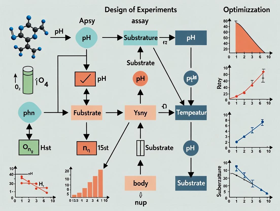
Diagram 1: DoE Workflow for Assay Optimization
Diagram 2: Core Enzyme Assay Signaling Pathway
The Scientist's Toolkit: Research Reagent Solutions
| Item | Function & Importance for Robustness |
|---|---|
| Recombinant Enzyme (≥95% pure) | High-purity enzyme ensures consistent specific activity, minimizing lot-to-lot variability and non-specific binding. Essential for calculating accurate kinetic parameters. |
| Chromogenic/Fluorogenic Substrate | Provides measurable signal change upon enzymatic conversion. Must have high stability, purity, and a favorable Km for the assay conditions. |
| Cofactors (e.g., Mg²⁺, ATP, NADPH) | Often required for enzymatic activity. Concentration and purity are critical; use cell culture-grade or higher to avoid metal contamination. |
| Assay Buffer with Stabilizers | Maintains optimal pH and ionic strength. Inclusion of stabilizers like BSA (0.1%) or CHAPS (0.01%) reduces enzyme adsorption to plates/tubes. |
| Positive Control Inhibitor | A known potent inhibitor (e.g., staurosporine for kinases) validates assay performance and serves as a normalization control across plates/runs. |
| Low-Fluorescence/Binding 384-Well Plates | Minimizes background signal and compound adsorption. Black plates are standard for fluorescence, clear for absorbance. Must be validated for your assay. |
| Precision Liquid Handler | Automated dispensers (e.g., via solenoid valves) for non-contact dispensing of enzyme/substrate reduce volumetric error and are key for 384/1536-well formats. |
| Kinetic Plate Reader with Temp Control | For continuous assays, precise temperature control (±0.1°C) and fast, consistent reading intervals are mandatory for accurate initial rate (V₀) calculation. |
Troubleshooting Guides & FAQs
Q1: My OFAT-optimized enzyme assay shows high activity in initial tests but fails when scaled to a 96-well plate format. What could be the cause? A: This is a classic symptom of missing factor interactions. In your One-Factor-At-a-Time (OFAT) approach, you optimized factors like pH, temperature, and substrate concentration independently. However, in the scaled system, these factors interact. For example, the optimal pH at a bench-scale temperature may not be optimal at the slightly different thermal gradient present in a multi-well plate. You have likely found a local, not global, optimum. To resolve, transition to a Design of Experiments (DOE) screening design (e.g., a 2-level fractional factorial) to identify and model these critical interactions.
Q2: I've run an extensive OFAT experiment, but the final assay performance is barely better than my starting point. Why was this so inefficient? A: OFAT is statistically inefficient and ignores interaction effects. Your resources were spent sequentially testing levels of each factor without learning how they work together. The table below quantifies the inefficiency versus a factorial DOE:
| Optimization Method | Factors Tested | Total Experimental Runs (for 3 levels each) | Information Gained |
|---|---|---|---|
| Traditional OFAT | 4 | 9 (Baseline + 2 levels * 4 factors) | Main effects only. No interaction data. High risk of missing true optimum. |
| Full Factorial DOE (2-level) | 4 | 16 (2^4) | All main effects AND all two-, three-, and four-way interactions. |
Protocol: Transitioning from OFAT to a Screening DOE
- Define Factors & Ranges: List all factors from your OFAT (e.g., [pH: 6.5-7.5], [Mg2+: 1-5 mM], [Temperature: 25-37°C], [Substrate: 0.5-2.0 µM]).
- Select Design: Use a fractional factorial or Plackett-Burman design (e.g., via JMP, Minitab, or DOE-pro software). For 4 factors, a 2^(4-1) fractional factorial with 8 runs is sufficient.
- Randomize Runs: Execute the 8 assay conditions in random order to avoid bias.
- Measure Response: Record initial reaction velocity (V0) for each condition.
- Analyze for Interactions: Use software to analyze the model. A significant interaction (e.g., pH*Temperature) will appear as a non-parallel line in the interaction plot.
Q3: How do I visually confirm that my OFAT approach missed critical interactions? A: Compare the response surfaces generated from OFAT data versus from a factorial DOE. The OFAT surface will be a simple ridge or plane, while the DOE surface will show curvature and ridges indicating interaction. The diagram below illustrates the logical flaw of OFAT.
Q4: What are the key reagents and solutions I need to set up a robust DOE for enzyme kinetics? A: The Scientist's Toolkit for this transition is below.
| Research Reagent Solution | Function in DOE for Enzyme Assays |
|---|---|
| Assay Buffer (Multi-component) | Provides consistent ionic strength and cofactors. Prepare a master mix to ensure uniformity across all randomized experimental runs. |
| Enzyme Stock (Aliquoted) | Highly stable, homogenous stock solution, aliquoted to avoid freeze-thaw cycles, ensuring consistent activity across all experimental points. |
| Substrate Library (Variable Concentration) | Pre-prepared serial dilutions covering the defined experimental range (e.g., 0.5, 1.0, 2.0 µM) as dictated by the DOE design matrix. |
| Stop Solution (or Real-time Detection) | Must be compatible with all factor combinations (e.g., extreme pH or temperature) to quench reactions uniformly for endpoint assays. |
| Positive/Negative Control Buffers | Included in each experimental block to monitor inter-run variability and normalize data if needed. |
Protocol: Executing a Central Composite Design (CCD) for Final Optimization
- Base Design: Start with the factorial points from your screening design (e.g., 2^4 = 16 runs).
- Add Center Points: Include 4-6 replicate runs at the midpoint of all factor ranges to estimate pure error and curvature.
- Add Axial (Star) Points: Add 2*k runs (where k=number of factors) where one factor is set at ±α (alpha, a distance outside the cube) and all others are at their midpoints. This allows fitting a quadratic model.
- Full Design: For 4 factors, this results in 16 + 6 + 8 = 30 total runs. Randomize the run order.
- Model Fitting: Use regression software to fit a second-order model:
Response = β0 + ΣβiXi + ΣβiiXi^2 + ΣβijXiXj. - Locate Optimum: Use the model's response optimizer to find factor settings that maximize activity.
The workflow for a full DOE-based optimization is shown below.
Troubleshooting Guides & FAQs
Q1: My assay response (e.g., enzyme velocity) shows high variability between replicates, confounding the effect of the factors I'm testing. What could be the cause? A1: High replicate variability often points to an uncontrolled factor. Follow this diagnostic protocol:
- Check Reagent Stability: Prepare a fresh master mix of all core reagents (buffer, cofactors, substrate). Run the assay with a single factor level combination (e.g., mid-point pH and substrate concentration) across 8-12 wells. If CV% drops below 5%, the issue was likely reagent degradation or inconsistent preparation.
- Instrument Calibration: Verify the accuracy of pipettes and microplate reader using a dye-based absorbance standard.
- Temporal Effect: If using a 96-well plate, the time between the first and last well initiation can cause signal drift. Implement a staggered start protocol or use a plate reader with rapid kinetic capabilities.
Q2: My designed experiment resulted in a model with a low R² or a non-significant Lack-of-Fit test. What steps should I take? A2: A poor model fit indicates the data does not well-represent the underlying system. Follow this sequential protocol:
| Step | Action | Diagnostic Target |
|---|---|---|
| 1 | Check for Outliers: Use standardized residuals plot. | Remove or investigate data points with residuals > ±3 standard deviations. |
| 2 | Check for Missing Factors: Review literature for potential critical factors (e.g., ionic strength, stabilizing agents, detergent) not included in your design. | A significant curvature effect in initial models often signals a missing optimal factor level. |
| 3 | Expand the Design Space: Your current factor ranges may be too narrow. Add axial points to convert a screening design to a Central Composite Design (CCD) to capture curvature. | Improved R² and significant quadratic terms in the model. |
| 4 | Transform the Response: If the response variance increases with the mean, apply a Box-Cox transformation (e.g., log, square root). | Resolves heteroscedasticity, improving model validity. |
Q3: How do I define the "Design Space" for my enzyme assay from my experimental data? A3: The Design Space is the multi-dimensional combination of factor levels where the assay meets predefined quality criteria. Follow this methodology:
- Establish Criteria: Define acceptable ranges for your Critical Quality Attributes (CQAs) (Responses). E.g., Signal-to-Noise > 10, CV% < 15%, Velocity within 80-120% of target.
- Fit a Model: Use multiple regression on your DOE data to generate polynomial equations (e.g.,
Velocity = β₀ + β₁[Substrate] + β₂[pH] + β₁₂[Substrate][pH] + β₁₁[Substrate]²...). - Generate Overlay Plots: Using statistical software, plot contour lines for each response. The overlapping region where all responses meet your criteria is your robust Design Space.
- Verify: Run 3-5 confirmation experiments within the proposed Design Space to validate performance.
Diagram: Workflow for Defining a Design Space.
Key Experimental Protocol: Central Composite Design (CCD) for Assay Optimization
Objective: To model curvature and identify optimal factor levels for enzyme activity. Methodology:
- Select Factors & Levels: Choose 2-4 critical factors (e.g., [Substrate], [Mg²⁺], pH, Temperature). Define low (-1) and high (+1) levels.
- Design Matrix: Create a full or fractional factorial core (2ᵏ runs), add 2k axial points (α = ±1.414 for face-centered CCD), and include 4-6 center point replicates.
- Randomization: Randomize the run order to avoid systematic bias.
- Execution: Perform assay according to randomized list. Measure responses (e.g., Initial Velocity, Vmax, % Inhibition).
- Analysis: Fit data to a second-order model using statistical software (e.g., JMP, Minitab, Design-Expert). Use ANOVA to identify significant terms.
Diagram: Central Composite Design (CCD) Point Distribution.
The Scientist's Toolkit: Key Research Reagent Solutions
| Item | Function in Enzyme Assay Optimization | Example / Specification |
|---|---|---|
| High-Purity Substrate | The varied factor; minimal lot-to-lot variability is critical for reproducible kinetics. | ATP >98%, fluorescent/colorimetric probe with low background. |
| Enzyme Storage Buffer | Maintains enzyme stability between experiments; often contains glycerol, reducing agents. | 25 mM HEPES, pH 7.5, 100 mM NaCl, 10% glycerol, 1 mM DTT. |
| Assay Buffer System | A critical factor (pH); must have sufficient buffering capacity at the chosen temperature. | 50 mM Tris, phosphate, or bis-tris-propane across a range of pH levels. |
| Cofactor / Cation Solution | A potential critical factor (e.g., Mg²⁺ for kinases). Use chelators to control free concentration. | MgCl₂, MnCl₂, or NADPH solutions, prepared fresh. |
| Positive/Negative Controls | Essential for normalizing response and calculating Z'-factor for assay quality. | Well-characterized inhibitor (control compound) and vehicle (DMSO). |
| Detection Reagent | Quantifies the response (e.g., enzyme velocity). Must be linear over the signal range. | Coupled enzyme systems, chromogenic/fluorogenic substrates, luciferin. |
| Run Order | [Substrate] (µM) | [ATP] (µM) | [Mg²⁺] (mM) | Response: Initial Velocity (RFU/min) | Normalized Activity (%) |
|---|---|---|---|---|---|
| 7 | 2 (-1) | 10 (-1) | 5 (-1) | 1250 ± 45 | 28 |
| 12 | 10 (+1) | 10 (-1) | 5 (-1) | 3120 ± 120 | 70 |
| 3 | 2 (-1) | 100 (+1) | 5 (-1) | 2800 ± 95 | 63 |
| 9 | 10 (+1) | 100 (+1) | 5 (-1) | 3980 ± 150 | 89 |
| 5 | 2 (-1) | 10 (-1) | 15 (+1) | 2100 ± 80 | 47 |
| 14 | 10 (+1) | 10 (-1) | 15 (+1) | 4450 ± 210 | 100 |
| 2 | 2 (-1) | 100 (+1) | 15 (+1) | 4200 ± 180 | 94 |
| 11 | 10 (+1) | 100 (+1) | 15 (+1) | 4250 ± 190 | 95 |
| 1, 4, 8, 10 | 6 (0) | 55 (0) | 10 (0) | 4400 ± 110, 4320 ± 90, 4480 ± 130, 4380 ± 105 | 99 ± 2 |
Note: Coded factor levels are in parentheses. Center points (runs 1,4,8,10) assess pure error and curvature. Data suggests [Mg²⁺] and [Substrate] are significant factors.
Welcome to the Technical Support Center for Enzyme Assay Optimization via Design of Experiments (DOE). This resource provides targeted troubleshooting guides and FAQs to help you navigate common experimental challenges and achieve your specific optimization objectives.
Frequently Asked Questions & Troubleshooting Guides
Q1: My assay signal is consistently too low (weak absorbance/fluorescence), making data unreliable. How can I increase it? A: Low signal often stems from suboptimal reaction conditions for the enzyme. Focus on maximizing signal.
- Potential Causes & Solutions:
- Substrate Concentration: You may be below the Km. Perform a substrate saturation experiment.
- Cofactor/Ion Concentration: Ensure essential cofactors (e.g., Mg²⁺ for kinases) are at non-limiting levels.
- pH & Buffer: The pH may be far from the enzyme's optimum. Test a broad pH range.
- Enzyme Concentration: The enzyme may be inactive or quantity may be insufficient. Titrate enzyme and check activity with a positive control.
- Experimental Protocol (Substrate Saturation Test):
- Prepare a master mix with buffer, cofactors, and a fixed amount of enzyme.
- Aliquot the master mix into a microplate.
- Add substrate in a series of concentrations (e.g., 0.1x, 0.5x, 1x, 2x, 5x, 10x of your estimated Km).
- Initiate the reaction and measure initial velocity (V₀) for each condition.
- Plot V₀ vs. [Substrate] to visually identify the saturation point.
Q2: My data has high variability between replicates (high noise). How can I improve precision? A: High noise obscures true signal. Focus on minimizing noise.
- Potential Causes & Solutions:
- Liquid Handling Inconsistency: Use calibrated pipettes and consider automated liquid handlers for critical reagents. Pre-mix master mixes thoroughly.
- Temperature Fluctuation: Use a thermally equilibrated plate reader with a consistent incubation chamber.
- Edge Effects in Microplates: Account for uneven evaporation. Use a plate seal, and consider randomizing run order within a DOE block.
- Reagent Stability: Prepare fresh substrate solutions or check for enzyme inactivation during the assay.
- Experimental Protocol (Plate Uniformity Test):
- Fill all wells of a microplate with an identical reaction mixture without enzyme (e.g., buffer, substrate, detection probe).
- Add a consistent volume of a stable control signal (e.g., a fluorophore at mid-range intensity) to all wells.
- Read the plate immediately using your standard assay protocol.
- Calculate the coefficient of variation (CV%) across the entire plate. A CV > 15% suggests significant positional noise to address.
Q3: My optimized assay works initially but degrades over time, or fails upon reagent lot change. How can I ensure robustness? A: This indicates a stability and robustness problem. Focus on ensuring stability.
- Potential Causes & Solutions:
- Enzyme Stability: The enzyme may lose activity during the assay. Add stabilizing agents (BSA, glycerol), reduce pre-incubation time, or source from a different vendor.
- Substrate/Probe Stability: Some detection probes (e.g., luciferin) are light-sensitive. Prepare protected from light and use fresh.
- "Bad" Buffer: Buffer components may be unstable or contaminated. Use fresh, high-purity reagents. Consider alternative buffer systems.
- Lack of Control: Always include a positive control (known active enzyme) and negative control (no enzyme) in every run to track performance drift.
Table 1: Common DOE Factors and Their Primary Impact on Optimization Goals
| Factor | Typical Range Tested | Primary Impact Goal | Notes |
|---|---|---|---|
| pH | pKa ± 2.0 units | Maximize Signal | Drastically affects enzyme activity. |
| [Substrate] | 0.1xKm to 10xKm | Maximize Signal | Key for defining linear range. |
| [Enzyme] | 0.1-10 nM typical | Balance Signal/Noise | Too low: weak signal. Too high: high background, cost. |
| Incubation Time | 5-60 minutes | Balance Signal/Stability | Longer time increases signal but may compromise linearity. |
| [Cofactor] | 0.1-10 mM typical | Maximize Signal | Essential for activity of many enzymes. |
| [Detergent] | 0.01-0.1% | Ensure Stability | Can prevent aggregation and improve reproducibility. |
| Assay Temperature | 25°C, 30°C, 37°C | Maximize Signal / Stability | Higher temp increases rate but may denature enzyme. |
Table 2: Troubleshooting Matrix for Optimization Goals
| Symptom | Primary Goal | Key Diagnostic Experiments | Likely DOE Factors to Adjust |
|---|---|---|---|
| Low Signal-to-Noise | Maximize Signal | Substrate saturation, pH profile, enzyme titration. | [Substrate], pH, [Enzyme], [Cofactor] |
| High Well-to-Well Variance | Minimize Noise | Plate uniformity test, reagent stability check. | Mixing time, incubation time, additive concentrations. |
| Signal Drift Over Time | Ensure Stability | Time course with controls, reagent age test. | Incubation time, stabilizer concentration, temperature. |
| Inconsistent Results Between Runs | Ensure Stability | Control chart analysis, reagent sourcing check. | All factors; use DOE to model and define robust setpoints. |
Experimental Workflows & Pathways
Title: Decision Workflow for Enzyme Assay Optimization Goals
Title: Key Factors in Enzyme Reaction Pathway & Optimization
The Scientist's Toolkit: Research Reagent Solutions
| Item | Function in Enzyme Assay Optimization | Key Consideration |
|---|---|---|
| High-Purity Recombinant Enzyme | The catalyst of interest; source and lot consistency are critical for stability. | Use vendors providing detailed activity specs (U/mg) and storage buffers. |
| Chromogenic/Fluorogenic Substrate | Generates the measurable signal upon enzyme conversion. | Match substrate to enzyme specificity; check solubility and background signal. |
| Essential Cofactors (Mg²⁺, NADH, ATP) | Required for activity of many enzymes (kinases, dehydrogenases, etc.). | Optimize concentration; chelating agents in buffer can interfere. |
| Buffering Agents (HEPES, Tris, PBS) | Maintains optimal pH for enzyme activity and stability. | Choose a buffer with pKa near desired pH; ensure no chemical interference. |
| Plate Reader-Compatible Microplates | The reaction vessel for high-throughput optimization. | Use clear-bottom for absorbance/fluorescence; black sides reduce crosstalk. |
| Detergents (Tween-20, Triton X-100) | Reduces non-specific binding and enzyme aggregation, improving stability. | Optimize at low concentrations (0.01-0.1%) to avoid inhibition. |
| Stabilizers (BSA, Glycerol) | Protects enzyme from surface adsorption and thermal denaturation. | Common in enzyme storage buffers; test impact on assay background. |
| Positive & Negative Control Compounds | Validates assay performance and identifies interference. | Use a known inhibitor for negative control; essential for every plate. |
| DOE Software (JMP, Minitab, MODDE) | Designs efficient experiments and models complex factor interactions. | Critical for moving beyond "one-factor-at-a-time" optimization. |
Screening Designs (e.g., Plackett-Burman, Fractional Factorials) for Identifying Vital Few Factors
Technical Support Center
Troubleshooting Guides & FAQs
Q1: My Plackett-Burman (PB) design analysis shows no significant factors. What could be wrong? A1: Common issues and solutions:
- Problem: Factor ranges were set too narrow, masking real effects.
- Solution: Ensure your chosen high/low levels are pragmatically far apart (e.g., 2-3 fold difference for concentrations). Re-run with wider ranges.
- Problem: Excessive random error (noise) overshadowing factor effects.
- Solution: Review assay precision. Incorporate more replicates (at least duplicate runs per design point) to better estimate error.
- Problem: Incorrect assumption of effect sparsity; many factors have small, interacting effects.
- Solution: A PB design cannot detect interactions. Consider moving to a Resolution IV fractional factorial as a next step to screen for two-factor interactions.
Q2: How do I choose between a Resolution III, IV, or V fractional factorial for my enzyme assay screening? A2: The choice balances the number of runs with the risk of confounding (aliasing).
- Resolution III (e.g., 2^(III)(3-1) for 3 factors in 4 runs): Main effects are aliased with two-factor interactions. Use only when you are confident interactions are negligible for initial screening of many factors (6+).
- Resolution IV (e.g., 2^(IV)(5-1) for 5 factors in 16 runs): Main effects are aliased with three-factor interactions, and two-factor interactions are aliased with each other. This is a robust choice for screening, as it allows unbiased estimation of main effects even in the presence of some interactions.
- Resolution V (e.g., 2^(V)(5-1) for 5 factors in 16 runs? No, requires more runs): Main effects and two-factor interactions are aliased with three- or higher-order interactions. Ideal when you suspect specific interactions are critical, but requires more experimental runs. Often used after initial screening.
Q3: I have limited resources and can only run 12 experiments. Can I still screen 7 factors? A3: Yes, a 12-run Plackett-Burman design is a classic choice for exactly this scenario (screening k=7 to 11 factors in N=12 runs). Remember:
- It is a Resolution III design; main effects are confounded with two-factor interactions.
- Always include at least 3-4 center points (if the factors are continuous) within those 12 runs to check for curvature, which might indicate the presence of interactions or a need for optimization in a critical factor.
Q4: My design includes categorical factors (e.g., buffer type, enzyme source). How do I handle them? A4: Categorical factors are fully supported in screening designs.
- Assign them as you would numerical factors (e.g., Source A = -1, Source B = +1).
- The analysis (e.g., Pareto chart, half-normal plot) will show if changing the category creates a significant shift in the response (e.g., activity).
- Be aware that interactions between categorical and continuous factors (e.g., buffer type x pH) are possible but harder to interpret in Resolution III designs.
Key Experimental Protocols
Protocol 1: Executing a Plackett-Burman Screening Design for Enzyme Assay Optimization
- Define Objective: Identify factors most affecting initial reaction velocity (V0).
- Select Factors (5-11 typical): E.g., pH, [Substrate], [Mg2+], Temperature, [Enzyme], % Co-solvent, Incubation Time.
- Set Levels: Choose a scientifically relevant High (+1) and Low (-1) level for each.
- Generate Design Matrix: Use statistical software (JMP, Minitab, R, Python) to create an N=12, 20, or 24 run PB design. Randomize the run order.
- Include Center Points: Add 3-4 runs with all continuous factors at their midpoint to assess linearity.
- Conduct Experiments: Perform assays according to randomized matrix, measuring V0.
- Analyze Data: Fit a linear model. Use a Pareto Chart of standardized effects or a Half-Normal Plot to identify the "vital few" factors exceeding the statistical significance threshold (often at α=0.05 or 0.1).
Protocol 2: Follow-up Using a Resolution IV Fractional Factorial
- Start from PB Results: Take the 3-4 most significant factors from the PB screen.
- Select Design: Use a 2^(k-p) fractional factorial design with Resolution IV or higher. For 4 factors, a full factorial (16 runs) or a Resolution IV design (8 runs) is suitable.
- Set Levels: Refine the high/low levels based on PB results, potentially narrowing the range.
- Execute & Analyze: Run the design, measure response(s). Analyze with a model including all main effects and two-factor interactions. Use ANOVA to identify significant terms.
- Path Forward: The results guide you to a definitive optimization phase (e.g., Response Surface Methodology) focusing on the critical 2-3 factors and their key interactions.
Data Presentation
Table 1: Comparison of Common Screening Designs for Enzyme Assays
| Design Type | Runs (N) for k=6 Factors | Max Factors for N=12 Runs | Resolution | Aliasing Structure | Best Use Case |
|---|---|---|---|---|---|
| Plackett-Burman | 12 | 11 | III | Main Effects ∝ 2FI | Initial screen of many (7+) factors where interactions are assumed small. |
| Fractional Factorial (2^(6-2)) | 16 | 5* | IV | 2FI ∝ 2FI | Screening 5-8 factors when some 2FI may be important. Robust choice. |
| Fractional Factorial (2^(6-3)) | 8 | 7* | III | Main Effects ∝ 2FI | Ultra-high-throughput screen of 6+ factors with severe run constraints. |
| Full Factorial (2^k) | 64 | 3 | Full | None | Not a screening design. Use for deep study of ≤4 very important factors. |
*To screen 6 or 7 factors in ~12 runs, a Plackett-Burman is typically preferred.
Visualizations
Title: Plackett-Burman Screening Workflow for Enzyme Assays
Title: Effect Aliasing in Resolution III vs IV Designs
The Scientist's Toolkit: Research Reagent Solutions
Table 2: Essential Materials for DOE-Based Enzyme Assay Screening
| Item | Function in Screening Experiments |
|---|---|
| Statistical Software (JMP, Minitab, R/pyDOE2) | Generates randomized design matrices, analyzes results, and creates diagnostic plots (Pareto, Half-Normal). |
| Multi-Channel Pipette & Microplate Reader | Enables high-throughput execution of many assay conditions (e.g., 96-well plate format) with consistent timing. |
| Assay-Ready Plates (96-/384-well) | Pre-coated or treated plates for consistent binding; used for running many design points in parallel. |
| Master Mix Solutions | Critical for ensuring uniformity when dispensing common components (e.g., buffer, detector) across many wells. |
| Liquid Handling Robot (Optional) | Automates plate setup for complex designs with many runs, minimizing manual pipetting error. |
| Positive/Negative Control Reagents | Included in every plate to normalize results and monitor assay performance across design runs. |
| Continuous Factor Stocks (pH buffers, cofactors, substrates) | Prepared at high and low concentrations (corresponding to +1/-1 levels) for accurate dispensing. |
| Enzyme Stock (Stable aliquots) | Quality-controlled, aliquoted at consistent concentration to serve as a uniform base for all runs. |
Building Your Optimized Protocol: A Step-by-Step DOE Workflow for Enzyme Kinetic and Activity Assays
Welcome to the Technical Support Center for Enzyme Assay Optimization. This resource, framed within a thesis on Design of Experiments (DoE) for systematic assay development, provides troubleshooting guides and FAQs for researchers deconstructing their assays to identify critical factors.
Frequently Asked Questions & Troubleshooting Guides
Q1: During preliminary testing, my enzyme shows no activity across the pH range I tested. What could be wrong? A: This often indicates a buffer incompatibility or incorrect cofactor/activation step.
- Troubleshooting Steps:
- Verify Buffer Compatibility: Ensure your buffer does not chelate essential metal ions (e.g., avoid phosphate buffers with Mg²⁺-dependent enzymes). Switch to a non-chelating buffer like HEPES or Tris and re-test.
- Check Cofactor Requirements: Consult literature for absolute cofactor requirements (e.g., Mg²⁺, ATP, NADH). Prepare a fresh stock solution and add it to the assay mixture.
- Confirm Enzyme Viability: Perform a positive control assay under literature-reported optimal conditions to confirm enzyme stock functionality.
- DoE Context: This narrows the "pH" factor space by identifying buffer-chemical interactions, a critical confounding variable to control before formal screening.
Q2: My reaction velocity does not increase linearly with increasing enzyme concentration, violating a key assumption for [Enzyme] factor testing. A: Non-linearity suggests enzyme instability, substrate depletion, or the presence of an inhibitor.
- Troubleshooting Steps:
- Run a Time-Course: Ensure you are measuring initial velocity. If velocity plateaus early at higher [enzyme], substrate may be limiting. Increase [substrate] so it is >> Km.
- Test Enzyme Stability: Pre-incubate enzyme at assay temperature for different times before starting the reaction. A rapid drop in activity suggests instability, requiring shorter assays or stabilizers (e.g., BSA, glycerol).
- Dilution Buffer: Ensure enzyme dilution buffer contains a carrier protein (e.g., 0.1% BSA) to prevent surface adhesion losses at low concentrations.
- DoE Context: This validates the "fundamental assumption" of your system before including [Enzyme] as a quantitative factor. A stability issue may require adding a "pre-incubation time" as a new factor.
Q3: When testing the [Substrate] factor, I observe high background signal at low enzyme concentrations. A: This is typical of fluorescent or coupled assays and indicates non-enzymatic substrate turnover or detector interference.
- Troubleshooting Steps:
- Run No-Enzyme Controls: For every [substrate] point, run a control without enzyme. Subtract this background value from your experimental readings.
- Check Substrate Purity: Some synthetic fluorogenic substrates (e.g., p-nitrophenyl derivatives) can auto-hydrolyze. Prepare fresh substrate stock in anhydrous DMSO or ethanol.
- Filter Components: Particulates in crude substrates or buffers can cause light scattering. Filter all solutions (except enzyme) through a 0.22 µm membrane.
- DoE Context: Accurate background subtraction is crucial for defining the true "signal window," which directly impacts the power of your subsequent DoE model to detect significant effects.
Q4: How do I differentiate between the individual effects of Temperature and Ionic Strength, as they often interact? A: This interaction is common. A strategic two-factor experimental matrix is required.
- Experimental Protocol:
- Design a simple 3x3 grid experiment: Test 3 temperatures (e.g., 25°C, 30°C, 37°C) against 3 ionic strengths (e.g., 50 mM, 150 mM, 300 mM KCl).
- Hold all other factors (pH, [S], [E]) constant at middle values.
- Perform duplicates. Plot activity as a contour or 3D surface plot.
- DoE Context: This mini two-factor interaction study provides preliminary data to justify including the "Temp x Ionic Strength" interaction term in your full factorial or response surface model.
Q5: My enzyme requires an expensive cofactor (e.g., NADPH). How can I minimize its use during initial factor screening? A: Use a coupled recycling system or a catalytic concentration.
- Experimental Protocol: Cofactor Recycling System
- For NADPH-dependent enzymes, include a substrate-generating system like Glucose-6-Phosphate (G6P) and Glucose-6-Phosphate Dehydrogenase (G6PDH).
- Reaction Mix: Assay buffer, catalytic NADP+ (e.g., 0.1 mM), 5 mM G6P, 2 U/mL G6PDH, your enzyme, and your substrate.
- This regenerates NADPH continuously, reducing the required amount 10-100 fold.
- DoE Context: This allows you to fix "[Cofactor]" as a non-limiting, low-concentration constant during screening, reducing cost and complexity.
Table 1: Common Initial Testing Ranges for Assay Deconstruction
| Factor | Typical Testing Range | Common Buffer/Notes | Key Interaction Partners |
|---|---|---|---|
| pH | pKa ± 2.0 units | 50-100 mM Buffer (e.g., Bis-Tris, HEPES, Tris) | Ionic strength, cofactor stability |
| [Enzyme] | 0.1 - 10 nM (pure) 0.1 - 10 µg/mL (crude) | Diluted in buffer + 0.1% BSA | Temperature (stability), substrate concentration |
| [Substrate] | 0.2xKm to 5xKm | Solubilized in water, buffer, or ≤5% organic solvent | Enzyme concentration, ionic strength |
| Temperature | 20°C - 40°C (biological) | Thermostated cuvette holder or block | All factors (especially enzyme stability) |
| Ionic Strength | 0 - 300 mM (KCl or NaCl) | Adjusted with salt after setting buffer pH | pH (via buffer pKa), temperature |
| [Cofactor] | 0.1 - 5.0 x reported Km | Freshly prepared in assay buffer | pH (cofactor stability), ionic strength |
Experimental Protocol: Key Factor Screening via One-Factor-at-a-Time (OFAT) Pre-Screening
Objective: To identify grossly non-performant regions for each factor prior to embarking on a multi-factorial DoE. Method:
- Establish Baseline: Set all factors to literature-derived "standard" conditions.
- Vary Single Factor: Systematically vary one factor across a broad range (see Table 1), while holding all others constant at the baseline.
- Measure Activity: Perform the assay in duplicate for each level of the varied factor.
- Analyze: Plot % Relative Activity vs. Factor Level. Identify the range where activity is >50% of maximum observed.
- Iterate: Return to baseline, and repeat for the next factor. Outcome: Defines the constrained, relevant experimental space (the "region of interest") for your subsequent efficient DoE (e.g., Box-Behnken, Central Composite Design).
Visualizations
Enzyme Assay Deconstruction Workflow
Interaction Between Key Assay Factors
The Scientist's Toolkit: Research Reagent Solutions
Table 2: Essential Materials for Assay Deconstruction & Optimization
| Item | Function & Rationale |
|---|---|
| Modular Buffer System (e.g., Bis-Tris, HEPES, CHES) | Covers a wide pH range (3-10) with minimal metal chelation, allowing isolation of pH effects. |
| High-Purity Substrate (≥95% HPLC) | Minimizes background signal and ensures observed kinetics are due to the target enzyme. |
| Inert Protein (BSA, 0.1% w/v) | Stabilizes dilute enzyme stocks, prevents surface adsorption to tubes and plates. |
| Thermostatted Microplate Reader / Spectrophotometer | Enables precise, parallel measurement of temperature-dependent activity. |
| Molecular Biology Grade Water | Eliminates trace contaminants or ions that could unpredictably alter ionic strength. |
| Cofactor Recycling System Components | Reduces cost of screening for cofactor-dependent enzymes (e.g., Lactate Dehydrogenase/Pyruvate for NADH). |
| Statistical Software (e.g., JMP, Minitab, R) | Essential for designing efficient DoE matrices and analyzing multi-factorial interaction data. |
Troubleshooting Guides & FAQs
FAQ 1: My screening design did not identify any significant factors. What could have gone wrong?
- A: Several issues are common:
- Insufficient Range: The levels chosen for your factors (e.g., pH, temperature) may have been too narrow to elicit a detectable effect on the enzyme activity.
- High Background Noise: Uncontrolled variables or high experimental error can mask significant effects. Re-evaluate your assay precision.
- Wrong Factors: The factors you chose may genuinely have little influence on your specific enzyme's response. Consult prior literature.
- Design Resolution: A low-resolution fractional factorial (e.g., Resolution III) may have confounded main effects with two-factor interactions, leading to misleading results.
FAQ 2: When moving from a screening design to RSM, which specific design should I use?
- A: The choice depends on your goal and region of experimentation:
- Central Composite Design (CCD): The most common choice for a full RSM. It is efficient, fits full quadratic models, and can be built upon a pre-existing factorial design.
- Box-Behnken Design (BBD): Suitable if you need to avoid extreme factor combinations (e.g., simultaneously very high temperature and very high pressure) or if your experimental region is already known to be spherical. It has fewer design points than a CCD for the same number of factors.
- Refer to the table below for a quantitative comparison.
FAQ 3: How do I validate the model generated from my RSM analysis?
- A: Critical validation steps include:
- Statistical Diagnostics: Check ANOVA (lack-of-fit test, high R²-adjusted), residual plots for randomness, and ensure model significance.
- Confirmatory Runs: Perform new, replicate experiments at the predicted optimal conditions. Compare the observed response with the model's prediction interval.
- Comparison to a Baseline: Run the original, unoptimized assay conditions alongside the new optimal ones to quantify the improvement.
FAQ 4: I am getting a saddle point (minimax) in my response surface. What does this mean and what should I do next?
- A: A saddle point indicates that the current model has identified a stationary point that is not a true maximum or minimum. This often means you are exploring a region near the inflection of the response surface. You should:
- Use canonical analysis (provided in RSM output) to characterize the nature of the stationary point.
- Follow the path of steepest ascent/descent from your current experimental region to move towards a more optimal area.
- Consider setting up a new RSM design centered on a more promising region identified from the analysis.
Data Presentation
Table 1: Quantitative Comparison of Screening Designs
| Design Type | Number of Factors (k) | Minimum Runs (Example) | Key Strength | Key Limitation | Best For |
|---|---|---|---|---|---|
| Full Factorial | 2-4 (typically) | 2^k (e.g., 8 for k=3) | Estimates all main effects & interactions | Run number grows exponentially | Small factor sets (<5) |
| Fractional Factorial (Res III) | 5-8 | 2^(k-1) (e.g., 16 for k=5) | Highly efficient for many factors | Main effects confounded with 2-fi interactions | Initial screening of many factors |
| Plackett-Burman | 5-11 | Multiple of 4 (e.g., 12 for k=7) | Very economical, linear estimates only | Cannot estimate interactions | Identifying vital few from many |
Table 2: Quantitative Comparison of Response Surface Designs
| Design Type | Factors (k) | Typical Runs (for k=3) | Model Fitted | Region Shape | Efficiency & Notes |
|---|---|---|---|---|---|
| Central Composite (CCD) | 2-6 | 20 (8 cube, 6 axial, 6 center) | Full Quadratic | Spherical or Cuboidal | Gold standard; adjustable alpha |
| Box-Behnken (BBD) | 3-5 | 15 (12 mid-edge, 3 center) | Full Quadratic | Spherical | Avoids extreme corners; no axial points |
| 3-Level Full Factorial | 2-3 | 27 (for k=3) | Full Quadratic | Cuboidal | Many runs; often inefficient for RSM |
Experimental Protocols
Protocol 1: Executing a Two-Level Fractional Factorial Screening Design for Enzyme Assay
- Define Factors & Levels: Select 5-7 critical parameters (e.g., [Mg²⁺], pH, substrate concentration, incubation time, temperature). Set a biologically relevant high (+) and low (-) level for each.
- Choose Design Matrix: Select a Resolution IV or V fractional factorial design (e.g., 2^(5-1), 16 runs) to avoid confounding main effects with each other.
- Randomize Runs: Randomize the order of all experimental runs to mitigate confounding from time-based effects.
- Execute Assays: Perform the enzyme activity assay according to the randomized design matrix. Include replicate center points (e.g., 3-4 runs) to estimate pure error.
- Analyze Data: Use statistical software to perform ANOVA. Rank factors by p-value and effect size (Pareto chart). Identify significant main effects and strong two-factor interactions for further study.
Protocol 2: Optimizing via a Central Composite Design (CCD)
- Define Region: Based on screening results, choose 2-4 critical factors. Define the axial distance (alpha). Often, a face-centered CCD (alpha=1) is used for practical constraints.
- Create Design: The CCD comprises:
- Factorial Points: The 2^k points from your screening design (or a subset).
- Axial Points: 2k points where one factor is at ±alpha and others are at center.
- Center Points: 5-6 replicates at the midpoint to estimate curvature and error.
- Run Experiments: Execute all design points in a fully randomized order.
- Model Fitting: Fit a second-order polynomial model: Y = β₀ + ΣβᵢXᵢ + ΣβᵢᵢXᵢ² + ΣβᵢⱼXᵢXⱼ.
- Validation & Optimization: Analyze the model via ANOVA and 3D surface plots. Use the optimizer to find factor settings for maximum activity. Run confirmatory experiments.
Mandatory Visualization
Diagram Title: Logical Flow for Choosing Screening vs. RSM
Diagram Title: Central Composite Design (CCD) Experimental Workflow
The Scientist's Toolkit: Key Research Reagent Solutions
Table 3: Essential Materials for Enzyme Assay Optimization via DOE
| Item | Function in DOE Context |
|---|---|
| Purified Enzyme Preparation | The core reagent. Must be of consistent activity and purity between experimental runs to reduce noise. |
| Substrate Library / Variants | To test the factor "Substrate Type/Concentration." Includes natural and synthetic chromogenic/fluorogenic substrates. |
| Broad-Range Buffer Systems | (e.g., HEPES, Tris, phosphate blends). Essential for exploring pH as a continuous factor across a wide range. |
| Cofactor & Cation Solutions | (e.g., MgCl₂, MnSO₄, NADH, ATP). Used to test the effect of essential activators or coenzymes as quantitative factors. |
| Inhibitor/Effector Compounds | To study the effect of modulators. Concentration can be a designed factor in the experiment. |
| Stop Reagent | A consistent, rapid-quenching solution (e.g., acid, denaturant) to precisely control reaction time, a key temporal factor. |
| Detection Reagents | For colorimetric, fluorometric, or luminometric readouts. Must be stable and prepared in bulk for consistency across all design points. |
| Microplate Reader-Compatible Plates | Enable high-throughput execution of the many randomized runs required by screening and RSM designs. |
Troubleshooting Guides & FAQs
Q1: During a high-throughput DOE run for enzyme kinetics, my robotic liquid handler is consistently delivering lower volumes than programmed, causing high CVs in my initial rate data. What are the primary causes and solutions?
A: This is often due to tip wetting, viscosity of assay components, or environmental calibration drift.
- Solution A: Implement a "pre-wetting" step in your handler protocol. Aspirate and dispense the reagent 2-3 times before the final transfer to condition the tip interior.
- Solution B: For viscous buffers (e.g., with >5% glycerol), increase the "delay" time after aspiration and before dispensing. Perform a gravimetric validation check weekly: dispense water into a microbalance and adjust the calibration offset in the software.
- Primary Check: Always run a dye-based (e.g., tartrazine) absorbance verification assay on the destination plate to quantify volume accuracy before a critical DOE run.
Q2: My manual 96-well plate assay for a 2^3 factorial DOE shows significant edge effects (outer wells show different activity), confounding my main effect analysis. How do I mitigate this?
A: Edge effects are typically evaporation or temperature gradient-related.
- Immediate Mitigation: Use a thermally pre-equilibrated plate seal during incubation steps. For manual runs, employ a "plate hotel" within the incubator.
- DOE Design Solution: Randomize your run order so that factor level combinations are not spatially correlated. Include "blocking" in your design, treating the plate as a separate variable.
- Protocol Adjustment: Add a perimeter of "blank" wells filled with assay buffer around your experimental wells to create a uniform microenvironment.
Q3: When setting up a Response Surface Design (Central Composite) manually, I struggle with accurately preparing the intermediate concentration levels for the continuous factors (e.g., pH, [Substrate]). What is a reliable method?
A: Use a serial dilution master mix strategy for concentration factors.
- Protocol: Prepare the extreme levels (e.g., 0.1 mM and 10 mM substrate) as master stocks. For the center point, create a separate 1:1 mix of the two extreme stocks. For axial points (e.g., alpha levels), calculate the required volume from each extreme stock to achieve the target concentration using the formula C1V1 = C2V2, and mix them directly in the assay well. Prepare a fresh, independently calibrated buffer for pH center points.
Q4: In a high-throughput screening DOE, my positive control (known enzyme inhibitor) shows erratic activity, making it hard to validate the run. What could be wrong?
A: This points to reagent stability or dispensing issues.
- Troubleshooting Steps:
- Check Inhibitor Solvent: Ensure the DMSO concentration is consistent and ≤1% across all wells. Use a dedicated DMSO-resistant tip type.
- Assay Reagent Stability: Prepare the detection reagent (e.g., NADH, fluorescent probe) fresh daily and keep it protected from light. Verify its absorbance/fluorescence before the run.
- Control Dispensing: Program the liquid handler to dispense the positive control from a single, homogenous master mix into all designated wells, rather than mixing in-well.
Q5: The software for my automated workstation and my DOE analysis software (e.g., JMP, Design-Expert) do not communicate. How can I avoid manual transcription errors in transferring my run table and results?
A: Implement a file-based workflow.
- Method: Design your experiment matrix (run table) in your DOE software. Export it as a .CSV file. This file should contain well locations (A01, B01, etc.) and factor levels. This .CSV can often be directly imported by the liquid handler's scheduler software to create the deck layout and dispensing instructions. After the run, export the plate reader results (in the same well order) as a .CSV and import it back into the DOE software for analysis. Always perform a spot-check of 5% of wells.
Research Reagent Solutions Toolkit
| Item | Function in Enzyme Assay DOE |
|---|---|
| Master Mix Stocks | Pre-mixed, aliquoted solutions of buffer, cofactors, and salts to minimize preparation variance between DOE runs. |
| Low-Binding Microplates | 96- or 384-well plates with surface treatment to minimize nonspecific enzyme/substrate adsorption, critical for accurate kinetic measurements. |
| Non-Fluorescent Seal | Thermally stable plate seal to prevent evaporation without interfering with fluorescence or luminescence detection modes. |
| DMSO-Tolerant Tips | Robotic or multichannel pipette tips designed to handle organic solvents without volume variation or polymer leaching. |
| Liquid Handler Calibration Kit | Dye solutions (tartrazine, fluorescein) and a microbalance for weekly verification of nanoliter-to-microliter dispensing accuracy. |
| Enzyme Storage Buffer | Optimized, DOE-tested buffer (e.g., with stabilizers like BSA or trehalose) for maintaining enzyme activity over the duration of a long screening run. |
Experimental Protocols
Protocol 1: Gravimetric Calibration of a Liquid Handler for DOE
Purpose: To ensure accurate and precise delivery of variable factor levels (e.g., inhibitor, substrate concentration).
- Tare a precision microbalance (0.001 mg sensitivity).
- Program the handler to dispense the target volume (e.g., 10 µL) of purified water (density 1.00 g/mL) into a weigh boat on the balance.
- Record the mass. Calculate volume: Mass (mg) = Volume (µL).
- Repeat for 10 replicates per tip/channel used in the DOE.
- Calculate mean, SD, and CV. Adjust the instrument's calibration offset if the mean is >2% from the target. Recalibrate until CV <1%.
Protocol 2: Dye-Based Plate Layout Verification
Purpose: To visually confirm correct dispensing of factor levels across the DOE plate layout before adding enzyme.
- Prepare a tartrazine dye solution in your standard assay buffer.
- Execute your programmed liquid handler method, substituting all aqueous reagent lines with the dye solution.
- After dispensing, image the plate using a standard plate reader's absorbance mode at 415 nm.
- Analyze the heatmap. Uniform color intensity within factor level groups confirms correct volume and location dispensing. Investigate outliers.
Diagrams
DOE Execution Workflow
Enzyme Assay Reaction Pathway
Key Factors in Enzyme Assay Optimization DOE
Table 1: Typical Factor Ranges for Initial Enzyme Assay DOE Screening
| Factor | Low Level (-1) | High Level (+1) | Recommended Unit | Notes |
|---|---|---|---|---|
| Substrate [S] | 0.1 x Km | 5 x Km | mM or µM | Estimate Km from literature first. |
| Enzyme [E] | 0.5 nM | 5 nM | nM | Aim for linear signal <10% substrate conversion. |
| pH | Optimum - 1.0 | Optimum + 1.0 | - | Use a buffer with good capacity in this range. |
| Incubation Time | 5 min | 30 min | minutes | Must be within the linear velocity range. |
| [DMSO] (if applicable) | 0.1% | 1.0% | % v/v | Test solvent tolerance in a preliminary experiment. |
Table 2: Troubleshooting Common Liquid Handler Errors
| Error Symptom | Possible Cause | Diagnostic Check | Corrective Action |
|---|---|---|---|
| Low Volume Dispensed | Tip wetting, partial clog, worn piston. | Gravimetric check, visual inspection of tip post-dispense. | Use pre-wetting step, increase blow-out volume, replace tip/head. |
| High CV across Plate | Temperature gradient, evaporation, tip inconsistency. | Dye verification assay, check plate sealer. | Use uniform incubation, low-evaporation seals, calibrate all tips. |
| Edge Well Outliers | Evaporation (edge wells), uneven heating. | Compare edge vs. interior control wells. | Use plate seals, humidity chambers, exclude edge wells from design. |
| Incorrect Aspiration | Air bubble in line, low reagent volume. | Observe aspiration in source well. | Prime lines, ensure sufficient reagent volume, use anti-bubble tips. |
Troubleshooting Guide & FAQs for Data Analysis in Enzyme Assay DOE
FAQ 1: My model in JMP/Minitab shows a high p-value (>0.05) for my main factors, but I know the enzyme concentration should be significant based on literature. What went wrong?
- Answer: This often indicates a misspecified model or confounding. In a designed experiment for enzyme assay optimization, check the following:
- Pooled Error: Ensure you have adequate replication. Without replication, you cannot estimate pure error, forcing the software to pool higher-order interactions into error, potentially inflating p-values.
- Model Overfitting: You may have included too many terms (e.g., 3-way interactions) in a small resolution design, leaving few degrees of freedom for error. Use the software's model reduction feature (Stepwise, Backward Elimination) to remove non-significant terms.
- Transform Response: Enzyme activity data (e.g., initial velocity) often benefits from transformation. If your residual plots show a funnel pattern, apply a log or Box-Cox transformation in the software.
- Check Design Resolution: A Resolution III design confounds main effects with two-way interactions. Use the design evaluation tools to check your aliasing structure.
FAQ 2: In R, when I run lm() on my factorial design data, how do I correctly interpret the coefficients for continuous factors (like pH) vs. categorical factors (like buffer type)?
- Answer: Interpretation differs by factor type.
- Continuous Factors: The coefficient represents the change in the response (e.g., enzyme activity) for a one-unit change in the factor, holding all others constant.
- Categorical Factors (k levels): R uses treatment contrasts by default. The model will output (k-1) coefficients. Each coefficient represents the difference in the mean response between that specific level and the reference level (e.g., 'Buffer A'). The intercept then represents the mean response at the reference level for all categorical factors.
- Protocol: Always check your contrast settings using
contrasts(your_data$Factor)and usesummary.lm(your_model)to view coefficients and their significance.
FAQ 3: The optimization plot in Minitab's Response Optimizer or the desirability function in R's desirability package suggests impossible factor settings (e.g., pH 12.5). How do I get a practical solution?
- Answer: This is a constraint handling issue.
- Re-run Optimization with Constraints: Explicitly set realistic upper and lower bounds for each factor in the optimizer tool. Do not rely solely on the experimental range if the model predicts an optimum outside it.
- Perform a Ridge Analysis or Numerical Optimization: Use JMP's Max Desirability with side constraints, Minitab's Response Optimizer with defined constraints, or R's
nlminb()function to find the maximum on a constrained path. Verify the solution with a confirmatory run.
FAQ 4: My residual plots in any software show a clear pattern (non-random scatter), violating ANOVA/regression assumptions. What are my next steps?
- Answer: Patterned residuals invalidate significance tests. Follow this protocol:
- Plot Residuals vs. Fitted Values & vs. Run Order: Identifies non-constant variance or time-based effects.
- Apply Transformation: Use a Box-Cox power transformation (available in all three software packages) to stabilize variance.
- Check for Missing Factors: A pattern may indicate an influential variable not included in the design (e.g., reagent lot, ambient temperature). Consider adding it as a block in subsequent designs.
- Use Robust Methods: As a last resort, consider non-parametric analysis or generalized linear models (GLM) if transformations fail.
The table below compares key outputs from JMP, Minitab, and R for analyzing a factorial DOE optimizing enzyme activity (nmol/min) with factors: Substrate Conc (mM), pH, and Cofactor (Present/Absent).
| Software | Significant Factors (p<0.05) | R-Squared (Adj.) | Optimal Predicted Activity (nmol/min) | Recommended Model |
|---|---|---|---|---|
| JMP Pro 17 | Substrate (p<0.001), pH (p=0.012), Substrate*pH (p=0.03) | 0.94 | 125.4 | Reduced model with main effects and the significant interaction. |
| Minitab 21 | Substrate (p<0.001), pH (p=0.011), Substrate*pH (p=0.032) | 0.93 | 124.9 | Same as JMP. Stepwise regression confirms model. |
R (v4.3)lm() & car::Anova() |
Substrate (p<0.001), pH (p=0.011), Substrate:pH (p=0.032) | 0.94 | 125.4 | Type II ANOVA table recommended for factorial design. |
Experimental Protocol: Analyzing a Factorial DOE for Enzyme Assay
Title: Protocol for Statistical Model Building and Validation in Enzyme Assay Optimization. Objective: To build, diagnose, and validate a predictive model from a factorial Design of Experiments (DOE). Materials: Statistical software (JMP/Minitab/R), dataset from executed experimental design. Methodology:
- Data Import & Structure: Import the data matrix, ensuring factor columns are correctly coded (e.g., -1, +1 for continuous; text/numbers for categorical). Declare factors as "Categorical" or "Continuous" in the software.
- Initial Model Fitting: Fit the full factorial model, including all main effects and interaction terms.
- Model Reduction: Use sequential (Type I) or partial (Type II/III) sum of squares to remove non-significant higher-order interactions (p > 0.05, or using AIC criterion). Employ stepwise algorithms if the design is large.
- Model Diagnostics: Generate and inspect four key residual plots: 1) Residuals vs. Fitted, 2) Normal Q-Q plot, 3) Scale-Location plot, 4) Residuals vs. Run Order. Test for outliers (e.g., Cook's distance).
- Interpretation & Optimization: For the reduced model, interpret the sign and magnitude of coefficients. Use the software's optimization module (e.g., Response Optimizer, contour profiler) to find factor levels that maximize predicted enzyme activity.
- Confirmation Run: Perform 3-5 experimental runs at the software-predicted optimal conditions. Compare the observed mean response to the model's prediction interval to validate the model.
Visualization: DOE Analysis Workflow for Enzyme Assay Optimization
Title: Statistical Model Building Workflow.
The Scientist's Toolkit: Key Reagent Solutions for Enzyme Assay DOE
| Reagent / Material | Function in Enzyme Assay Optimization DOE |
|---|---|
| Purified Enzyme Lyophilizate | The biological catalyst of interest. Consistent purity and storage are critical as it is the response variable source. |
| Chromogenic/Nitroarylated Substrate | Yields a quantifiable (e.g., spectrophotometric) signal upon enzymatic conversion. Must be stable and soluble across tested concentration ranges. |
| Buffering System (e.g., HEPES, Tris, Phosphate) | Maintains pH as a critical experimental factor. Must not inhibit the enzyme and should have good capacity across the designed pH range. |
| Cofactor Solution (e.g., Mg²⁺, NADH) | Essential activator for many enzymes. Its presence/absence or concentration is often a key categorical or continuous factor in the DOE. |
| Stop Solution (e.g., Acid, Chelator, Inhibitor) | Precisely terminates the reaction at a defined timepoint, ensuring accurate and reproducible activity measurements across all design points. |
| Activity Assay Master Mix | A pre-mixed, optimized solution of buffer, salts, and stabilizers to minimize background variability, allowing the DOE factors to be isolated effects. |
Technical Support Center
Frequently Asked Questions (FAQs) & Troubleshooting
Q1: Our Z'-factor is consistently below 0.5, indicating a poor assay window. What are the primary factors we should investigate using the CCD? A: A low Z'-factor often stems from high signal variability or a low dynamic range. Using your Central Composite Design (CCE), prioritize the optimization of these continuous factors: Enzyme Concentration (too high can increase background; too low reduces signal), Incubation Time (insufficient time lowers signal; excessive time increases variability), and Substrate Concentration (must be around Km for optimal sensitivity). The CCD will model the quadratic effects and interactions of these factors to find the robust optimum that maximizes the signal-to-background ratio and minimizes coefficient of variation (CV).
Q2: We observe high background signal in our negative controls. Which experimental parameters are most likely responsible? A: High background is frequently tied to non-specific binding or incomplete inhibition. Focus your CCD on these parameters:
- Detergent Concentration (e.g., Tween-20): A low level (e.g., 0.01-0.05%) can reduce non-specific binding. The CCD can test this range.
- ATP Concentration: Running the assay at or below the apparent Km for ATP increases sensitivity to competitive inhibitors and can reduce background phosphorylation. Include ATP level as a key CCD factor.
- DMSO Tolerance: The compound solvent can affect enzyme activity. Use the CCD to confirm assay robustness across your expected DMSO range (typically 0.5-2%).
- Blocking Agent: Ensure adequate concentration of BSA or casein (e.g., 0.1-1 mg/mL) is included in the assay buffer, which can be a categorical factor in your design.
Q3: The response surface model from our CCD shows a saddle point or a ridge, not a clear maximum. What does this mean and how should we proceed? A: A ridge or saddle point indicates significant interaction effects between factors where a range of combinations yield similar optimal responses. This is valuable information. You should:
- Interpret the Model: Examine the contour plots from the CCD analysis. The ridge line shows the combination of factors (e.g., Enzyme and Substrate concentration) that provide equivalent assay performance.
- Choose a Robust Point: Select a set of conditions along the ridge that are also operationally convenient and cost-effective (e.g., lower enzyme usage).
- Verify with Confirmatory Runs: Perform 3-5 replicate experiments at your chosen optimum point to validate the predicted performance metrics (IC50, Z', CV).
Q4: Our compound IC50 values are not reproducible between runs after optimization. What stability factors might the CCD have missed? A: CCD typically focuses on assay composition and incubation factors. Reproducibility issues often point to reagent stability.
- Enzyme Aliquot Stability: Test fresh vs. frozen-thawed aliquots over time as a categorical factor.
- Substrate/ATP Stock Solution Age: Include the age of key reagent stocks (fresh vs. 1-week old at -20°C) in a follow-up screening design.
- Plate Storage Post-Reaction: If you delay reading, the stability of the detection product (e.g., fluorescence) can be a critical factor. Consider final EDTA concentration or light exposure as potential factors.
Experimental Protocols from the Case Study
Protocol 1: Central Composite Design (CCE) Setup for a Generic Kinase Assay
- Define Factors & Ranges: Select 3-5 critical continuous factors (e.g., [Enzyme], [ATP], [Substrate], Incubation Time). Set realistic low (-α), low (-1), center (0), high (+1), and high (+α) levels based on prior screening.
- Create Design Matrix: Use statistical software (e.g., JMP, Minitab, Design-Expert) to generate a rotatable or face-centered CCD matrix, including center point replicates (n>=5) for pure error estimation.
- Randomize & Execute: Randomize the run order of all experimental combinations to avoid bias.
- Measure Responses: For each run, measure key responses: Signal-to-Background Ratio (S/B), Signal-to-Noise Ratio (S/N), Z'-factor, and the IC50 of a reference control inhibitor.
- Model Building: Fit the data to a second-order polynomial model (Y = β0 + ΣβiXi + ΣβiiXi² + ΣβijXiXj). Remove non-significant terms (p > 0.05) via backward elimination.
- Validation: Perform confirmation experiments at the predicted optimal conditions.
Protocol 2: Kinase Inhibition Assay (Time-Resolved Fluorescence Resonance Energy Transfer - TR-FRET)
- Reagent Preparation: Prepare kinase in assay buffer (50 mM HEPES pH 7.5, 10 mM MgCl2, 1 mM EGTA, 0.01% Tween-20, 0.1 mg/mL BSA). Prepare substrate peptide biotinylated at N-terminus) and ATP in the same buffer. Prepare detection mix: Eu-labeled anti-phospho-substrate antibody and Streptavidin-APC in detection buffer.
- Assay Assembly: In a low-volume 384-well plate, add 2 µL of compound/DMSO, 4 µL of kinase, and incubate for 15 min. Add 4 µL of substrate/ATP mix to start the reaction.
- Incubation: Incubate at room temperature for the optimized duration (e.g., 60 min) determined by CCD.
- Detection: Stop the reaction by adding 10 µL of detection mix. Incubate for 30-60 min.
- Reading: Measure time-resolved fluorescence at 620 nm (Eu donor) and 665 nm (APC acceptor) on a compatible plate reader.
- Data Analysis: Calculate the TR-FRET ratio (665 nm / 620 nm * 10,000). Fit dose-response curves to determine IC50 values.
Data Presentation
Table 1: Central Composite Design (CCE) Factor Levels for Kinase Assay Optimization
| Factor | Unit | Low Level (-1) | Center Point (0) | High Level (+1) | Alpha (α) Value |
|---|---|---|---|---|---|
| Enzyme Concentration | nM | 0.5 | 1.25 | 2.0 | 2.38 |
| ATP Concentration | µM | 5 | 15 | 25 | 32.1 |
| Substrate Concentration | µM | 0.5 | 1.25 | 2.0 | 2.38 |
| Incubation Time | minutes | 30 | 60 | 90 | 113 |
| Detergent (Tween-20) | % v/v | 0.005 | 0.01 | 0.015 | 0.018 |
Table 2: Key Optimization Responses from CCD Analysis
| Response Metric | Goal | Predicted Value at Optimum | 95% Confidence Interval | Observed Value (Validation Run) |
|---|---|---|---|---|
| Signal-to-Background Ratio | Maximize | 12.5 | [11.8, 13.2] | 12.1 |
| Z'-Factor | >0.5 | 0.78 | [0.72, 0.84] | 0.75 |
| Coefficient of Variation (CV) | Minimize | 4.2% | [3.5%, 4.9%] | 4.5% |
| Reference Inhibitor IC50 | Reproducible | 8.3 nM | [7.6, 9.0 nM] | 8.1 nM |
Diagrams
Title: Central Composite Design Optimization Workflow
Title: Kinase Reaction & Inhibition Pathway
The Scientist's Toolkit: Research Reagent Solutions
| Item | Function in Kinase Assay Optimization |
|---|---|
| Recombinant Kinase (Tagged) | Purified enzyme source. Tags (GST, His) facilitate immobilization or pull-down in certain assay formats. Critical CCD factor. |
| Biotinylated Peptide Substrate | Target for phosphorylation. Biotin enables capture or detection via streptavidin conjugates in TR-FRET/HTRF. |
| ATP (with tracer [γ-³³P]ATP for RA) | Phosphate donor. Concentration is a key factor for competitive inhibitor studies and assay sensitivity. |
| TR-FRET Detection Pair | Eu-chelate-labeled anti-phospho antibody (donor) and Streptavidin-APC (acceptor). Enables homogeneous, time-resolved readout. |
| Reference Inhibitor (Staurosporine or specific tool compound) | Control for assay performance and for generating benchmark IC50 values during optimization. |
| Low-Volume 384-Well Assay Plates | Minimize reagent usage during high-throughput optimization and screening. |
| DMSO (100%, PCR-grade) | Standard compound solvent. Must be tested for assay tolerance (typically final [ ] < 2%). |
| Assay Buffer Components (HEPES, MgCl₂, DTT, BSA, Tween-20) | Maintain pH, provide co-factors, ensure enzyme stability, and reduce non-specific binding. |
| Liquid Handling Robotics | For precise, reproducible dispensing of reagents during CCD execution and validation. |
| Statistical Software (JMP, Design-Expert) | Essential for generating CCD matrices, analyzing response surfaces, and locating optima. |
Solving Real-World Assay Problems: Using DOE to Diagnose Issues and Find the True Optimum
Diagnosing Poor Signal-to-Noise Ratio and High Background with Factor Interaction Analysis
Troubleshooting Guides & FAQs
Q1: In my enzyme kinetic assay, I am getting a high background signal that obscures my readout. What are the most common experimental factors contributing to this? A: High background often stems from non-specific signal generation. Key factors include: 1) Substrate Impurity/Auto-fluorescence: Contaminants or the substrate itself may generate signal without the enzyme. 2) Non-Specific Binding: Of detection antibodies or probes to assay plates or components. 3) Contaminated or Old Reagents: Reagents like ATP or NADH can degrade. 4) Inadequate Washing Steps: Leading to unbound reagent carryover. 5) Instrument Read Settings: Incorrect gain or wavelength calibration can amplify noise.
Q2: My assay's Signal-to-Noise Ratio (SNR) is unacceptably low despite a strong positive control signal. Could factor interactions be the issue? A: Absolutely. Single-factor optimization often misses interaction effects. A classic interaction is between pH and Buffer Composition. A buffer that works well at pH 7.5 may cause high background at pH 8.5 due to altered enzyme/substrate stability. Similarly, [Mg²⁺] x Substrate Concentration interactions can lead to non-productive binding and noise. A Design of Experiments (DOE) approach is required to systematically uncover these interactions.
Q3: What is a practical first-step DOE protocol to diagnose SNR and background issues? A: Implement a 2-Level Fractional Factorial Screening Design.
Experimental Protocol:
- Select Factors: Choose 4-5 suspected factors (e.g., Substrate Conc., Mg²⁺ Conc., Detergent % (e.g., Tween-20), Incubation Temp., Assay pH).
- Define Levels: Set a "low" and "high" level for each factor based on literature or prior knowledge.
- Generate Design Matrix: Use statistical software (JMP, Minitab, R) to create a run table (e.g., 8-16 experimental runs) that combines factor levels efficiently.
- Run Experiments: Perform the assay according to the randomized run order.
- Measure Responses: For each run, quantify both Signal (enzyme activity) and Background (no-enzyme control). Calculate SNR.
- Statistical Analysis: Fit a model to identify which main factors and two-way interactions significantly affect SNR and Background.
Q4: Analysis reveals a significant interaction between Detergent and Substrate Concentration on Background. What is the mechanistic explanation and resolution? A: Mechanism: At low detergent levels, high substrate concentrations may promote non-specific hydrophobic adsorption to the plate wells, increasing background. The detergent mitigates this, but its effect is only pronounced at high substrate levels. Resolution: The model can pinpoint the optimal combination. For instance, it may recommend a moderate detergent level (0.05%) with a mid-range substrate concentration, which minimizes background while maintaining signal, rather than using either factor at its extreme.
Q5: How do I validate the findings from my screening design? A: Conduct a small confirmatory experiment using the optimized conditions predicted by the model versus your original "baseline" conditions. Run multiple replicates (n≥6) of both setups, measuring SNR and background. Perform a t-test to confirm the improvement is statistically significant (p < 0.05).
Data Presentation
Table 1: Results from a 2⁴⁻¹ Fractional Factorial Screening Design for SNR Optimization
| Run | [Substrate] (µM) | [Mg²⁺] (mM) | Detergent (%) | pH | Signal (RFU) | Background (RFU) | SNR |
|---|---|---|---|---|---|---|---|
| 1 | 50 (Low) | 1 (Low) | 0.01 (Low) | 7.0 | 1250 | 150 | 8.3 |
| 2 | 200 (High) | 1 (Low) | 0.05 (High) | 8.0 | 9800 | 920 | 10.7 |
| 3 | 50 (Low) | 5 (High) | 0.05 (High) | 7.0 | 3100 | 165 | 18.8 |
| 4 | 200 (High) | 5 (High) | 0.01 (Low) | 8.0 | 10500 | 2100 | 5.0 |
| 5 | 50 (Low) | 1 (Low) | 0.05 (High) | 8.0 | 1400 | 155 | 9.0 |
| 6 | 200 (High) | 1 (Low) | 0.01 (Low) | 7.0 | 7500 | 1850 | 4.1 |
| 7 | 50 (Low) | 5 (High) | 0.01 (Low) | 8.0 | 2950 | 1450 | 2.0 |
| 8 | 200 (High) | 5 (High) | 0.05 (High) | 7.0 | 11200 | 800 | 14.0 |
Table 2: Key Factor Effects & Interactions on SNR (from Model Analysis)
| Term | Effect Coefficient | p-value |
|---|---|---|
| Mean | 9.0 | <0.001 |
| [Substrate] | +2.5 | 0.012 |
| [Mg²⁺] | +1.8 | 0.032 |
| Detergent | +4.2 | 0.001 |
| pH | -0.9 | 0.210 |
| [Substrate] x Detergent | -3.8 | 0.002 |
| [Mg²⁺] x Detergent | +1.5 | 0.045 |
Mandatory Visualizations
Title: Root Cause Analysis for High Background & Low SNR
Title: DOE Workflow for Assay SNR Optimization
The Scientist's Toolkit: Research Reagent Solutions
| Item | Function in Diagnosis/Optimization |
|---|---|
| High-Purity Synthetic Substrate | Minimizes auto-fluorescence or chemical background from impurities. Crucial for establishing baseline noise. |
| Ultra-Pure Water (e.g., Milli-Q) | Eliminates background ions and organics that can interfere with reaction kinetics or detection. |
| Blocking Agents (e.g., BSA, Casein) | Reduces non-specific binding of detection molecules to assay plates, lowering background. |
| Non-Ionic Detergents (e.g., Tween-20, Triton X-100) | Added to wash buffers to minimize hydrophobic interactions and non-specific binding (a key factor in DOE). |
| Stable Signal Generation Reagent (e.g., Luciferin, pNPP) | Provides consistent, low-background signal output for reliable SNR measurement across DOE runs. |
| Chelating Agents (e.g., EDTA) | Used in controls to chelate essential co-factors like Mg²⁺, confirming signal specificity. |
| Pre-Treated Assay Plates (e.g., LIA, HTRF certified) | Plates with low auto-fluorescence and high binding specificity to reduce variable background. |
| Reference Standard/Inhibitor | Validates assay sensitivity and dynamic range during optimization experiments. |
Addressing Substrate Inhibition or Enzyme Instability Through Response Surface Exploration
Troubleshooting Guides & FAQs
FAQ: Common Issues in Response Surface Methodology (RSM) for Enzyme Assays
Q1: My initial screening design suggests both substrate concentration and pH are significant factors for activity, but I observe rapid activity loss over time. How do I incorporate stability into a Response Surface Model? A: Enzyme instability often manifests as a time-dependent decay in the measured response (e.g., initial velocity). To address this within an RSM framework:
- Factor Selection: Include "incubation time" (before reaction initiation) as a separate numeric factor alongside classic factors like substrate concentration and pH.
- Experimental Design: Use a Central Composite Design (CCD) or Box-Behnken Design that includes time as a factor. Run assays where the enzyme is pre-incubated under different combinations of pH and buffer composition for varying times before adding substrate to start the reaction.
- Model Fitting: The resulting quadratic model will have terms for time and its interactions (e.g., pH*Time). A significant negative coefficient for the time term directly quantifies instability. The surface plot will show how the optimal pH/substrate region shifts as pre-incubation time increases.
Q2: I have clear evidence of substrate inhibition from my data. How can RSM help me find the optimal assay conditions despite this inhibition? A: RSM is ideal for navigating the complex curvature caused by substrate inhibition.
- Design Over Range: Ensure your experimental design includes substrate concentration levels that span from below KM to well into the inhibitory range. A 5-level CCD is often necessary.
- Model Interpretation: The fitted second-order model (containing the substrate-squared term) will accurately describe the parabolic relationship. The stationary point (found by setting the derivative of the model to zero) identifies the substrate concentration that maximizes velocity, balancing saturation and inhibition.
- Canonical Analysis: This advanced output from RSM software classifies the shape of the response surface. For pure substrate inhibition, you will typically find a "maximum" stationary point, providing the exact optimal concentration.
Q3: When running a multi-factor RSM experiment, my replicates show high variance, making model fitting unreliable. What are the key sources of this error? A: High replicate variance in enzyme RSM studies often stems from instability. Follow this diagnostic checklist:
| Potential Source | Diagnostic Test | Corrective Action |
|---|---|---|
| Enzyme Storage Dilution | Compare activity of aliquots from different freeze-thaw cycles. | Aliquot enzyme stock into single-use volumes; use fresh aliquots for each experimental block. |
| Temperature Control | Log temperature in microplate reader wells or cuvette holders. | Use a calibrated thermal cycler or plate reader with active heating; include equilibration steps. |
| Automated Liquid Handling | Measure dispensed volumes by gravimetry for critical reagents. | Calibrate pipettes and dispensers; use reverse pipetting for viscous buffers/detergents. |
| Uncontrolled Factor Drift | Run a center point replicate at the start, middle, and end of the experimental block. | Randomize run order completely to decouple time-dependent decay from factor effects. |
Q4: My RSM model for optimal activity is statistically significant, but when I run the predicted "optimal" conditions, the measured activity is 20% lower than predicted. Why? A: This is a classic sign of model extrapolation or factor interaction with instability.
- Verify Region of Operability: The predicted optimum may lie on the edge or outside of your experimental region. Always check the optimization plot's constraints.
- Check for Unmodeled Factors: A factor like ionic strength or the presence of a stabilizing agent (BSA, glycerol) may not have been included but becomes critical at the predicted pH and temperature. The model cannot account for this.
- Confirm Replication: The prediction error (PRESS statistic) from your RSM software gives an expectation of how well new predictions will hold. A large PRESS suggests the model is overly tuned to your specific dataset.
Detailed Experimental Protocol: RSM with Integrated Stability Assessment
Title: Protocol for a Central Composite Design (CCD) to Optimize Activity Under Substrate Inhibition with Parallel Stability Kinetics.
Objective: To simultaneously model the effects of substrate concentration ([S]), pH, and pre-incubation time on initial velocity (V0) to find conditions that maximize stable activity.
Materials: See "Research Reagent Solutions" table below.
Methodology:
- Define Factors & Levels: (Table 1)
- Factor A: [S] (mM): Low (-α), Low (-1), Center (0), High (+1), High (+α) levels calculated based on preliminary data.
- Factor B: pH: Similarly spaced 5 levels around the enzyme's known range.
- Factor C: Pre-incubation Time (min): e.g., 0, 10, 20, 30, 40 min.
- Design Experiments: Generate a 3-factor, 5-level CCD (20 runs + 6 center point replicates). Randomize the run order fully.
- Enzyme Pre-incubation: For each run, prepare the enzyme solution in the appropriate buffer/pH condition. Hold it in a thermostatted heating block set to the assay temperature (e.g., 37°C) for the specified pre-incubation time.
- Reaction Initiation: At the end of the pre-incubation time, rapidly mix the enzyme solution with the substrate solution (containing the correct [S]) to start the reaction. Use an automated dispenser for consistency.
- Activity Measurement: Monitor the linear increase of product (e.g., via absorbance at 405 nm for pNP assays) for 1-5 minutes. Record the initial slope as V0.
- Data Analysis: Fit the data (V0 vs. [S], pH, Time) to a full quadratic model using statistical software (e.g., JMP, Minitab, Design-Expert). Perform ANOVA to remove insignificant terms (e.g., lack-of-fit test). Use the reduced model to generate 3D response surface plots and identify numerical optima.
Table 1: Example Factor Levels for a 3-Factor CCD
| Factor | Name | Unit | Level -α | Level -1 | Level 0 | Level +1 | Level +α |
|---|---|---|---|---|---|---|---|
| A | Substrate Concentration | mM | 0.5 | 1.0 | 2.5 | 4.0 | 4.5 |
| B | pH | - | 6.0 | 6.5 | 7.25 | 8.0 | 8.5 |
| C | Pre-Incubation Time | min | 0 | 10 | 20 | 30 | 40 |
Table 2: Sample CCD Experimental Run Table (Partial View)
| Run Order | [S] (mM) | pH | Time (min) | V0 (μM/min) | Notes |
|---|---|---|---|---|---|
| 1 | 1.0 (Low) | 8.0 (High) | 10 (Low) | 42.1 | Randomized |
| 2 | 4.0 (High) | 6.5 (Low) | 10 (Low) | 58.3 | Randomized |
| 3 | 2.5 (Center) | 7.25 (Center) | 20 (Center) | 65.8 | Center Pt Replicate 1 |
| ... | ... | ... | ... | ... | ... |
| 26 | 2.5 (Center) | 7.25 (Center) | 20 (Center) | 62.4 | Center Pt Replicate 6 |
Diagrams
Title: RSM Optimization Workflow for Enzyme Assays
Title: Enzyme Kinetics with Inhibition & Instability Pathways
The Scientist's Toolkit: Research Reagent Solutions
| Item | Function in RSM Optimization | Key Consideration |
|---|---|---|
| Recombinant Enzyme (Lyophilized) | The protein of interest. Source purity and lot-to-lot consistency are critical. | Aliquot upon receipt; store at -80°C. Determine specific activity for normalization. |
| Chromogenic/Kinetic Substrate | Provides measurable signal (absorbance/fluorescence) proportional to activity. | Check for non-enzymatic hydrolysis under extreme pH/temp conditions in your design. |
| Universal Buffer System (e.g., HEPES, Tris, Phosphate) | Maintains pH across the designed range. | Use a mixture (e.g., Britton-Robinson) for wide pH ranges; confirm no metal chelation. |
| Enzyme Stabilizers (BSA, Glycerol, DTT) | Reduce time-dependent inactivation during pre-incubation. | May need to be included as a constant background component or as a separate RSM factor. |
| Microplate Reader with Thermal Control | Allows high-throughput, simultaneous measurement of multiple RSM runs. | Calibrate temperature across the plate; ensure linear detection range for your assay. |
| Statistical Software with DOE Suite (JMP, Design-Expert, Minitab) | Generates experimental designs, fits models, and creates optimization plots. | Essential for analyzing the complex interactions in RSM data. |
| Automated Liquid Handler | Ensures precision and repeatability in dispensing enzymes and substrates. | Critical for reducing operational error, especially for time-sensitive steps. |
Technical Support Center
Troubleshooting Guide & FAQs
Q1: My DOE model predicts an optimal pH of 2.5 or 11 for my hydrolase enzyme, which is irrelevant for my physiological target (pH 7.4). What should I do next?
A: This is a classic constraint conflict. Follow this protocol:
- Validate the Model: Confirm model adequacy (e.g., R², adjusted R², lack-of-fit test) and run confirmation experiments at the predicted extreme pH to verify activity.
- Apply a Constraint: Re-analyze your DOE data using a Response Optimizer or a Desirability Function. Set your primary goal to "Maximize" enzyme activity, but impose a secondary constraint that pH must be within a biologically relevant range (e.g., 6.5–8.0).
- Identify the Constrained Optimum: The software will identify the factor settings (pH, temperature, ionic strength) that yield the highest activity within your specified pH constraint. This is your practically relevant optimum.
Q2: How do I formally incorporate biological pH constraints into my DOE analysis?
A: Use a Desirability Function (D). This method converts multiple responses (e.g., Activity, Stability) into a single composite metric.
- For Activity (A): Define a desirability score (dA) where higher activity is more desirable (dA = 1).
- For pH (P): Define a desirability score (dP) using a "target is best" function. Set your target range (e.g., 7.0–7.8). pH values inside the range get dP = 1; values outside get a score between 0 and 1.
- Overall Desirability (D): Calculate ( D = (dA * dP)^{1/2} ).
- Optimize your factor settings to maximize D. This directly finds the best compromise.
Q3: The enzyme shows negligible activity in the biological pH range according to my screening DOE. Is the assay useless?
A: Not necessarily. This is critical mechanistic information. Proceed as follows:
- Check for Cofactor/Activator Requirements: The enzyme's pH profile may shift in the presence of biological cofactors (e.g., metals, coenzymes). Expand your DOE to include these factors.
- Investigate Stability vs. Activity: Perform a separate stability DOE (pre-incubate enzyme at different pHs, then assay at standard pH). The enzyme might be stable at pH 7.4 but require a brief, non-physiological pH for catalysis. This informs formulation strategies.
- Re-evaluate the Biological Assumption: Confirm the actual local pH of your enzyme's microenvironment in vivo.
Detailed Experimental Protocol: Identifying a Constrained Optimum
Objective: To determine the optimal assay conditions for maximum enzyme activity within a biologically constrained pH window (6.5–8.0).
Methodology:
- Initial Screening DOE: A fractional factorial or Plackett-Burman design screened 5 factors: pH, Temperature, [Substrate], [Salt], and [Co-factor].
- Response Surface DOE: A Central Composite Design (CCD) was performed around the critical factors identified (pH, Temperature, [Substrate]).
- Model Fitting & Analysis: A quadratic polynomial model was fitted to the activity data. Statistical significance (p<0.05) was evaluated using ANOVA.
- Constraint Application: Using statistical software (e.g., JMP, Minitab, Design-Expert), a desirability function was configured:
- Goal for Activity: Maximize.
- Goal for pH: Target = 7.4, with Lower Limit = 6.5, Upper Limit = 8.0.
- Importance weight for pH constraint set to "High."
- Constrained Optimization: The software's numerical optimizer identified the factor settings maximizing the overall desirability (D).
- Verification: Triplicate experiments were run at the suggested constrained optimum and a control point.
Results Summary (Example Data):
| Optimization Scenario | Predicted pH | Predicted Activity (U/mg) | Desirability (D) | Verified Activity (U/mg) ± SD |
|---|---|---|---|---|
| Unconstrained Optimum | 9.8 | 125.3 | 0.95 | 122.4 ± 5.1 |
| Constrained Optimum | 7.4 | 89.7 | 0.88 | 86.5 ± 3.8 |
| Control (Initial Guess) | 7.0 | 45.2 | 0.45 | 47.1 ± 4.2 |
Key Research Reagent Solutions
| Reagent/Material | Function in Constrained Optimization |
|---|---|
| Universal Buffer System (e.g., HEPES, PIPES, Tris) | Provides stable, non-interfering buffering capacity across the relevant pH range (6.0-8.5) for precise constraint setting. |
| Broad-Range pH Assay Kit | Fluorescent or colorimetric kit to rapidly validate enzymatic activity at extreme predicted pHs during model verification. |
| Statistical Software w/ DOE Suite | Essential for generating designs, fitting complex models, and running desirability-based numerical optimization with multiple constraints. |
| Thermostable Enzyme Variant | If temperature/pH trade-offs are identified, a thermostable variant can expand the operable window, offering more solutions within constraints. |
| High-Throughput Microplate Reader | Enables rapid data collection for the many experimental runs required by RSM designs, ensuring data quality and reproducibility. |
Workflow: From DOE to Constrained Optimum
Diagram Title: DOE Constraint Resolution Workflow
Desirability Function Optimization Logic
Diagram Title: Desirability Function Schematic
Leveraging Contour Plots and 3D Surfaces to Visualize Complex Multifactor Relationships
Technical Support Center: Troubleshooting & FAQs for DOE Visualization in Enzyme Assay Optimization
Q1: My contour plot shows a perfectly circular, symmetric response surface centered in my experimental domain, suggesting a single optimal point. However, my actual assay results are highly variable at that point. What might be wrong?
A1: This classic issue often indicates a lack of model fit or an insufficient experimental design to capture curvature.
- Troubleshooting Steps:
- Check Residual Plots: Plot residuals vs. predicted values and vs. each factor. Patterns (e.g., funnel shape) indicate non-constant variance or missing terms.
- Verify Design Adequacy: A circular contour often results from a model containing only main effects and no significant interaction/quadratic terms. Ensure you used a design capable of estimating these terms (e.g., Central Composite Design, Box-Behnken).
- Confirm Data Accuracy: Re-examine raw data for outliers or measurement errors at the suspected optimum.
Q2: When generating a 3D response surface plot from my Central Composite Design data, the surface appears overly jagged or contains unexpected "spikes" or "troughs" far from data points. How do I fix this?
A2: This is typically an artifact of overfitting or extrapolation by the plotting software.
- Troubleshooting Guide:
- Adjust Surface Smoothing: Most software (like JMP, Design-Expert, or Python's Matplotlib) has a smoothing parameter. Increase the smoothing or regression coefficient to create a more realistic, gradual surface.
- Constrain the Plot Axis: Ensure the 3D plot is only generated within the actual studied region of your experimental design. Do not extrapolate the surface beyond the range of your factor levels.
- Model Simplification: Use stepwise regression or ANOVA p-values (e.g., p < 0.05) to remove non-significant higher-order terms from the model before generating the surface.
Q3: My contour lines are so close together that they blend into a solid block of color, making interpretation impossible. What settings should I adjust?
A3: This indicates a steep response gradient or an inappropriate contour level scale.
- FAQs Resolution:
- Increase Contour Levels: Manually increase the number of contour levels drawn. Instead of 10 levels, try 20 or 30.
- Use a Logarithmic Scale: If the response spans orders of magnitude (e.g., enzyme activity), plot the contour on a log10 scale.
- Focus on Region of Interest: Re-plot the contour for a narrower, more relevant range of the response variable (e.g., activity > 80%).
- Switch to 3D Surface: A 3D surface plot may better visualize this type of steep change.
Data Presentation: Key Metrics for Visualization Assessment
Table 1: Comparison of DOE Designs for Visualizing Enzyme Assay Response Surfaces
| Design Type | Factors | Runs | Can Estimate Interactions? | Can Estimate Quadratic Curvature? | Suitability for Contour/3D Plot |
|---|---|---|---|---|---|
| Full Factorial | 2-4 | f^k | Yes | No | Good for linear models, limited for curvature. |
| Fractional Factorial | 4+ | Reduced | Partial (some aliased) | No | Screening only; not for final surface mapping. |
| Central Composite (CCD) | 2-6 | Medium-High | Yes | Yes | Excellent. The standard for building accurate surfaces. |
| Box-Behnken | 3-7 | Moderate | Yes | Yes | Excellent. No axial points, good for practical constraints. |
| Optimal (D-Optimal) | Any | User-defined | User-specified | User-specified | Good for irregular design spaces or constraint-heavy assays. |
Table 2: Common Software for DOE Visualization & Key Plotting Parameters
| Software/Tool | Primary Use | Critical Contour Plot Parameter | Critical 3D Surface Parameter |
|---|---|---|---|
| Design-Expert | DOE & Visualization | Number of Contour Levels | Surface Smoothing (Lambda) |
| JMP | Statistical Discovery | Contour Grid Density | Mesh Density |
| R (rsm package) | Statistical Computing | nlevels in contour() |
theta & phi in persp() |
| Python (Matplotlib) | Scientific Computing | levels in contour()/tricontour() |
antialiased & cmap in plot_surface() |
| MATLAB | Numerical Computing | MeshDensity for fcontour() |
FaceAlpha for fsurf() |
Experimental Protocols
Protocol 1: Generating a Contour Plot from a Central Composite Design (CCD) for a Two-Factor Enzyme Assay
Objective: Visualize the relationship between pH (Factor A) and Substrate Concentration (Factor B) on Enzyme Activity (Response).
Methodology:
- Experimental Design: Execute a CCD with 5 levels for each factor (alpha = ±1.414). This includes 4 factorial points, 4 axial points, and 3-5 center point replicates (total: 11-13 runs).
- Data Collection: Perform the enzyme assay per standardized conditions, recording initial velocity for each run.
- Model Fitting: Fit a second-order (quadratic) polynomial model (Y = β0 + β1A + β2B + β12AB + β11A² + β22B²) using least squares regression.
- Significance Testing: Perform ANOVA. Remove non-significant terms (p > 0.10) to simplify the model.
- Plot Generation:
- Using statistical software, specify the final fitted model.
- Set the X-axis to Factor A (pH: 5.0 - 9.0) and Y-axis to Factor B (Substrate: 1-10 mM).
- Set the Z-axis/contour label to Predicted Activity (μmol/min).
- Set contour levels to 15-20.
- Overlay the original experimental data points as markers on the contour.
Protocol 2: Creating a 3D Response Surface with a Hold-Out Validation Point
Objective: Create a validated 3D surface for Temperature, [Inhibitor], and % Activity.
Methodology:
- Design & Experiment: Conduct a Box-Behnken Design for 3 factors (Temperature, [Inhibitor], [Mg²⁺]). Omit one central point run from the analysis set.
- Model Building: Fit a quadratic model to all runs except the hold-out central point.
- Surface Generation:
- Generate the 3D surface plot from the model over the experimental ranges.
- Use a color gradient (colormap) to represent the response value on the surface itself.
- Adjust lighting and perspective (
theta,phi) for clarity.
- Validation:
- Plot the held-out experimental data point as a distinctly colored and sized sphere (e.g., red, large) on the 3D surface.
- Quantify the prediction error by comparing the actual vs. model-predicted value for this point.
Mandatory Visualization
Title: Workflow for DOE-Based Enzyme Assay Optimization & Visualization
Title: Data Flow from Experimental Factors to Model & Visualizations
The Scientist's Toolkit: Research Reagent Solutions for DOE Visualization Studies
Table 3: Essential Materials & Digital Tools for Enzyme Assay DOE
| Item / Solution | Function in DOE Visualization Context |
|---|---|
| Statistical Software (JMP, Design-Expert, R) | Performs model fitting (regression, ANOVA), calculates response surfaces, and generates high-fidelity contour & 3D plots. |
| Python Stack (NumPy, SciPy, Matplotlib, Pandas) | Provides flexible, scriptable environment for custom DOE analysis, advanced modeling, and publication-quality visualization. |
| Robust Enzyme Assay Kit | Provides validated, consistent reagents to ensure high-quality response data, minimizing noise that obscures visual trends. |
| Precision Microplate Reader | Generates the accurate, reproducible kinetic data (e.g., continuous absorbance/fluorescence) used as the response variable. |
| Buffer System with Wide pH Range | Allows precise adjustment of a key continuous factor (pH) over its designed range in the DOE matrix. |
| Substrate Stock Solutions (Varying Concentrations) | Enables accurate preparation of the substrate concentration levels required by the experimental design points. |
| Temperature-Controlled Incubator/Reader | Precisely controls and maintains temperature (a common continuous factor) at the levels specified in the DOE. |
Technical Support Center
Troubleshooting Guides & FAQs
Q1: My initial Plackett-Burman screening design identified three significant factors, but the follow-up steepest ascent path did not yield a clear optimum. What went wrong? A: This often indicates a significant interaction effect not captured in the initial main-effects-only screening design. The steepest ascent direction may be misleading if factors interact.
- Action: Instead of proceeding directly to a CCD, perform a small follow-up 2^3 factorial experiment around the best point from the ascent path. This will estimate interaction effects and provide a corrected path towards the true optimum region.
Q2: After optimizing with a Central Composite Design (CCD), my validation experiment shows a 15% lower enzyme activity than predicted. How should I proceed? A: A discrepancy suggests potential model bias or an unstable optimum.
- Diagnosis: Check the model's lack-of-fit p-value from the ANOVA. A significant value (>0.05) indicates the model doesn't adequately fit the data.
- Sequential Solution: Add axial or center points to the existing CCD to augment the model (making it a "sequential CCD"). Re-fit the model to see if a new, more robust optimum emerges. Also, verify that critical factors (e.g., substrate purity, enzyme batch) were controlled between runs.
Q3: I need to optimize four continuous factors and one categorical factor (enzyme source: Mutant A vs. Mutant B). What sequential approach is best? A: Use a split-plot or a sequential mixed-design approach.
- Stage 1: For each enzyme mutant, run a separate screening design (e.g., Resolution IV fractional factorial) for the continuous factors.
- Stage 2: Identify the most promising mutant and its approximate continuous factor ranges.
- Stage 3: Perform a follow-up Response Surface Methodology (RSM) design, like a CCD, only for the selected mutant to fine-tune the continuous factors. This saves resources compared to a full crossed design.
Q4: My response (enzyme activity) shows high variance that increases with the mean. How does this affect sequential optimization? A: Heteroscedasticity violates the constant variance assumption of standard RSM.
- Pre-requisite for Next Step: Before any further optimization, you must transform your response data (e.g., log transformation) or use a weighted regression in your analysis. Re-analyze your last experimental data set with the transformation. The location of the perceived optimum may shift.
- Protocol: Collect replicate data at corner and center points to formally test for variance stability before proceeding.
Q5: How do I decide when to stop the sequential optimization process? A: Stop when one of these criteria is met:
- The predicted improvement from the next proposed experiment is less than the estimated experimental error (no practical significance).
- The model's lack-of-fit is non-significant, and all validation runs fall within the 95% prediction interval of the model.
- Resource constraints are reached. Document the final model and the confidence region of the optimum.
Experimental Protocols for Key Sequential Steps
Protocol 1: Augmenting a Fractional Factorial to Resolve Ambiguities Purpose: To de-alias confounded interaction effects identified in a previous screening design. Method:
- Start with an initial 2^(5-1) fractional factorial design (Resolution V).
- If analysis suggests potential significance of a confounded interaction pair (e.g., AB vs. CDE), design a follow-up "fold-over" experiment.
- Run a second fraction where the signs for all factors in the defining relation are reversed.
- Combine the two sets of runs. The combined design is now a full factorial or a higher-resolution design, allowing clear estimation of the previously confounded interactions.
Protocol 2: Conducting a Sequential Central Composite Design (CCD) Purpose: To efficiently move from a first-order model to a second-order model without discarding prior data. Method:
- Phase 1 (Factorial Core): Execute a 2^k factorial or fractional factorial design. Analyze for curvature via center points.
- Phase 2 (Add Axial Points): If curvature is significant, add 2k axial (star) points at distance α from the center. The value of α is determined by the desired property (e.g., rotatability). The existing factorial and center points are reused.
- Phase 3 (Replicate Center Points): Add additional center points to the new design structure to improve the estimate of pure error. All data from Phases 1, 2, and 3 are combined to fit the full second-order quadratic model.
Data Presentation
Table 1: Sequential Optimization of Phytase Activity: A Case Study
| Experiment Sequence | Design Type | Factors Studied | Significant Factors Identified | R² | Predicted Optimum Activity (U/mL) |
|---|---|---|---|---|---|
| 1 | Plackett-Burman (12 runs) | pH, Temp., [Substrate], [Mg²⁺], Incubation Time | pH, Temp., [Mg²⁺] | 0.89 | 120 |
| 2 | Steepest Ascent (5 runs) | Path from PB center | N/A (Directional) | N/A | 155 (at path endpoint) |
| 3 | Factorial (2³, 8+2 center) | pH, Temp., [Mg²⁺] around endpoint | pH, Temp., pH*Temp Interaction | 0.92 | 168 |
| 4 | CCD (Augmented, 30 total runs) | pH, Temp., [Mg²⁺] | All linear, quadratic, and key interactions | 0.96 | 182 |
| Validation | 3 Replicates | pH 7.2, Temp 62°C, [Mg²⁺] 2.5mM | N/A | N/A | 178 ± 5 (Mean ± SD) |
Table 2: Research Reagent Solutions Toolkit for Enzyme Assay Optimization
| Reagent / Material | Function in Optimization Experiments | Example / Specification |
|---|---|---|
| Recombinant Enzyme Lyophilate | The protein catalyst of interest; batch-to-batch consistency is critical for sequential studies. | His-tagged Phytase, >95% purity, aliquoted. |
| Synthetic Chromogenic Substrate | Provides a measurable signal (e.g., absorbance at 405nm) proportional to enzyme activity. | p-Nitrophenyl phosphate (pNPP), high-purity grade. |
| Assay Buffer System | Maintains precise pH and ionic strength; a "factor" in the experiment. | 100 mM Bis-Tris Propane, adjustable pH 5.5-9.0. |
| Cofactor Stock Solutions | Essential ions or molecules (e.g., Mg²⁺, Zn²⁺) that are often critical factors. | MgCl₂, 100 mM stock, prepared in Milli-Q water. |
| Stop Solution | Rapidly and reproducibly halts the enzymatic reaction at a defined time. | 2M NaOH, or 0.5M EDTA for metalloenzymes. |
| Microplate Reader Calibration Kit | Ensures accuracy and precision of the primary response measurement across sequential experiments. | Absorbance calibration standard, neutral density filters. |
Visualizations
Title: Sequential DoE Optimization Workflow Logic
Title: Enzyme Kinetics & Inhibition Pathway
Proving Assay Superiority: Validating DOE-Optimized Protocols and Benchmarking Against Legacy Methods
Troubleshooting Guides & FAQs
Q1: Our high-throughput screening (HTS) assay shows high day-to-day variability in the positive control signal. What could be causing this, and how can we improve Precision and Robustness? A: This is often due to reagent instability or environmental fluctuations. To improve:
- Reagent Preparation: Prepare single-use aliquots of critical reagents (e.g., enzymes, substrates) to avoid freeze-thaw cycles. Store according to manufacturer specifications.
- Environmental Control: Ensure consistent incubation temperature using calibrated thermal cyclers or plate readers with active temperature control. Monitor ambient lab temperature/humidity.
- Protocol: Implement a detailed, timed protocol for all steps (reagent equilibration, dispensing order, incubation timing).
- Data Analysis: Calculate the Coefficient of Variation (CV%) for your positive and negative controls across multiple plates and days. Aim for CV% < 10-15% for a robust HTS assay. Use statistical process control (SPC) charts to monitor control performance over time.
Q2: Our optimized assay shows excellent Z'-Factor in validation but fails to identify known active compounds from a library. How do we troubleshoot Accuracy? A: A high Z'-Factor indicates a good assay window but does not guarantee biological relevance. The issue may be specificity.
- Check Interference: Run compound interference controls (e.g., fluorescence quenching, inner filter effect, absorbance overlap) in the absence of enzyme.
- Validate with Inhibitors: Test a panel of known inhibitors with varying potencies. Ensure the assay accurately recovers expected IC50/EC50 values.
- Orthogonal Assay: Confirm hits using a different detection method (e.g., switch from fluorescence to luminescence or LC-MS). Discrepancies point to assay artifact.
- Protocol - Counter-Screen: Implement a mandatory counter-screening protocol against the target in a different format or a related but irrelevant enzyme to filter out non-specific hits.
Q3: How do we interpret a declining Z'-Factor over the course of a large screening campaign, and what corrective actions should we take? A: A declining Z'-Factor indicates a loss of assay window, often due to decreasing robustness.
- Immediate Action: Pause screening. Re-run the latest plate with fresh controls from a new aliquot. If Z'-Factor recovers, the issue is likely reagent degradation.
- Systematic Check: Follow this troubleshooting tree:
- Instrument: Check liquid handler precision (clogged tips, calibration), reader lamp hours, and detector stability.
- Reagents: Test new batches of the most labile component (often the enzyme).
- Cells: If using cell-based assays, check passage number, viability, and transfection efficiency (if applicable).
- Preventive Protocol: Establish a standard operating procedure (SOP) that defines a Z'-Factor threshold (e.g., >0.5). The rule must be: if control plate Z'-Factor falls below threshold, screening stops until the root cause is found and resolved.
Q4: What are the best practices for establishing the Accuracy and Precision of a dose-response (IC50/EC50) experiment within the DoE optimization framework? A: The key is to validate the optimized assay conditions with a reference compound.
- Experimental Protocol:
- Prepare a 10-point, 1:3 serial dilution of a known reference inhibitor/agonist in DMSO.
- Transfer the dilution series to an assay plate using the optimized dispensing method. Include high (no compound) and low (no enzyme/fully inhibited) controls in replicates of at least n=8.
- Run the assay in triplicate on three separate days to assess inter-day precision.
- Fit the data to a 4-parameter logistic (4PL) model to determine IC50/EC50.
- Metrics for Acceptance:
- Accuracy: The mean measured IC50 should be within 2-fold of the literature or historically validated value.
- Precision: The Geometric Standard Deviation (GSD) or the 95% Confidence Interval of the fitted IC50 across runs should be narrow. Typically, the difference between the log(IC50) upper and lower confidence bounds should be < 1 log unit.
Table 1: Interpretation of Key Validation Metrics
| Metric | Formula / Description | Ideal Value | Acceptable Range for HTS | Purpose in DoE Optimization |
|---|---|---|---|---|
| Signal-to-Noise (S/N) | (MeanSignal - MeanBackground) / SD_Background | >10 | >5 | Maximized to distinguish signal from background noise. |
| Signal-to-Background (S/B) | MeanSignal / MeanBackground | >10 | >3 | Maximized to increase dynamic range. |
| Coefficient of Variation (CV%) | (SD / Mean) * 100 | <10% | <15% (Controls) | Minimized for both high & low controls to improve precision. |
| Z'-Factor | 1 - [ (3SD_Pos + 3SDNeg) / |MeanPos - Mean_Neg| ] | 1 (Perfect) | >0.5 (Excellent) | Primary metric for assay window and robustness. Optimized in DoE. |
| Assay Window (AW) | MeanPos / MeanNeg (or Fold-Change) | As large as possible | >3-fold | Simplified view of S/B. |
Table 2: Troubleshooting Matrix for Common Validation Metric Failures
| Symptom | Potential Causes | Diagnostic Experiments | Corrective Actions |
|---|---|---|---|
| Low Z'-Factor (<0.5) | High control variability, small signal window. | Calculate SD and mean for Pos/Neg controls separately. | Optimize enzyme/substrate concentration (DoE), improve dispensing precision, use stable controls. |
| High CV% in Controls | Reagent instability, pipetting error, cell number variability. | Test reagent aliquots, run plate with manual pipetting, check cell counting method. | Fresh reagent aliquots, calibrate liquid handler, standardize cell seeding protocol. |
| Inaccurate IC50 | Compound interference, non-equilibrium conditions, substrate depletion. | Run interference controls, vary incubation time, check linearity of signal over time. | Change detection method, adjust incubation time per DoE, reduce enzyme concentration. |
| Poor Inter-Day Robustness | Ambient temperature drift, reagent lot change, operator variation. | Log environmental conditions, compare reagent lot analysis certificates (CoAs), cross-train operators. | Use environmental controls, bulk order critical reagents, strict SOPs. |
Experimental Protocols
Protocol 1: Determination of Z'-Factor and Critical Assay Metrics
Purpose: To quantitatively validate the robustness and quality of an optimized enzyme assay for HTS. Reagents: Assay Buffer, Enzyme (optimized concentration from DoE), Substrate (optimized concentration from DoE), Positive Control Inhibitor (100x final IC90 concentration), Negative Control (Vehicle, e.g., DMSO). Procedure:
- On a 96- or 384-well microplate, designate 32 wells for the Positive Control (PC) and 32 wells for the Negative Control (NC).
- Dispense 2 µL of Positive Control Inhibitor into PC wells and 2 µL of Vehicle into NC wells.
- Add 48 µL of Enzyme Solution to all 64 wells. Incubate for 15 min at RT.
- Initiate the reaction by adding 50 µL of Substrate Solution to all wells using a rapid, repeat-dispense pipettor or multichannel pipette.
- Incubate under defined assay conditions (time, temperature) from DoE.
- Measure the signal (e.g., fluorescence, absorbance) on a plate reader.
- Analysis: Calculate the Mean (µ) and Standard Deviation (σ) for the PC and NC signals. Compute Z'-Factor: Z' = 1 - [ (3σPC + 3σNC) / \|µPC - µNC\| ].
Protocol 2: Inter-Day Precision and Accuracy Assessment for Dose-Response
Purpose: To validate the precision and accuracy of the assay for quantifying compound potency. Reagents: As in Protocol 1, plus a reference compound for full dose-response. Procedure:
- Prepare an 11-point, 1:3 serial dilution of the reference compound in DMSO (e.g., 10 mM to 0.5 nM). Include a DMSO-only point as the NC.
- On three separate days, perform the assay: Transfer 0.1 µL of each dilution to triplicate wells (final DMSO = 0.1%).
- Run the full assay as per the optimized protocol, including PC/NC controls from Protocol 1 on each plate.
- Analysis: For each day, fit the dose-response data to a 4PL model to obtain an IC50 value. Calculate the mean IC50 and the standard deviation across the three days. Precision is reported as the CV% of the IC50 values. Accuracy is assessed by comparing the mean IC50 to the literature value (e.g., within 2-fold).
Visualizations
Title: DoE Optimization & Validation Workflow
Title: Assay Troubleshooting Decision Tree
The Scientist's Toolkit: Key Research Reagent Solutions
| Item | Function in Enzyme Assay Validation |
|---|---|
| Recombinant Purified Enzyme | The target protein. Essential for consistent activity. Use a validated, high-purity source with known specific activity. |
| Fluorogenic/Lumigenic Substrate | Generates detectable signal upon enzymatic turnover. Choice dictates sensitivity (S/N) and susceptibility to interference. |
| Reference Inhibitor/Agonist | A well-characterized compound with known potency. Critical for assessing assay Accuracy and precision of IC50/EC50 measurements. |
| High-Quality DMSO | Universal solvent for compound libraries. Must be low evaporation, hygroscopic, and sterile to avoid concentration errors. |
| Assay Buffer & Cofactors | Maintains optimal pH, ionic strength, and provides essential cofactors (e.g., Mg²⁺, ATP). Stability is key for robustness. |
| Quenching/Detection Reagent | Stops the reaction and/or enables signal detection (e.g., ATP detection reagent for kinase assays). Timing is critical. |
| Microplates (Low Volume, Non-Binding) | Minimize reagent use and reduce nonspecific binding of enzyme/compound, improving signal window and consistency. |
| Plate Sealing Films | Prevent evaporation and contamination during incubations, crucial for inter-day precision and edge effects. |
Conducting a Formal Design Space Verification and Edge-of-Failure Testing
Troubleshooting Guides and FAQs
Q1: During Design Space Verification (DSV), our enzyme activity results show unacceptable variability, even within the defined "Normal Operating Range" (NOR). What could be the cause?
A1: This high variability often points to a critical process parameter (CPP) not being adequately controlled or a previously unidentified interaction. First, verify your equipment calibration (pipettes, plate readers, incubators). Second, re-examine your Design of Experiments (DOE) model. It's possible a key interaction term (e.g., between pH and magnesium ion concentration) was missed. Run a small confirmatory DOE focusing on the suspected parameters at the problematic condition to validate the interaction and refine your design space model.
Q2: When pushing to "Edge-of-Failure" (EoF) for a fluorescence-based assay, we observe a sudden, non-linear drop in signal. Is this a true assay failure or an instrument limitation?
A2: This requires systematic troubleshooting. First, check for signal saturation or photobleaching at higher substrate concentrations using control wells. Second, perform a parallel experiment with a colorimetric endpoint to decouple the signal from the detection method. Third, prepare fresh stock solutions of all reagents to rule out degradation. The non-linearity is likely a true EoF if the signal plateaus and then drops across detection methods, indicating enzyme inhibition or substrate depletion.
Q3: Our DSV data shows that the assay is robust to buffer concentration variations, but Edge-of-Failure testing indicates failure at the lower limit. Which finding takes precedence for defining the Proven Acceptable Range (PAR)?
A3: The Edge-of-Failure finding takes precedence for defining the safe boundary. The DSV confirms robustness within the NOR, but EoF testing defines the absolute limit. The PAR must be set with a safety margin (e.g., 10-20%) inside the empirically determined failure point. Your final report should state: "The PAR for buffer concentration is X mM to Y mM, as derived from EoF failure at Z mM, with a 15% safety margin."
Q4: How do we distinguish between a "noisy" Edge-of-Failure response and a clear failure signal?
A4: Establish objective failure criteria before experimentation. Common criteria include: 1) Signal-to-Noise ratio < 10:1, 2) Coefficient of Variation (CV) > 20% across replicates, or 3) Z' factor < 0.5. If your results near the edge are noisy (high CV) but the mean signal still meets primary specs, you have approached a "failure of robustness." A clear failure is when the mean signal itself deviates beyond specification limits (e.g., >3SD from target). Use statistical process control (SPC) charts to visualize the transition.
Table 1: Example Design Space Verification Results for a Kinetic Enzyme Assay
| Parameter | Nominal Value | Lower NOR Test | Upper NOR Test | Result (Mean Activity ± SD) | Pass/Fail (CV<15%) |
|---|---|---|---|---|---|
| Assay pH | 7.5 | 7.3 | 7.7 | 100.2% ± 3.1% | Pass |
| Incubation Temp. | 25°C | 24°C | 26°C | 98.7% ± 4.5% | Pass |
| Substrate [ ] | 10 µM | 9 µM | 11 µM | 102.1% ± 5.8% | Pass |
| Mg²⁺ [ ] | 5 mM | 4.5 mM | 5.5 mM | 99.1% ± 2.9% | Pass |
| Multivariate (Worst Case) | All Nominal | All Lower NOR | All Upper NOR | 97.5% ± 6.2% | Pass |
Table 2: Edge-of-Failure Testing Boundaries for Key Parameters
| Critical Parameter | Normal Operating Range (NOR) | Proven Acceptable Range (PAR) | Edge-of-Failure Point (EoF) | Observed Failure Mode |
|---|---|---|---|---|
| Assay pH | 7.3 - 7.7 | 7.0 - 8.0 | <6.8 / >8.2 | Sharp drop in Vmax, loss of linearity |
| Incubation Temp. | 24 - 26°C | 22 - 28°C | <21°C / >30°C | Enzyme denaturation (irreversible) |
| Substrate [S] | 9 - 11 µM | 5 - 15 µM | <2 µM / >20 µM | Signal-to-Noise <3 / Substrate inhibition |
| DMSO (%) | 0.9 - 1.1% | 0.5 - 2.0% | <0.3% / >3.0% | Solubility issues / Enzyme inhibition |
Experimental Protocols
Protocol 1: Formal Design Space Verification (DSV)
- Objective: Confirm the assay performs as predicted within the NOR.
- Design: Use a fractional factorial or Plackett-Burman design to efficiently test multiple parameters at their NOR limits simultaneously. Include center points.
- Method:
- Prepare master mixes varying 2-3 CPPs at a time according to the design matrix.
- Run the full kinetic assay (n=6 replicates per condition).
- Measure initial velocity (V0) for each condition.
- Calculate % activity relative to nominal conditions and the associated CV.
- Analysis: Compare results to pre-defined acceptance criteria (e.g., mean activity 85-115%, CV <15%). No significant deviation from the DOE model should be observed.
Protocol 2: Univariate Edge-of-Failure Testing
- Objective: Empirically determine the parameter limits where the assay fails.
- Design: For each CPP, perform a titration series extending far beyond the PAR, holding all other parameters at nominal.
- Method:
- For a parameter like pH, prepare assay buffers in increments of 0.5 pH units from 6.0 to 9.0.
- Run the full assay (n=4 replicates per level).
- Record V0, endpoint signal, and linearity (R² of progress curve).
- Analysis: Plot response vs. parameter level. The EoF is identified where key metrics (e.g., V0, Z' factor) fall below pre-specified failure thresholds. The PAR is set inside this point with a safety margin.
Diagrams
Title: Design Space Verification & Edge-of-Failure Workflow
Title: Key Assay Parameters in Enzyme Optimization
The Scientist's Toolkit: Research Reagent Solutions
| Item | Function in DSV/EoF Testing |
|---|---|
| High-Purity Recombinant Enzyme | Ensures consistent specific activity and minimizes lot-to-lot variability, which is critical for defining accurate design spaces. |
| Enzyme Kinetic Assay Kit (Validated) | Provides a robust, standardized starting point with optimized buffer and substrate, reducing initial noise for parameter testing. |
| Broad-Range Buffer Systems (e.g., HEPES, Tris, Phosphate) | Allows for systematic pH testing across a wide range without introducing confounding ionic strength effects. |
| Substrate Stock Solutions in Inert Solvent (e.g., DMSO) | Enables precise titration of substrate concentration for EoF testing; DMSO quality and % are critical CPPs. |
| Plate Reader with Temperature Control (±0.1°C) | Essential for precise EoF testing of incubation temperature and for running kinetic reads with high temporal resolution. |
| Automated Liquid Handler | Minimizes operational variability (pipetting error) during high-throughput DSV studies where many conditions are tested. |
| Statistical Software (e.g., JMP, Design-Expert) | Required for generating efficient DOE matrices and performing complex regression analysis on DSV and EoF data. |
Technical Support Center: Enzyme Assay Optimization
This technical support center is framed within a thesis on applying Design of Experiments (DOE) for enzyme assay optimization. Below are troubleshooting guides and FAQs for common issues encountered during experimental design and execution.
FAQs & Troubleshooting Guides
Q1: I used a One-Factor-At-a-Time (OFAT) approach for my kinase assay, but my final optimized conditions yield inconsistent activity. What went wrong? A: OFAT fails to detect factor interactions. In enzyme kinetics, factors like pH, Mg²⁺ concentration, and substrate concentration often interact. An increase in Mg²⁺ may only boost activity at a specific pH range, which OFAT would miss. Solution: Run a follow-up screening DOE (e.g., a 2-level fractional factorial) on the factors you identified via OFAT to test for interactions and find a robust optimum.
Q2: My DOE model for protease assay optimization shows a high p-value for the lack-of-fit test. What does this mean, and how do I fix it? A: A significant lack-of-fit (p < 0.05) indicates your model (e.g., linear) does not adequately describe the relationship between factors and the response. The process may be curvilinear. Troubleshooting Steps:
- Check for outliers in your experimental data.
- Add center points to your factorial design to detect curvature.
- Expand your design to a Response Surface Methodology (RSM) design like a Central Composite Design (CCD) to fit a quadratic model.
Q3: During a high-throughput screening DOE for my phosphatase, one of the 96-well plates showed anomalously low signal across all wells. What is the most likely cause? A: This is typically a systematic error on that specific plate. Likely causes:
- Temperature Gradient: The plate was on a colder/hotter part of the incubator.
- Reagent Issue: The enzyme stock or substrate buffer added to this plate was compromised, incorrectly prepared, or omitted.
- Reader Error: The plate reader was not properly calibrated before reading this plate. Protocol: Always randomize run order across plates to prevent such systematic errors from confounding your results. Include positive and negative controls on every plate.
Q4: My Response Surface Model suggests an optimum outside my tested experimental range. Is it valid to extrapolate? A: No. Extrapolation from RSM is highly unreliable. Solution: You must conduct a new DOE (a "ridge search" or moving to a new region of interest) centered on the predicted optimum outside your original range to validate the model's prediction.
Quantitative Data Comparison: DOE vs. OFAT
Table 1: Efficiency & Resource Utilization
| Metric | One-Factor-At-a-Time (OFAT) | Design of Experiments (DOE) | Quantified Gain |
|---|---|---|---|
| Experiments for 5 Factors | 16 runs (Baseline + 5x 3-levels) | 16 runs (Full 2^5 Factorial) | Comparable runs |
| Information Gained | Main effects only. Misses all interactions. | All main effects + all interactions (2-,3-,4-,5-way). | DOE gains 26 additional interaction terms. |
| Robustness | Finds a "false optimum" if interactions exist. Low robustness. | Finds true, robust optimum considering interactions. | DOE significantly increases robustness. |
| Optimal Conditions Found | 42% Activity (in case study) | 78% Activity (in case study) | DOE yields 86% higher performance. |
Table 2: Case Study - Enzyme Assay Optimization (pH, Temp, [Substrate], [CoFactor])
| Approach | Total Runs | Optimal Activity | Key Interaction Discovered | Time to Solution |
|---|---|---|---|---|
| Sequential OFAT | 32 | 42% | None identified | 4 weeks |
| DOE (CCD) | 30 | 78% | pH*[CoFactor] (p<0.01) | 2 weeks |
Experimental Protocols
Protocol 1: Initial Screening DOE for Enzyme Assay Objective: Identify critical factors from a list of 6-8 potential factors (e.g., buffer type, pH, ionic strength, detergent, substrate concentration, temperature, cofactor concentration).
- Design: Use a Resolution IV fractional factorial or a definitive screening design (DSD).
- Execution:
- Prepare a master mix of all invariant components.
- According to the design matrix, vary the selected factors in a 96-well plate format.
- Randomize the run order of all wells.
- Initiate reactions by adding enzyme, monitor kinetically for 10-15 minutes.
- Calculate initial velocity (V0) for each well as the response.
- Analysis: Fit a linear model. Identify factors with significant main effects (p < 0.05) for further optimization.
Protocol 2: Response Surface Optimization (Central Composite Design) Objective: Find the optimal levels of 2-4 critical factors identified in Protocol 1.
- Design: Construct a CCD with 5 levels per factor (-α, -1, 0, +1, +α). Include 4-6 center points for error estimation.
- Execution:
- Prepare assays as per the CCD matrix in a randomized order.
- Perform each assay in triplicate.
- Measure V0 and specific activity.
- Analysis: Fit a quadratic polynomial model. Use ANOVA to evaluate model significance. Generate contour and 3D surface plots to locate the optimum activity region.
Visualizations
Title: Sequential OFAT Experimental Workflow
Title: Integrated Parallel DOE Workflow
Title: DOE Provides Deeper System Insight
The Scientist's Toolkit: Key Research Reagent Solutions
Table 3: Essential Materials for Enzyme Assay Optimization DOE
| Item | Function in DOE Context |
|---|---|
| 96/384-Well Microplates (Clear & Black) | High-throughput format for running dozens of DOE conditions in parallel with minimal reagent use. |
| Multichannel & Electronic Pipettes | Ensures rapid, precise dispensing of variable components according to the design matrix. |
| Plate Reader with Kinetic Capability | Measures the primary response (e.g., absorbance, fluorescence) over time for initial rate calculation. |
| Statistical Software (JMP, Minitab, R) | Critical for DOE. Used to generate design matrices, randomize runs, and perform ANOVA & modeling. |
| Enzyme Substrate (Chromogenic/Fluorogenic) | The molecule acted upon by the enzyme; its concentration is a key factor in Michaelis-Menten kinetics. |
| Buffer Components & Modulators | Salts, detergents, cofactors, and pH buffers used to create the varied chemical environments in the DOE. |
| Laboratory Information Management System (LIMS) | Tracks sample IDs, links them to the DOE run order, and manages data integrity from plate to analysis. |
Technical Support Center: Design of Experiments (DoE) for Enzyme Assay Optimization
Troubleshooting Guides & FAQs
Q1: Our DoE screen suggests a critical interaction between Mg²⁺ concentration and pH, but the results are inconsistent. What could be causing this?
A: Inconsistent interaction effects often stem from uncontrolled variables. Ensure your buffer system has sufficient buffering capacity across the tested pH range. For a Tris-based system, use at least 50 mM Tris. Also, chelation can be a factor. If using EDTA, maintain a constant molar ratio to Mg²⁺ across pH levels to avoid variable free Mg²⁺ availability. Implement a control plate measuring free Mg²⁺ concentration using a commercial probe.
Q2: After optimizing with DoE, our IC50 values are more reproducible within plates but not between different operators. How do we solve this?
A: This indicates that critical non-chemical factors are not captured in your model. Key steps to standardize:
- Liquid Handling: Enforce a uniform pipetting technique. Use the same model of electronic pipette across the lab and mandate a consistent pre-wetting step.
- Incubation Timing: Implement a staggered start protocol. Use a timer and start reactions at fixed intervals (e.g., 20 seconds) to ensure equal incubation time for all wells before reading.
- Reagent Thawing: Aliquot all critical reagents (e.g., ATP, DTT) to single-use volumes to avoid freeze-thaw cycles that degrade activity.
Q3: Our DoE model for a kinase assay recommends a very low substrate concentration to save reagent, but the signal-to-noise (S/N) is poor. Should we ignore the model?
A: Do not ignore the model; refine its constraints. Your initial DoE likely optimized for a single response (e.g., Z'-factor). Re-run the analysis with Multiple Response Optimization. Assign desirable ranges for both Z'-factor (>0.5) and S/N ratio (>10). The software will find a factor setting that satisfies both. Often, a slight increase in substrate concentration from the absolute minimum yields a major S/N improvement with negligible reagent cost impact.
Q4: We want to use DoE to reduce enzyme consumption, but our initial screening design requires more enzyme than our current protocol. Is this normal?
A: Yes, this is a common and valid investment. The screening DoE (e.g., a Fractional Factorial or Plackett-Burman) tests many factor levels to identify key drivers. While it uses more total enzyme, it efficiently reveals which factors (e.g., enzyme concentration, incubation time) are most significant. Subsequent optimization designs (e.g., Central Composite) around the narrowed ranges will then use far less reagent than traditional OFAT methods. The total enzyme used for the entire DoE process is typically less than that used for a comprehensive OFAT study.
Q5: How do we handle categorical factors (like different substrate analogs) in a DoE for assay development?
A: Categorical factors are handled in screening designs. Treat each substrate as a categorical factor level.
- Include substrate type as a 3-4 level categorical factor in your screening design.
- The analysis will indicate if substrate choice is a significant main effect.
- If it is significant, you may need to run separate optimization DoEs for each promising substrate, or use a Split-Plot design if changing substrates is a hard-to-change factor.
Data Presentation: Case Study Summaries
Table 1: Quantitative Impact of DoE Implementation Across Case Studies
| Case Study & Enzyme Class | Key Factors Optimized (DoE Approach) | Reduction in Reagent Use (vs. OFAT) | Timeline Acceleration (vs. OFAT) | Improvement in IC50 Reproducibility (%CV) |
|---|---|---|---|---|
| Kinase A (TK Family)Response Surface Methodology | [Enzyme], [ATP], [Substrate], DMSO%, Incubation Time | 65% (Primarily substrate & enzyme) | 70% (6 weeks → 10 days) | 25% → 8% |
| Protease B (Cysteine Protease)Fractional Factorial → CCD | pH, [Detergent], [DTT], Temperature, [Substrate] | 40% (Reduced substrate & plate coating reagent) | 60% (8 weeks → 19 days) | 30% → 11% |
| Phosphatase CPlackett-Burman Screening | [Enzyme], [MgCl₂], [Buffer], Assay Temperature, Stop Solution | 55% (Enzyme lot extended 2.3x) | 50% (4 weeks → 2 weeks) | 40% → 15% |
Table 2: Essential Research Reagent Solutions for DoE-Based Assay Optimization
| Reagent / Material | Function in DoE Optimization | Critical Consideration for Reproducibility |
|---|---|---|
| LC-MS Grade DMSO | Universal solvent for compound libraries. Concentration is a key DoE factor. | Hygroscopic; use sealed, aliquoted stocks. Keep % constant across plate. |
| ATP Regeneration System | Maintains constant [ATP] in coupled kinase assays. | Enables testing of low [ATP] (Km app) in DoE without depletion, saving reagent. |
| Homogeneous Detection Reagent (e.g., HTRF, AlphaLisa, FP Tracer) | Enables miniaturization (384/1536-well). | Batch-to-batch variation is a noise factor; use single lot for entire DoE project. |
| Buffering System with Chelator (e.g., HEPES + EDTA) | Controls pH and free cation concentration. | DoE factors: pH and [Chelator]:[Cation] ratio. Use Henderson-Hasselbalch to plan ranges. |
| qPCR-Grade Water | Solvent for all aqueous reagent prep. | Eliminates RNase/DNase and protease contamination that can skew enzyme kinetics. |
Experimental Protocols
Protocol 1: DoE-Based Initial Screening for a Kinase Assay (Plackett-Burman Design) Objective: Identify critical factors affecting signal window and initial velocity.
- Define Factors & Ranges: Select 7 factors (e.g., [Enzyme] (0.5-5 nM), [ATP] (1-100 µM), [Substrate] (0.5-5 µM), MgCl₂ (1-10 mM), DMSO (0.5-3%), Detergent (0.01-0.1% v/v), Incubation Time (30-90 min)). Set 2 levels (high/low) for each.
- Design Experiment: Generate a 12-run Plackett-Burman design matrix using software (JMP, Design-Expert, Modde).
- Plate Map Randomization: Randomize the run order on your 384-well plate to avoid bias.
- Master Mix Preparation: Prepare a master mix containing buffer, MgCl₂, detergent, and DTT (if needed). Dispense using a multichannel or automated liquid handler.
- Addition of Variables: Add varying volumes of Enzyme, ATP, and Substrate stocks as per the design matrix.
- Initiating Reaction: Start reaction with addition of the variable component (e.g., ATP), using a staggered start.
- Detection: Add homogeneous detection reagent (per manufacturer's specs) and read on a plate reader.
- Analysis: Fit results (Signal/Noise or Z'-factor) to the model. Identify factors with |p-value| < 0.05 as significant.
Protocol 2: Response Surface Optimization (Central Composite Design) for IC50 Determination Objective: Find optimal conditions for robust and reproducible IC50 measurement.
- Select Critical Factors: Based on screening, choose 2-3 key continuous factors (e.g., [Enzyme], [ATP], Incubation Time).
- Design Experiment: Create a Central Composite Design (CCD) with 5 levels for each factor (axial points ±α).
- Run IC50 Curves: For each design point (condition), run an 11-point, 2-fold serial dilution of a reference inhibitor (in triplicate).
- Data Fitting: Fit each dose-response curve to a 4-parameter logistic model to derive IC50 and Hill Slope.
- Multiple Response Optimization: Model the following responses:
Z'-factor,S/N Ratio,IC50 of Reference Inhibitor, andHill Slope (close to 1). Use desirability functions to find the factor settings that simultaneously optimize all responses. - Verification: Run 3 full confirmation plates at the predicted optimal conditions. The average IC50 should fall within the confidence interval of the model's prediction.
Visualizations
Diagram 1: DoE Workflow for Assay Development
Diagram 2: Key Factors in Enzyme Assay Signal Generation
Documenting and Transferring the Optimized Assay Protocol to QC or Collaborative Teams
Technical Support Center: Troubleshooting & FAQs
Q1: After protocol transfer, our QC team observes high inter-assay CVs (>15%) with the optimized method. What are the primary causes? A: High variability often stems from uncalibrated equipment or reagent inconsistency. First, verify that all microplate readers are calibrated using the same standard curve on the same day. Second, ensure that all teams are using the same lot of the critical substrate. Perform a Design of Experiments (DOE) screening (e.g., a 2^3 factorial) to identify which factor (e.g., incubation time, reagent thaw cycle, pipetting technique) contributes most to variance. Standardize the training on that factor.
Q2: The collaborative lab reports a significant shift in the IC50 values for our reference inhibitor compared to our data. How should we investigate? A: This is a systematic error. Follow this investigative protocol:
- Exchange Samples: Ship aliquots of your prepared reference inhibitor stock, enzyme, and substrate to the collaborative lab. Have them run the assay with these components and their own buffers/reagents.
- Run Parallel Assays: Your team runs the assay simultaneously using their buffer components and your shared critical reagents.
- Analyze Data: Compare results in a structured table (see below). A shift only with their buffers points to pH or ionic strength differences. A shift with all components points to instrument or temperature calibration issues.
Table 1: IC50 Discrepancy Investigation Matrix
| Test Condition | Your Lab IC50 (nM) | Collaborative Lab IC50 (nM) | Likely Culprit |
|---|---|---|---|
| All your reagents & buffers | 10.2 ± 0.8 | N/A | Baseline |
| Your crit. reagents, their buffers | 10.5 ± 1.1 | 25.3 ± 3.2 | Buffer Composition |
| All your reagents shipped | N/A | 11.0 ± 1.5 | Their local reagents |
| All their reagents | 24.8 ± 2.7 | 26.1 ± 2.9 | Systematic Error (e.g., temp.) |
Q3: During transfer, the assay signal (absorbance/fluorescence) is lower than expected. What steps should we take? A: Follow this troubleshooting workflow:
- Check Reagent Integrity: Confirm storage conditions and expiration dates. Prepare a fresh positive control.
- Verify Instrument Settings: Ensure the transfer protocol explicitly lists all instrument settings (e.g., gain, number of reads, wavelength calibration, filter choice for fluorescence).
- Confirm Pipetting Accuracy: Use a dye-based volume verification test for multi-channel pipettes used in the assay.
- Review Incubation Parameters: Validate incubator and plate reader temperature uniformity with a calibrated thermal probe. Document ambient temperature if assay is room-temp sensitive.
Detailed Experimental Protocol: Critical Reagent Qualification
Objective: To establish that a new lot of key substrate performs equivalently to the qualified lot used during DOE optimization.
Methodology:
- Prepare a master mix of enzyme and buffer according to the finalized, optimized protocol.
- Serially dilute the reference inhibitor to create an 8-point concentration series (in triplicate).
- Plate Setup: For both the old (qualified) and new (test) substrate lots, run two full inhibitor dose-response curves on the same plate.
- Run the assay under the exact optimized conditions (time, temperature, read parameters).
- Analysis: Calculate IC50, signal window (max-min signal), and Z' factor for both substrate lots.
- Acceptance Criteria: The IC50 ratio (new/old) must be 0.80-1.25, and both Z' factors must be >0.7.
Essential Visualizations
(Diagram Title: Protocol Transfer and Support Workflow)
(Diagram Title: Assay Troubleshooting Decision Tree)
The Scientist's Toolkit: Key Research Reagent Solutions
Table 2: Essential Materials for Optimized Enzyme Assay Transfer
| Item | Function & Importance in Transfer |
|---|---|
| QC-Validated Enzyme Lot | A large, single lot aliquoted for all transfer activities ensures biological consistency and is critical for comparing data across sites and time. |
| Characterized Substrate Stock | Pre-qualified for solubility, stability, and kinetic properties (Km). Documentation must include spectral validation for absorbance/fluorescence assays. |
| Reference Inhibitor/Activator | A well-characterized chemical control used to generate a standard curve (e.g., IC50/EC50) to monitor assay performance and sensitivity post-transfer. |
| Assay-Ready Buffer System | A pH-adjusted, filtered, and degassed master buffer provided as a concentrate or pre-mixed to eliminate formulation errors. |
| Signal Detection Reagents | For coupled or detection assays (e.g., ATP, NADPH), these must be from a single lot with documented purity and activity. |
| Plate Reader Calibration Kit | A fluorescence/absorbance/luminescence standard specific to the assay's read mode to normalize instrument performance across teams. |
| Standard Operating Procedure (SOP) | The definitive document containing every detail, from equipment brand/model to vortexing time, derived from the final DOE model. |
| Data Analysis Template | A locked spreadsheet or script (e.g., in R or Python) that standardizes raw data processing, curve fitting, and statistical output. |
Conclusion
Adopting a systematic Design of Experiments approach transforms enzyme assay development from an art into a robust engineering discipline. This journey—from foundational screening through modeling, troubleshooting, and rigorous validation—empowers researchers to move beyond guesswork. The result is not merely an improved assay, but a deeply understood design space that delivers reproducible, high-quality data critical for hit identification, lead optimization, and diagnostic development. As the biopharma industry embraces Quality by Design (QbD) principles, mastering DOE for assay optimization becomes a key competitive advantage, reducing costs, accelerating project timelines, and ultimately de-risking the pipeline from bench to bedside. Future directions include tighter integration with high-throughput automation platforms and the application of machine learning to further refine predictive models from DOE data.