DNA Shuffling and Gene Recombination: A Comprehensive Guide for Modern Directed Evolution
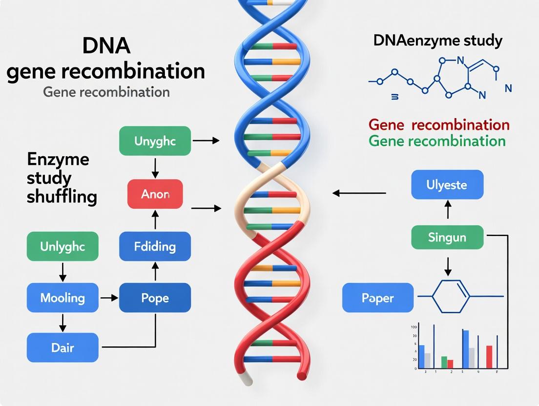
This article provides a comprehensive overview of DNA shuffling and gene recombination techniques, essential tools in directed evolution for researchers, scientists, and drug development professionals.
DNA Shuffling and Gene Recombination: A Comprehensive Guide for Modern Directed Evolution
Abstract
This article provides a comprehensive overview of DNA shuffling and gene recombination techniques, essential tools in directed evolution for researchers, scientists, and drug development professionals. It begins by exploring the foundational principles and history behind mimicking natural evolution in vitro. It then details core methodologies, advanced applications in protein and enzyme engineering, and biotherapeutics development. The guide addresses common troubleshooting and optimization strategies for maximizing library diversity and quality. Finally, it offers a comparative analysis of validation techniques and next-generation sequencing approaches to assess shuffled libraries, concluding with future implications for biomedical research.
What is DNA Shuffling? The Foundation of In Vitro Evolution
Within the broader thesis of advancing protein engineering and directed evolution, DNA shuffling and gene recombination represent foundational methodologies. These techniques accelerate the laboratory mimicry of natural evolution by recombining genetic elements from multiple parental sequences to generate novel, optimized variants. The core principle involves the fragmentation of homologous genes followed by their reassembly into full-length chimeric genes through a polymerase cycling assembly. This process introduces crossovers at regions of sequence homology, generating diversity that can be screened for improved or novel functions. Recent advancements integrate machine learning for in silico library design and next-generation sequencing for high-throughput fitness landscape analysis.
Application Notes: Comparative Analysis of Recombination Methods
The following table summarizes key quantitative parameters for contemporary DNA shuffling and recombination techniques, crucial for selecting an appropriate strategy in drug development pipelines.
Table 1: Comparison of DNA Shuffling and Gene Recombination Methods
| Method | Principle | Avg. Crossover Frequency (per kB) | Library Diversity (Theoretical) | Optimal Parent Homology | Primary Application |
|---|---|---|---|---|---|
| Classical DNA Shuffling | DNase I fragmentation + PCR reassembly | 4-10 | High (~10⁸) | >70% | Family shuffling of homologous genes |
| Staggered Extension Process (StEP) | Template switching during PCR | 1-5 | Moderate (~10⁶) | >50% | Low-homology recombination |
| Yeast Homologous Recombination | In vivo recombination in yeast | High (user-defined) | Very High (~10¹⁰) | >30 bp homology arms | Assembly of large pathways & megabases DNA |
| Sequence Homology-Independent Protein Recombination (SHIPREC) | Linker-based fusion of fragments | 1 (fixed) | Moderate (~10⁵) | None required | Recombination of unrelated genes |
| Rationally Designed Libraries (e.g., SISDC) | Computational design of crossover points | Programmable | Focused (~10⁴) | Variable | Targeted exploration of sequence space |
Experimental Protocols
Protocol 3.1: Standard DNA Shuffling for Family of Homologous Genes
Objective: To create a chimeric library from 3-5 parental genes with >70% sequence identity for directed evolution of enzymatic activity.
Materials:
- DNA Parents: 100-500 ng of each purified gene fragment.
- DNase I: (0.015 U/µL final concentration) in 50 mM Tris-HCl, 10 mM MnCl₂, pH 7.4.
- PCR Reagents: Taq DNA Polymerase (or high-fidelity polymerase), dNTPs, MgCl₂, appropriate primers flanking the gene.
- Agarose Gel Electrophoresis System for size selection (50-100 bp fragments).
- Thermocycler.
Procedure:
- Fragment Generation:
- Combine parental DNA in equimolar ratios (total 1-2 µg).
- Digest with DNase I at 25°C for 10-20 minutes. Quench with 10 mM EDTA.
- Separate fragments on 2% agarose gel. Excise and purify fragments in the 50-100 bp range.
Reassembly PCR:
- Assemble reaction without primers: 10-50 ng purified fragments, 0.2 mM dNTPs, 2.5 mM MgCl₂, 2.5 U Taq polymerase, 1x PCR buffer.
- Thermocycling: 95°C for 2 min; then 35 cycles of [94°C for 30 sec, 50-55°C for 30 sec, 72°C for 30 sec + 5 sec/cycle]; final 72°C for 5 min. This allows priming and extension of fragments on homologous templates.
Amplification of Full-Length Products:
- Use 1 µL of reassembly product as template in a standard PCR with gene-specific flanking primers.
- Clone the resulting PCR product into your desired expression vector for library creation.
Protocol 3.2:In VivoDNA Shuffling via Yeast Homologous Recombination
Objective: To recombine large DNA fragments or pathways (>5 kB) with high efficiency for metabolic engineering.
Materials:
- S. cerevisiae strain with high recombination efficiency (e.g., BY4741).
- Linearized Vector and PCR-amplified gene fragments with 30-50 bp homology overlaps.
- Lithium Acetate (LiAc)/PEG Transformation reagents.
- Synthetic Drop-out Agar Plates for selection.
Procedure:
- Preparation of DNA Parts:
- Amplify each gene fragment with primers that create 30-50 bp overlaps with adjacent fragments and the linearized vector ends.
- Purify all fragments and the linearized vector.
Yeast Transformation:
- Follow standard LiAc/PEG yeast transformation protocol.
- Combine 100-200 ng of linearized vector with a 2-3x molar excess of each overlapping gene fragment.
- Co-transform the DNA mixture into competent yeast cells.
Selection and Library Recovery:
- Plate transformation on appropriate synthetic drop-out media.
- Incubate at 30°C for 2-3 days.
- Harvest yeast colonies, perform colony PCR or plasmid rescue (to E. coli) to obtain the recombined DNA library.
Visualizations
Title: Classical DNA Shuffling Experimental Workflow
Title: Yeast Homologous Recombination Assembly
The Scientist's Toolkit: Essential Research Reagent Solutions
Table 2: Key Reagents and Materials for DNA Shuffling Experiments
| Item | Function & Role in Experiment | Example/Catalog Consideration |
|---|---|---|
| High-Fidelity DNA Polymerase | Accurate amplification of parental genes and final chimeric products; reduces spurious mutations. | Q5 (NEB), KAPA HiFi, Phusion. |
| DNase I (RNase-free) | Controlled digestion of parental DNA into random fragments for classical shuffling. | Worthington Biochemical, Roche. |
| Homologous Recombination Kit (Yeast) | Streamlines in vivo assembly, increasing transformation efficiency and colony yield. | Yeast Maker, Gibson Assembly Master Mix (can be adapted). |
| Gel Extraction & PCR Purification Kits | Critical for size-selecting fragmented DNA and purifying assembly products. | Qiagen, Zymoclean, Monarch kits. |
| E. coli Cloning Strain | High-efficiency chemical competent cells for library construction after in vitro shuffling. | NEB 10-beta, DH5α, TOP10. |
| Next-Generation Sequencing Service | Deep sequencing of input libraries and evolved populations to map crossovers and identify hits. | Illumina MiSeq, services from Genewiz or Azenta. |
| Robotic Liquid Handling System | Enables high-throughput library preparation, transformation, and screening assays. | Beckman Coulter Biomek, Opentrons OT-2. |
1. Historical Evolution and Quantitative Milestones The development of DNA shuffling and gene recombination techniques represents a paradigm shift from observing natural evolution to directing it in the laboratory. The table below summarizes key historical milestones and their quantitative impacts.
Table 1: Key Milestones in Directed Evolution & DNA Shuffling
| Year | Pioneer(s)/Group | Technology/Method | Key Quantitative Outcome | Ref. |
|---|---|---|---|---|
| 1970s | R. K. Saiki et al. | Polymerase Chain Reaction (PCR) | Amplified DNA fragments by a factor of 2^30 (>1 billion copies). | [1] |
| 1994 | Willem P. C. Stemmer | DNA Shuffling (Sexual PCR) | Increased β-lactamase activity 32,000-fold over wild-type after 3 rounds. | [2] |
| 1998 | Frances H. Arnold | Directed Evolution of Enzymes | Evolved subtilisin E for activity in 60% DMF; 256-fold improvement. | [3] |
| 2001 | C. H. Kim et al. | Family Shuffling | Created chimeric P450 enzymes with 20-fold higher activity than parents. | [4] |
| 2010s | D. R. Liu et al. | Phage-Assisted Continuous Evolution (PACE) | Achieved >300 rounds of protein evolution in a single 10-day experiment. | [5] |
| 2020s | Various (e.g., D. Baker) | Machine Learning-Guided Diversification | Designed novel enzymes with >100-fold efficiency improvements over initial designs. | [6] |
2. Application Notes & Core Protocols
2.1. Protocol: Stemmer's Classical DNA Shuffling (DNase I-Based) Objective: Recombine homologous DNA sequences to generate a library of chimeric genes for directed evolution.
Materials & Reagents:
- DNA Parental Genes: Pool of homologous gene sequences (≥ 70% identity).
- DNase I: To fragment DNA randomly.
- Taq DNA Polymerase: For primerless reassembly PCR.
- dNTPs: Deoxynucleotide triphosphates.
- PCR Primers: Gene-specific primers flanking the shuffled region.
- Gel Extraction Kit: For purification of DNA fragments.
Procedure:
- Fragmentation: Combine 1–10 µg of pooled DNA in 100 µL of DNase I digestion buffer (e.g., 50 mM Tris-HCl, pH 7.5, 10 mM MnCl₂). Add 0.015–0.15 units of DNase I and incubate at 15°C for 10–20 min. Goal: generate random fragments of 50–200 bp.
- Purification: Run fragments on a 2% agarose gel. Excise and purify the 50–200 bp smear.
- Reassembly PCR: Set up a 100 µL PCR reaction without primers. Use 10–100 ng of purified fragments, 0.2 mM dNTPs, 2.5 U of Taq polymerase, and standard PCR buffer. Thermocycle: 95°C for 2 min; then 35–45 cycles of [94°C for 30 sec, 50–60°C for 30 sec, 72°C for 30 sec + 5 sec/cycle]; final extension at 72°C for 7 min. This allows homologous fragments to prime each other.
- Amplification: Dilute the reassembly product 10-fold. Use 2–5 µL as template in a standard 50 µL PCR with flanking primers to amplify full-length chimeric genes.
- Cloning & Selection: Clone the amplified library into an expression vector and screen/select for desired functional improvements.
2.2. Protocol for In Silico Shuffling and Machine Learning-Guided Design (Contemporary Approach) Objective: Use computational tools to design a focused, high-potential variant library.
Procedure:
- Sequence Alignment & Analysis: Curate a multiple sequence alignment (MSA) of the target protein family. Identify conserved and variable regions.
- Fitness Prediction Model: Train a machine learning model (e.g., Gaussian process, neural network) on experimental data from a prior, smaller library linking sequence to function (e.g., fluorescence, activity).
- In Silico Library Generation: Use algorithms (e.g., SCHEMA, PROSS) to computationally recombine parental sequences or generate point mutations, predicting stability and function.
- Variant Ranking: Score all in silico generated variants using the trained model. Select the top 100–1000 predicted best variants for synthesis.
- Empirical Testing: Synthesize the gene library (via oligo pool synthesis), express, and assay. Feed new data back into the model for iterative rounds of improvement.
3. The Scientist's Toolkit: Key Research Reagent Solutions Table 2: Essential Materials for DNA Shuffling Experiments
| Reagent/Material | Function/Application | Example Product/Note |
|---|---|---|
| High-Fidelity DNA Polymerase | Error-free amplification of parental genes and final shuffled products. | Phusion U Hot Start DNA Polymerase. |
| DNase I (RNase-free) | Controlled random digestion of DNA for classical shuffling. | Requires Mn²⁺ to create random double-strand breaks. |
| Next-Generation Sequencing Kit | Deep mutational scanning to map sequence-function relationships. | Illumina DNA Prep kits for library preparation. |
| Golden Gate Assembly Mix | Efficient, seamless assembly of shuffled fragments into vectors. | BsaI-HFv2 based systems for modular cloning. |
| Phosphorothioate-modified dNTPs | Used in some shuffling methods to bias crossover points and enhance diversity. | Increases resistance to exonuclease digestion. |
| In silico Design Software | Predicts protein stability, folding, and functional landscapes. | Rosetta, FoldX, ProteinMPNN. |
4. Visualized Workflows & Pathways
Classical DNA Shuffling Experimental Workflow
ML-Guided Directed Evolution Cycle
From Natural to Directed Evolution Principle
This application note, framed within a broader thesis on directed evolution via gene recombination, details the comparative advantages of DNA shuffling for protein engineering. It provides quantitative comparisons, practical protocols, and essential resources for researchers and drug development professionals.
Comparative Analysis: DNA Shuffling vs. Alternative Methods
Table 1: Key Methodological and Outcome Comparison
| Parameter | Random Mutagenesis (e.g., error-prone PCR) | Rational Design (e.g., site-directed mutagenesis) | DNA Shuffling (Family/Chimeragenesis) |
|---|---|---|---|
| Primary Basis | Stochastic nucleotide substitution | Pre-existing structural/mechanistic knowledge | Recombination of functional genetic diversity |
| Library Diversity | Point mutations (low complexity, often deleterious). | Targeted, precise changes (very low complexity). | Combinatorial assembly of beneficial mutations/segments (high functional complexity). |
| Evolutionary Mimicry | Low; mimics point mutation only. | None; purely computational/structural. | High; mimics sexual recombination, accelerating natural evolution. |
| Probability of Improved Variants | Low; "hill-climbing" limited by single mutational steps. | Variable; entirely dependent on accuracy of model and hypothesis. | High; combines beneficial mutations from different parents in single step. |
| Throughput Requirement | Very high (to find rare beneficial combinations). | Low (tests specific designs). | High, but with higher frequency of improved clones. |
| Key Limitation | Accumulation of neutral/deleterious mutations; rarely crosses fitness valleys. | Requires extensive, often imperfect, knowledge of structure-function. | Requires starting sequence diversity (homology >60-70% often needed). |
| Typical Fold Improvement* | 2-10 fold | Can be infinite if design is correct, but often 0-fold (failure). | 100-10,000 fold (cumulative from multiple cycles) |
Data synthesized from recent literature (e.g., *ACS Synth. Biol. 2023, 12, 4, 1089–1103) and historical benchmarks (Stemmer, 1994). Improvements are property-dependent (e.g., enzyme activity, thermostability, binding affinity).
Protocol: Standard DNA Shuffling for Enzyme Thermostability
Objective: To recombine homologous genes from mesophilic and thermophilic organisms to generate chimeric enzymes with enhanced thermostability.
Materials & Workflow:
Diagram Title: DNA Shuffling Protocol Core Workflow
Detailed Protocol Steps:
- Gene Preparation: Isolate or synthesize 2-4 homologous parent genes (e.g., ~1kb each, >70% identity). Purify via agarose gel electrophoresis.
- DNase I Fragmentation:
- Combine 1-10 µg total DNA in 100 µL of 50 mM Tris-HCl (pH 7.4), 10 mM MnCl₂.
- Add 0.15 U of DNase I (RNase-free). Incubate at 15°C for 10-20 min.
- Monitor fragment size (aim for 10-50 bp) by analyzing 5 µL aliquots on a 3% agarose gel.
- Stop reaction by heating to 90°C for 10 min in the presence of 10 mM EDTA.
- Fragment Purification: Purify fragments using a silica-membrane-based kit (e.g., Qiagen QIAquick PCR Purification Kit). Elute in 30 µL nuclease-free water.
- PCR Assembly (Self-Priming):
- Mix: 30 µL purified fragments, 5 µL 10X PCR buffer (no Mg²⁺), 1 µL dNTPs (10 mM each), 2.5 µL MgCl₂ (50 mM), 0.5 µL Taq DNA polymerase (5 U/µL). Add water to 50 µL.
- Cycle: 94°C for 2 min; then 40-60 cycles of [94°C for 30 sec, 50-55°C for 30 sec, 72°C for 30 sec + 5 sec/cycle]; final 72°C for 5 min. This step allows homologous fragments to prime each other.
- Full-Length Gene Amplification:
- Use 1 µL of the assembly product as template in a 50 µL standard PCR with gene-specific primers flanking the ORF.
- Gel-purify the correctly sized product.
- Library Construction & Screening: Clone the shuffled pool into an expression vector. Transform into competent E. coli. Screen colonies for thermostability via a high-throughput assay (e.g., residual activity after heat challenge vs. a standard assay).
The Scientist's Toolkit: Key Reagent Solutions
Table 2: Essential Research Reagents for DNA Shuffling
| Reagent/Material | Function & Critical Note |
|---|---|
| DNase I (Grade I, RNase-free) | Creates random double-stranded breaks. Critical: Use Mn²⁺ buffer to generate blunt-ended fragments, not Mg²⁺. |
| Homologous Parent Genes | Source of diversity. Can be natural variants, engineered mutants, or synthetic designed libraries. Optimal homology: 70-95%. |
| Proofreading DNA Polymerase (e.g., Q5, Phusion) | Used for final amplification to minimize introduction of new point mutations during PCR. |
| Non-Proofreading Polymerase (e.g., Taq) | Used in the assembly PCR step due to its higher tolerance for mismatched primers (fragments). |
| High-Efficiency Cloning Kit (e.g., Gibson Assembly, Golden Gate) | For seamless, high-efficiency assembly of shuffled products into expression vectors, maximizing library size. |
| High-Throughput Screening Substrate | Fluorogenic or chromogenic substrate compatible with cell lysates or culture supernatants for rapid activity detection. |
| Thermocycler with Gradient Function | Essential for optimizing annealing temperatures during the assembly and amplification steps. |
Conceptual Advantage: Crossing Fitness Valleys
The principal advantage of shuffling is its ability to combine mutations that are individually neutral or deleterious but collectively beneficial—a process nearly impossible for sequential random mutagenesis.
Diagram Title: Shuffling Crosses Fitness Valleys
Conclusion: DNA shuffling remains a cornerstone of directed evolution because it harnesses the power of recombination. It systematically outperforms random mutagenesis in discovering synergistic mutations and bypasses the knowledge bottlenecks of rational design, providing a robust, nature-inspired engine for protein optimization in therapeutic and industrial applications.
Within the broader thesis on DNA shuffling and gene recombination techniques, the Shuffle-Select-Amplify cycle represents the foundational, iterative engine of in vitro directed evolution. This paradigm mimics Darwinian evolution at the molecular level, enabling researchers to evolve proteins, ribozymes, or entire pathways with novel or enhanced functions for drug discovery, biocatalysis, and synthetic biology. The cycle consists of three core phases: the creation of genetic diversity (Shuffle), the application of a functional screen or selection (Select), and the recovery and preparation of genetic material for the next iteration (Amplify). This document provides detailed application notes and protocols for implementing this cycle, grounded in current methodologies.
Application Notes
The Shuffle Phase: Generating Diversity
The "Shuffle" phase involves creating a combinatorial library of variant genes. The key is to balance diversity with the retention of beneficial mutations and structural integrity.
- DNA Shuffling (Stemmer Method): The classic method uses DNase I to fragment a pool of homologous parent genes, followed by a reassembly PCR without primers. This allows homologous recombination of fragments, swapping blocks of sequence between parents.
- Family Shuffling: Uses genes from natural homologous families as parents for DNA shuffling, accessing a broader functional landscape.
- Site-Saturation Mutagenesis & CASTing: Focuses diversity to specific residues or regions (e.g., around the active site), often used in conjunction with shuffling.
- Modern Techniques: Methods like Golden Gate shuffling, USER assembly, and CRISPR-assisted editing enable more precise and seamless assembly of large gene blocks.
Note: Library quality is paramount. Use computational tools to model library size and diversity. Aim for a library size that exceeds the theoretical diversity by at least 10-fold to ensure coverage.
The Select Phase: Interrogating Function
The "Select" phase applies the selective pressure. The stringency and throughput of this step determine the success of the evolution campaign.
- In vivo Selection: Linking gene function to cell survival (e.g., antibiotic resistance, auxotrophy complementation). Offers extreme throughput (>10^10 variants) but is limited to functions compatible with host biology.
- Phage/yeast/ribosome Display: Physically linking the gene (in a viral genome or on a ribosome) to its encoded protein product. Allows panning against immobilized targets. Common for evolving antibody affinities.
- Microfluidic-based Screening (FACS, droplet sorting): Enables ultra-high-throughput (10^7-10^9) screening of fluorescent or enzymatic activities in picoliter compartments.
- In vitro Compartmentalization (IVC): Emulsion-based technology that creates cell-like compartments for transcription/translation, linking genotype to phenotype without host cells.
Note: The selection pressure must be carefully tuned. Too stringent, and no variants survive; too relaxed, and the background noise drowns out improved clones. Iterative rounds with gradually increasing stringency are often most effective.
The Amplify Phase: Regenerating the Pool
The "Amplify" phase recovers the genetic material from selected variants for analysis or the next shuffling cycle.
- PCR Amplification: Standard method to recover genes from selected clones or pools. Use high-fidelity polymerases to minimize the introduction of spurious mutations.
- Pooled Plasmid Recovery: For selection methods that retain plasmids (e.g., some in vivo selections), simply extracting and transforming the plasmid pool is sufficient.
- Next-Generation Sequencing (NGS) Analysis: Critical modern tool. Sequencing the pool post-selection identifies enriched mutations and clonal families, informing the design of parent genes for the next shuffle round (data-driven evolution).
Protocols
Protocol 1: DNA Shuffling and Reassembly
Objective: To create a shuffled library from 2-4 parental genes with >70% homology.
Materials:
- Purified parental DNA fragments (PCR-amplified genes, 0.5-1 kb each).
- DNase I (RNase-free, 1 U/µL).
- MnCl2 (10 mM).
- DTT (0.1 M).
- Taq DNA Polymerase (or similar non-proofreading polymerase).
- dNTP mix (10 mM each).
- Primers flanking the gene of interest.
- PCR purification kit.
- Agarose gel electrophoresis equipment.
Procedure:
- Fragment Generation:
- Pool 1-5 µg of total parental DNA.
- In a 100 µL reaction, add 0.5-1.0 U of DNase I, 2 mM MnCl2, and 1x DNase I buffer.
- Incubate at 15°C for 10-20 min. Monitor fragmentation by running 10 µL on a 2.5% agarose gel. Ideal fragment size is 50-200 bp.
- Stop reaction by heating to 90°C for 10 min.
- Purify fragments using a PCR cleanup kit.
Reassembly PCR (Primerless):
- Set up a 50 µL reaction: 100-200 ng purified fragments, 0.2 mM dNTPs, 2.5 U Taq polymerase, 1x PCR buffer (with Mg2+).
- Run the following program:
- 94°C for 2 min.
- [94°C for 30 sec, 50-60°C for 30 sec, 72°C for 30-60 sec] for 35-45 cycles.
- 72°C for 5 min.
- Analyze 5 µL on a 1% agarose gel. A smear or a band at the expected full-length size should appear.
Amplification of Full-Length Products:
- Dilute 1 µL of the reassembly product 1:50.
- Perform a standard PCR with flanking primers.
- Gel-purify the band corresponding to the correct size.
- Clone into your desired expression vector for the Select phase.
Protocol 2: Microtiter Plate-Based Screening for Enzyme Activity
Objective: To screen ~10^4 clones from a shuffled library for improved enzymatic activity.
Materials:
- E. coli clones transformed with the shuffled library, arrayed in 96- or 384-well plates.
- LB media with selective antibiotic.
- Induction agent (e.g., IPTG).
- Lysis buffer (e.g., BugBuster Master Mix).
- Transparent flat-bottom assay plates.
- Plate reader with kinetic capability.
- Enzyme-specific substrate.
Procedure:
- Culture and Expression:
- Inoculate clones in deep-well plates with 0.5-1 mL media. Grow overnight at 37°C, 300 rpm.
- Dilute cultures 1:50 into fresh media in assay plates. Grow to mid-log phase.
- Induce protein expression with optimal concentration of inducer (e.g., 0.1-1 mM IPTG). Incubate for 4-16 hours at appropriate temperature.
Cell Lysis:
- Pellet cells by centrifugation (2000 x g, 10 min).
- Resuspend in 100-200 µL of lysis buffer per well. Incubate with shaking for 20-30 min.
Activity Assay:
- Transfer 20-50 µL of lysate (or clarified supernatant) to a fresh assay plate.
- Initiate reaction by adding 100-200 µL of substrate solution (prepared in appropriate buffer).
- Immediately place plate in a pre-warmed plate reader.
- Measure product formation (e.g., absorbance, fluorescence) kinetically over 5-30 minutes.
- Calculate initial velocities for each well.
Hit Identification:
- Normalize activities to cell density (e.g., OD600 of culture pre-lysis).
- Select clones showing >2-3 standard deviations above the average activity of the parental controls for sequence analysis and further validation.
Data Presentation
Table 1: Comparison of Key Shuffling & Selection Techniques
| Technique | Typical Library Size | Throughput (Variants Screened) | Key Advantages | Best For |
|---|---|---|---|---|
| DNA Shuffling | 10^6 - 10^8 | 10^4 - 10^7 | Recombines beneficial mutations; mimics natural recombination. | General protein optimization, enzyme evolution. |
| Golden Gate Shuffling | 10^3 - 10^6 | 10^3 - 10^6 | Scarless, precise, order-of-operations control. | Pathway assembly, domain swapping, multi-gene circuits. |
| Phage Display | 10^8 - 10^11 | 10^10 - 10^13 | Direct physical genotype-phenotype link; very high library size. | Protein-protein interactions (antibodies, peptides). |
| FACS-based Screening | 10^7 - 10^9 | 10^7 - 10^9 per hour | Quantitative, multi-parameter, ultra-high-throughput. | Enzymes with fluorescent or cell-surface readouts. |
| Droplet Sorting | 10^7 - 10^10 | 10^7 - 10^9 per day | Compartmentalization allows assay of diverse chemistries. | Any reaction where substrate/product can be coupled to fluorescence. |
Table 2: Example Evolution Campaign Metrics for a Hydrolase
| Round | Shuffling Method | Selection Pressure | Library Size | Hits Identified | Best kcat/Km Improvement (vs. WT) |
|---|---|---|---|---|---|
| 1 | Family Shuffling (4 parents) | 0.1 mM Substrate analog in vivo | 5 x 10^6 | 45 | 2.5x |
| 2 | Staggered Extension (SEP) from Round 1 hits | 0.5 mM Substrate analog in vivo | 2 x 10^7 | 12 | 12x |
| 3 | Site-saturation at 3 hot-spot residues | Microtiter plate screen for activity at pH 5.0 | 3 x 10^4 | 3 | 40x |
Visualizations
Diagram Title: The Core Shuffle-Select-Amplify Cycle
Diagram Title: DNA Shuffling Protocol Workflow
The Scientist's Toolkit
Table 3: Essential Research Reagent Solutions for Directed Evolution
| Item | Function in the Cycle | Example/Notes |
|---|---|---|
| High-Fidelity & Taq DNA Polymerases | Amplify parent genes (high-fidelity) and drive recombination in primerless assembly (Taq). | KAPA HiFi for fidelity; wild-type Taq for shuffling reassembly. |
| DNase I (for classic shuffling) | Randomly cleaves parent genes to generate fragments for recombination. | Must be used with Mn2+ to generate random, not staggered, ends. |
| Golden Gate Assembly Mix | Modern shuffling method using Type IIs restriction enzymes for seamless assembly. | Esp3I or BsaI-HFv2, T7 Ligase. Enables precise modular cloning. |
| Microfluidic Encapsulation Reagent | Forms monodisperse water-in-oil droplets for ultra-high-throughput screening. | Fluorinated oil/surfactant systems (e.g., from Sphere Fluidics, Bio-Rad). |
| Phage Display Kit (M13) | Provides the system for in vitro selection of binding proteins/peptides. | Commercial kits from NEB, Thermo Fisher simplify library construction and panning. |
| Fluorescent/Chromogenic Substrates | Report on enzymatic activity in microtiter plate or droplet-based screens. | Must be cell-permeable or used with lysis for intracellular enzymes. |
| Next-Generation Sequencing Kit | Deep sequencing of variant pools to identify enriched mutations and map diversity. | Illumina MiSeq kits for short reads; Oxford Nanopore for full-length gene analysis. |
| Lysis Reagent (for cell-based screens) | Releases intracellular enzyme for activity assays in microtiter plates. | BugBuster, PopCulture, or lysozyme-based buffers. |
Within a broader research thesis on DNA shuffling and gene recombination techniques, understanding homologous sequences and gene families is foundational. These concepts provide the raw genetic material—evolutionarily related sequences with conserved functions or structures—for recombination-based protein engineering. Directed evolution methods, such as DNA shuffling, rely on recombining homologous genes from a family to generate novel chimeric proteins with improved or new properties, accelerating drug development and biocatalyst design.
Definitions & Key Concepts
Homologous Sequences: Sequences descended from a common ancestor. They can be:
- Orthologs: Homologs separated by a speciation event (e.g., the same gene in human and mouse).
- Paralogs: Homologs separated by a gene duplication event within a genome (e.g., beta-globin and myoglobin in humans).
- Xenologs: Homologs transferred horizontally between organisms.
Gene Family: A set of several similar genes, formed by duplication of a single original gene, and generally with similar biochemical functions. They are clusters of paralogs within and across genomes.
Quantitative Data: Metrics for Homology Analysis
Table 1: Key Quantitative Metrics for Analyzing Homologous Sequences
| Metric | Description | Typical Threshold for Homology Inference | Tool Example |
|---|---|---|---|
| Percent Identity | Percentage of identical residues between two aligned sequences. | >25-30% often suggests common ancestry. | BLAST, Clustal Omega |
| E-value | The number of expected hits of similar quality (score) by chance. Lower is better. | <1e-5 to <1e-3 is considered significant. | BLAST |
| Bit Score | A normalized score representing alignment quality, independent of database size. Higher is better. | Higher scores indicate more significant matches. | BLAST, HMMER |
| Coverage | The fraction of the query sequence length aligned to a target sequence. | High coverage with significant identity strengthens homology claim. | BLAST |
| Substitution Rate (dN/dS) | Ratio of non-synonymous to synonymous nucleotide substitutions. | dN/dS < 1: purifying selection; =1: neutral; >1: positive selection. | PAML, HyPhy |
Application Notes & Protocols
Note: These protocols are framed within the context of creating a diverse parental gene library for DNA shuffling.
Protocol 1: Identifying a Gene Family and Retrieving Homologous Sequences
Objective: To compile a set of homologous gene sequences from public databases for use as parents in DNA shuffling.
Materials & Reagents:
- Computer with internet access.
- NCBI BLAST suite (online or local installation).
- Sequence retrieval tools (efetch, BioPython Entrez module).
- Multiple Sequence Alignment (MSA) software (e.g., MAFFT, Clustal Omega).
Procedure:
- Seed Sequence: Start with a protein or DNA sequence of interest (the "seed").
- Homology Search: Use the seed to perform a BLASTP (for protein) or TBLASTN (protein vs. translated nucleotide) search against the non-redundant (nr) database.
- Filter Results: Apply filters: E-value < 1e-10, query coverage > 70%, and percent identity across a range (e.g., 40-90%) to capture functional diversity.
- Retrieve Sequences: Download the top 5-20 significant hits, ensuring they represent diverse taxonomic sources.
- Construct MSA: Align the retrieved sequences using MAFFT with default parameters. Visually inspect the alignment for conserved blocks (potential functional domains) and variable regions (potential for recombination diversity).
- Confirm Gene Family: Analyze the MSA phylogenetically (e.g., with FastTree) to confirm evolutionary relationships and identify major paralogous groups.
Protocol 2: In Silico Analysis of Recombination Potential
Objective: To analyze homologous sequences for optimal crossover points prior to experimental DNA shuffling.
Materials & Reagents:
- MSA from Protocol 1.
- Software for identity plot generation (e.g., Geneious, custom Python/R scripts).
- DNA shuffling simulation software (e.g., SHIPREC simulator, in-house scripts).
Procedure:
- Calculate Sequence Identity Plot: Generate a sliding-window plot of percentage identity across the MSA. Regions of high identity (>70%) are predicted to allow for efficient crossovers during shuffling.
- Map Functional Domains: Annotate the MSA with known domain architecture (from Pfam/InterProScan) to ensure crossovers do not consistently disrupt critical functional units.
- Simulate Shuffling: Use in silico shuffling algorithms to model the theoretical diversity of chimeric libraries generated from your homologous set. This helps assess if the family provides sufficient diversity for the engineering goal.
- Select Parental Sequences: Based on steps 1-3, select a final subset (e.g., 4-6 genes) that offers balanced diversity and high cross-over compatibility for experimental work.
Visualization of Concepts & Workflows
Title: Origin of Homologs: Orthologs, Paralogs, Xenologs
Title: Gene Family to DNA Shuffling Workflow
The Scientist's Toolkit
Table 2: Essential Research Reagents & Solutions for Homology Analysis and Shuffling
| Item | Function/Application in Context |
|---|---|
| High-Fidelity DNA Polymerase (e.g., Phusion) | For accurate amplification of homologous parent genes from genomic or cDNA templates prior to shuffling. |
| DNase I (for classical shuffling) | Randomly fragments homologous DNA sequences to generate primers for reassembly in early DNA shuffling protocols. |
| Restriction Enzymes & Ligase | For formal recombination methods like STEP (Staggered Extension Process) or in silico-defined block swapping. |
| Homology Detection Software (BLAST, HMMER) | To identify and retrieve homologous sequences from databases based on statistical significance (E-value). |
| Multiple Sequence Alignment Tool (MAFFT, Clustal Omega) | Aligns homologous sequences to visualize conserved/variable regions and plan recombination points. |
| Chimera Library Assembly Kit (e.g., Gibson Assembly Master Mix) | Seamlessly assembles homologous fragments generated by PCR-based shuffling methods into full-length chimeric genes. |
| Error-Prone PCR Kit | Sometimes used in conjunction with shuffling to introduce additional point mutations within homologous blocks. |
| Expression Vector & Competent Cells | To clone and express the library of shuffled chimeric genes for functional screening (e.g., for drug target activity). |
How to Shuffle DNA: Core Protocols and Cutting-Edge Applications
Within the broader research on in vitro directed evolution, DNA shuffling stands as a cornerstone methodology for gene recombination. This protocol overview details two seminal techniques: Staggered Extension Process (StEP) and DNase I-based DNA shuffling. These methods facilitate the rapid generation of genetic diversity by recombining homologous sequences, enabling the evolution of proteins with improved or novel functions for therapeutic and industrial applications.
Key Research Reagent Solutions
| Reagent / Material | Function in Protocol |
|---|---|
| DNase I (Grade I, RNase-free) | Randomly cleaves dsDNA templates to generate small fragments for reassembly. Critical for classic DNA shuffling. |
| MgCl₂ / MnCl₂ Solution | Divalent cations. Mg²⁺ is standard for DNase I; Mn²⁺ can be used to produce smaller, more random fragments. |
| Taq DNA Polymerase | Thermostable polymerase used in StEP for primer extension and fragment reassembly without added primers. |
| dNTP Mix | Nucleotide building blocks essential for the polymerase-driven extension and reassembly phases. |
| GeneFamily Parental DNA Templates | Homologous genes (≥70% identity) serving as the source of diversity for recombination. |
| Thermocycler | Instrument for precise temperature cycling required for StEP reassembly and PCR amplification. |
| Gel Electrophoresis System | For analyzing fragment size distribution post-DNase I digestion and for purifying reassembled products. |
| QIAquick Gel Extraction Kit | For purification of DNA fragments from agarose gels post-digestion and post-reassembly. |
Table 1: Critical Reaction Conditions for DNase I Shuffling
| Parameter | Typical Range | Optimal Value / Note |
|---|---|---|
| DNase I Concentration | 0.001 - 0.1 U/µg DNA | Must be titrated for each enzyme lot. |
| Digestion Temperature | 15-25°C | Room temperature (22°C) is standard. |
| Digestion Time | 2 - 10 minutes | Time influences fragment size distribution. |
| Fragment Size Target | 10 - 50 bp | Small fragments ensure high crossover frequency. |
| DNA Template Amount | 0.1 - 1 µg per digestion | Higher amounts aid fragment purification. |
Table 2: Critical Cycling Parameters for StEP Shuffling
| Parameter | Typical Range | Function |
|---|---|---|
| Denaturation Temperature | 94 - 96°C | Separates DNA strands. |
| Annealing/Extension Temp | 50 - 65°C | Lowers for very short primer alignment & extension. |
| Extension Time | 5 - 15 seconds | Key parameter; very short to promote template switching. |
| Number of Cycles | 80 - 120 | High cycle count accumulates full-length genes. |
| Parental Template Mix | 10 - 100 ng total | Provides homologous sequences for recombination. |
Detailed Experimental Protocols
Protocol 1: Classic DNase I Shuffling
Objective: To recombine multiple homologous parent genes via random fragmentation and reassembly.
- Template Preparation: Pool 0.5-1 µg of each purified parental DNA sequence (≥70% homology).
- DNase I Digestion:
- Prepare digestion buffer: 50 mM Tris-HCl (pH 7.4), 10 mM MnCl₂ (or 1 mM MgCl₂ for larger fragments).
- Add pooled DNA to buffer on ice.
- Add diluted DNase I (e.g., 0.01 U/µg DNA) and incubate at 22°C for 2-10 minutes.
- Immediately stop reaction by heating to 90°C for 10 minutes (if Mg²⁺ used) or adding 10 mM EDTA.
- Fragment Purification: Resolve digested fragments (target 20-50 bp) on a 2-3% agarose gel. Excise and purify using a gel extraction kit.
- Reassembly PCR:
- Assemble reaction: Purified fragments (10-100 ng), 0.2 mM dNTPs, 2.5 U Taq polymerase, standard PCR buffer.
- Cycle without primers: 94°C for 60s; then 40 cycles of [94°C for 30s, 50-55°C for 30s, 72°C for 30s]; final 72°C for 5 min. Fragments prime each other.
- Amplification: Use 1-5 µL of reassembly product as template in a standard PCR with gene-specific primers to amplify full-length chimeric genes.
- Cloning & Selection: Clone amplified products into an expression vector for functional screening.
Protocol 2: Staggered Extension Process (StEP) Shuffling
Objective: To recombine parent genes in a single tube reaction through repeated very short annealing/extension cycles.
- Template Mix: Combine 10-50 ng of each parental DNA template in a thin-walled PCR tube.
- StEP Reassembly Reaction:
- Prepare a master mix: 1X PCR buffer, 0.2 mM dNTPs, 2.5 U Taq polymerase. No primers added.
- Add master mix to template. Total volume: 50 µL.
- Thermocycling for Reassembly:
- Initial Denaturation: 94°C for 2 min.
- StEP Cycles (80-120 repeats): 94°C for 30s (denaturation) followed by 55°C for 5-15s (annealing/extension). The critical short extension time causes polymerase to dissociate and re-anneal to different templates.
- Full-Length Product Amplification: After reassembly, add gene-specific primers (0.2-1.0 µM final) directly to the tube. Perform 20-25 standard PCR cycles to amplify the recombined full-length products.
- Analysis & Cloning: Analyze PCR product by gel electrophoresis. Purify, clone, and screen the library.
Visualized Workflows
DNase I Shuffling Protocol Workflow
StEP Shuffling Mechanism and Workflow
Application Notes
Within the broader thesis exploring the evolution of DNA shuffling and gene recombination techniques, modern library creation methods address key limitations of classical homologous recombination. ITCHY (Incremental Truncation for the Creation of Hybrid enzymes), SCRATCHY (ITERative SCRATCHY), and RACHITT (Random ChimeraGenesis on Transient Templates) represent pivotal advancements for recombining genes with low homology or for achieving more controlled crossover distributions. Sequence-independent methods further extend the toolbox, enabling fusion without any homology. These techniques are critical in protein engineering for drug development, particularly for creating novel antibodies, enzymes, and biosynthetic pathways.
Key Methods Comparison
Table 1: Comparison of Modern Gene Recombination Methods
| Method | Core Principle | Homology Requirement | Crossover Control | Typical Library Size | Primary Application |
|---|---|---|---|---|---|
| ITCHY | Incremental truncation of gene fragments followed by blunt-end ligation. | None | Single, random fusion point; controlled by truncation granularity. | 10^3 – 10^5 | Creating hybrid genes from unrelated parents; functional domain swapping. |
| SCRATCHY | Iterative application of ITCHY to create multi-crossover libraries. | None | Multiple, random crossover points. | 10^5 – 10^7 | Extensive shuffling of non-homologous genes for deep exploration of sequence space. |
| RACHITT | Annealing of fragmented single-stranded DNA onto a full-length transient template, followed by gap filling and ligation. | Low to High | High frequency of crossovers; template-driven. | 10^7 – 10^9 | High-density shuffling of families with moderate homology for directed evolution. |
| Sequence-Independent (e.g., SISDC, uSEC) | Use of linkers, overlap primers, or specific enzymatic handles (e.g., Type IIs endonucleases). | None | Precisely defined fusion junctions or random via designed linkers. | 10^3 – 10^6 | Fusion of arbitrary DNA fragments, modular cloning, and combinatorial assembly. |
Experimental Protocols
Protocol 1: ITCHY Library Construction
Objective: Create a comprehensive library of single-crossover hybrids between two genes (Gene A and Gene B) with no sequence homology.
Research Reagent Solutions:
- Exonuclease III (ExoIII): Processive 3'→5' exonuclease for controlled truncation.
- S1 Nuclease: Single-strand specific endonuclease to polish ExoIII-generated overhangs.
- Alkaline Phosphatase (CIP): Removes 5'-phosphate to prevent re-circularization of vector.
- T4 DNA Ligase: Joins blunt-ended truncated fragments.
- pTRC-HisA Vector or similar: Expression vector with in-frame start codon and selection marker.
Methodology:
- Fragment Preparation: PCR amplify Gene A and Gene B with primers that introduce flanking, non-complementary restriction sites (e.g., NcoI on Gene A 5', XhoI on Gene B 3').
- Vector Digestion: Digest the expression vector with NcoI and XhoI. Treat with CIP to dephosphorylate.
- Incremental Truncation:
- Digest Gene A with NcoI and a blunt-end generating enzyme (e.g., EcoRV) to create a 5' overhang and a blunt 3' end. Similarly, digest Gene B with XhoI and a different blunt-end cutter to create a 3' overhang and a blunt 5' end.
- Separately, treat the digested Gene A and Gene B with ExoIII at a constant temperature (e.g., 22°C). Remove aliquots at regular timepoints (e.g., every 30 seconds for 10 minutes) and quench in a formamide/EDTA buffer.
- Pool timepoint aliquots for each gene. Treat with S1 nuclease to create blunt ends. Purify.
- Ligation & Cloning: Ligate the pool of truncated Gene A fragments to the pool of truncated Gene B fragments at a 1:1 molar ratio. Then, ligate the resulting hybrid fragments into the prepared vector.
- Transformation: Transform the ligation mixture into high-efficiency E. coli competent cells and plate on selective media to generate the ITCHY library.
Protocol 2: RACHITT Library Construction
Objective: Generate a high-crossover density library from a family of homologous genes (≥70% identity).
Research Reagent Solutions:
- Gene 32 Protein (gp32): Single-stranded DNA binding protein to prevent secondary structure formation.
- DNase I: For random fragmentation of single-stranded DNA (ssDNA).
- T4 DNA Polymerase: For gap filling and repair synthesis.
- DNA Ligase: For sealing nicks post-repair.
- Uracil-DNA Glycosylase (UDG): For selective degradation of the uracil-containing template strand.
- dUTP: Incorporated during PCR to make template strand labile.
Methodology:
- Template Preparation: PCR amplify the primary parental gene using dUTP/dNTP mix to create a uracil-containing ssDNA template. Biotinylate one end and immobilize on streptavidin magnetic beads.
- Donor Fragmentation: Generate ssDNA from the pool of homologous donor genes. Treat with DNase I under controlled conditions to produce random fragments (50-300 bp). Denature and purify.
- Annealing: Mix the donor ssDNA fragments with the immobilized template in a large molar excess (≥100:1) in the presence of gp32. Anneal by slow cooling.
- Template Degradation: Treat the annealed mixture with UDG and a chemical (e.g., piperidine) or enzyme (Endonuclease VIII) to cleave the uracil-containing template backbone, leaving the annealed donor fragments as the new scaffold.
- Gap Filling & Ligation: Incubate with dNTPs, T4 DNA Polymerase, and DNA Ligase to fill any gaps and seal nicks, creating full-length hybrid genes.
- PCR Amplification: Release the full-length hybrids from beads and PCR amplify with flanking primers for subsequent cloning into an expression vector.
Visualizations
ITCHY Workflow: Creating Hybrid Genes by Incremental Truncation
RACHITT Workflow: Template-Mediated High-Density Shuffling
The Scientist's Toolkit
Table 2: Essential Research Reagents for Modern DNA Shuffling
| Reagent / Material | Function in Protocol |
|---|---|
| Exonuclease III (ExoIII) | Core enzyme for ITCHY/SCRATCHY; enables controlled, time-dependent truncation of DNA from the 3' end. |
| Uracil-DNA Glycosylase (UDG) | Critical for RACHITT; enables selective removal of the uracil-containing template strand after donor fragment annealing. |
| Gene 32 Protein (gp32) | Used in RACHITT to coat ssDNA, preventing secondary structure formation and promoting efficient annealing of fragments. |
| Type IIs Restriction Enzyme (e.g., SapI, BsaI) | Enables sequence-independent cloning (e.g., Golden Gate assembly) by cutting outside recognition sites, creating unique, designable overhangs. |
| T4 DNA Polymerase | Used in RACHITT for gap filling; possesses 3'→5' exonuclease and 5'→3' polymerase activity for precise repair synthesis. |
| S1 Nuclease | Converts the staggered ends generated by ExoIII truncation in ITCHY into blunt ends suitable for ligation. |
| Alkaline Phosphatase (CIP/AP) | Prevents vector self-ligation by removing 5'-phosphate groups, a standard step in cloning fragmented libraries. |
| Magnetic Streptavidin Beads | Provides a solid support for immobilizing biotinylated DNA templates (RACHITT) for easy buffer exchange and template removal. |
Software and In Silico Tools for Designing Shuffling Experiments
1. Introduction and Context within DNA Shuffling Research
Within the broader thesis on advancing gene recombination techniques, in silico tools have become indispensable for the rational design of DNA shuffling experiments. Moving beyond purely random recombination, these software platforms enable researchers to simulate shuffling outcomes, predict chimeric library diversity, select optimal fragment assembly strategies, and prioritize variants for synthesis and screening. This application note details current tools, their quantitative benchmarks, and provides executable protocols for integrating computational design into experimental workflows.
2. Quantitative Comparison of Key Software Tools
Table 1: Feature and Performance Comparison of In Silico Shuffling Software
| Tool Name | Primary Function | Input Requirements | Key Algorithm/Output | Reported Library Efficiency Gain | Access |
|---|---|---|---|---|---|
| SCHEMA | Identify recombination- tolerant breakpoints | 3D protein structure or homology model | Computes disruption scores for chimeras; identifies fragments minimizing structural disruption. | Up to 10-fold increase in functional chimera yield vs. random. | MATLAB scripts, Web server. |
| DNAWorks | Optimize oligonucleotide design for gene synthesis | Amino acid sequence, target %GC, codon usage. | Algorithm for de novo gene design via thermodynamically balanced PCR assembly. | >90% synthesis success rate for genes <1 kb. | Web server, standalone. |
| GLUE-IT / PriFi | Design primers for sequence homology-independent recombination | Parent DNA sequences (FASTA). | Identifies recombination sites and designs primers for seamless assembly (e.g., SISDC, USER). | N/A (enables creation of highly diverse libraries). | Web server. |
| Gene Designer | Integrated platform for synthetic gene design and optimization | Sequence, organism-specific parameters. | Codon optimization, restriction site management, oligonucleotide design for assembly. | N/A (streamlines entire design process). | Desktop application. |
| CASTER | Predict crossovers in DNA shuffling | Multiple aligned parent sequences. | Simulates in vitro shuffling process; predicts crossover locations and library diversity. | Accurately models in vitro results (R² >0.85 for crossover prediction). | Web server. |
3. Detailed Experimental Protocols
Protocol 3.1: In Silico Library Design Using SCHEMA and DNAWorks
Objective: Design a chimeric gene family library from three homologous parental genes with optimized codons for E. coli expression.
Materials:
- Parental protein sequences (P1, P2, P3) in FASTA format.
- Homology model or PDB file for one parent.
- SCHEMA (web server or scripts).
- DNAWorks 3.2 (web server).
- Standard desktop computer.
Procedure:
- Sequence Alignment: Generate a robust multiple sequence alignment (MSA) of the three parental protein sequences using Clustal Omega or MUSCLE.
- SCHEMA Analysis: a. Submit the MSA and the structural data to the SCHEMA server. b. Set parameters (e.g., fragment size range: 50-150 amino acids). c. Run the analysis to obtain a list of optimal "breakpoints" that minimize structural disruption upon recombination. d. Export the selected block pattern (e.g., Break at residues 45, 98, 150).
- Chimera Sequence Generation: a. Translate the block pattern into all possible chimeric combinations (e.g., P1-P2-P3, P2-P1-P3, etc.). b. Generate the amino acid sequences for each theoretical chimera.
- Oligonucleotide Design with DNAWorks: a. Input a chimera's amino acid sequence into DNAWorks. b. Set parameters: Host organism (E. coli), desired melting temperature (Tm ~60°C), oligonucleotide length (40-60 bases), optimization for %GC content. c. Run the design. The output will be a list of overlapping oligonucleotides covering the entire gene, optimized for assembly PCR. d. Repeat for all chimeric variants targeted for synthesis.
- Output: A finalized set of oligonucleotide sequences for solid-phase synthesis, ready for experimental assembly.
Protocol 3.2: Simulating Shuffling Outcomes with CASTER
Objective: Predict the statistical diversity and crossover distribution of a traditional DNAse I-based shuffling experiment in silico.
Materials:
- Aligned DNA sequences of parent genes (FASTA format).
- CASTER web server.
Procedure:
- Prepare Input: Ensure parent nucleotide sequences are accurately aligned. The alignment defines regions of homology where crossovers can occur.
- Configure CASTER Parameters: a. Upload the aligned FASTA file. b. Set simulation parameters: Number of in silico progeny (e.g., 10,000), fragment size distribution (e.g., 50-200 bp), homology threshold for recombination (default: >95% identity in overlap region). c. Select the shuffling model ("DNAse I-based fragment assembly").
- Execute Simulation: Run the CASTER algorithm.
- Analyze Results: a. Review the output table showing predicted crossover hotspots and coldspots. b. Analyze the histogram of the number of crossovers per chimeric gene. c. Assess the theoretical library coverage (percentage of all possible chimeras generated).
- Decision Point: Use the simulation data to decide if experimental parameters (e.g., fragment size, parent ratio) need adjustment to achieve desired library diversity.
4. Visualization of Workflows and Logical Relationships
Title: Integrated In Silico Design Workflow for Gene Shuffling
5. The Scientist's Toolkit: Research Reagent Solutions
Table 2: Essential Materials for Computational-Experimental Shuffling Pipeline
| Item / Reagent | Function in Workflow | Example / Notes |
|---|---|---|
| Homology Modeling Software | Generates 3D protein structure from sequence when no PDB exists. Required for SCHEMA. | SWISS-MODEL, AlphaFold2, I-TASSER. |
| High-Fidelity DNA Polymerase | Accurately assembles designed oligonucleotides into full-length chimeric genes. | Phusion U Green, Q5 High-Fidelity. |
| Cloning Vector with Selection | Allows for the ligation and propagation of assembled genes in a host organism. | pET series (for E. coli expression), linearized yeast display vectors. |
| Competent Cells | For transformation of the assembled and ligated library. High efficiency is critical for diversity capture. | NEB 5-alpha (cloning), BL21(DE3) (expression), electrocompetent cells. |
| NGS Library Prep Kit | Validates final library sequence diversity and crossover locations post-assembly. | Illumina Nextera XT, Swift Accel-NGS 2S. |
| Automated Liquid Handler | Enables high-throughput pipetting for setting up assembly PCRs and library transformations. | Beckman Coulter Biomek, Opentrons OT-2. |
Thesis Context: This application note details protocols for enzyme engineering, framed within a broader research thesis exploring advanced DNA shuffling and gene recombination techniques. These methods are pivotal for accelerating the directed evolution of biocatalysts with enhanced industrial properties.
Application Notes
The directed evolution of enzymes via gene recombination mimics natural evolution in the laboratory, enabling the development of biocatalysts with improved activity, stability, and solvent tolerance for industrial applications (e.g., chemical synthesis, pharmaceutical production, and biomass conversion). Recent advances in DNA shuffling methodologies have significantly increased library quality and functional hit rates.
Quantitative Data Summary: Evolution of a Model Lipase for Thermostability
Table 1: Performance Metrics of Parent vs. Evolved Lipase Variants
| Variant | Parent | Variant A3 | Variant D7 | Assay Conditions |
|---|---|---|---|---|
| Half-life (t₁/₂) at 60°C | 15 min | 120 min | 95 min | In 50 mM Tris-HCl, pH 8.0 |
| Melting Temp (Tm) Δ | 0 °C | +12.5 °C | +9.1 °C | DSF measurement |
| Specific Activity | 100% | 145% | 88% | p-NP palmitate hydrolysis |
| Organic Solvent Tolerance | 100% | 210% | 165% | Residual activity after 1h in 25% (v/v) DMSO |
Experimental Protocols
Protocol 1: Staggered Extension Process (StEP) DNA Shuffling Objective: Generate a recombined gene library from a pool of homologous parent genes (e.g., lipase genes from thermophilic organisms). Materials: Parent plasmid DNA templates, gene-specific primers, Taq DNA polymerase (lacking proofreading), dNTP mix, PCR purification kit. Procedure:
- Fragment Generation: Set up a standard PCR (94°C for 30s, 55°C for 30s, 72°C for 1 min/kb) with ≤15 cycles to amplify parent genes. Do not purify.
- StEP Recombination: Dilute the PCR product 1:50. Perform StEP cycling: 94°C for 30s, followed by 50-100 cycles of 94°C for 5s and 55°C for 5s. The very short extension time promotes template switching.
- Full-Length Assembly: Add 0.5 µL of Taq polymerase to the StEP product. Run 5 cycles of standard PCR (94°C for 30s, 55°C for 30s, 72°C for 2 min/kb) to assemble full-length chimeric genes.
- Final Amplification: Add gene-specific primers (0.4 µM final) and perform 25 cycles of standard PCR. Purify the product and clone into your expression vector.
Protocol 2: High-Throughput Screening for Thermostability & Activity Objective: Identify improved variants from a shuffled library using a coupled kinetic assay. Materials: Lysates of E. coli clones expressing the library, p-nitrophenyl ester substrate (e.g., p-NP palmitate), clear 96-well assay plates, multi-channel pipettes, plate reader capable of 405 nm absorbance. Procedure:
- Heat Challenge: Aliquot 50 µL of clarified cell lysate into two sets of a 96-well PCR plate. Incubate one set at the challenge temperature (e.g., 60°C) for 30 minutes. Keep the other set on ice.
- Activity Assay: Transfer 10 µL from each well to a clear 96-well assay plate containing 90 µL of assay buffer (50 mM Tris-HCl, pH 8.0, 0.1% Triton X-100).
- Kinetic Read: Start the reaction by adding 100 µL of pre-warmed p-NP substrate (0.5 mM in isopropanol). Immediately measure the increase in absorbance at 405 nm (A₄₀₅) for 5 minutes at 30°C.
- Analysis: Calculate residual activity for each variant: (Activityheated / Activityunheated) * 100%. Rank clones by both residual activity and total initial activity.
Visualizations
The Scientist's Toolkit
Table 2: Key Research Reagent Solutions for Enzyme Engineering
| Reagent / Material | Function / Purpose |
|---|---|
| High-Fidelity & Taq Polymerase Mix | For initial gene amplification (high-fidelity) and subsequent StEP shuffling (Taq for low processivity). |
| p-Nitrophenyl (p-NP) Ester Substrates | Chromogenic substrates for high-throughput kinetic screening of hydrolytic enzyme (e.g., lipase, esterase) activity. |
| His-Tag Purification Resin (Ni-NTA) | Rapid, standardized purification of His-tagged enzyme variants for detailed biochemical characterization. |
| Thermal Shift Dye (e.g., SYPRO Orange) | For Differential Scanning Fluorimetry (DSF) to quickly estimate protein melting temperature (Tm) changes. |
| Error-Prone PCR Kit | Used in combination with shuffling to introduce de novo point mutations and expand sequence diversity. |
| Golden Gate or Gibson Assembly Master Mix | For seamless, efficient cloning of shuffled gene fragments into expression vectors. |
This application note is situated within a broader thesis on advancing DNA shuffling and gene recombination techniques for protein engineering. The directed evolution of antibodies and therapeutic proteins represents a cornerstone application of these technologies. By harnessing stochastic recombination and rational design, researchers can rapidly traverse vast sequence spaces to identify variants with enhanced affinity, specificity, stability, and developability—parameters critical for successful biotherapeutics.
Table 1: Comparative Analysis of Protein Engineering Techniques
| Technique | Typical Library Size | Key Screening Throughput (variants/week) | Primary Application | Typical Affinity Improvement (KD) | Timeline to Candidate (months) |
|---|---|---|---|---|---|
| Error-Prone PCR | 10^6 - 10^8 | 10^3 - 10^4 (microtiter) | Affinity maturation, stability | 2-10 fold | 6-12 |
| DNA Shuffling (Family) | 10^7 - 10^12 | 10^4 - 10^5 (FACS) | Humanization, multi-parameter optimization | 10-100 fold | 4-8 |
| Yeast Surface Display | 10^7 - 10^9 | 10^7 - 10^8 (FACS) | Antibody affinity, stability | 10-1000 fold | 3-6 |
| Phage Display | 10^9 - 10^11 | 10^7 - 10^8 (panning) | Nanobody/scFv discovery, peptide libraries | 10-100 fold | 2-5 |
| Machine Learning-Guided Library Design | 10^4 - 10^6 | 10^3 - 10^4 (rational) | De novo design, solubility optimization | Predictable multi-parameter gains | 2-4 |
Table 2: Benchmarking of Developed Therapeutic Protein Attributes
| Protein Class | Starting Affinity (nM) | Evolved Affinity (pM) | Thermal Stability (Tm °C Increase) | Aggregation Propensity (% Reduction) | Developability Score (Silico) |
|---|---|---|---|---|---|
| Anti-TNFα mAb | 5.2 | 22 | +8.5 | 65% | High |
| IL-2 Variant (Nektar-like) | N/A (activity) | N/A | +12.1 | 80% | Optimized |
| AAV Capsid (Gene Therapy) | N/A (tropism) | >100x specificity | +5.7 | 40% | N/A |
| CAR T-binding Domain | 310 | 4.5 | +6.3 | 55% | High |
Detailed Experimental Protocols
Protocol 3.1: DNA Shuffling for Antibody Affinity Maturation
Objective: Recombine homologous parent antibody genes (e.g., from immunized animals or initial hits) to create a diverse library for selecting high-affinity clones.
Materials:
- DNase I: For random fragmentation of parent genes.
- PCR reagents: dNTPs, Taq polymerase (no proofreading), primers.
- Purification kits: Gel extraction and PCR clean-up.
- Expression vector: e.g., pComb3X for phage display or pYD1 for yeast display.
Procedure:
- Gene Pool Preparation: Amplify 2-5 homologous antibody VH and VL genes (≥70% identity) using gene-specific primers. Purify and quantify.
- Fragmentation: Digest 1-2 µg of pooled DNA with 0.15 U DNase I in 10 µL of 50 mM Tris-HCl (pH 7.4), 10 mM MnCl2 at 15°C for 10-20 min. Quench with 10 mM EDTA. Analyze fragments (20-50 bp) on agarose gel.
- Reassembly PCR: Perform PCR without primers: 2-10 ng fragments, 0.2 mM dNTPs, 2.5 U Taq polymerase in 50 µL. Cycle: 95°C 2 min; then 35-45 cycles of [94°C 30s, 50-55°C 30s, 72°C 30s]; final 72°C 5 min.
- Amplification: Add outer primers (1 µM final) to 5 µL of reassembly product. Run standard PCR (20-25 cycles) to amplify full-length shuffled genes.
- Cloning & Selection: Digest amplified product and vector, ligate, transform into appropriate host (e.g., E. coli TG1 for phage). Proceed to panning (Protocol 3.2) or FACS screening.
Protocol 3.2: Yeast Surface Display for Multi-Parameter Screening
Objective: Simultaneously screen for antigen binding affinity and thermal stability.
Materials:
- Yeast strain: EBY100 (S. cerevisiae).
- Induction media: SGCAA and SGLCAA.
- Labeling reagents: Biotinylated antigen, fluorescent streptavidin (e.g., SA-PE), anti-c-myc-FITC antibody.
- FACS sorter.
Procedure:
- Library Transformation: Transform the shuffled library (from Protocol 3.1, cloned into pYD1) into EBY100 electrocompetent cells. Plate on SDCAA to determine library size.
- Induction: Inoculate library into SDCAA, grow at 30°C to OD600 ~6. Pellet, wash, resuspend in SGCAA to OD600=1.0. Induce at 20°C, 250 rpm for 20-24h.
- Dual-Parameter Labeling:
- For Affinity: Serially dilute biotinylated antigen (1 nM to 1 µM). Label 1e7 induced yeast with antigen dilutions on ice for 1h. Wash, label with SA-PE.
- For Stability: Aliquot labeled yeast. Heat shock one aliquot at desired temperature (e.g., 65°C, 70°C) for 5-10 min, keep control on ice.
- Surface Expression: Label all samples with anti-c-myc-FITC.
- FACS Gating & Sorting: Gate on FITC+ (expression-positive) cells. For the non-heat-shocked sample, sort the top 1-2% of PE+ (high-affinity) binders at a low antigen concentration (e.g., 10 nM). For the heat-shocked sample, sort PE+ cells that retain fluorescence post-heat shock.
- Recovery & Analysis: Grow sorted populations in SDCAA, repeat induction and sorting for 2-3 rounds. Isolate plasmid DNA from final sorted pool, sequence individual clones, and characterize.
Diagrams and Visualizations
The Scientist's Toolkit: Research Reagent Solutions
Table 3: Essential Materials for Shuffling & Display Experiments
| Item | Function & Key Attribute | Example Product/Catalog |
|---|---|---|
| DNase I (RNase-free) | Creates random DNA fragments for shuffling. Critical for controlling fragment size distribution. | Thermo Scientific EN0521 |
| Taq DNA Polymerase | Used in reassembly PCR; lack of proofreading allows incorporation of mismatches, promoting diversity. | NEB M0273 |
| Yeast Display Vector | Episomal vector for surface display; contains Aga2p fusion, selection markers (e.g., TRP1), c-myc tag. | Addgene pYD1 |
| Biotinylated Antigen | Essential for labeling during FACS or panning. Requires site-specific biotinylation to avoid epitope masking. | Biotinylation kit: Thermo 21435 |
| Magnetic Streptavidin Beads | For phage or yeast panning; captures biotinylated antigen and bound clones. | Dynabeads M-280 Streptavidin |
| Anti-c-myc-FITC Antibody | Detects surface expression level on yeast, enabling normalization of binding signal. | Miltenyi Biotec 130-116-485 |
| Electrocompetent E. coli TG1 | High-efficiency transformation for phage display library construction. | Lucigen 60502 |
| Electrocompetent S. cerevisiae EBY100 | Strain engineered for efficient surface display via the Aga1/Aga2 system. | Invitrogen C303003 |
| Next-Generation Sequencing (NGS) Service | Deep sequencing of library pools pre- and post-selection to track enrichment. | Illumina MiSeq |
| Protein A/G Biosensor Chips | For label-free kinetic analysis (KD, kon, koff) of purified antibodies via SPR/BLI. | Sartorius Octet SA/AR2G |
This application note details the practical implementation of DNA shuffling, a directed evolution technique based on in vitro homologous recombination, for the optimization of two critical biotechnology products: vaccine antigens and biosensor recognition elements. The work is framed within a broader thesis on gene recombination techniques, which posits that the iterative fragmentation and reassembly of homologous gene sequences, followed by stringent selection, is a powerful paradigm for generating biomolecules with enhanced properties. This case study validates that thesis by demonstrating measurable improvements in immunogenicity and binding affinity.
Application Note: Optimizing a Hemagglutinin (HA) Antigen for Influenza Vaccine Development
Objective: To generate influenza virus hemagglutinin (HA) variants with broader neutralizing antibody response and higher expression yield in cell culture systems.
Background: The high mutation rate of influenza HA necessitates annual vaccine updates. DNA shuffling of HA genes from multiple circulating strains can create chimeric antigens presenting conserved epitopes.
Experimental Protocol for HA Shuffling
Step 1: Gene Library Preparation
- Isolate cDNA encoding the HA1 domain (approx. 1 kb) from five distinct H3N2 strains (A/Victoria/2570/2019, A/Darwin/9/2021, etc.).
- Purify PCR products using a commercial clean-up kit. Quantify DNA concentration via spectrophotometry (Nanodrop). Pool equimolar amounts (100 ng each) for shuffling.
Step 2 DNAse I Fragmentation & Reassembly
- In a 100 µL reaction, combine: 500 ng pooled HA DNA, 0.15 U DNAse I (in 10 mM MnCl₂ buffer), 1X Tris-MgCl₂ buffer.
- Incubate at 15°C for 10 min to generate random fragments of 50-200 bp. Heat-inactivate at 80°C for 10 min.
- Perform reassembly PCR without primers: 1X PCR buffer, 0.2 mM dNTPs, 2.5 mM MgCl₂, 0.5 U/µL Taq Polymerase. Use cycling: 94°C for 2 min; 40 cycles of [94°C for 30s, 50-55°C for 30s, 72°C for 30s]; 72°C for 5 min.
Step 3: Primer-Based Amplification
- Amplify full-length shuffled products using primers specific to the conserved 5’ and 3’ ends of the HA1 domain. Clone into a mammalian expression vector (e.g., pcDNA3.1+).
Step 4: Selection & Screening
- Transfect library into HEK293F cells (in triplicate). Harvest supernatant at 72h.
- Primary Screen (Yield): Quantify HA expression by sandwich ELISA. Select top 20% expressing variants.
- Secondary Screen (Breadth): Evaluate purified HA variants in a microneutralization assay against a panel of 6 heterologous H3N2 strains. Select clones demonstrating >50% neutralization in ≥4 strains.
Key Results and Quantitative Data
Table 1: Characterization of Shuffled HA Antigen Candidates
| Variant ID | Expression Yield (µg/mL) | Neutralization Breadth (# of strains/6) | Average IC₅₀ (µg/mL) vs. Panel |
|---|---|---|---|
| Wild-type (A/Vic) | 12.5 ± 1.8 | 2 | 5.2 ± 1.1 |
| ShHA-12 | 45.3 ± 4.1 | 4 | 3.1 ± 0.7 |
| ShHA-17 | 38.7 ± 3.5 | 6 | 1.8 ± 0.4 |
| ShHA-23 | 41.2 ± 3.9 | 5 | 2.4 ± 0.5 |
Application Note: Engineering a Biosensor Protein for Cortisol Detection
Objective: Enhance the sensitivity and specificity of a cortisol-binding protein (CBP) for use in a point-of-care diagnostic electrochemical biosensor.
Background: The native cortisol receptor has moderate affinity (Kd ~ 10 nM). DNA shuffling of homologous steroid-binding domains can improve affinity and reduce cross-reactivity with cortisone.
Experimental Protocol for CBP Shuffling
Step 1: Library Creation from Homologs
- Select four homologous genes: human glucocorticoid receptor ligand-binding domain (LBD), progesterone receptor LBD, and two engineered CBPs from literature.
- Use error-prone PCR on individual genes (0.1 mM MnCl₂, biased dNTP ratios) to introduce low-level point mutations (0.2-0.5% per kb).
- Mix PCR products equally and subject to DNA shuffling as per Section 2.1, Step 2.
Step 2: Phage Display Selection
- Clone shuffled library into a phage display vector (e.g., pIII of M13).
- Perform 5 rounds of panning against cortisol-BSA conjugate immobilized on plates.
- Counter-selection: After rounds 2 and 4, pre-incubate phage pool with cortisone-BSA conjugate to remove cross-reactive binders.
- Elute specific binders with free cortisol (100 µM).
Step 3: Biosensor Integration & Testing
- Subclone selected CBP variants into an expression vector with a C-terminal AviTag.
- Purify proteins, biotinylate, and immobilize on streptavidin-coated screen-printed carbon electrodes.
- Measure electrochemical impedance spectroscopy (EIS) response to cortisol in synthetic saliva (range: 1 pM – 1 µM).
Key Results and Quantitative Data
Table 2: Performance of Shuffled Cortisol-Binding Proteins
| Variant ID | Affinity Kd (nM) | Cross-reactivity with Cortisone (%) | EIS Signal ΔRct per decade (kΩ) | Dynamic Range |
|---|---|---|---|---|
| Wild-type CBP | 9.8 ± 1.5 | 35 ± 5 | 1.2 ± 0.2 | 10 nM – 1 µM |
| shCBP-4 | 2.1 ± 0.3 | 28 ± 4 | 2.5 ± 0.3 | 1 nM – 1 µM |
| shCBP-9 | 0.5 ± 0.1 | 8 ± 2 | 4.8 ± 0.5 | 100 pM – 100 nM |
| shCBP-15 | 1.2 ± 0.2 | 15 ± 3 | 3.1 ± 0.4 | 500 pM – 500 nM |
Visualization of Workflows and Pathways
Title: DNA Shuffling Workflow for HA Antigen Optimization
Title: Selection Pathway for Cortisol Biosensor Engineering
The Scientist's Toolkit: Essential Research Reagents and Materials
Table 3: Key Reagent Solutions for DNA Shuffling Experiments
| Reagent/Material | Function/Application | Example Product/Note |
|---|---|---|
| DNase I (Grade I) | Creates random fragments of parental genes for shuffling. Critical for diversity. | Roche, #10104159001. Use in Mn²⁺ buffer for random cleavage. |
| High-Fidelity PCR Mix | For initial gene amplification and final assembly of shuffled products. Minimizes spurious mutations. | NEB Q5 Hot Start Mix. |
| Mammalian Expression Vector | For cloning and expressing shuffled antigen libraries in eukaryotic cells. | pcDNA3.1+/C-(K)DYK from Genscript. Includes tags for purification. |
| Phage Display System | For panning shuffled libraries against immobilized targets (e.g., cortisol-BSA). | M13KE-derived vector from NEB (#E8101S). |
| Electrochemical Cell & Electrodes | For biosensor characterization. Measures impedance change (ΔRct) upon analyte binding. | Screen-printed carbon electrodes (Metrohm Dropsens). |
| Cortisol-BSA Conjugate | Critical for immobilizing the small molecule target during biosensor protein selection. | Sigma-Aldritch, C8537-10MG. Used for phage panning and sensor surface prep. |
| HEK293F Cells | Suspension cell line for high-yield transient expression of shuffled antigen proteins. | Gibco FreeStyle 293-F Cells. Grown in serum-free media. |
| Sandwich ELISA Kit | For rapid, quantitative screening of protein expression levels (e.g., HA yield). | Custom pairs of anti-tag or anti-protein antibodies required. |
Optimizing DNA Shuffling: Troubleshooting Library Diversity and Quality
Application Notes
DNA shuffling, a cornerstone of directed evolution, accelerates the development of proteins with enhanced functions for therapeutic and industrial applications. However, its efficacy is often compromised by three persistent pitfalls: the generation of libraries with Low Diversity, the disproportionate representation of sequences from one parent (Parental Bias), and the introduction of Frameshift Errors that render clones non-functional. Within the broader thesis on advancing gene recombination techniques, understanding and mitigating these pitfalls is critical for generating high-quality, diverse libraries capable of yielding true evolutionary breakthroughs.
Low Diversity arises from inefficient fragmentation and reassembly, leading to a limited exploration of sequence space. Recent studies (2023-2024) indicate that suboptimal DNase I concentration or digestion time can result in over 60% of shuffled clones representing fewer than 5 unique crossover events, severely constricting diversity.
Parental Bias occurs when homologous recombination favors one template sequence due to differences in GC content, sequence length, or melting temperature. Quantitative analysis shows bias can exceed a 4:1 ratio of progeny from one parent versus another, skewing library representation.
Frameshift Errors are introduced when staggered ends from digestion or incorrect ligation disrupt the open reading frame. Protocols lacking rigorous size selection or frame-check steps report frameshift rates as high as 30-40%, drastically reducing the pool of functional proteins.
The following tables summarize key quantitative findings from recent investigations into these pitfalls.
Table 1: Impact of Fragmentation Conditions on Library Diversity
| DNase I (units/µg DNA) | Avg. Fragment Size (bp) | Unique Crossovers/Clone | % Library with <5 Crossovers |
|---|---|---|---|
| 0.05 | 250 | 8.2 | 18% |
| 0.10 | 150 | 12.7 | 9% |
| 0.20 | 75 | 9.5 | 22% |
| 0.50 | <50 | 4.1 | 65% |
Table 2: Parental Bias Under Different Homology Conditions
| Parental Sequence %GC Difference | Reassembly PCR Polymerase | Observed Progeny Bias (Parent A : Parent B) |
|---|---|---|
| <5% | Standard Taq | 1.2 : 1 |
| <5% | High-Fidelity | 1.1 : 1 |
| 15% | Standard Taq | 4.3 : 1 |
| 15% | High-Fidelity | 2.8 : 1 |
Table 3: Frameshift Error Rates by Method
| Reassembly Method | Size Selection | Frame-Check PCR | Measured Frameshift Error Rate |
|---|---|---|---|
| DNase I Shuffling | No | No | 35% |
| DNase I Shuffling | Yes (100-300 bp) | No | 18% |
| PCR-based Staggered Extension | N/A | No | 22% |
| Any Method | Yes | Yes | <5% |
Experimental Protocols
Protocol 1: Optimized DNase I Shuffling with Diversity Enhancement
This protocol is designed to maximize crossover frequency and minimize bias.
Materials: Purified parental DNA genes (≥95% homology), DNase I (RNase-free), 100 mM MnCl₂, Stop Solution (200 mM EDTA, pH 8.0), S1 Nuclease, DNA Clean-Up Kit, Taq DNA Polymerase, dNTPs, Primers flanking gene.
Procedure:
- Fragmentation: Combine 10 µg of pooled parental DNA in 100 µL of 50 mM Tris-HCl, pH 7.4, with 10 mM MnCl₂. Place on ice.
- Add 0.1 unit of DNase I per µg DNA. Incubate at 15°C for 10 minutes.
- Immediately add 10 µL of Stop Solution and heat to 90°C for 10 minutes to inactivate DNase I.
- Size Selection: Purify fragments using a clean-up kit. Separate fragments on a 2% agarose gel. Excise and purify DNA in the 50-150 bp range.
- Reassembly PCR: In a 50 µL reaction without primers, combine 100 ng of purified fragments, 0.2 mM dNTPs, 2.5 mM MgCl₂, 1x Taq buffer, and 2.5 U Taq polymerase.
- Run the following thermocycler program: 95°C for 2 min; 40 cycles of [94°C for 30 sec, 50-55°C for 30 sec, 72°C for 30 sec + 5 sec/cycle]; 72°C for 5 min.
- Dilute reassembly product 1:10. Use 1 µL as template for Frame-Check PCR with gene-flanking primers and a high-fidelity polymerase under standard conditions to amplify full-length, in-frame sequences.
- Clone the Frame-Check PCR product for library construction.
Protocol 2: Bias-Correction via Synthetic Oligonucleotide Stitching
This protocol uses synthetic chimeric oligonucleotides to ensure equal representation of parental sequences.
Materials: Designed 60-mer oligonucleotides with 30 bp homology to each parent at alternating segments, High-Fidelity DNA Polymerase, dNTPs.
Procedure:
- Oligo Design: For a 1 kb gene, design 20-30 overlapping 60-mer oligonucleotides that collectively cover the entire sequence, alternating parental templates at predefined crossover points (every ~50 bp).
- Gene Synthesis PCR: Perform two-step assembly PCR. Step 1: In separate tubes, assemble oligonucleotides into 200-300 bp segments using 5-10 overlapping oligos per segment.
- Step 2: Use the purified segments from Step 1 as overlapping megaprimers in a final PCR with outermost primers to assemble the full-length, bias-minimized chimeric gene.
Visualization
DNA Shuffling Pitfalls and Mitigation Pathways
Optimized Shuffling with Frame-Check Workflow
The Scientist's Toolkit
Table 4: Essential Research Reagent Solutions for DNA Shuffling
| Reagent/Material | Function & Rationale |
|---|---|
| RNase-free DNase I | Creates random double-stranded breaks in parental DNA for fragment generation. RNase-free grade prevents RNA contamination. |
| Manganese Chloride (MnCl₂) | Cofactor for DNase I. Prefer over MgCl₂ as it produces more random fragments with fewer single-strand nicks. |
| S1 Nuclease | Trims single-stranded overhangs from DNase I fragments to create blunt ends for more efficient reassembly. |
| High-Fidelity DNA Polymerase (e.g., Q5, Phusion) | Used in final Frame-Check PCR to minimize point mutations while amplifying correctly assembled, in-frame genes. |
| Standard Taq Polymerase | Used in the primerless reassembly step for its ability to promote fragment annealing via low-fidelity strand displacement and mismatch tolerance. |
| Agarose (High-Resolution) | For precise gel extraction of fragment sizes (e.g., 50-150 bp) critical for controlling crossover density and reducing frameshifts. |
| Synthetic Chimeric Oligonucleotides (60-80 mers) | To synthetically define crossover points and eliminate parental bias by ensuring equal representation of sequences. |
| Frame-Check Primers | Primers binding to conserved regions flanking the shuffled gene to selectively amplify only full-length, in-frame chimeras. |
Application Notes & Protocols
Within the Thesis Context: This work forms a core experimental chapter of a broader thesis investigating the mechanistic drivers of efficiency in in vitro homologous recombination methods, specifically DNA shuffling. The goal is to define rational, rather than empirical, parameters for library generation to maximize diversity and functional output in directed evolution pipelines for drug development.
DNA shuffling efficiency, measured as crossover frequency, is critically dependent on the size of the starting DNA fragments and the specific conditions under which they are reassembled. Optimal parameters balance sufficient homology for priming with fragment diversity to enable multiple crossovers per gene. This protocol details a systematic approach to determine these optima for any gene family.
Table 1: Effect of Fragment Size on Crossover Rate and Reassembly Efficiency
| DNase I Digestion Time (min) | Average Fragment Size (bp) | Crossover Rate (events/kb)* | Full-Length Product Yield (ng/µL) |
|---|---|---|---|
| 1 | 200-300 | 3.8 ± 0.4 | 15.2 ± 3.1 |
| 2 | 80-150 | 5.1 ± 0.5 | 45.5 ± 6.7 |
| 5 | 50-80 | 4.2 ± 0.3 | 32.1 ± 4.9 |
| 10 | 30-50 | 2.1 ± 0.2 | 8.8 ± 2.4 |
*Crossover rate determined by sequencing 20 randomly selected clones from a model GFPuv/BFP gene system.
Table 2: Optimization of PCR-Based Reassembly Conditions
| Condition Variable | Tested Range | Optimal Value | Impact on Crossover Rate vs. Standard |
|---|---|---|---|
| Mg²⁺ Concentration | 1.0 - 3.5 mM | 2.5 mM | +25% |
| dNTP Concentration | 0.1 - 0.4 mM | 0.2 mM | +10% |
| Polymerase Blend* | Taq, Phusion, Mix (1:1) | Taq:Phusion (95:5) | +40% |
| Template Concentration | 10 - 100 ng/µL | 50 ng/µL | +18% |
| Cycle Number | 25 - 45 | 35 | +15% (vs. 25), -20% (vs. 45) |
Blend: Taq polymerase provides low-fidelity, gap-tolerant extension; high-fidelity polymerase checks errors. *Standard conditions: 2.0 mM Mg²⁺, 0.2 mM dNTPs, pure Taq polymerase, 25 ng/µL template, 35 cycles.
Experimental Protocols
Protocol 3.1: Determination of Optimal Fragment Size via Controlled DNase I Digestion Objective: To generate a gradient of DNA fragments for reassembly testing. Materials: Purified parental gene(s) (pool, 100 µg/mL in 10 mM Tris-HCl, pH 7.5), DNase I (1 U/µL, in storage buffer), 10X Digestion Buffer (100 mM Tris-HCl pH 7.5, 25 mM MgCl₂, 5 mM CaCl₂), 0.5 M EDTA, agarose gel equipment. Procedure:
- Prepare four 50 µL reactions on ice, each containing 5 µL purified gene pool, 5 µL 10X Digestion Buffer, and 38 µL nuclease-free water.
- Add 2 µL of 1:1000 diluted DNase I (final ~0.0004 U/µL) to each tube.
- Incubate at 25°C for 1, 2, 5, and 10 minutes respectively.
- Stop each reaction immediately by adding 5 µL of 0.5 M EDTA (final 50 mM) and heating at 80°C for 10 min.
- Analyze 20 µL of each sample on a 3% agarose gel. Excise and purify fragments in the target size range (e.g., 50-150 bp) using a gel extraction kit.
- Quantify purified fragment concentration via fluorometry.
Protocol 3.2: Primerless PCR Reassembly under Optimized Conditions Objective: To reassemble purified fragments into full-length chimeric genes. Materials: Purified DNA fragments (from Protocol 3.1), 10X PCR Buffer (with Mg²⁺), 50 mM MgSO₄, 10 mM dNTP mix, Taq DNA Polymerase (5 U/µL), Phusion High-Fidelity DNA Polymerase (2 U/µL), thermocycler. Procedure:
- Set up a 50 µL reassembly PCR: 10-100 ng purified fragments, 1X PCR Buffer, 2.5 mM Mg²⁺ (final, adjust using 50 mM MgSO₄), 0.2 mM dNTPs, 0.05 U/µL Taq polymerase, 0.0025 U/µL Phusion polymerase.
- Run the following thermocycler program:
- Denaturation: 95°C for 2 min.
- Reassembly Cycles (35x): 95°C for 30 sec, 50-60°C (gradient test recommended) for 30 sec, 72°C for 1 min/kb of target full-length product.
- Final Extension: 72°C for 7 min.
- Hold: 4°C.
- Analyze 5 µL of product on a 1% agarose gel. A smear leading to a band at the expected full-length size is typical.
- Use 1 µL of this reassembly product as template for a standard PCR amplification (with external primers) to generate sufficient quantities for cloning and sequencing analysis.
Diagrams
Title: DNA Shuffling Workflow for High Crossover
Title: Impact of DNA Fragment Size on Shuffling
The Scientist's Toolkit: Research Reagent Solutions
Table 3: Essential Materials for Fragment Shuffling Optimization
| Reagent / Material | Function & Rationale | Example/Note |
|---|---|---|
| DNase I (Grade I) | Controlled, random fragmentation of dsDNA. Requires precise dilution and timing for reproducible fragment size distribution. | Roche, Sigma-Aldrich. Must be RNase-free. |
| Proofreading & Non-Proofreading Polymerase Blend | The blend enables gap-tolerant extension (Taq) while providing fidelity checks (Phusion) to balance crossover frequency and error rate. | Taq:Phusion at 95:5 unit ratio. |
| Mg²⁺ Optimization Kit | A set of solutions (e.g., 25-100 mM MgCl₂/MgSO₄) for fine-tuning cation concentration, critical for primer annealing and polymerase activity. | Often included with PCR optimization kits. |
| High-Sensitivity DNA Assay/Kits | Accurate quantification of low-concentration fragmented DNA and final library DNA. Fluorometric methods are essential. | Qubit dsDNA HS Assay, Picogreen. |
| Size-Selective Purification Beads | For clean recovery of target fragment sizes post-digestion and final library purification. | SPRIselect/AMPure XP beads at varying ratios. |
| Thermostable Pyrophosphatase (Optional) | Degrades pyrophosphate produced during PCR, which can inhibit polymerization and lower reassembly yield. | Can be added to difficult reassemblies. |
Strategies for Shuffling Low-Homology Genes and Single Genes
DNA shuffling, a cornerstone of directed evolution, enables the rapid generation of genetic diversity by recombining homologous sequences. However, a significant challenge arises when evolving genes with low sequence homology (<70-80%) or when attempting to evolve a single gene in the absence of natural homologs. These scenarios are common in drug development, where one may wish to improve the stability, affinity, or expression of a unique therapeutic protein. This Application Note details contemporary strategies to overcome these limitations, framed within ongoing thesis research aimed at expanding the toolbox of gene recombination techniques for creating novel biomolecules.
Core Strategies and Quantitative Comparison
Table 1: Comparison of Strategies for Low-Homology and Single-Gene Shuffling
| Strategy | Principle | Optimal Homology Range | Key Advantage | Key Limitation | Typical Library Size |
|---|---|---|---|---|---|
| Family SHIPREC | Forced recombination via single-gene fragmentation and re-ligation based on fragment size. | N/A (Single Gene) | Generates chimeras from a single parent; no homology required. | Limited crossover events; bias towards parental sequence. | 10⁴ - 10⁵ |
| SCRATCHY | ITCHY + DNA shuffling hybrid. Creates incremental truncation libraries which are then shuffled. | <60% (After ITCHY) | Enables recombination where homology is too low for standard shuffling. | Protocol is labor-intensive, multi-stage. | 10⁶ - 10⁷ |
| RACHITT | Use of a single-stranded DNA template to scaffold fragments from multiple parents for gap repair. | 50-80% | High crossover frequency (~14 per gene), efficient use of fragments. | Requires DNase I fragmentation and careful template handling. | 10⁷ - 10⁸ |
| Nucleotide Exchange & Excision Technology (NExT) | Use of dUTP incorporation and uracil DNA glycosylase to create random breaks for recombination. | N/A (Single Gene) | Applicable to single genes; creates diversity via point mutations and recombination. | Mutation rate can be high and difficult to fine-tune. | 10⁵ - 10⁶ |
| Structure-Guided Recombination (e.g., SHREC) | Uses protein structural data to design crossover points in regions of structural alignment, not sequence. | <50% (Structural homology required) | Breaks the sequence homology dependency. | Requires known 3D structures; computationally intensive. | 10⁴ - 10⁵ |
Detailed Experimental Protocols
Protocol 3.1: Family SHIPREC for Single-Gene Evolution
Objective: To create a library of chimeric genes from a single parent gene by random fragmentation and size-selection-driven reassembly.
Materials: See Scientist's Toolkit (Section 5).
Procedure:
- Gene Fragmentation: Digest 5-10 µg of the purified target gene (e.g., in a plasmid) with DNase I in a reaction containing 10mM Tris-HCl (pH 7.5) and 2.5mM MnCl₂ at 15°C for 2-5 minutes. Optimize time to yield fragments of 50-200 bp.
- Size Fractionation: Purify fragments and separate by agarose gel electrophoresis. Excise the region corresponding to 50-100 bp.
- Blunt-Ending & Phosphorylation: Purify eluted DNA. Treat with T4 DNA polymerase and dNTPs to create blunt ends, followed by T4 polynucleotide kinase to add 5'-phosphates.
- Circularization: Perform a blunt-ended ligation at low DNA concentration (<10 ng/µL) with T4 DNA ligase at 16°C for 16 hours to promote intramolecular circularization of fragments.
- Linearization & Amplification: Digest the circularized molecules with a restriction enzyme that cuts in the original plasmid backbone. Purify the linearized chimeric genes and amplify by PCR using primers flanking the original gene insertion site.
- Cloning & Selection: Clone the PCR products into an expression vector and transform into E. coli for library creation and subsequent functional screening.
Protocol 3.2: RACHITT for Low-Homology Gene Family Shuffling
Objective: To recombine multiple parent genes with moderate-to-low homology using a single-stranded DNA template to guide homology.
Procedure:
- Template Preparation: Generate a single-stranded DNA (ssDNA) template of one parent gene using biotinylated primers and streptavidin bead separation or phage-derived systems.
- Donor Fragmentation: Digest the other parent gene(s) (donors) with DNase I as in Protocol 3.1, Step 1. Gel-purify fragments in the 10-50 bp range.
- Hybridization: Phosphorylate donor fragments. Mix a molar excess of donor fragments with the immobilized ssDNA template in hybridization buffer. Anneal by heating to 95°C and slow cooling to 45°C over 45 minutes.
- Gap Repair and Synthesis: Add a mixture of Taq DNA polymerase (lacks 3'→5' exonuclease), T4 DNA polymerase (has 3'→5' exonuclease), and T4 DNA ligase. Incubate at 37°C for 60-90 minutes. The enzymes will fill gaps, trim overlaps, and ligate nicks.
- Strand Displacement & Release: Raise temperature to 72°C to release the newly synthesized, recombined strand from the template via strand displacement.
- Amplification and Cloning: PCR-amplify the eluted product using outer primers. Clone into an expression vector to generate the library.
Mandatory Visualizations
Family SHIPREC Workflow for Single Gene
RACHITT Method for Low-Homology Genes
The Scientist's Toolkit: Research Reagent Solutions
Table 2: Essential Materials for Featured Protocols
| Item | Function & Role in Protocol | Example Product/Catalog # (Typical) |
|---|---|---|
| DNase I (Grade I) | Creates random double-stranded breaks in DNA for fragmentation. Critical for SHIPREC, RACHITT. | Roche, #10104159001 |
| MnCl₂ Solution (25mM) | Cofactor for DNase I; used with Mn²⁺ to generate random fragments (vs. Mg²⁺ for nicking). | Invitrogen, AM9530G |
| T4 DNA Polymerase | Blunts ends by 3'→5' exonuclease & 5'→3' polymerase activity. Used in SHIPREC blunt-ending, RACHITT gap repair. | NEB, #M0203S |
| T4 Polynucleotide Kinase (PNK) | Adds 5'-phosphate groups to DNA fragments, essential for subsequent ligation steps. | NEB, #M0201S |
| T4 DNA Ligase | Catalyzes phosphodiester bond formation. Used for circularization (SHIPREC) and nick ligation (RACHITT). | NEB, #M0202S |
| Uracil DNA Glycosylase (UDG) | For NExT Protocol: Excises uracil bases to create abasic sites and subsequent strand breaks for recombination. | NEB, #M0280S |
| High-Fidelity PCR Mix | For error-free amplification of recombined genes prior to cloning to avoid introducing additional noise. | Thermo Fisher, #F531L |
| Streptavidin Magnetic Beads | For RACHITT: Used to immobilize biotinylated ssDNA template for separation and hybridization steps. | Thermo Fisher, #65601 |
| Structure Prediction Software | For SHREC: Enables identification of structurally conserved regions for designing crossovers. | Rosetta, MODELLER |
Balancing Mutation Rate and Functional Diversity in Final Libraries
Application Notes & Protocols Framed within a thesis on DNA shuffling and gene recombination techniques.
In directed evolution via DNA shuffling, the primary challenge is optimizing the mutational load to maximize the probability of discovering improved variants without compromising library fitness or oversampling non-functional sequences. This protocol outlines a systematic approach to balance mutation rate with functional diversity, ensuring libraries are enriched with viable, diverse candidates for downstream screening in drug development pipelines.
Table 1: Impact of Mutation Rate on Library Characteristics
| Mutation Rate (nucleotide substitutions/gene) | % Functional Clones | Unique Variants in 10^6 Clone Library | Optimal Screening Depth (Clones) | Typical Hit Rate (%) |
|---|---|---|---|---|
| 1-3 | 65-85% | 5.0 x 10^5 - 7.5 x 10^5 | 1.0 x 10^5 | 0.5 - 2.0 |
| 4-7 | 30-60% | 2.5 x 10^5 - 5.0 x 10^5 | 2.5 x 10^5 | 0.1 - 1.0 |
| 8-12 | 10-25% | 8.0 x 10^4 - 2.0 x 10^5 | 1.0 x 10^6 | 0.01 - 0.5 |
| >13 | <5% | < 5.0 x 10^4 | > 1.0 x 10^7 | < 0.01 |
Table 2: Comparison of Shuffling Method Efficiencies
| Method | Avg. Crossovers/Gene | Mutation Introduction Control | Library Size for 95% Coverage | Best Use Case |
|---|---|---|---|---|
| StEP (Staggered Extension) | 2-4 | Low (error-prone PCR based) | 1 x 10^6 | Exploring local minima |
| ITCHY (Incremental Truncation) | 1 | High (controlled truncation) | 1 x 10^7 | Domain fusion, no homology |
| SHIPREC (Sequence Homology) | 3-6 | Medium (homology-dependent) | 5 x 10^6 | Family shuffling |
| RID (Random Insertion/Deletion) | Variable | Low | 1 x 10^8 | Indel diversity generation |
| CRISPR-assisted shuffling | 4-8 | High (targeted) | 1 x 10^6 | Large gene families, pathways |
Core Experimental Protocols
Protocol 3.1: Tunable Error-Prone PCR & Shuffling for Rate Optimization
Objective: Generate a shuffled library with a defined range of mutation rates.
Materials: See "Scientist's Toolkit" (Section 5). Procedure:
- Fragmentation: Dilute 1-5 µg of parent gene(s) (≥4 sequences with 60-90% homology) in 50 µL TE buffer. Subject to DNase I digestion (0.15 U/µL) in 10 mM MnCl₂ at 15°C for 10-20 min. Quench with 10 µL 0.5 M EDTA.
- Size Selection: Purify fragments (Qiagen kit). Separate on 2% agarose gel. Excise and extract DNA fragments in the 50-150 bp range.
- Reassembly PCR: In a 50 µL reaction: combine 100 ng fragments, 0.2 mM dNTPs, 2.5 mM MgCl₂, 1x Taq buffer, 0.5 µM outer primers, 2.5 U Taq polymerase. Use cycling: 94°C (2 min); [94°C (30 s), 50-55°C (30 s), 72°C (1 min)] for 40-45 cycles; 72°C (5 min).
- Mutation Rate Tuning (Parallel Reactions): Set up separate Error-Prone PCR amplifications of the reassembled product using different conditions to skew rate:
- Low Rate (1-3 mut/gene): 0.2 mM dNTPs, 0.1 mM MnCl₂, 7 mM MgCl₂, 0.5 µM primers.
- Medium Rate (4-7 mut/gene): Use commercial kit (e.g., GeneMorph II) with 1-10 ng template.
- High Rate (8-12 mut/gene): 0.2 mM dATP/dGTP, 1 mM dCTP/dTTP, 0.5 mM MnCl₂, 7 mM MgCl₂. Cycle: 94°C (2 min); [94°C (30 s), 55°C (30 s), 72°C (1 min/kb)] for 25-30 cycles.
- Cloning & Analysis: Gel-purify PCR products. Clone into expression vector via restriction digest/ligation or Gibson assembly. Transform high-efficiency electrocompetent E. coli (≥ 1 x 10^9 cfu/µg). Sequence 20-50 random clones to calculate actual mutation rate and crossover frequency.
Protocol 3.2: Functional Pre-Selection via FACS or Phage Display
Objective: Enrich library for functional clones prior to high-throughput screening, increasing effective diversity.
Procedure:
- Library Expression: For phage display, clone shuffled library into phage vector (e.g., pIII or pVIII). Propagate in E. coli helper strain (e.g., TG1) with M13KO7 helper phage. For FACS-based pre-selection, clone into a mammalian display vector (e.g., pDisplay).
- Binding Selection: Incubate phage or cell library (10^10 - 10^12 diversity) with biotinylated target antigen (10-100 nM) for 1-2 h. For negative selection, pre-clear against immobilized non-target proteins.
- Capture & Elution: Capture binding clones on streptavidin-coated magnetic beads. Wash stringently (3-5x with PBS + 0.1% Tween-20). Elute phage with glycine-HCl (pH 2.2) or trypsin; elute cells via enzymatic cleavage (TEV protease) for FACS sorting.
- Amplification & Iteration: Infect/transform eluted material into fresh E. coli for phage; expand sorted cells for FACS. Repeat selection for 2-4 rounds with increasing wash stringency.
- Characterization: Isolate individual clones from final round. Sequence to assess post-selection diversity and mutation distribution. Test binding/activity.
Visualizations
Diagram Title: Balancing Mutation Rate in DNA Shuffling Workflow
Diagram Title: The Mutation Rate-Diversity Balance
The Scientist's Toolkit: Research Reagent Solutions
Table 3: Essential Materials for Shuffling & Library Construction
| Item/Category | Specific Example(s) | Function & Rationale |
|---|---|---|
| Nucleases for Fragmentation | DNase I (Mn²⁺), Fragmentase | Creates random double-stranded breaks for shuffling fragments. Mn²⁺ produces more random fragments than Mg²⁺. |
| Polymerases for Reassembly/EP-PCR | Taq DNA Pol (standard), Mutazyme II, GeneMorph II Random Mutagenesis Kit | Taq for low-fidelity reassembly; specialized blends (e.g., Mutazyme) offer tunable, spectrum-controlled mutation rates. |
| Cloning & Assembly Master Mix | Gibson Assembly Master Mix, NEBuilder HiFi DNA Assembly | Enables seamless, high-efficiency assembly of shuffled PCR products into linearized vectors, critical for large libraries. |
| Competent Cells | Electrocompetent E. coli (e.g., MC1061, NEB 10-beta), ≥ 1x10^9 cfu/µg | Maximizes transformation efficiency to capture full library diversity. Electroporation is standard for library construction. |
| Selection & Display Systems | M13KO7 Helper Phage, Streptavidin Magnetic Beads, FACS Sorting Buffer Kits | Enables functional pre-selection to remove non-functional clones, enriching library quality before resource-intensive screening. |
| Quantification & QC Kits | NEBNext Ultra II FS DNA Library Prep Kit for Illumina, Qubit dsDNA HS Assay | Prepares library samples for NGS to quantitatively analyze mutation rates, crossover points, and diversity pre- and post-selection. |
Best Practices for Library Transformation and Host Selection (E. coli, Yeast Display)
This Application Note details optimized protocols for transforming combinatorial libraries generated via DNA shuffling and related gene recombination techniques. Selecting the appropriate host system—E. coli for soluble expression or Yeast Display for surface-anchored screening—is critical for the success of directed evolution campaigns aimed at drug discovery. The methodologies herein are framed within a thesis investigating the correlation between recombination efficiency, library diversity, and functional output in different host environments.
Quantitative Host Comparison
Table 1: Comparison of E. coli and Yeast Display Host Systems
| Parameter | E. coli (e.g., BL21(DE3), SHuffle) | Yeast Display (S. cerevisiae, e.g., EBY100) |
|---|---|---|
| Typical Library Size | 10^8 – 10^10 | 10^7 – 10^9 |
| Transformation Efficiency | >10^9 cfu/µg (Electro) | 10^5 – 10^7 cfu/µg (LiAc) |
| Expression Timeframe | Hours (3-24h) | Days (2-3 days) |
| Key Advantage | High diversity, fast screening (soluble lysates) | Direct phenotype-genotype link, eukaryotic folding/secretion |
| Key Limitation | Lack of post-translational modifications | Lower transformation efficiency, slower growth |
| Best For | Enzymes, intracellular targets, high-diversity pre-screening | Antibodies, scaffolds requiring disulfides, cell-surface receptors |
Experimental Protocols
Protocol 3.1: High-Efficiency Electroporation of Shuffled Libraries intoE. coli
Objective: Achieve maximum transformation efficiency to preserve library diversity. Materials: See "Research Reagent Solutions" (Section 5).
Steps:
- DNA Preparation: Desalt or ethanol-precipitate the shuffled library DNA. Resuspend in nuclease-free water or 10 mM Tris-HCl (pH 8.0). Aim for >100 ng/µL.
- Cell Preparation: Use commercially competent cells (e.g., NEB 10-beta) or prepare electrocompetent cells in-house.
- Grow culture to mid-log phase (OD600 ~0.5-0.7).
- Chill cells on ice, pellet, and wash 3x with ice-cold 10% glycerol.
- Resuspend in a minimal volume of 10% glycerol. Aliquot and flash-freeze.
- Electroporation:
- Thaw competent cells on ice. Mix 1 µL of library DNA (10-100 ng) with 25-50 µL of cells.
- Transfer to a pre-chilled 1 mm electroporation cuvette.
- Apply pulse (typical settings: 1.8 kV, 200 Ω, 25 µF for E. coli).
- Immediately add 1 mL of pre-warmed SOC medium.
- Recovery & Plating:
- Transfer to a tube and incubate with shaking at 37°C for 1 hour.
- Plate serial dilutions on selective agar to calculate efficiency.
- For library amplification, grow the entire recovery culture in selective liquid medium for plasmid harvest.
Protocol 3.2: Lithium Acetate Transformation of Shuffled Libraries intoS. cerevisiaefor Display
Objective: Generate a yeast display library with high representation of shuffled variants. Materials: See "Research Reagent Solutions" (Section 5).
Steps:
- Plasmid & Strain: Clone shuffled library into a yeast display vector (e.g., pYD1) containing Aga2p fusion. Use strain EBY100.
- Cell Preparation:
- Grow yeast overnight in YPD to OD600 ~1.0.
- Inoculate 50 mL YPD to OD600 0.2-0.3 and grow to OD600 ~0.6-0.8.
- Pellet cells, wash with 25 mL sterile water, then with 1 mL 0.1M LiAc.
- Resuspend final pellet in 500 µL 0.1M LiAc.
- Transformation Mix:
- For each reaction, combine in order:
- 240 µL 50% PEG 3350
- 36 µL 1.0M LiAc
- 50 µL single-stranded carrier DNA (2 mg/mL, sheared and denatured)
- 34 µL sterile water
- 1-5 µg library plasmid DNA (in ≤10 µL volume)
- 50 µL yeast cell suspension.
- Vortex vigorously for 1 minute.
- For each reaction, combine in order:
- Heat Shock & Recovery:
- Incubate at 42°C for 40 minutes.
- Pellet cells, resuspend in 1 mL YPD or SD-CAA medium.
- Incubate at 30°C with shaking for 1-2 hours.
- Induction for Display:
- Pellet cells and resuspend in SG-CAA medium (contains galactose to induce expression).
- Culture at 20-30°C for 18-48 hours to allow surface display.
- Confirm display via anti-c-myc tag staining and flow cytometry.
Visualizations
Diagram Title: Host Selection & Transformation Workflow for Shuffled Libraries
Diagram Title: Key Steps in Yeast and E. coli Transformation Protocols
The Scientist's Toolkit: Research Reagent Solutions
Table 2: Essential Materials for Library Transformation
| Item | Function | Example Product/Catalog # (If Applicable) |
|---|---|---|
| Electrocompetent E. coli | High-efficiency library uptake via electroporation. | NEB 10-beta Electrocompetent E. coli (C3020K) |
| Yeast Display Strain | Engineered for Aga1p expression and inducible display. | S. cerevisiae EBY100 (Thermo Fisher) |
| Yeast Display Vector | Plasmid for Aga2p-fusion cloning and selection. | pYD1 (Thermo Fisher V83501) |
| Lithium Acetate (LiAc) | Critical reagent for yeast cell wall permeabilization. | Sigma L4158 |
| Polyethylene Glycol 3350 (PEG) | Acts as a molecular crowding agent to facilitate DNA uptake in yeast. | Sigma 202444 |
| Sheared Carrier DNA | Competes for non-specific DNA binding, enhancing plasmid uptake in yeast. | Salmon Sperm DNA (Thermo Fisher 15632011) |
| Electroporation Cuvettes (1mm gap) | Precision chamber for applying electric field to cells. | Bio-Rad 1652089 |
| SOC Recovery Medium | Rich, non-selective medium for cell recovery post-electroporation. | Various manufacturers (per lab recipe) |
| SD-CAA / SG-CAA Media | Selective and induction media for yeast display growth and expression. | Defined synthetic media with Casamino Acids. |
Validating Shuffled Libraries: Analytical Methods and Technology Comparisons
Within a research thesis focused on advancing DNA shuffling and gene recombination techniques, rigorous quality control (QC) is paramount. These methods rely on the assembly of randomized gene fragments to create novel chimeric libraries. Each intermediate step—from initial PCR amplification and restriction digest of parent genes to the final cloning of shuffled constructs—must be validated to ensure library integrity and diversity. This application note details the essential QC protocols of restriction analysis, gel electrophoresis, and cloning verification, which collectively confirm fragment sizes, purities, and correct recombinant assembly before downstream expression and screening.
Essential Research Reagent Solutions
The following table lists key reagents and materials critical for the described QC workflows in a gene shuffling pipeline.
| Reagent/Material | Primary Function |
|---|---|
| Type IIS Restriction Enzymes (e.g., BsaI-HFv2) | Create non-palindromic overhangs for seamless, scarless assembly of shuffled fragments in Golden Gate or similar cloning. |
| High-Fidelity DNA Polymerase (e.g., Q5) | Amplify parent gene fragments with ultra-low error rates to minimize spurious mutations during library construction. |
| DNA Clean & Concentrator Kits | Purify PCR products and restriction digests, removing enzymes, salts, and primers that interfere with downstream steps. |
| High-Resolution DNA Ladders (e.g., 100 bp, 1 kb+) | Accurately size DNA fragments on agarose gels for QC of digests and assembly products. |
| FastDigest Restriction Enzymes | Rapidly verify cloned plasmid inserts by diagnostic digest, often in a universal buffer. |
| T4 DNA Ligase | Ligate restriction-digested vector and insert fragments to form the final recombinant plasmid. |
| Chemically Competent E. coli (High Efficiency) | Transform assembled plasmids for propagation and library amplification. |
| Agarose (High Resolution) | Matrix for gel electrophoresis to separate DNA fragments by size. |
| SYBR Safe DNA Gel Stain | Safer, non-ethidium bromide stain for visualizing DNA under blue light. |
| Plasmid Miniprep Kit | Isolate high-quality plasmid DNA from bacterial colonies for verification. |
Protocols for Quality Control
Protocol: Analytical Restriction Digest for Fragment Verification
Purpose: To verify the identity and purity of DNA fragments (e.g., parent genes, shuffled constructs) prior to assembly.
- Setup Reaction:
- In a 0.2 mL PCR tube, combine:
- DNA (PCR product or purified fragment): 100-500 ng
- Appropriate 10x Reaction Buffer: 2 µL
- Restriction Enzyme(s) (5-10 U/µL): 0.5-1 µL each
- Nuclease-free water to a final volume of 20 µL.
- In a 0.2 mL PCR tube, combine:
- Incubation: Mix gently and centrifuge briefly. Incubate at the enzyme's optimal temperature (typically 37°C) for 15-60 minutes.
- Termination: Heat-inactivate at 65-80°C for 20 minutes (if enzyme is heat-labile), or proceed directly to gel analysis.
- Analysis: Resolve the digest alongside an uncut control and an appropriate DNA ladder using agarose gel electrophoresis (see Protocol 3.2).
Protocol: Agarose Gel Electrophoresis for Size Analysis
Purpose: To separate, visualize, and approximate the size of DNA fragments from restriction digests, PCRs, or ligations.
- Gel Preparation: Prepare a 0.8-2.0% agarose solution (w/v) in 1x TAE buffer. Microwave to dissolve, cool slightly, add nucleic acid stain (e.g., 1x SYBR Safe), and pour into a casting tray with a comb.
- Sample Loading: Once set, place gel in an electrophoresis chamber filled with 1x TAE. Mix 5-10 µL of each DNA sample with 6x loading dye. Load samples and an appropriate DNA ladder into wells.
- Electrophoresis: Run gel at 4-10 V/cm (distance between electrodes) for 30-60 minutes, until dye front has migrated sufficiently.
- Visualization: Image gel using a blue light transilluminator or gel documentation system. Analyze band sizes by comparison to the ladder.
Protocol: Diagnostic Digest for Clone Verification
Purpose: To confirm the correct insertion and orientation of shuffled gene constructs in plasmid vectors.
- Plasmid Isolation: Pick 3-5 bacterial colonies from a transformation plate. Inoculate small cultures, grow overnight, and isolate plasmid DNA using a miniprep kit.
- Digest Design: Select restriction enzymes that cut in the vector backbone and insert, producing a unique banding pattern for positive clones versus empty vector.
- Reaction Setup: Perform a restriction digest as in Protocol 3.1, using 200-500 ng of miniprep DNA.
- Analysis: Resolve the digest on an agarose gel. Compare the observed band sizes to the expected pattern for the correct recombinant plasmid.
Table 1: Expected Fragment Sizes from Diagnostic Digest of a Shuffled Gene Construct in pET-28a(+) Plasmid map assumes a ~750 bp shuffled gene insert. Vector size: 5369 bp.
| Digest Enzyme(s) | Expected Bands for Correct Clone | Expected Bands for Empty Vector |
|---|---|---|
| BamHI & XhoI (Double Digest) | ~750 bp (insert), ~5369 bp (linearized vector) | Single band at ~5369 bp |
| EcoRI (Single Cut in Vector) | Single band at ~6119 bp (vector + insert) | Single band at ~5369 bp |
| Insert-Specific Internal Cutter (e.g., NdeI) | 2-3 bands (sum ~6119 bp), pattern depends on insert sequence | Single band at ~5369 bp |
Table 2: Typical Performance Metrics for QC Steps in DNA Shuffling Workflow
| QC Step | Key Metric | Target/Acceptance Criterion | Typical Yield/Result |
|---|---|---|---|
| Parent Gene PCR | Product Purity (A260/A280) | 1.8 - 2.0 | 1.85 - 1.95 |
| Fragment Purification | DNA Recovery | > 70% | 70-90% |
| Restriction Digest (Analytical) | Completion | > 95% of DNA cleaved | Complete digest in 30 min (FastDigest) |
| Ligation | Colony Forming Units (CFUs) | > 1000 CFU/µg vector (cloning efficiency) | 1 x 10^3 - 1 x 10^6 CFU/µg |
| Diagnostic Digest | Correct Clone Identification Rate | > 90% of picked colonies | 70-95% (depends on assembly efficiency) |
Visualized Workflows and Pathways
Title: DNA Shuffling QC Workflow
Title: Diagnostic Digest Protocol Flow
Within a broader thesis on advancing gene recombination techniques, the accurate assessment of library diversity post-DNA shuffling is paramount. DNA shuffling drives directed evolution by mimicking natural recombination, generating vast variant libraries. This application note details how Next-Generation Sequencing (NGS) provides a quantitative, high-resolution analysis of shuffled library composition, diversity, and enrichment, critical for applications in protein engineering and drug development.
Table 1: Key NGS Output Metrics for Library Assessment
| Metric | Description | Typical Target Range for a Quality Shuffled Library |
|---|---|---|
| Total Sequencing Reads | Raw number of sequences obtained. | >1 million reads for statistical robustness. |
| Unique Variants | Count of distinct DNA sequences. | High (e.g., >10^5), ideally close to library theoretical size. |
| Shannon Diversity Index (H') | Measures richness and evenness of variants. | >8.0 for highly diverse, complex libraries. |
| Coverage Depth | Average number of reads per unique variant. | >50x to ensure reliable frequency estimation. |
| Mutation Frequency | Average number of mutations per variant relative to parent. | Variable, typically 1-15 mutations/kb, set by shuffling parameters. |
| Recombination Events | Average crossover count per variant. | >2 per variant to confirm effective shuffling. |
Table 2: Comparative Analysis of Pre- and Post-Selection Libraries
| Parameter | Naïve Library (Pre-Selection) | Enriched Library (Post-Selection) | Interpretation |
|---|---|---|---|
| Variant Richness | High | Significantly Reduced | Selection for functional clones. |
| Variant Evenness | Even | Skewed | Specific high-fitness variants dominate. |
| Mutation Hotspots | Random distribution | Clusters in functional regions (e.g., active site) | Identifies regions critical for improved function. |
| Consensus Sequence | Matches parent sequence | Deviates, showing selected mutations | Defines a superior, evolved sequence. |
Experimental Protocols
Protocol 1: Library Preparation for Illumina Sequencing
Objective: To generate amplicon libraries suitable for Illumina NGS from a shuffled DNA pool.
PCR Amplification with Adapter Addition:
- Design primers that anneal to the constant regions flanking the shuffled gene and contain overhangs with full Illumina P5 and P7 adapter sequences, including indices for multiplexing.
- Reaction Setup: 50 ng shuffled library DNA, 1x High-Fidelity PCR Master Mix, 0.5 µM each primer. Total volume: 50 µL.
- Cycling Conditions: 98°C for 30 sec; 18 cycles of (98°C for 10 sec, 65°C for 30 sec, 72°C for 30 sec/kb); 72°C for 5 min. Keep cycles low to prevent skewing.
Purification: Clean up the PCR product using a spin column-based kit (e.g., AMPure XP beads). Use a 0.8x bead-to-sample ratio to remove primer dimers and short fragments.
Library Quantification and Normalization:
- Quantify using a fluorometric method (e.g., Qubit dsDNA HS Assay).
- Assess size distribution via capillary electrophoresis (e.g., Bioanalyzer).
- Pool multiple libraries (if multiplexed) at equimolar concentrations (e.g., 4 nM each).
Sequencing: Load onto an Illumina MiSeq or HiSeq system using a v2 or v3 reagent kit, aiming for a minimum of 500,000 paired-end reads (2x300 bp recommended) per library.
Protocol 2: Bioinformatic Analysis Pipeline for Diversity Assessment
Objective: To process raw NGS data and calculate key diversity and recombination metrics.
Demultiplexing and Quality Control: Use
bcl2fastq(Illumina) to generate FASTQ files. Assess read quality withFastQC.Read Processing:
- Trim adapters and low-quality bases using
Trimmomatic. - Merge paired-end reads with
PEARorFLASHif overlap exists.
- Trim adapters and low-quality bases using
Alignment and Variant Calling:
- Align processed reads to a reference parent sequence using
BWAorBowtie2. - Generate a sorted BAM file using
SAMtools. - Call variants and generate a consensus for each unique sequence using
BCFtools.
- Align processed reads to a reference parent sequence using
Diversity and Recombination Analysis:
- Use a custom Python script (
Biopython) to:- Cluster identical sequences and count unique variants.
- Calculate Shannon Diversity Index: H' = -Σ(pi * ln(pi)) where p_i is the frequency of variant i.
- Identify crossover points by scanning aligned reads for blocks of sequence identity to different parent genes.
- Compute average mutation frequency and crossover events per variant.
- Use a custom Python script (
Visualization: Generate plots (rank-abundance, mutation maps) using
R(ggplot2) orPython(Matplotlib).
Visualization: Diagrams and Workflows
NGS Workflow for Assessing Shuffled Library Diversity
Logic for Identifying Recombination Crossovers
The Scientist's Toolkit: Research Reagent Solutions
Table 3: Essential Materials for NGS-Based Library Assessment
| Item | Function/Explanation | Example Product/Kit |
|---|---|---|
| High-Fidelity DNA Polymerase | For error-minimized amplification of shuffled library prior to NGS. | KAPA HiFi HotStart ReadyMix, Q5 Hot Start DNA Polymerase. |
| Illumina-Compatible Adapter Primers | Custom oligos to attach sequencing adapters and sample indices via PCR. | TruSeq-style custom primers from IDT. |
| SPRIselect Beads | Size-selective magnetic beads for PCR purification and library size selection. | Beckman Coulter AMPure XP. |
| Fluorometric DNA Quant Kit | Accurate quantification of dilute NGS libraries without interferences. | Invitrogen Qubit dsDNA HS Assay. |
| Library Quantification Standards | For qPCR-based absolute quantification of library molarity pre-sequencing. | Illumina Library Quantification Kits. |
| MiSeq Reagent Kit v3 | Provides reagents for cluster generation and sequencing on the MiSeq platform. | Illumina MiSeq Reagent Kit v3 (600-cycle). |
| Bioinformatics Software Suite | Tools for processing, aligning, and analyzing NGS data. | FastQC, Trimmomatic, BWA, SAMtools, custom Python/R scripts. |
Within a broader thesis on gene recombination techniques, this application note provides a comparative analysis of three cornerstone methods for directed evolution and synthetic gene construction: DNA shuffling, error-prone PCR (epPCR), and gene synthesis via oligo assembly. Each method facilitates the generation of genetic diversity, yet their mechanisms, applications, and outcomes differ significantly. This document details protocols and applications, aiding researchers in selecting the optimal strategy for protein engineering, pathway optimization, or novel biomolecule development.
Table 1: Core Characteristics and Quantitative Metrics
| Parameter | DNA Shuffling | Error-Prone PCR (epPCR) | Oligo Synthesis & Assembly |
|---|---|---|---|
| Primary Principle | Homologous recombination of fragmented DNA. | Low-fidelity PCR with nucleotide misincorporation. | Chemical synthesis and assembly of oligonucleotides. |
| Diversity Type | Recombination of existing mutations/variants. | Primarily point mutations. | Designed, precise sequences; can include all mutation types. |
| Mutation Rate (Controllable Range) | N/A (depends on parent genes). | 0.1 - 2 mutations/kb per round. | 100% design-defined. |
| Library Size (Typical) | 10⁴ - 10⁷ clones. | 10⁵ - 10⁸ clones. | Limited only by assembly efficiency (10³ - 10⁶ common). |
| Sequence Length Capacity | High (multi-kb genes, pathways). | Medium-High (limited by PCR, typically <5 kb). | High (genes to genomes via hierarchical assembly). |
| Key Advantage | Recombines beneficial mutations; explores sequence space efficiently. | Simple, fast, requires no sequence information. | Complete control over every base pair; codons, regulatory elements. |
| Key Limitation | Requires high sequence homology (>70%). | Bias in mutation spectrum (A/T, G/C transitions). | Cost for large constructs; requires perfect design. |
| Best For | Family shuffling, improving evolved proteins. | Initial diversification of a single gene. | De novo gene construction, codon optimization, library design with defined variance. |
Detailed Protocols
Protocol 1: DNA Shuffling (Family Shuffling Variant)
Objective: To generate a chimeric library from a family of homologous parent genes.
Materials (Research Reagent Solutions):
- DNase I (RNase-free): For random fragmentation of parental DNA.
- DpnI Restriction Enzyme: To digest template plasmid DNA post-PCR.
- Taq DNA Polymerase (or similar): For primerless reassembly PCR.
- High-Fidelity DNA Polymerase: For final amplification with primers.
- GeneRuler DNA Ladder Mix: For fragment size analysis.
- PCR Purification Kit & Gel Extraction Kit: For DNA cleanup.
- Cloning Vector & Competent Cells: For library construction.
Method:
- Template Preparation: Amplify 2-5 homologous parent genes (70-95% identity) using gene-specific primers. Purify PCR products.
- Random Fragmentation: Digest 1-5 µg of pooled PCR products with 0.15 U of DNase I in 100 µL of 50 mM Tris-HCl (pH 7.4), 10 mM MnCl₂ for 10-20 minutes at 25°C. Quench with 10 µL of 0.5 M EDTA.
- Size Selection: Purify fragments and run on a 2% agarose gel. Excise and extract fragments in the 50-150 bp range.
- Reassembly PCR: In a 50 µL reaction, combine 10-100 ng of purified fragments, 0.2 mM dNTPs, 2.5 mM MgCl₂, 5 µL 10x PCR buffer, and 2.5 U of Taq polymerase. Use a thermocycler program: 95°C for 2 min; 35-45 cycles of (94°C for 30 sec, 50-60°C for 30 sec, 72°C for 30 sec + 5 sec/cycle); 72°C for 7 min.
- Amplification: Dilute reassembly product 10-fold. Use 1 µL as template in a standard PCR with gene-specific primers and high-fidelity polymerase to amplify full-length chimeric genes.
- Cloning & Screening: Digest and ligate the final PCR product into an expression vector. Transform into competent cells to create the library for screening.
Protocol 2: Error-Prone PCR (epPCR) Using Mutagenic Nucleotides
Objective: To introduce random point mutations into a target gene.
Materials (Research Reagent Solutions):
- Mutazyme II DNA Polymerase (or similar): A proprietary low-fidelity polymerase blend optimized for random mutagenesis.
- Mutagenic dNTP Mix: Commercially available or prepared mix with biased ratios (e.g., elevated dGTP, dATP).
- Target Plasmid DNA: Purified, high-quality template.
- DpnI Restriction Enzyme: Critical for digesting methylated template DNA post-PCR.
- PCR Purification Kit: For cleaning the mutagenized product.
Method:
- Reaction Setup: Prepare a 50 µL PCR containing: 1-10 ng plasmid template, 1x Mutazyme reaction buffer, 0.5 mM each dGTP and dATP, 0.2 mM each dCTP and dTTP, 10 pmol each forward and reverse primer, and 2.5 U of Mutazyme II polymerase.
- Thermocycling: Run: 95°C for 2 min; 25-30 cycles of (95°C for 45 sec, 55°C for 45 sec, 72°C for 1 min/kb); 72°C for 10 min.
- Template Removal: Add 10 U of DpnI directly to the PCR product and incubate at 37°C for 1-2 hours to digest the methylated parental plasmid.
- Purification: Purify the DpnI-treated PCR product using a PCR cleanup kit. The resulting DNA contains a pool of mutated genes, ready for cloning into an expression vector.
Protocol 3: Gene Synthesis via Oligo Assembly (PCR-Based)
Objective: To assemble a synthetic gene from overlapping oligonucleotides.
Materials (Research Reagent Solutions):
- Designed Oligonucleotides (40-80 nt): Overlapping sense and antisense oligos covering the entire gene, with 15-20 bp overlaps.
- High-Fidelity DNA Polymerase: For precise assembly and amplification.
- T5 Exonuclease (or similar): For chew-back assembly methods.
- Gibson Assembly Master Mix: A commercial blend of exonuclease, polymerase, and ligase for seamless assembly.
- Cloning Vector, linearized: Compatible with the assembly method chosen.
Method (Two-Step PCR Assembly):
- Oligo Annealing & Extension: In a 20 µL reaction, combine 0.5 pmol of each oligonucleotide, 0.2 mM dNTPs, and 0.5 U of high-fidelity polymerase. Run program: 95°C for 2 min; 40°C for 2 min; ramp to 72°C at 0.1°C/sec; 72°C for 10 min.
- Full-Length Gene Amplification: Dilute the first reaction 100-fold. Use 1 µL as template in a second PCR with outermost forward and reverse primers (including restriction sites or homology arms for cloning). Run a standard high-fidelity PCR (25 cycles).
- Cloning: Purify the final PCR product. Clone into the desired vector via restriction digestion/ligation or seamless assembly (e.g., using Gibson Assembly with the linearized vector).
Visualizations
Diagram 1: DNA Shuffling Workflow
Title: DNA Shuffling Process Steps
Diagram 2: Error-Prone PCR Mechanism
Title: Error-Prone PCR Workflow
Diagram 3: Oligo Synthesis & Assembly Logic
Title: Gene Synthesis by Oligo Assembly
The Scientist's Toolkit: Key Reagents & Materials
Table 2: Essential Research Reagent Solutions
| Reagent/Material | Function in Protocols |
|---|---|
| DNase I (RNase-free) | Creates random double-stranded breaks in DNA for fragmentation in DNA shuffling. |
| DpnI Restriction Enzyme | Digests methylated E. coli template DNA post-PCR, crucial for reducing background in epPCR and shuffling protocols. |
| Mutazyme II DNA Polymerase | Engineered polymerase blend for efficient and random nucleotide misincorporation during epPCR. |
| Gibson Assembly Master Mix | All-in-one enzymatic mix for seamless, one-pot assembly of multiple DNA fragments (oligos or PCR products) into a vector. |
| High-Fidelity Polymerase | For accurate amplification of reassembled (shuffling) or synthesized (oligo assembly) genes prior to cloning. |
| PCR Purification Kit | Rapid cleanup of DNA from enzymes, salts, and nucleotides between protocol steps. |
| Gel Extraction Kit | Isolates DNA fragments of a specific size range (e.g., 50-150 bp fragments for shuffling). |
| Competent Cells (High Efficiency) | For transformation of constructed DNA libraries to achieve sufficient clone numbers for screening. |
Within the broader thesis on advancing DNA shuffling and gene recombination techniques, this analysis compares two pivotal directed evolution strategies: Family Shuffling and Site-Saturation Mutagenesis (SSM). Family shuffling recombines multiple homologous parent genes to create chimeric libraries, exploiting natural diversity. In contrast, SSM systematically targets specific residues, replacing them with all possible amino acids to explore local sequence space with high precision. This document provides application notes, protocols, and quantitative comparisons to guide researchers in selecting the optimal strategy for protein engineering and drug development campaigns.
Table 1: Core Methodological and Outcome Comparison
| Parameter | Family Shuffling | Site-Saturation Mutagenesis (SSM) |
|---|---|---|
| Genetic Basis | Recombination of homologous gene sequences (>70% identity). | Targeted randomization of a single codon or defined set of codons. |
| Library Diversity Source | Crossovers of natural sequence variation from multiple parents. | All 20 amino acids (or a subset) at chosen position(s). |
| Library Size & Complexity | Large (~10⁴–10⁶); diverse in both point mutations and recombination events. | Focused; 20 variants per position (or 32 codon NNk library). |
| Best Application | Improving complex traits (e.g., thermostability, enantioselectivity), exploring distant sequence space. | Fine-tuning active sites, substrate specificity, or probing functional roles of specific residues. |
| Key Advantage | Accelerated evolution by combining beneficial mutations from different parents. | Pinpoint control over mutated positions, minimal disruption to protein scaffold. |
| Primary Challenge | Requires multiple parent genes; crossovers may break beneficial combinations. | Requires prior structural or functional knowledge to select target sites. |
Table 2: Quantitative Performance Metrics from Recent Studies (2020-2024)
| Study Focus (Enzyme) | Method | Library Size Screened | Hit Rate (%) | Fold Improvement (vs. WT) | Reference Key Metric |
|---|---|---|---|---|---|
| Thermostable Lipase | Family Shuffling (4 parents) | 1.2 x 10⁴ | 0.8 | 12x (Tm +14°C) | 85% chimeras functional |
| Antibody Affinity Maturation | Family Shuffling (CDR shuffling) | 5.0 x 10⁵ | 0.05 | 150x (KD reduction) | 10-15 crossovers per variant |
| Cytochrome P450 Activity | SSM (Active site residues) | 3,200 (5 sites) | 2.1 | 8x (activity) | 95% coverage of diversity |
| Glycosidase Substrate Scope | SSM (Substrate pocket, 3 sites) | 6,400 | 1.5 | 20x (new substrate) | <1% non-functional variants |
Experimental Protocols
Protocol 1: Family Shuffling via DNAse I Digestion and Reassembly Objective: Generate a chimeric library from multiple homologous parent genes.
- Parent Gene Preparation: Amplify 2-5 homologous genes (≥70% identity) via PCR. Purify and quantify equimolar amounts (total 2-5 µg).
- Fragmentation: Digest pooled DNA with DNAse I (0.15 U/µg) in 50 mM Tris-HCl, 10 mM MnCl₂ (pH 7.4) at 25°C for 10-15 min. Quench with 10 mM EGTA on ice.
- Size Selection: Run fragments on 2% agarose gel. Excise and purify fragments in the 50-200 bp range.
- Reassembly PCR: Assemble fragments (100 ng/µL) in a PCR mix without primers. Use thermocycling: 94°C (2 min); then 35 cycles of [94°C (30s), 50-55°C (30s), 72°C (30s)]; final 72°C (5 min). This allows homologous fragments to prime each other.
- Amplification: Add gene-specific flanking primers to the reassembly product. Perform standard PCR to amplify full-length chimeric genes.
- Cloning & Library Construction: Digest and ligate products into expression vector. Transform into competent E. coli. Plate to determine library size (aim for >10⁵ CFU).
Protocol 2: Site-Saturation Mutagenesis via NNK Codon Design Objective: Create a library where a specific residue is randomized to all 20 amino acids.
- Primer Design: Design forward and reverse primers that anneal to the target site. The forward primer should contain the degenerate codon NNK (N = A/T/G/C; K = G/T) at the codon position(s) to be randomized.
- PCR Mutagenesis: Perform a whole-plasmid PCR (e.g., using a high-fidelity polymerase) with the mutagenic primers and the template plasmid (50 ng).
- Template Digestion: Treat the PCR product with DpnI restriction enzyme (37°C, 2 hrs) to digest the methylated parental template DNA.
- Transformation: Directly transform the DpnI-treated DNA into competent E. coli cells.
- Library Quality Control: Sequence 10-20 random colonies to assess mutation rate and diversity. An NNK library provides 32 codons covering all 20 amino acids.
Diagrams and Workflows
Title: Family Shuffling Experimental Workflow
Title: Site-Saturation Mutagenesis (SSM) Workflow
Title: Method Selection Decision Tree
The Scientist's Toolkit: Research Reagent Solutions
Table 3: Essential Materials for Directed Evolution Experiments
| Item | Function in Experiment | Example Product/Catalog |
|---|---|---|
| High-Fidelity DNA Polymerase | Accurate amplification of parent genes and for SSM PCR to minimize off-target mutations. | Q5 High-Fidelity (NEB), KAPA HiFi HotStart. |
| DNase I (RNase-free) | Controlled fragmentation of parent DNA for family shuffling. | DNase I, Amplification Grade (Invitrogen). |
| DpnI Restriction Enzyme | Selective digestion of methylated template plasmid post-SSM PCR, crucial for background reduction. | FastDigest DpnI (Thermo Scientific). |
| NNK Degenerate Oligos | Primers encoding all 20 amino acids for SSM library construction. | Custom synthesis from IDT, Twist Bioscience. |
| Gel Extraction Kit | Purification of correctly sized DNA fragments (50-200 bp) during family shuffling. | Zymoclean Gel DNA Recovery Kit. |
| Cloning-Compatible Vector | Expression vector with appropriate tags and selection markers for library construction. | pET series (Novagen), pBAD (Invitrogen). |
| High-Efficiency Competent Cells | Essential for achieving large, representative library sizes after transformation. | NEB 5-alpha (for cloning), BL21(DE3) (for expression). |
| Next-Generation Sequencing (NGS) Service | For comprehensive analysis of library diversity and population dynamics pre-/post-selection. | Illumina MiSeq, PacBio SEQUEL. |
Application Notes
Within DNA shuffling and gene recombination research, generating vast variant libraries necessitates robust High-Throughput Screening (HTS) methods to identify clones with improved function. This document contrasts two primary paradigms: Functional Screening and Selection, detailing their applications, quantitative performance, and integration into directed evolution workflows.
Functional Screening involves assaying individual library members for a desired activity, typically using a detectable signal (e.g., fluorescence, absorbance, luminescence). It allows for the quantification of a spectrum of activities but is throughput-limited by assay speed and automation. Selection imposes a conditional growth or survival advantage directly linking the desired function to host cell propagation, enabling the evaluation of extremely large libraries (>10^9 variants) but often only providing a binary (pass/fail) output.
The choice between screening and selection hinges on library size, the biochemical activity of interest, and the availability of a genetically tractable link between function and survival/reporting.
Quantitative Comparison of HTS Methods
Table 1: Key Parameters of Functional Screening vs. Selection
| Parameter | Functional Screening | Selection |
|---|---|---|
| Typical Throughput | 10^4 - 10^7 variants | 10^8 - 10^12 variants |
| Primary Readout | Analog signal (e.g., fluorescence intensity) | Digital growth/survival |
| Activity Resolution | Quantitative, can rank variants | Primarily binary, enrichment-based |
| False Positive Rate | Moderate to High (assay-dependent) | Typically Low (direct linkage) |
| Key Limitation | Throughput and assay development | Requires a selectable phenotype |
| Common Applications | Enzyme activity, binding affinity, promoter strength | Antibiotic resistance, metabolic pathway engineering, protein solubility |
Table 2: Common Assay Technologies for Functional Screening
| Technology | Detectable Signal | Typical Assay Format (for Enzymes) | Dynamic Range |
|---|---|---|---|
| Fluorescence | Fluorescence (FITC, GFP, etc.) | Fluorogenic substrate cleavage | 2-3 orders of magnitude |
| Absorbance | Colorimetric change | Chromogenic substrate (e.g., pNP derivatives) | 1-2 orders of magnitude |
| Luminescence | Light emission (RLU) | Luciferase-coupled ATP detection, BRET | 3-6 orders of magnitude |
| Fluorescence Polarization | Polarized fluorescence | Binding event (small molecule-protein) | -- |
| FACS-based | Cell fluorescence | Surface display (yeast, mammalian) | Limited by sorter speed (~50,000 events/sec) |
Experimental Protocols
Protocol 1: Functional Screening of a Shuffled Hydrolase Library via Microplate Fluorescence Assay
Objective: To identify improved hydrolase variants from a DNA-shuffled library using a fluorogenic substrate in a 96-well or 384-well microplate format.
Materials: See "Research Reagent Solutions" table.
Workflow:
- Library Transformation & Cultivation: Transform the shuffled gene library into an appropriate expression host (e.g., E. coli BL21). Plate on selective agar to obtain isolated colonies. Pick colonies into 96-well deep-well plates containing 500 µL of auto-induction medium with antibiotic. Seal with breathable film and incubate at 30°C, 800 rpm for 40 hours for expression.
- Cell Lysis & Clarification: Centrifuge plates at 3000 x g for 15 min. Decant supernatant. Resuspend cell pellets in 200 µL of lysis buffer (e.g., BugBuster Master Mix). Shake for 15 min at room temperature. Centrifuge at 4000 x g for 20 min to pellet debris.
- Fluorogenic Assay Setup: Transfer 20 µL of clarified lysate supernatant from each well to a new black-walled, clear-bottom 384-well assay plate. Prepare a negative control (lysis buffer only) and a positive control (wild-type enzyme lysate). Initiate the reaction by automated addition of 80 µL of 100 µM fluorogenic substrate (e.g., 4-Methylumbelliferyl (4-MU) derivative) in assay buffer.
- Kinetic Measurement: Immediately place the plate in a pre-warmed (30°C) plate reader. Measure fluorescence (ex: 360 nm, em: 460 nm) every 60 seconds for 30 minutes.
- Data Analysis: Calculate the initial velocity (V0) for each well from the linear portion of the kinetic curve. Normalize V0 to the cell density (OD600) of the culture prior to lysis. Identify top-performing variants (typically >3 standard deviations above the library mean) for sequence analysis and validation.
Protocol 2: Selection for Antibiotic Resistance from a Shuffled β-Lactamase Library
Objective: To enrich for β-lactamase variants with increased ampicillin resistance from a shuffled library using a gradient plate selection.
Materials: See "Research Reagent Solutions" table.
Workflow:
- Gradient Plate Preparation: Prepare two 25 mL aliquots of LB agar. Add a high concentration of ampicillin (e.g., 1000 µg/mL) to one aliquot. Pour the antibiotic-free agar into a square bioassay plate, tilting it to create a sloped surface. Allow it to solidify at an angle. Then, pour the ampicillin-containing agar on top, creating a linear antibiotic concentration gradient after horizontal solidification.
- Library Transformation & Plating: Transform the shuffled ampC β-lactamase library into competent E. coli cells lacking endogenous β-lactamase (e.g., E. coli DH5α). After recovery, concentrate cells and spread ~10^8 CFU evenly across the surface of the gradient plate.
- Selection & Isolation: Incubate the plate at 37°C for 24-48 hours. Observe growth distribution. Pick colonies from the high-antibiotic concentration zone (where the parent strain cannot grow). Streak these isolates onto a fresh LB agar plate with a fixed, high ampicillin concentration (e.g., 500 µg/mL) to confirm resistance.
- Characterization: Inoculate confirmed isolates in liquid culture and perform minimum inhibitory concentration (MIC) assays in a 96-well format with serial dilutions of ampicillin to quantify the resistance level gained.
Visualizations
Workflow for Functional HTS Screening
Workflow for Selection-Based HTS
Decision Logic: Screening vs. Selection
The Scientist's Toolkit
Table 3: Essential Research Reagent Solutions for HTS
| Item | Function & Application |
|---|---|
| Fluorogenic Substrates (e.g., 4-MU derivatives) | Non-fluorescent pro-substrates that yield a highly fluorescent product upon enzymatic hydrolysis. Core reagent for functional screening of hydrolases. |
| Chromogenic Substrates (e.g., p-Nitrophenyl (pNP) derivatives) | Yield a colored, spectrophotometrically detectable product upon cleavage. Used for absorbance-based screening. |
| BugBuster or B-PER Master Mix | Ready-to-use, non-denaturing detergent formulations for efficient bacterial cell lysis and soluble protein extraction in multi-well plates. |
| Auto-induction Media (e.g., Overnight Express) | Media formulations that automatically induce protein expression at high cell density, eliminating the need for manual IPTG addition in deep-well plates. |
| Black-walled, Clear-bottom Microplates (384-well) | Optimized for fluorescence assays; black walls minimize cross-talk, clear bottoms allow for OD measurement if needed. |
| Gradient Plate Maker (or Bioassay Dish) | Specialized tray for creating antibiotic or chemical gradient agar plates for selection experiments. |
| Competent Cells for Library Construction (e.g., XL10-Gold) | High-efficiency, high-transformation-capacity E. coli cells essential for ensuring full library representation without bias. |
| Phusion High-Fidelity DNA Polymerase | Critical for performing DNA shuffling (gene recombination) and subsequent PCR amplification with low error rates to avoid spurious mutations. |
Integrating Shuffling with Machine Learning for Predictive Protein Design
This document provides application notes and protocols for integrating directed evolution techniques, specifically DNA shuffling, with modern machine learning (ML) to advance predictive protein design. This work is situated within a broader thesis on DNA shuffling and gene recombination techniques, which posits that the synergistic combination of physical library generation (shuffling) and in silico predictive modeling represents the next paradigm in efficient protein engineering. The goal is to move from iterative, screening-heavy cycles to intelligent, prediction-driven design.
Table 1: Comparison of Traditional Shuffling vs. ML-Integrated Shuffling Outcomes
| Metric | Traditional DNA Shuffling (Avg.) | ML-Guided Shuffling (Reported Improvement) | Key Supporting Study/Model |
|---|---|---|---|
| Library Size Required | 10^6 - 10^9 variants | 10^3 - 10^5 variants (10-1000x reduction) | Surovtsev et al., 2023 (Nature Comm.) |
| Hit Rate (Improved Function) | 0.01% - 0.1% | 1% - 10% (10-100x increase) | Wittmann et al., 2021 (Science) |
| Rounds to Optimization | 5-10 rounds | 2-3 rounds (50-70% reduction) | Model-based shuffling protocols |
| Sequence Space Explored | Local, recombination-driven | Focused exploration of predicted high-fitness regions | Gaussian Process & DNN-guided shuffling |
Table 2: Common ML Models in Predictive Protein Design
| Model Type | Primary Use Case | Input Data | Strengths | Limitations |
|---|---|---|---|---|
| Variational Autoencoder (VAE) | Latent space exploration & generation | Sequence, MSAs | Generates novel, diverse sequences; smooth latent space. | Can generate non-functional "hallucinations". |
| Protein Language Model (e.g., ESM-2) | Fitness prediction, zero-shot design | Single sequences or MSA | Captures evolutionary constraints; requires no explicit labels. | Computationally intensive; black-box predictions. |
| Gaussian Process (GP) | Bayesian optimization | Sequence-activity pairs | Quantifies uncertainty; data-efficient. | Scales poorly with very large datasets (>10^4 points). |
| Convolutional Neural Network (CNN) | Structure-aware prediction | Structural embeddings (e.g., voxels, graphs) | Captures spatial relationships. | Requires accurate structural data or predictions. |
Experimental Protocols
Protocol 3.1: ML-Guided Primer Design for Focused Shuffling
Objective: To generate a shuffled library enriched in variants predicted by an ML model to have high fitness.
Materials: See "The Scientist's Toolkit" (Section 6). Pre-Protocol Step: Train a regression model (e.g., CNN or GP) on a historical dataset of sequence-activity pairs for your protein family.
Procedure:
- In Silico Sequence Generation: Use the trained ML model to score a vast in silico library of all possible single and double mutants within the parental sequences.
- Identify Hotspots: Select 10-20 contiguous amino acid regions ("blocks") that contain a high density of predicted beneficial mutations.
- Primer Design: Design staggered, overlapping primers for each block. Forward primers for block n should contain a 20-25bp overlap with the reverse complement of primers for block n-1. Incorporate degenerate codons (NNK) at positions where the ML model identified high-probability beneficial mutations.
- Fragment Amplification: Perform PCR on parental genes using the designed block-specific primers to generate a pool of gene fragments.
- Shuffling Assembly: Assemble the full-length gene via Primerless PCR (StEP):
- Mix fragments without added primers.
- Thermocycling: 95°C for 3 min; then 100 cycles of: 95°C for 30 sec, 50-60°C (gradient) for 30 sec, 72°C for 45 sec/kb.
- Final extension: 72°C for 5 min.
- Amplification & Cloning: Add outer primers and run 25 cycles of standard PCR. Purify and clone into your expression vector.
Protocol 3.2: Training a Predictive Model from a Preliminary Shuffling Round
Objective: To create a dataset and train a simple, interpretable model to guide subsequent shuffling rounds.
Procedure:
- Generate & Screen Initial Library: Perform one round of standard DNA shuffling with 4-6 diverse parent genes. Screen 200-500 clones for your desired activity (e.g., fluorescence, enzymatic rate).
- Create Training Dataset: For each screened clone, record:
- Sequence (full AA or DNA).
- Fitness Score (normalized activity metric).
- Features: Calculate per-position amino acid frequencies, physicochemical property averages (hydrophobicity, charge), and pairwise co-occurrence metrics.
- Model Training (Random Forest Example):
- Use one-hot encoded amino acid identities at variable positions as input features (X).
- Use normalized fitness scores as the target (y).
- Split data 80/20 for training/test.
- Train a Random Forest regressor (scikit-learn). Use GridSearchCV to optimize
n_estimatorsandmax_depth. - Evaluate using Pearson's R on the test set.
- Feature Interpretation: Extract feature importance scores from the trained model. Identify which sequence positions and which amino acid substitutions are most predictive of high fitness.
- Inform Next Library: Use the top 5-10 important positions/alleles to design a "smart" library via Protocol 3.1, focusing shuffling and diversity on these key positions.
Visualizations
Title: ML-Guided Directed Evolution Workflow
Title: VAE for Sequence Generation & Fitness Prediction
The Scientist's Toolkit: Research Reagent Solutions
Table 3: Essential Materials for ML-Integrated Shuffling Experiments
| Item | Function & Rationale | Example/Supplier |
|---|---|---|
| High-Fidelity DNA Polymerase | Accurate amplification of parental genes and block fragments to minimize spurious mutations. | Q5 (NEB), KAPA HiFi |
| Restriction Enzymes & Cloning Vector | For efficient, directional cloning of shuffled libraries. | Gibson Assembly Master Mix (NEB) is often preferred. |
| Competent Cells (High-Efficiency) | Essential for obtaining large, representative library transformation sizes (>10^6 CFU). | NEB 5-alpha or similar (≥1x10^8 cfu/μg). |
| Next-Generation Sequencing Kit | For deep sequencing of input libraries and output populations to train and validate ML models. | Illumina MiSeq Reagent Kit v3. |
| Microplate Reader & Assay Reagents | For quantitative, medium-throughput functional screening to generate fitness labels for ML. | Tecan Spark, Promega luminescence/fluorescence assays. |
| ML Software Environment | Libraries for data processing, model training, and analysis. | Python with PyTorch/TensorFlow, scikit-learn, pandas. |
| Cloud Computing Credits | For training large protein language models or running extensive in silico simulations. | AWS, Google Cloud Platform, Azure. |
Conclusion
DNA shuffling and related gene recombination techniques have matured from pioneering concepts into indispensable, high-throughput tools for directed evolution. By understanding the foundational principles (Intent 1), mastering the methodological nuances and applications (Intent 2), implementing robust troubleshooting and optimization (Intent 3), and employing rigorous validation and comparative strategies (Intent 4), researchers can reliably engineer biomolecules with novel and enhanced functions. The future of these techniques lies in their tighter integration with AI-driven *in silico* design, next-generation sequencing for deep library analysis, and automation. This synergy promises to dramatically accelerate the development of novel enzymes, targeted therapeutics, diagnostic tools, and sustainable biocatalysts, solidifying directed evolution's central role in solving complex challenges in biomedicine and industrial biotechnology.