ELISA Protocol Guide: Principles, Applications, and Troubleshooting for Researchers
This comprehensive guide provides researchers, scientists, and drug development professionals with an in-depth explanation of the Enzyme-Linked Immunosorbent Assay (ELISA).
ELISA Protocol Guide: Principles, Applications, and Troubleshooting for Researchers
Abstract
This comprehensive guide provides researchers, scientists, and drug development professionals with an in-depth explanation of the Enzyme-Linked Immunosorbent Assay (ELISA). Covering foundational concepts from antibody-antigen interactions to assay design principles, the article details step-by-step protocols for direct, indirect, sandwich, and competitive ELISA formats. It addresses common troubleshooting scenarios, optimization strategies for sensitivity and specificity, and critical validation parameters. Furthermore, it compares ELISA to modern techniques like MSD and Luminex, discussing its evolving role in biomarker discovery, clinical diagnostics, and therapeutic development. This resource serves as a methodological cornerstone for robust and reproducible immunoassay data generation.
ELISA Fundamentals: Understanding the Core Principles of Immunoassay Design
What is ELISA? Defining the Enzyme-Linked Immunosorbent Assay
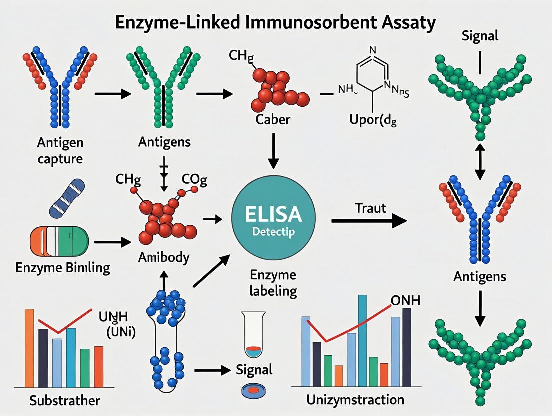
Enzyme-Linked Immunosorbent Assay (ELISA) is a foundational immunoassay technique used for the quantitative and qualitative detection of antigens or antibodies. Operating on the principle of specific antigen-antibody binding coupled with an enzymatic colorimetric reaction, ELISA is a cornerstone in diagnostics, drug development, and basic biomedical research. This whitepaper details the core principles, assay formats, experimental protocols, and data analysis, contextualizing its pivotal role in quantitative biological measurement.
ELISA immobilizes an antigen or antibody to a solid surface (typically a polystyrene microplate well). A specific antibody conjugated to an enzyme (e.g., Horseradish Peroxidase, Alkaline Phosphatase) is then introduced. After adding a chromogenic substrate, the enzyme catalyzes a reaction producing a measurable color change, the intensity of which is proportional to the target analyte concentration in the sample.
Primary Assay Formats: A Comparative Analysis
| Assay Format | Target | Immobilized Phase | Detection Antibody | Key Applications | Sensitivity (Typical) |
|---|---|---|---|---|---|
| Direct ELISA | Antigen | Antigen | Enzyme-linked Primary Antibody | Antigen screening, low-complexity targets. | Low (pg/mL range) |
| Indirect ELISA | Antibody | Antigen | Enzyme-linked Secondary Antibody | Serology (HIV, Lyme), autoimmune antibody detection. | High |
| Sandwich ELISA | Antigen | Capture Antibody | Enzyme-linked Detection Antibody | Cytokine measurement, biomarker quantitation (e.g., TNF-α, IL-6). | Very High (fg/mL - pg/mL) |
| Competitive/Inhibition ELISA | Small Antigens | Antigen (or Antibody) | Enzyme-linked Antigen (or Antibody) | Hormones (insulin), drugs of abuse, haptens. | High |
Table 1: Summary of core ELISA formats, their configurations, and performance characteristics. Sensitivity ranges are format- and target-dependent.
Detailed Protocol: Sandwich ELISA for Cytokine Quantification
This protocol is standard for quantifying soluble protein targets like cytokines or biomarkers in serum, plasma, or cell culture supernatant.
Day 1: Coating
- Dilute the capture antibody in carbonate-bicarbonate coating buffer (pH 9.6) to a concentration typically between 1-10 µg/mL.
- Add 100 µL per well to a 96-well microplate.
- Seal plate and incubate overnight at 4°C.
Day 2: Blocking, Sample & Detection
- Wash: Discard coating solution. Wash plate 3x with 300 µL PBS containing 0.05% Tween-20 (PBST) per well using a microplate washer or manual manifold.
- Block: Add 300 µL blocking buffer (e.g., 5% non-fat dry milk or 1% BSA in PBS) per well. Incubate for 1-2 hours at room temperature (RT).
- Wash: Repeat wash step as in 2.1.
- Sample Addition: Add 100 µL of standard (serial dilutions of recombinant protein), sample, or blank (diluent buffer) per well in duplicate or triplicate. Incubate 2 hours at RT or 1 hour at 37°C.
- Wash: Repeat wash step 5 times thoroughly.
- Detection Antibody Addition: Add 100 µL of biotinylated or enzyme-linked detection antibody (diluted in blocking buffer as per manufacturer's datasheet) per well. Incubate 1-2 hours at RT.
- Wash: Repeat wash step 5 times.
Day 2 (Continued): Signal Development & Readout
- Enzyme Conjugate: If using a biotinylated detection antibody, add 100 µL of Streptavidin-HRP conjugate (diluted in blocking buffer) per well. Incubate 30 minutes at RT, protected from light. Wash 5-7 times.
- Substrate Addition: Add 100 µL of chromogenic substrate (e.g., TMB for HRP) per well. Incubate for 5-30 minutes at RT, protected from light, until color develops adequately.
- Stop Reaction: Add 100 µL of stop solution (e.g., 1M H2SO4 for TMB) per well. The color will change from blue to yellow.
- Read Absorbance: Immediately measure absorbance at the appropriate wavelength (e.g., 450nm for TMB, with 570nm or 620nm as a reference) using a microplate reader.
Data Analysis and Interpretation
- Generate a standard curve by plotting the mean absorbance (y-axis) against the known concentration of the standard (x-axis) using a 4- or 5-parameter logistic (4PL/5PL) curve fit.
- Use the curve fit equation to interpolate the concentration of unknown samples from their mean absorbance values.
- Report data within the linear range of the standard curve. Typical performance metrics are summarized below.
| Parameter | Definition | Acceptable Range |
|---|---|---|
| Lower Limit of Detection (LLOD) | Lowest conc. distinguishable from zero. | Typically 2-3 SD above blank mean. |
| Lower Limit of Quantification (LLOQ) | Lowest conc. quantified with acceptable precision (CV <20%) and accuracy (80-120%). | Defined per assay. |
| Dynamic Range | Span between LLOQ and ULOQ. | Often 2-3 logs of concentration. |
| Intra-assay CV | Precision within a single plate. | <10% (preferably <8%). |
| Inter-assay CV | Precision between different plates/runs. | <15% (preferably <12%). |
| Spike Recovery | Accuracy of measuring added known analyte. | 80-120% of expected value. |
Table 2: Key validation parameters for a quantitative ELISA.
Visualizing ELISA Workflows and Signaling
Diagram 1: Sandwich ELISA Experimental Workflow
Diagram 2: Four Core ELISA Formats and Their Logical Steps
The Scientist's Toolkit: Essential Reagents & Materials
| Category | Item | Primary Function & Notes |
|---|---|---|
| Core Consumables | 96-Well Microplate (High-Binding) | Polystyrene plate for protein immobilization via passive adsorption. |
| Coating Buffer (Carbonate-Bicarbonate, pH 9.6) | Optimizes passive adsorption of antibodies/antigens to the plate. | |
| Wash Buffer (PBS with 0.05% Tween 20, PBST) | Removes unbound reagents; Tween-20 reduces non-specific binding. | |
| Blocking Buffer (e.g., 1% BSA, 5% Milk in PBS) | Covers residual protein-binding sites to minimize background signal. | |
| Detection System | Capture & Detection Antibody Pair | Matched monoclonal antibodies targeting different epitopes on the antigen. |
| Enzyme Conjugate (HRP or AP) | Catalyzes substrate conversion. Often linked to detection Ab or streptavidin. | |
| Chromogenic Substrate (TMB, OPD, pNPP) | Produces measurable color change upon enzymatic action. | |
| Stop Solution (e.g., 1M H2SO4) | Halts enzymatic reaction, stabilizing signal for measurement. | |
| Instrumentation | Microplate Washer | Ensures consistent and thorough washing between steps. |
| Microplate Reader (Spectrophotometer) | Measures absorbance at specific wavelengths for quantification. | |
| Reference | Recombinant Protein Standard | Pure antigen for generating the standard curve for quantitation. |
| Control Samples (Positive/Negative) | Validates assay performance in each run. |
Table 3: Essential research reagent solutions and materials for a robust ELISA.
This whitepaper delineates the biochemical principles governing antibody-antigen interactions, a cornerstone of immunoassay development. Framed within ongoing research to elucidate the Enzyme-Linked Immunosorbent Assay (ELISA) method, this guide details the thermodynamic, kinetic, and structural determinants of this specific molecular recognition. The insights herein are critical for researchers, scientists, and drug development professionals designing and interpreting high-sensitivity immunoassays.
Fundamental Principles of Molecular Recognition
Antibody-antigen interaction is a specific, reversible binding event driven by non-covalent forces. The binding site, the paratope on the antibody, complements a region on the antigen called the epitope. Affinity, the strength of this single-site interaction, is quantified by the dissociation constant (KD).
Thermodynamic and Kinetic Parameters
The interaction is governed by the association rate constant (kon), dissociation rate constant (koff), and the resultant KD (koff/kon). High-affinity antibodies (low KD) are essential for sensitive ELISA protocols.
Table 1: Representative Kinetic and Affinity Parameters for Antibody Classes
| Antibody Type | Typical KD Range (M) | kon (M-1s-1) | koff (s-1) | Primary Role in ELISA |
|---|---|---|---|---|
| Monoclonal IgG | 10-9 – 10-12 | 105 – 106 | 10-5 – 10-3 | Capture/Detection agent |
| Polyclonal IgG | 10-7 – 10-10 | 104 – 106 | 10-3 – 10-1 | Broad-target detection |
| IgM (naïve) | 10-5 – 10-7 | 103 – 105 | 10-1 – 101 | Rare; early-phase detection |
Structural Complementarity and Epitope Types
Epitopes are categorized as linear (continuous amino acid sequence) or conformational (discontinuous, dependent on 3D structure). Monoclonal antibodies often target specific conformational epitopes, while polyclonal sera recognize multiple linear and conformational epitopes.
Experimental Protocols for Characterizing Interactions
Surface Plasmon Resonance (SPR) for Kinetic Analysis
Protocol: This label-free technique measures binding kinetics in real-time.
- Chip Preparation: Immobilize a ligand (antigen or antibody) onto a dextran-coated gold sensor chip via amine coupling.
- Baseline Establishment: Flow running buffer (e.g., HBS-EP) over the chip to establish a stable baseline.
- Association Phase: Inject a series of analyte concentrations over the ligand surface. Monitor the change in resonance units (RU) versus time.
- Dissociation Phase: Switch flow to running buffer alone to monitor complex dissociation.
- Regeneration: Inject a mild acidic or basic buffer to remove bound analyte, regenerating the ligand surface.
- Data Analysis: Fit the resulting sensorgrams globally to a 1:1 binding model to extract kon, koff, and KD.
Isothermal Titration Calorimetry (ITC) for Thermodynamic Profiling
Protocol: This method directly measures the heat change during binding.
- Sample Preparation: Thoroughly dialyze both antibody and antigen into identical, degassed buffer.
- Experiment Setup: Load the antibody solution (typically 50-200 µM) into the sample cell. Fill the syringe with antigen at a concentration 10-20 times higher.
- Titration: Perform a series of automated injections of antigen into the antibody cell while maintaining constant stirring.
- Data Measurement: The instrument records the heat required to maintain a temperature difference of zero after each injection.
- Analysis: Integrate the heat peaks and fit the data to a binding model to derive stoichiometry (N), enthalpy change (ΔH), entropy change (ΔS), and the binding constant (KA = 1/KD).
The Scientist's Toolkit: Key Research Reagent Solutions
Table 2: Essential Reagents for Antibody-Antigen Interaction Studies & ELISA Development
| Reagent/Material | Function & Importance |
|---|---|
| High-Affinity Monoclonal Antibodies | Provide exquisite specificity for a single epitope, reducing cross-reactivity and background in sandwich ELISA. |
| Polyclonal Antibody Sera | Recognize multiple epitopes, enhancing signal amplification and detection sensitivity in indirect ELISA formats. |
| Recombinant Protein Antigens | Defined, pure antigens essential for assay standardization, calibration curve generation, and specificity testing. |
| Horseradish Peroxidase (HRP) / Alkaline Phosphatase (AP) | Enzyme conjugates for antibody labeling; catalyze colorimetric, chemiluminescent, or fluorescent signals in ELISA. |
| Chromogenic Substrates (e.g., TMB, PNPP) | Produce a measurable color change upon enzymatic reaction, enabling spectrophotometric detection. |
| Chemiluminescent Substrates (e.g., luminol-based) | Offer higher sensitivity and broader dynamic range than colorimetric substrates for low-abundance target detection. |
| Blocking Agents (e.g., BSA, Casein, Non-fat Dry Milk) | Saturate non-specific binding sites on the solid phase (microplate) to minimize background noise. |
| High-Binding ELISA Plates (Polystyrene) | Provide a hydrophobic surface for passive adsorption of proteins (antigens or capture antibodies). |
Visualization of Core Concepts
Forces Driving Antibody-Antigen Binding
Sandwich ELISA Workflow Steps
This document, framed within a broader thesis on the principles and applications of immunoassays, details the pivotal transition from Radioimmunoassay (RIA) to the Enzyme-Linked Immunosorbent Assay (ELISA). This evolution represents a paradigm shift in bioanalytical chemistry, driven by the need for safer, more stable, and quantitatively robust detection methodologies. Understanding this progression is crucial for appreciating the design logic, versatility, and foundational protocols that underpin modern immunoassay development in research and drug discovery.
The Foundation: Radioimmunoassay (RIA)
Developed by Rosalyn Yalow and Solomon Berson in the late 1950s, RIA was the first technique to use antibodies for the precise quantification of minute amounts of biological substances, such as hormones (e.g., insulin), in complex mixtures.
Core Principle: Competitive binding between a radiolabeled antigen (tracer) and an unlabeled antigen (sample analyte) for a limited number of specific antibody binding sites. The amount of radioactivity in the antibody-bound fraction is inversely proportional to the concentration of unlabeled antigen in the sample.
Detailed RIA Protocol (Typical)
- Preparation: A fixed, limiting concentration of specific antibody is incubated with known concentrations of unlabeled antigen standards and a constant, known amount of radiolabeled (e.g., Iodine-125) antigen.
- Competitive Incubation: The mixture is incubated to equilibrium, allowing competitive binding to the antibody.
- Separation (B/F Separation): The antibody-bound antigen is separated from the free antigen. Common methods included:
- Precipitation: Using a second antibody (anti-IgG) or polyethylene glycol (PEG).
- Charcoal Dextran: Activated charcoal adsorbs free antigen, which is then centrifuged.
- Measurement: The radioactivity in the bound (or free) fraction is measured using a gamma counter.
- Data Analysis: A standard curve is plotted (Bound radioactivity vs. log concentration of standard). The concentration of antigen in unknown samples is interpolated from this curve.
The Evolutionary Leap to ELISA
The desire to eliminate hazardous radioisotopes and complex disposal requirements, coupled with the need for more stable reagents and easier automation, drove the development of enzyme-based detection. The first ELISA was described independently by Engvall & Perlmann and Van Weemen & Schuurs in 1971.
Core Innovations:
- Label: Replacement of radioisotopes with enzymes (e.g., Horseradish Peroxidase - HRP, Alkaline Phosphatase - ALP).
- Detection: Enzyme activity is measured colorimetrically by adding a substrate (e.g., TMB for HRP, pNPP for ALP), producing a colored product.
- Solid-Phase: Widespread adoption of the 96-well microplate, pioneered by researchers like Catt and Tregear, for efficient separation via washing and high-throughput processing.
Quantitative Comparison: RIA vs. Direct/Indirect ELISA
Table 1: Comparative Analysis of RIA and Core ELISA Formats
| Feature | Radioimmunoassay (RIA) | Direct ELISA | Indirect ELISA |
|---|---|---|---|
| Detection Label | Radioisotope (e.g., ¹²⁵I) | Enzyme conjugated to primary antibody | Enzyme conjugated to secondary antibody |
| Signal Measured | Gamma radiation | Colorimetric (Absorbance) | Colorimetric (Absorbance) |
| Typical Assay Time | 24-72 hours | 2-4 hours | 3-5 hours |
| Key Advantage | High sensitivity; established gold standard | Fast, simple protocol | Amplification via multiple secondary antibodies; versatile |
| Major Disadvantage | Radiation hazard; short reagent shelf-life | Less sensitive; lower signal amplification | Additional incubation step |
| Approx. Cost per Sample | High (radioisotopes, licensing) | Low | Very Low |
| Modern Prevalence | Largely obsolete for routine use | Common for antigen detection | Most common format for antibody detection |
Detailed Modern ELISA Protocol: Indirect ELISA for Antibody Detection
This protocol exemplifies the standard workflow widely used in serology and immunology research.
1. Coating: Dilute the target antigen in carbonate/bicarbonate coating buffer (pH 9.6). Add 100 µL/well to a polystyrene microplate. Incubate overnight at 4°C or 1-2 hours at 37°C. 2. Washing & Blocking: Discard coating solution. Wash plate 3x with PBS containing 0.05% Tween-20 (PBST). Add 200-300 µL/well of blocking buffer (e.g., 1-5% BSA or non-fat dry milk in PBS). Incubate 1-2 hours at 37°C. Wash 3x with PBST. 3. Primary Antibody Incubation: Serially dilute the sample (serum, hybridoma supernatant) in blocking buffer. Add 100 µL/well. Include blank (buffer only) and negative control wells. Incubate 1-2 hours at 37°C. Wash 3-5x with PBST. 4. Secondary Antibody Incubation: Add 100 µL/well of enzyme-conjugated species-specific anti-IgG antibody (e.g., Goat anti-Human IgG-HRP) diluted in blocking buffer. Incubate 1 hour at 37°C. Wash 3-5x thoroughly with PBST. 5. Detection: Add 100 µL/well of substrate solution (e.g., TMB for HRP). Incubate in the dark for 10-30 minutes at room temperature. 6. Stop & Read: Add 50-100 µL/well of stop solution (e.g., 1M H₂SO₄ for TMB). Measure absorbance immediately at the appropriate wavelength (e.g., 450 nm for TMB).
Key Methodological Pathways and Workflows
Figure 1: Comparative Workflow of RIA and Indirect ELISA
Figure 2: ELISA Signal Generation Cascade
The Scientist's Toolkit: Essential ELISA Research Reagent Solutions
Table 2: Key Reagents and Materials for Modern ELISA
| Item | Function & Technical Note |
|---|---|
| Polystyrene Microplate | Solid phase for protein adsorption. High-binding plates (e.g., Nunc MaxiSorp) are coated with modified polymers to maximize protein binding capacity. |
| Coating Buffer (Carbonate-Bicarbonate, pH 9.6) | Alkaline buffer optimizes passive adsorption of most proteins (antigens/antibodies) to the polystyrene surface via hydrophobic interactions. |
| Blocking Agent (BSA, Casein, Non-fat Dry Milk) | Inert protein or detergent solution that saturates uncovered plastic surfaces to prevent non-specific binding of detection antibodies, reducing background noise. |
| Wash Buffer (PBS with 0.05% Tween 20) | Isotonic buffer with a mild non-ionic detergent (Tween 20) to remove unbound reagents while maintaining protein structure and antibody-antigen interactions. |
| Detection Antibody Conjugates (HRP/ALP) | Secondary antibodies covalently linked to reporter enzymes (Horseradish Peroxidase or Alkaline Phosphatase). Critical for signal generation. |
| Chromogenic Substrate (TMB, OPD, pNPP) | Enzyme-specific, non-toxic chemical that yields a colored, soluble reaction product upon enzymatic catalysis. TMB is the most common for HRP. |
| Stop Solution (e.g., 1M H₂SO₄, 2M H₂SO₄) | Acidic solution that rapidly denatures the enzyme, halting the substrate reaction and stabilizing the final signal intensity for measurement. |
| Microplate Spectrophotometer (Reader) | Instrument to measure the absorbance (Optical Density - OD) of light by the colored product in each well, typically at 450 nm for TMB or 405 nm for pNPP. |
The transition from RIA to ELISA fundamentally transformed biomedical research and diagnostics, providing a safe, robust, and adaptable platform. This evolution, rooted in the core thesis of immunoassay principles, underscores the continuous innovation in detection modalities—a trajectory that continues today with advancements in chemiluminescence, electrochemiluminescence (ECL), and digital ELISA, pushing the limits of sensitivity and multiplexing ever further.
The Enzyme-Linked Immunosorbent Assay (ELISA) remains a cornerstone analytical technique in biomedical research, diagnostic development, and therapeutic discovery. Its robustness and versatility stem from the precise interplay of its four core components: plates, antibodies, enzymes, and substrates. This whitepaper provides an in-depth technical analysis of these components, framed within a broader thesis that optimizing these elements is fundamental to enhancing assay sensitivity, specificity, and reproducibility. Advances in material science, protein engineering, and detection chemistry continue to refine ELISA performance, directly impacting critical areas such as biomarker validation, pharmacokinetic studies, and drug immunogenicity assessment.
The Solid Phase: Microplates
The microplate serves as the foundational solid phase, providing the surface for antigen immobilization and subsequent molecular interactions.
Key Characteristics and Recent Data:
| Plate Property | Optimal Specification | Impact on Assay Performance | Common Material/Coating |
|---|---|---|---|
| Binding Capacity | High (> 400 ng IgG/cm²) | Increases dynamic range, reduces hook effect. | Polystyrene, Modified Polystyrene |
| Well-to-Well Uniformity | CV < 5% | Ensures reproducibility across the plate. | Precision molding techniques |
| Background Optical Density | OD < 0.1 at 450 nm | Improves signal-to-noise ratio. | Black plates for fluorescence, clear for colorimetry |
| Surface Chemistry | Amino, Streptavidin, GST | Enables oriented capture, reduces denaturation. | Covalent linkage to functional groups |
Protocol: Plate Coating Optimization
- Antigen/Coating Antibody Dilution: Prepare a checkerboard serial dilution (e.g., 0.5 – 10 µg/mL) of the capture protein in carbonate-bicarbonate coating buffer (pH 9.6).
- Coating: Dispense 100 µL per well. Incubate plate sealed overnight at 4°C or for 1-2 hours at 37°C.
- Washing: Aspirate and wash plate 3x with 300 µL PBS containing 0.05% Tween 20 (PBST) using an automated plate washer.
- Blocking: Add 300 µL of blocking buffer (e.g., 1-5% BSA or casein in PBS) per well. Incubate for 1-2 hours at room temperature on a plate shaker.
- Storage: Wash once with PBST. Plates can be dried and sealed for future use or used immediately.
The Specificity Agents: Antibodies
Antibodies confer the assay's specificity. The choice between monoclonal and polyclonal, along with engineering advancements, is critical.
Quantitative Comparison of Antibody Types:
| Parameter | Monoclonal Antibody | Polyclonal Antibody | Recombinant Antibody |
|---|---|---|---|
| Specificity | High (single epitope) | Moderate (multiple epitopes) | High (engineered) |
| Affinity (K_D) | Consistent, can be very high (pM-nM) | Variable mixture, often high | Can be engineered to pM range |
| Production & Lot Consistency | Excellent, immortal hybridoma | Variable, depends on animal | Excellent, from sequenced gene |
| Cross-Reactivity Risk | Low | Higher | Very low (highly defined) |
| Typical Application | Capture antibody in sandwich ELISA | Detection antibody (broad signal) | Both capture and detection |
Protocol: Antibody Pair Screening for Sandwich ELISA
- Capture Antibody Coating: Coat plates with candidate capture antibodies (2-10 µg/mL) as per the protocol above.
- Antigen Incubation: Block plate. Add a dilution series of the target antigen and a negative control sample. Incubate 2 hours at RT.
- Detection Antibody Titration: Prepare serial dilutions of candidate detection antibodies in blocking buffer. Add to wells post-wash. Incubate 1 hour at RT.
- Signal Development: Use a standardized enzyme conjugate and substrate development step (see below). Analyze signal-to-background ratio and assay midpoint for each pair. The optimal pair shows high signal for antigen and minimal background.
The Signal Generators: Enzymes and Substrates
The enzyme conjugate amplifies the specific binding event, and the substrate conversion yields a measurable signal.
Performance Data of Common Enzyme-Substrate Pairs:
| Enzyme | Common Substrate | Detection Mode | Sensitivity (approx.) | Kinetics | Quenching Required? |
|---|---|---|---|---|---|
| Horseradish Peroxidase (HRP) | TMB (3,3',5,5'-Tetramethylbenzidine) | Colorimetric (450 nm) | 1-10 pg/well | Fast | Yes (Acid) |
| HRP | ADHP (Amplex UltraRed) | Fluorometric (Ex 570/Em 585) | 0.1-1 pg/well | Fast | No |
| Alkaline Phosphatase (AP) | pNPP (p-Nitrophenyl Phosphate) | Colorimetric (405 nm) | 10-100 pg/well | Moderate | Yes (Base) |
| AP | AttoPhos / CDP-Star | Chemiluminescent | 0.01-0.1 pg/well | Very Fast | No |
Protocol: Substrate Development and Signal Stopping
- Substrate Preparation: Prepare working solution of substrate immediately before use. For TMB, use a stabilized, ready-to-use solution. For chemiluminescent substrates, follow manufacturer's instructions precisely regarding timing.
- Development: Add 100 µL per well to washed plate containing the enzyme conjugate. Incubate at RT for a consistent, predetermined time (e.g., 10-30 minutes) in the dark.
- Stopping (for colorimetric): For TMB, add 100 µL of 1M H₂SO₄ or HCl to stop the reaction, converting the blue product to yellow. For pNPP, add 50 µL of 3M NaOH.
- Reading: Read absorbance immediately on a plate reader at the appropriate wavelength (TMB: 450 nm; pNPP: 405 nm). Chemiluminescence is read immediately without a stop solution.
The Scientist's Toolkit: Key Research Reagent Solutions
| Item | Function & Importance |
|---|---|
| High-Binding, Low-Noise Microplates | Provides consistent protein adsorption with minimal non-specific binding, critical for baseline stability. |
| Matched Antibody Pairs (Monoclonal) | Pre-optimized capture and detection antibodies targeting non-overlapping epitopes ensure sensitive, specific sandwich assays. |
| Recombinant Protein Standards | Highly purified, quantitated antigen for generating the standard curve, essential for accurate sample quantification. |
| HRP or AP Conjugation Kits | Enable consistent labeling of detection antibodies with enzymes, maintaining antibody affinity and enzyme activity. |
| Stabilized TMB Substrate | Single-component, ready-to-use chromogenic substrate offers convenience and consistent development kinetics. |
| Blocking Buffer (Protein-Based) | 5% BSA or specialty commercial blockers reduce non-specific binding, lowering background noise. |
| Wash Buffer Concentrate (20-25X) | Consistent formulation of PBS with detergent (Tween 20) ensures efficient removal of unbound reagents. |
| Plate Sealers | Prevent evaporation and contamination during incubations, ensuring well-to-well consistency. |
Diagrams
The Enzyme-Linked Immunosorbent Assay (ELISA) remains a cornerstone technique in biomedical research and diagnostic development. Within the broader thesis of "ELISA method explained" research, a critical understanding of assay architecture is paramount. The four principal types—Direct, Indirect, Sandwich, and Competitive—represent fundamental immunological strategies for quantifying analytes, each with distinct advantages in sensitivity, specificity, multiplexing potential, and required reagents. This whitepaper provides an in-depth technical comparison, guiding researchers and drug development professionals in selecting and optimizing the appropriate format for their specific antigen-antibody system.
Core Principles and Comparative Analysis
All ELISA types rely on the specific binding of an antibody to its target antigen and the subsequent detection of this complex via an enzyme-linked conjugate that catalyzes a measurable colorimetric, chemiluminescent, or fluorescent signal. The key differentiating factor is the sequence and configuration of antibody-antigen interactions.
Table 1: High-Level Comparison of the Four Main ELISA Types
| Feature | Direct ELISA | Indirect ELISA | Sandwich ELISA | Competitive ELISA |
|---|---|---|---|---|
| Principle | Antigen is immobilized; detected directly by a labeled primary antibody. | Antigen is immobilized; detected by an unlabeled primary antibody, then a labeled secondary antibody. | Antigen is captured by an immobilized antibody, then detected by a second, labeled antibody. | Sample antigen competes with a reference antigen for binding to a limited amount of labeled antibody. |
| Steps | 1. Coat antigen.2. Add labeled primary antibody.3. Add substrate. | 1. Coat antigen.2. Add unlabeled primary antibody.3. Add labeled secondary antibody.4. Add substrate. | 1. Coat capture antibody.2. Add sample antigen.3. Add labeled detection antibody.4. Add substrate. | 1. Coat antigen (or capture antibody).2. Co-incubate sample antigen and labeled antibody (or sample antigen, reference antigen, and labeled antibody).3. Add substrate. |
| Key Advantage | Speed; minimal steps; no cross-reactivity from secondary antibody. | High sensitivity due to signal amplification; flexibility with one labeled secondary for many primaries. | High specificity (two antibodies); excellent for complex samples; wide dynamic range. | Best for small antigens; robust in complex matrices; less prone to hook effect. |
| Key Disadvantage | Low sensitivity; every primary antibody must be individually labeled. | Potential for cross-reactivity from secondary antibody; extra step required. | Requires two antibodies against different epitopes; optimization can be complex. | Inverse signal relationship (low signal = high analyte); narrower dynamic range. |
| Typical Sensitivity Range | Moderate (ng/mL to µg/mL) | High (pg/mL to ng/mL) | Highest (pg/mL) | High (pg/mL to ng/mL) |
| Best For | Large antigens with high abundance; antibody screening. | General purpose; high-throughput serology (e.g., antibody titer). | Quantitative measurement of proteins, cytokines, biomarkers in serum, cell lysates. | Haptens, small molecules (hormones, drugs), antigens with only one epitope. |
Table 2: Quantitative Performance Metrics (Typical Values)
| Assay Type | Time to Completion | Approx. Cost per Sample | Signal Amplification | Minimum Sample Volume Required |
|---|---|---|---|---|
| Direct ELISA | ~2 hours | Low | None | 50-100 µL |
| Indirect ELISA | ~3 hours | Low-Moderate | High (≥ 2x Direct) | 50-100 µL |
| Sandwich ELISA | ~4 hours | High | Very High | 50-100 µL |
| Competitive ELISA | ~2.5 hours | Moderate | None | 25-50 µL |
Detailed Experimental Protocols
General Reagents: All protocols require: Coating Buffer (e.g., 0.1 M Carbonate-Bicarbonate, pH 9.6), Wash Buffer (PBS or Tris with 0.05% Tween 20, PBST), Blocking Buffer (e.g., 1-5% BSA or non-fat dry milk in PBST), Substrate (e.g., TMB for HRP, pNPP for AP), and Stop Solution (e.g., 1M H₂SO₄ for TMB).
Direct ELISA Protocol
- Coating: Dilute purified antigen in coating buffer. Add 50-100 µL/well to a 96-well microplate. Incubate overnight at 4°C or 1-2 hours at 37°C.
- Washing: Aspirate and wash plate 3x with 200-300 µL/well of wash buffer.
- Blocking: Add 150-200 µL/well of blocking buffer. Incubate 1-2 hours at 37°C or overnight at 4°C. Wash 3x.
- Primary Antibody Incubation: Add 50-100 µL/well of enzyme-conjugated (e.g., HRP) primary antibody diluted in blocking buffer. Incubate 1-2 hours at 37°C. Wash 3-5x.
- Detection: Add 50-100 µL/well of substrate solution. Incubate in the dark for 10-30 minutes.
- Stop & Read: Add 50 µL/well of stop solution. Measure absorbance immediately at the appropriate wavelength (e.g., 450nm for TMB).
Indirect ELISA Protocol
- Coating & Blocking: Perform as per Direct ELISA steps 1-3.
- Primary Antibody Incubation: Add 50-100 µL/well of unlabeled primary antibody diluted in blocking buffer. Incubate 1-2 hours at 37°C. Wash 3-5x.
- Secondary Antibody Incubation: Add 50-100 µL/well of enzyme-conjugated secondary antibody (specific to the host species of the primary) diluted in blocking buffer. Incubate 1 hour at 37°C. Wash 3-5x.
- Detection, Stop & Read: Perform as per Direct ELISA steps 5-6.
Sandwich ELISA Protocol
- Capture Antibody Coating: Dilute the capture antibody in coating buffer. Add 50-100 µL/well. Incubate and wash as in 3.1.
- Blocking: Perform as per Direct ELISA step 3.
- Antigen Incubation: Add 50-100 µL/well of sample or standard containing the antigen, diluted in blocking buffer. Incubate 2 hours at 37°C or overnight at 4°C. Wash 3-5x.
- Detection Antibody Incubation: Add 50-100 µL/well of enzyme-conjugated detection antibody (specific to a different epitope on the antigen) diluted in blocking buffer. Incubate 1-2 hours at 37°C. Wash 3-5x.
- Detection, Stop & Read: Perform as per Direct ELISA steps 5-6.
Competitive ELISA Protocol (Antigen-Coated Format)
- Reference Antigen Coating & Blocking: Coat plate with a known quantity of purified antigen. Perform blocking as per Direct ELISA steps 1-3.
- Competitive Incubation: Pre-mix a constant, limited amount of enzyme-conjugated detection antibody with serially diluted sample (containing unknown antigen) or standard. Add this mixture to the coated wells. Incubate 1-2 hours at 37°C. The free antigen in the sample competes with the plate-bound antigen for the labeled antibody. Wash 3-5x.
- Detection, Stop & Read: Perform as per Direct ELISA steps 5-6. Note: The signal is inversely proportional to the analyte concentration in the sample.
Visualized Workflows and Pathways
Diagram 1: Direct vs. Indirect ELISA Workflow
Diagram 2: Sandwich vs. Competitive ELISA Workflow
The Scientist's Toolkit: Key Research Reagent Solutions
Table 3: Essential Materials for ELISA Development
| Item | Function & Critical Considerations |
|---|---|
| High-Binding Polystyrene Microplates | Provides a hydrophobic surface for passive adsorption of proteins (antigens or antibodies). Plate uniformity is critical for assay precision. |
| Purified Antigen & Antibody Pairs | The core reagents. For Sandwich ELISA, a matched pair recognizing non-overlapping epitopes is mandatory. Affinity and specificity define assay limits. |
| Enzyme-Conjugated Detection Reagents | HRP (Horseradish Peroxidase) and AP (Alkaline Phosphatase) are most common. Conjugates include labeled primary antibodies (Direct/Competitive) or secondary antibodies (Indirect/Sandwich). |
| Chromogenic/Luminescent Substrate | TMB (3,3',5,5'-Tetramethylbenzidine) for HRP is a common chromogen. Chemiluminescent substrates offer higher sensitivity. Must be matched to the enzyme. |
| Precision Liquid Handling System | Multi-channel and single-channel pipettes for reproducible reagent addition. Automated washers and plate handlers are essential for high-throughput. |
| Plate Washer | Ensures consistent and thorough removal of unbound reagents, a key factor in reducing background noise and variability. |
| Microplate Spectrophotometer (Reader) | Measures absorbance of chromogenic products. For fluorescent or luminescent endpoints, respective plate readers are required. |
| Blocking Agent (BSA, Casein, etc.) | Saturates unused protein-binding sites on the plate to prevent nonspecific adsorption of detection reagents, lowering background. |
| Sample Diluent/Assay Buffer | Optimized buffer (often containing blockers and detergents) for diluting samples and reagents to maintain stability and minimize matrix interference. |
| Data Analysis Software | For curve fitting (typically 4- or 5-parameter logistic for Sandwich, linear for Competitive) and sample concentration interpolation from standard curves. |
Within the context of ELISA method explained research, the performance and reliability of the assay are governed by four critical analytical parameters: Sensitivity, Specificity, Dynamic Range, and Limit of Detection (LoD). This whitepaper provides an in-depth technical guide to these parameters, framing their importance for researchers, scientists, and drug development professionals who rely on precise and accurate immunoassays for diagnostic and therapeutic applications.
Sensitivity and Specificity
Sensitivity is defined as the ability of an ELISA to correctly identify true positive samples. It is calculated as the proportion of actual positives that are correctly identified. Specificity is the ability of the assay to correctly identify true negative samples, calculated as the proportion of actual negatives that are correctly identified.
Defining Equations
- Sensitivity (%) = [TP / (TP + FN)] × 100
- Specificity (%) = [TN / (TN + FP)] × 100
- TP = True Positives; FN = False Negatives; TN = True Negatives; FP = False Positives.
Experimental Protocol for Determination
A validated reference method (e.g., mass spectrometry, a gold-standard clinical test) is used to classify a panel of known samples (N > 100 recommended). Each sample is then tested using the investigational ELISA. Results are compared to the reference method to populate the confusion matrix.
Table 1: Example Confusion Matrix for Sensitivity/Specificity Calculation
| Reference Method (Gold Standard) | ELISA Positive | ELISA Negative | Total |
|---|---|---|---|
| Disease Positive | 95 (TP) | 5 (FN) | 100 |
| Disease Negative | 3 (FP) | 97 (TN) | 100 |
| Total | 98 | 102 | 200 |
- Calculated Sensitivity = (95 / 100) × 100 = 95%
- Calculated Specificity = (97 / 100) × 100 = 97%
Dynamic Range
The Dynamic Range (or Analytical Measurement Range) is the interval between the upper and lower concentrations of an analyte for which the ELISA demonstrates a linear, reproducible response with acceptable accuracy and precision.
Protocol for Establishing Dynamic Range
A standard curve is prepared using a serial dilution of the analyte of known concentration. Each standard is assayed in replicate (n≥3). The mean absorbance is plotted against the analyte concentration. Linear regression analysis is performed on the linear portion of the curve (typically R² > 0.99). The Lower Limit of Quantification (LLOQ) and Upper Limit of Quantification (ULOQ) define the range.
Table 2: Example Data for Dynamic Range Determination
| Standard Concentration (pg/mL) | Mean Absorbance (450 nm) | CV (%) |
|---|---|---|
| 0 (Blank) | 0.052 | 5.2 |
| 15.6 | 0.108 | 4.8 |
| 31.3 | 0.215 | 3.5 |
| 62.5 | 0.420 | 3.1 |
| 125 | 0.805 | 2.9 |
| 250 | 1.560 | 3.4 |
| 500 | 2.850 (Signal Saturation) | 8.7 |
| 1000 | 3.100 (Signal Saturation) | 10.1 |
- Dynamic Range (Example): 15.6 pg/mL (LLOQ) to 250 pg/mL (ULOQ). The LLoQ is defined as the lowest concentration with a signal ≥ blank + 10SD and a CV ≤ 20%.
Limit of Detection (LoD)
The Limit of Detection (LoD) is the lowest concentration of analyte that can be consistently distinguished from a blank sample (zero analyte). It is a measure of detection capability, not quantification.
Protocol for LoD Determination (ICH Guidelines)
The LoD is typically determined by analyzing multiple replicates (n ≥ 10) of the blank (zero standard) and a low-concentration sample. Two common methods are:
- Signal-to-Noise: LoD is the concentration yielding a signal 2.5 to 3 times the standard deviation of the blank.
- Standard Deviation of Response & Slope: LoD = (3.3 × σ) / S, where σ is the standard deviation of the blank response, and S is the slope of the calibration curve.
Table 3: Example Data for LoD Calculation
| Parameter | Value |
|---|---|
| Mean Absorbance of Blank (n=20) | 0.051 |
| SD of Blank Absorbance (σ) | 0.005 |
| Slope of Calibration Curve (S) | 0.012 absorbance/(pg/mL) |
| LoD (3.3σ/S) | 1.38 pg/mL |
Visualization: ELISA Parameter Determination Workflow
The Scientist's Toolkit: Key Research Reagent Solutions
Table 4: Essential Materials for Robust ELISA Development
| Item | Function & Importance |
|---|---|
| High-Affinity, Monoclonal Capture Antibody | Drives assay specificity by selectively binding the target epitope. Critical for minimizing cross-reactivity. |
| Matched Detection Antibody (Biotin or HRP conjugated) | Completes the "sandwich," providing the signal. Must bind a different, non-overlapping epitope than the capture antibody. |
| Recombinant Purified Antigen Standard | Used to generate the calibration curve. Must be highly pure and accurately quantified to define assay sensitivity and range. |
| Low-Binding Microplates | Minimizes non-specific adsorption of proteins (antibodies, samples), reducing background noise and improving LoD. |
| High-Sensitivity Chromogenic/Luminescent Substrate (e.g., TMB, SuperSignal) | Generates the measurable signal. Choice impacts dynamic range and sensitivity (luminescent > chromogenic). |
| Precision Liquid Handling System | Ensures reproducible dispensing of standards, samples, and reagents, which is fundamental for achieving low CVs and a reliable LoD. |
| Validated Sample Diluent/Matrix | Mimics the sample environment (e.g., serum, cell lysate) to control for matrix effects that can alter antibody binding and specificity. |
| Plate Reader with Temperature Control | Provides accurate and consistent endpoint or kinetic readings. Stable temperature is vital for consistent enzymatic reaction rates. |
Step-by-Step ELISA Protocol: From Plate Coating to Data Analysis
The Enzyme-Linked Immunosorbent Assay (ELISA) remains a cornerstone analytical technique in life science research, clinical diagnostics, and drug development. Within the broader thesis of "ELISA method explained," this guide focuses on the critical strategic decision of format selection. The choice of ELISA format—direct, indirect, sandwich, or competitive—is not arbitrary but is dictated by the biochemical properties of the target analyte, the availability of specific binding reagents, and the required assay performance characteristics. This decision fundamentally impacts sensitivity, specificity, dynamic range, time-to-result, and cost. An improperly formatted ELISA can yield misleading data, wasting resources and impeding project timelines. This technical guide provides a systematic framework for researchers to align their target's attributes with the optimal ELISA architecture.
Core ELISA Formats: Principles and Applications
Direct ELISA
Principle: The antigen of interest is immobilized directly onto the plate. A single, primary antibody conjugated to an enzyme (e.g., HRP) is then used for detection. Workflow Diagram:
Title: Direct ELISA Workflow
Best For: High-throughput screening of purified antigens or when conjugate availability is not a constraint. It is simple and fast but generally offers lower sensitivity.
Indirect ELISA
Principle: The antigen is immobilized. An unlabeled primary antibody binds, followed by an enzyme-conjugated secondary antibody that recognizes the primary antibody's Fc region. Workflow Diagram:
Title: Indirect ELISA Workflow
Best For: Immunogenicity testing (detection of antibodies in serum), or when signal amplification is needed. It offers enhanced sensitivity over direct ELISA due to multiple secondary antibodies binding per primary.
Sandwich ELISA
Principle: Requires two antibodies that bind non-overlapping epitopes on the target antigen. A capture antibody is immobilized, the antigen is "sandwiched," and a second detection antibody (often conjugated or detected indirectly) provides the signal. Workflow Diagram:
Title: Sandwich ELISA Workflow
Best For: Complex samples (serum, cell lysates) where high specificity and sensitivity for a protein target are required. It is the gold standard for quantitative cytokine and biomarker analysis.
Competitive/Inhibition ELISA
Principle: Used for detecting small molecules (haptens) or when only one specific antibody is available. The sample antigen competes with a labeled reference antigen for a limited number of antibody binding sites. Workflow Diagram:
Title: Competitive ELISA Workflow
Best For: Measuring small molecules (hormones, drugs), anti-drug antibodies, or targets with limited epitopes. Signal is inversely proportional to analyte concentration.
Comparative Analysis of ELISA Formats
Table 1: Key Characteristics and Applications of ELISA Formats
| Format | Sensitivity | Specificity | Complexity | Time | Typical Applications | Key Requirement |
|---|---|---|---|---|---|---|
| Direct | Low-Medium | Medium | Low | Short (~2-3 hrs) | High-throughput screening of purified proteins; Epitope mapping. | High-affinity, labeled primary antibody. |
| Indirect | Medium-High | High | Medium | Medium (~3-4 hrs) | Serology (antibody detection); General protein detection with amplification. | Species-specific conjugated secondary antibody. |
| Sandwich | Very High | Very High | High | Long (~4-5 hrs) | Biomarker/cytokine quantification in complex matrices; Clinical diagnostics. | Matched antibody pair to non-overlapping epitopes. |
| Competitive | Medium-High | High | Medium-High | Medium (~3-4 hrs) | Haptens/small molecules (e.g., cortisol, T3/T4); Anti-drug antibodies. | Pure, labeled antigen or specific antibody. |
Table 2: Format Selection Guide Based on Target Properties
| Target Property | Recommended Format(s) | Rationale |
|---|---|---|
| Large Protein (>10 kDa) | Sandwich, Indirect, Direct | Multiple epitopes allow for sandwich or indirect detection. |
| Small Molecule/Hapten (<1 kDa) | Competitive | Limited epitope size necessitates competition. |
| Unknown/Antibody Target | Indirect (for Ab detection) | Ideal for detecting immunoglobulins in samples. |
| High-Abundance in Complex Mix | Direct, Indirect | Simpler formats suffice if sensitivity is not limiting. |
| Low-Abundance in Complex Mix | Sandwich | Capture step purifies and concentrates analyte, maximizing signal-to-noise. |
| Only One Specific Ab Available | Competitive, Direct | Sandwich not possible; competition or direct conjugate are options. |
| Need for Maximum Sensitivity | Sandwich (with amplification) | Signal amplification via enzymatic cascade and use of multiple labels. |
Detailed Experimental Protocol: Sandwich ELISA for Cytokine Quantification
This protocol exemplifies the most common high-performance ELISA format.
A. Materials & Reagents
- Capture Antibody: Monoclonal antibody specific to target cytokine.
- Detection Antibody: Biotinylated monoclonal antibody binding a different epitope.
- Standards: Recombinant cytokine of known concentration.
- Microplate: 96-well, high-binding polystyrene.
- Coating Buffer: 0.2 M carbonate-bicarbonate buffer, pH 9.4.
- Blocking Buffer: 1X PBS with 1% BSA or 5% non-fat dry milk.
- Wash Buffer: 1X PBS with 0.05% Tween-20 (PBST).
- Streptavidin-HRP Conjugate: Enzyme reporter.
- Substrate: TMB (3,3',5,5'-Tetramethylbenzidine).
- Stop Solution: 1 M H₂SO₄ or 1 M HCl.
- Plate Reader: Capable of measuring absorbance at 450 nm (and 570 nm for reference).
B. Step-by-Step Protocol
- Coating: Dilute capture antibody in coating buffer to 1-10 µg/mL. Add 100 µL per well. Seal plate and incubate overnight at 4°C.
- Washing: Aspirate liquid. Wash plate 3 times with ≥300 µL/well wash buffer (manual or automated). Blot on absorbent paper.
- Blocking: Add 300 µL blocking buffer per well. Incubate for 1-2 hours at room temperature (RT). Wash as in Step 2.
- Sample & Standard Addition: Prepare serial dilutions of the cytokine standard in sample diluent (e.g., blocking buffer). Add 100 µL of standards (in duplicate) and prepared samples to appropriate wells. Include blank wells (diluent only). Incubate 2 hours at RT. Wash 3 times.
- Detection Antibody: Add 100 µL of biotinylated detection antibody (diluted per manufacturer's recommendation in diluent). Incubate 1-2 hours at RT. Wash 3 times.
- Streptavidin-HRP: Add 100 µL of Streptavidin-HRP conjugate (diluted in diluent). Incubate 30 minutes at RT, protected from light. Wash 5 times thoroughly.
- Substrate Development: Add 100 µL of TMB substrate per well. Incubate in the dark at RT for 5-30 minutes, monitoring for color development.
- Stop Reaction: Add 100 µL stop solution per well. The color will change from blue to yellow.
- Measurement: Read absorbance at 450 nm within 30 minutes. Subtract any reference absorbance (e.g., 570 nm).
C. Data Analysis Generate a standard curve by plotting the mean absorbance (y-axis) against the standard concentration (x-axis) using a 4- or 5-parameter logistic (4PL/5PL) curve fit. Use the resulting equation to interpolate sample concentrations.
The Scientist's Toolkit: Essential Research Reagent Solutions
Table 3: Key Reagents and Their Functions in ELISA Development
| Reagent Category | Specific Example | Critical Function |
|---|---|---|
| Solid Phase | High-Binding Polystyrene Microplate | Provides hydrophobic surface for passive adsorption of proteins (antibodies/antigens). |
| Capture Molecule | Monoclonal Anti-Human IL-6 Antibody | Provides specificity by immobilizing the target analyte from the sample matrix. |
| Detection Molecule | Biotinylated Anti-Human IL-6 Antibody | Binds a distinct epitope on the captured analyte, enabling specific detection. |
| Signal Amplification System | Streptavidin-Poly-HRP Conjugate | Exploits high biotin-streptavidin affinity and carries multiple enzyme molecules, greatly amplifying signal. |
| Enzyme Substrate | TMB (3,3',5,5'-Tetramethylbenzidine) | Colorimetric HRP substrate producing a soluble blue product measurable at 450nm. |
| Blocking Agent | Bovine Serum Albumin (BSA) or Casein | Saturates non-specific binding sites on the plate and wells to reduce background noise. |
| Wash Solution Additive | Polysorbate 20 (Tween-20) | A non-ionic detergent that reduces non-specific hydrophobic interactions during washing steps. |
| Standard | Recombinant Human IL-6, Lyophilized | Provides a known quantity of pure analyte for generating the calibration curve, enabling quantitative analysis. |
This technical guide provides an in-depth walkthrough of the Sandwich ELISA (Enzyme-Linked Immunosorbent Assay) protocol, a cornerstone technique in quantitative immunodetection. Framed within a broader thesis on ELISA method explained research, this document serves as a primary resource for quantifying target antigens with high specificity and sensitivity. The sandwich format is distinguished by its use of two antibodies, enhancing target selectivity and reducing background, making it indispensable for detecting complex analytes in serum, plasma, and cell culture supernatants in drug development and clinical research.
The Sandwich ELISA is predicated on the immobilization of a capture antibody onto a solid phase, typically a polystyrene microplate. The sample containing the target antigen is then added, allowing the antigen to be specifically bound. Following washing, a detection antibody, conjugated to an enzyme such as Horseradish Peroxidase (HRP) or Alkaline Phosphatase (AP), is introduced, forming an antibody-antigen-antibody "sandwich." Subsequent addition of a chromogenic substrate yields a measurable signal proportional to the antigen concentration. This guide details the protocol, optimized for robust quantitative analysis.
Detailed Experimental Protocol
Materials and Reagents
- Microplate: 96-well polystyrene plate, high protein-binding capacity.
- Capture Antibody: Monoclonal or affinity-purified polyclonal antibody specific to the target antigen.
- Blocking Buffer: 1-5% Bovine Serum Albumin (BSA) or non-fat dry milk in PBS or TBS.
- Wash Buffer: PBS or TBS with 0.05% Tween 20 (PBST/TBST).
- Antigen Standard: Purified antigen for generating a standard curve.
- Detection Antibody: Enzyme-conjugated antibody recognizing a different epitope on the target antigen.
- Enzyme Substrate: TMB (3,3’,5,5’-Tetramethylbenzidine) for HRP, or pNPP (p-Nitrophenyl Phosphate) for AP.
- Stop Solution: 1M or 2M sulfuric acid (for HRP/TMB reaction).
- Plate Reader: Capable of measuring absorbance (e.g., 450 nm for TMB).
Step-by-Step Procedure
Day 1: Coating and Blocking
- Coating: Dilute the capture antibody in carbonate-bicarbonate coating buffer (pH 9.6) or PBS. Dispense 100 µL per well. Seal the plate and incubate overnight at 4°C.
- Washing: Aspirate the coating solution. Wash each well 3 times with 300 µL wash buffer using a multichannel pipette or automated plate washer. Blot plate on clean absorbent paper.
- Blocking: Add 200-300 µL of blocking buffer per well. Incubate for 1-2 hours at room temperature or overnight at 4°C. Wash as in Step 2.
Day 2: Antigen Capture and Detection
- Antigen Incubation: Prepare serial dilutions of the antigen standard and add diluted samples to the plate (100 µL/well). Include blank wells (diluent only). Incubate for 2 hours at room temperature or overnight at 4°C. Wash 3 times.
- Detection Antibody Incubation: Add the appropriately diluted enzyme-conjugated detection antibody (100 µL/well). Incubate for 1-2 hours at room temperature. Wash 3-5 times thoroughly to remove unbound antibody.
- Substrate Development: Add the enzyme substrate (e.g., TMB, 100 µL/well). Incubate in the dark for 5-30 minutes, monitoring color development.
- Stop Reaction: Add stop solution (e.g., 50-100 µL of 1M H₂SO₄ for TMB). The color will change from blue to yellow.
- Read Plate: Measure the absorbance immediately using a plate reader at the appropriate wavelength (e.g., 450 nm for TMB, with a 620 nm reference).
Data Analysis
Generate a standard curve by plotting the mean absorbance of the standard dilutions against their known concentrations. Fit the data using a four- or five-parameter logistic (4PL/5PL) curve fitting model, which is optimal for the sigmoidal response of immunoassays. Interpolate the concentration of unknown samples from the standard curve.
Table 1: Typical Optimization Ranges for Key Sandwich ELISA Parameters
| Parameter | Recommended Range | Purpose & Notes |
|---|---|---|
| Capture Antibody Conc. | 1–10 µg/mL in 100 µL | High-affinity antibodies can be used at lower concentrations. |
| Coating Time/Temp | Overnight at 4°C or 2h at 37°C | Overnight at 4°C often yields more uniform coating. |
| Blocking Agent | 1-5% BSA or 1-5% non-fat milk | BSA is preferred for phosphorylated targets; milk may contain biotin. |
| Antigen Incubation | 2h at RT or Overnight at 4°C | Longer incubation can increase sensitivity for low-abundance targets. |
| Detection Antibody Conc. | 0.5–5 µg/mL in 100 µL | Must be determined via checkerboard titration with antigen. |
| Substrate Incubation (TMB) | 5–30 minutes at RT | Stop reaction before saturation (high absorbance) for linear range. |
Table 2: Common ELISA Enzyme-Substrate Systems
| Enzyme | Substrate | Product Color | Absorbance (nm) | Stop Solution |
|---|---|---|---|---|
| Horseradish Peroxidase (HRP) | TMB | Blue → Yellow | 450, 650* | 1-2M H₂SO₄ |
| Horseradish Peroxidase (HRP) | OPD (o-phenylenediamine) | Orange | 492 | 1M H₂SO₄ |
| Alkaline Phosphatase (AP) | pNPP | Yellow | 405–415 | 1-3M NaOH |
*TMB can also be read at 650 nm before stopping.
The Scientist's Toolkit: Research Reagent Solutions
Table 3: Essential Materials for Sandwich ELISA
| Item | Function & Critical Notes |
|---|---|
| High-Binding 96-Well Plate | Polystyrene plates with high protein affinity ensure efficient antibody coating. |
| Matched Antibody Pair | A pair of monoclonal (or purified polyclonal) antibodies binding non-overlapping epitopes on the target antigen. |
| Recombinant Antigen Standard | Highly purified, quantified protein for generating the standard curve. Essential for accurate quantification. |
| HRP-Conjugated Detection Antibody | Provides signal amplification. Anti-species secondary antibodies are common if the detection antibody is unconjugated. |
| Chromogenic Substrate (e.g., TMB) | Enzyme substrate that produces a colorimetric change upon catalysis. |
| Microplate Washer | Ensures consistent and thorough removal of unbound reagents, critical for reducing background noise. |
| Spectrophotometric Plate Reader | Precisely measures the absorbance of each well to quantify the enzymatic reaction product. |
Protocol Visualization
Diagram Title: Sandwich ELISA Workflow
Diagram Title: Sandwich ELISA Molecular Principle
Within the comprehensive thesis on ELISA methodology, the paramount importance of robust and reproducible sample preparation cannot be overstated. The accuracy of any immunoassay, including ELISA, is fundamentally constrained by the quality of the input sample. This technical guide provides an in-depth examination of contemporary protocols for preparing the four most common biological matrices in drug development and biomedical research: serum, plasma, cell lysates, and culture supernatants. Proper handling mitigates pre-analytical variables such as protease activity, analyte degradation, and interferent introduction, which are critical for generating reliable, publication-quality data.
Serum and Plasma: Collection and Processing
Serum and plasma, derived from whole blood, are rich sources of soluble biomarkers. Their preparation must prevent coagulation (for plasma) or control it (for serum) while maintaining analyte stability.
Experimental Protocol: Preparation of Plasma (EDTA)
- Collection: Draw blood using a vacuum phlebotomy system into a tube pre-coated with K₂EDTA (1.5-2.2 mg/mL blood). Invert tube 8-10 times gently.
- Processing Delay: Process within 30 minutes of collection for labile analytes (e.g., phosphorylated signaling proteins). For stable analytes, process within 2 hours. Hold at 4°C during delay.
- Centrifugation: Spin at 1,500-2,000 x g for 10 minutes at 4°C in a swinging-bucket rotor. Critical: Maintain brake in the OFF position to avoid disturbing the pellet.
- Aliquotting: Using a sterile pipette, carefully transfer the top plasma layer (approximately 55% of total volume) into pre-chilled polypropylene tubes. Avoid the buffy coat (white cell layer) and platelets.
- Storage: Flash-freeze aliquots in liquid nitrogen or a dry-ice/ethanol bath. Store at ≤ -70°C. Avoid repeated freeze-thaw cycles.
Experimental Protocol: Preparation of Serum
- Collection: Draw blood into a serum-separator tube (SST) or plain tube without anticoagulant. Invert 5 times.
- Clot Formation: Allow blood to clot at room temperature for 30-60 minutes. Do not exceed 60 minutes to minimize cell lysis.
- Centrifugation: Spin at 1,500-2,000 x g for 10 minutes at room temperature. Clot formation allows use of the brake.
- Aliquotting: Transfer the clear serum supernatant to a fresh tube. Avoid aspirating any cellular material.
- Storage: Flash-freeze and store at ≤ -70°C.
Key Differences & Considerations
- Anticoagulant Choice: EDTA plasma is standard for most assays. Heparin can interfere in some antibody-based assays. Citrate plasma has a significant dilution factor (1:9).
- Hemolysis: Ruptured red blood cells release interferents (e.g., hemoglobin, proteases, intracellular analytes). Visibly hemolyzed samples should be noted and potentially excluded.
Table 1: Comparison of Serum and Plasma Preparation
| Parameter | Serum | Plasma (EDTA) |
|---|---|---|
| Collection Tube | Plain or Serum Separator Tube (SST) | Tube with anticoagulant (K₂EDTA, Citrate, Heparin) |
| Clotting Time | 30-60 min at RT | Not required |
| Typical Yield | ~40-50% of blood volume | ~55-60% of blood volume |
| Critical Step | Complete clot formation | Immediate, gentle mixing; brake-off centrifugation |
| Common Interferents | Fibrin clots, platelet factors, longer exposure to cells | Residual platelets, anticoagulant interference |
| Preferred for | Many routine chemistry tests, autoantibodies | Proteomics, peptidomics, labile phospho-proteins |
Cell Lysates: Extraction of Intracellular Targets
Cell lysis liberates intracellular proteins, phospho-proteins, and nucleic acids for detection. The method must be tailored to analyte localization and fragility.
Experimental Protocol: RIPA Buffer-Based Lysis for Total Protein
- Cell Washing: Culture adherent or suspension cells. Wash monolayer 2x with ice-cold PBS. Aspirate completely.
- Lysis: Add ice-cold RIPA buffer (150 mM NaCl, 1% NP-40, 0.5% sodium deoxycholate, 0.1% SDS, 50 mM Tris, pH 8.0) supplemented with 1x protease and phosphatase inhibitors (e.g., PMSF, sodium orthovanadate, cocktail tablets). Use 100-200 µL per 10⁶ cells.
- Harvesting: Scrape adherent cells on ice. Transfer lysate (suspension cells are directly lysed) to a pre-chilled microcentrifuge tube.
- Incubation: Rock lysate at 4°C for 30 minutes.
- Clarification: Centrifuge at 14,000 x g for 15 minutes at 4°C.
- Collection: Transfer supernatant (cleared lysate) to a fresh pre-chilled tube. Determine protein concentration via BCA assay.
- Storage: Aliquot and store at -70°C. For phospho-proteins, analyze immediately.
Diagram: Workflow for Cell Lysate Preparation
Title: Cell Lysate Preparation Workflow
Culture Supernatants: Harvesting Secreted Analytes
Culture supernatants contain secreted proteins (cytokines, antibodies, metabolites). Preparation aims to remove cells and debris without loss of analyte.
Experimental Protocol: Harvesting from Adherent Cell Cultures
- Collection: At assay endpoint, gently swirl culture vessel and pipette supernatant into a centrifuge tube. Avoid disturbing the cell monolayer.
- Low-Speed Spin: Centrifuge at 300-500 x g for 5 minutes at 4°C to pellet any dislodged cells or large debris.
- Clarification: Transfer supernatant to a fresh tube. For complete debris removal, a second spin at 2,000 x g for 10 minutes is optional but recommended.
- Aliquotting & Storage: Aliquot into small volumes to avoid repeated freeze-thaws. Flash-freeze and store at -70°C. For short-term (<48h), store at 4°C.
Universal Pre-ELISA Considerations
- Protein Quantification: Normalize cell lysates to total protein concentration (e.g., BCA assay). For supernatants/sera/plasma, equal volume loading is common, but total protein normalization can be used.
- Sample Dilution: Use the recommended ELISA assay diluent to match matrix composition and reduce non-specific background. Typical starting dilutions: serum/plasma (1:10-1:100), culture supernatant (neat-1:10), cell lysate (diluted to 1-2 mg/mL).
- Interference Mitigation: Lipemic, hemolyzed, or icteric samples may require additional clearing steps (e.g., ultracentrifugation, filtration). Assess spike-and-recovery and linearity of dilution for each new matrix.
Table 2: Summary of Critical Parameters by Sample Type
| Sample Type | Key Inhibitor(s) to Add | Optimal Processing Temp | Maximum Hold Pre-Process | Storage Recommendation | Primary Interference Risk |
|---|---|---|---|---|---|
| Serum | Let clot form | Room Temp (for clot) | 1 hour (RT) | ≤ -70°C, single-use aliquots | Hemolysis, incomplete clotting |
| Plasma (EDTA) | EDTA, Protease inhibitors | 4°C | 30 min (labile), 2h (stable) | ≤ -70°C, single-use aliquots | Platelet contamination, hemolysis |
| Cell Lysates | Protease/Phosphatase inhibitors | 4°C (always) | Immediate lysis preferred | ≤ -70°C; phospho-targets: immediate use | Incomplete/inconsistent lysis, degradation |
| Culture Supernatant | Optional: protease inhibitors | 4°C | Immediate processing ideal | ≤ -70°C for long term | Cellular contamination, evaporation |
The Scientist's Toolkit: Essential Research Reagent Solutions
Table 3: Key Reagents for Sample Preparation
| Item | Function & Rationale |
|---|---|
| K₂EDTA Blood Collection Tubes | Prevents coagulation by chelating calcium; preferred for plasma for most immunoassays. |
| Serum Separator Tubes (SST) | Contains a gel barrier that moves during centrifugation to separate serum from clotted cells. |
| Protease Inhibitor Cocktail (Tablets/Liquid) | Broad-spectrum inhibition of serine, cysteine, and metalloproteases to prevent protein degradation. |
| Phosphatase Inhibitor Cocktail | Inhibits serine/threonine and tyrosine phosphatases to preserve phosphorylation states. |
| RIPA Lysis Buffer | A robust, denaturing buffer effective for total protein extraction from mammalian cells. |
| Non-denaturing Lysis Buffer | Mild detergent-based buffer (e.g., with Triton X-100) to preserve protein complexes and native conformation. |
| BCA Protein Assay Kit | Colorimetric, detergent-compatible method for accurate total protein quantification post-lysis. |
| Polypropylene Cryotubes | Resistant to cracking at ultra-low temperatures; prevents sample loss and contamination. |
| Sterile, Pyrogen-/Protein-Free Pipettes | Minimizes introduction of contaminants that could interfere with sensitive immunoassays. |
Meticulous sample preparation is the foundational step that governs the success of subsequent ELISA analysis. Standardizing protocols for serum, plasma, cell lysates, and culture supernatants—as detailed in this guide—directly enhances data reproducibility, minimizes pre-analytical variance, and strengthens the conclusions drawn within ELISA-focused research. Integrating these practices ensures that the analytical performance of the ELISA truly reflects the underlying biology, a core tenet of rigorous scientific investigation in drug development.
Within the broader context of ELISA method optimization research, the specific processes of coating, blocking, and incubation are critical determinants of assay performance. This technical guide examines the kinetic and thermodynamic principles governing these steps, providing a framework for optimizing sensitivity, specificity, and dynamic range in diagnostic and drug development applications. Precise control of these foundational phases directly impacts the signal-to-noise ratio and the reliability of quantitative data.
The Coating Process: Immobilization Dynamics
Coating involves the passive adsorption of a capture molecule (typically an antibody or antigen) onto a solid polystyrene surface. The process is governed by hydrophobic and ionic interactions, with efficiency dependent on protein concentration, buffer composition, pH, ionic strength, and temperature.
Key Optimization Parameters:
- Protein Concentration: A saturation curve must be established. Too little leads to low signal; too much can cause multi-layer formation or steric hindrance.
- Buffer: Carbonate-bicarbonate buffer (pH 9.6) is standard, as the high pH increases hydrophobicity and promotes adsorption. PBS (pH 7.4) is sometimes used for antigens sensitive to alkaline conditions.
- Time and Temperature: Standard protocols use 4°C overnight (16-18 hours) for maximum uniformity and binding. Shorter incubations (1-2 hours at 37°C) are faster but may reduce consistency.
Table 1: Optimization of Coating Conditions
| Parameter | Standard Condition | Optimized Range | Impact on Assay Performance |
|---|---|---|---|
| Capture [Protein] | 1-10 µg/mL | 2-5 µg/mL (empirically determined) | Defines upper limit of detection; minimizes cost |
| Buffer pH | 9.6 (Carbonate) | 7.4-9.6 (solute-dependent) | Influences binding efficiency & protein stability |
| Incubation Time | Overnight (16h) | 1h (37°C) to 24h (4°C) | Longer times increase uniformity |
| Incubation Temp | 4°C | 4°C (uniform) or 37°C (fast) | 4°C minimizes evaporation & protein degradation |
Detailed Protocol: Coating Optimization Experiment
- Prepare a series of capture antibody dilutions in coating buffer (e.g., 0.5, 1, 2, 5, 10 µg/mL).
- Dispense 100 µL/well into a 96-well microplate. Include wells with buffer only for background control.
- Seal plate and incubate under test conditions (e.g., 1h/37°C, 2h/RT, overnight/4°C).
- Remove solution and wash plate 3x with wash buffer (e.g., PBS + 0.05% Tween 20).
- Proceed immediately to blocking. Analyze final assay signal vs. concentration to determine optimal coating condition.
The Blocking Process: Minimizing Non-Specific Binding
Blocking saturates remaining protein-binding sites on the plastic surface after coating. Inadequate blocking results in high background noise and poor specificity.
Blocking Agent Selection:
- Protein-Based: Bovine Serum Albumin (BSA), casein, non-fat dry milk, or serum. Offer good blocking but can sometimes cross-react.
- Polymer-Based: Blocking reagents like Thermo Scientific SuperBlock or StartingBlock. Often provide low background and stability.
Table 2: Comparison of Common Blocking Agents
| Blocking Agent | Typical Conc. | Key Advantages | Potential Drawbacks |
|---|---|---|---|
| BSA | 1-5% (w/v) | Highly purified, consistent, low interference | Can be antigen in some systems; cost |
| Non-Fat Dry Milk | 1-5% (w/v) | Inexpensive, effective | Contains endogenous biotin/phosphatases; perishable |
| Casein | 1-3% (w/v) | Effective, low background in chromogenic assays | Can be insoluble; requires heating to dissolve |
| Synthetic/Protein-Free | As per mfr. | No cross-reactivity, long shelf life, stable | Higher cost; performance is target-dependent |
Detailed Protocol: Blocking Efficiency Test
- Coat plates with a sub-saturating level of antigen (e.g., 1 µg/mL).
- Divide plates and block with different agents (e.g., 1% BSA, 3% BSA, 5% milk, commercial blocker) for 1 hour at room temperature.
- Wash 3x.
- Add detection antibody (without target analyte) at working concentration. Incubate 1h.
- Add enzyme conjugate and substrate. Measure signal in wells with no analyte.
- The blocker yielding the lowest background signal (OD) while maintaining the highest specific signal is optimal.
The Incubation Process: Kinetic Optimization
Incubation steps (for sample and detection reagents) are governed by the law of mass action. The goal is to reach equilibrium binding efficiently.
Critical Factors:
- Time: Longer incubations increase binding but extend total assay time.
- Temperature: Higher temperature (37°C) increases kinetic energy and shortens time to equilibrium but may increase non-specific binding and evaporation.
- Agitation: Orbital shaking can reduce diffusion limitations, cutting incubation times by up to 50%.
Table 3: Incubation Condition Optimization
| Incubation Step | Standard Condition | Optimized Approach | Rationale |
|---|---|---|---|
| Sample/Analyte | 2h, RT or 1h, 37°C | 1h with agitation, RT | Shaking promotes contact, improves precision |
| Detection Antibody | 1-2h, RT | 30-60 min with agitation | Reduces total assay time |
| Enzyme Conjugate | 30 min, RT | 30 min, RT, protected from light | Stable; over-incubation increases background |
Detailed Protocol: Kinetic Incubation Study
- Perform coating and blocking under standardized optimal conditions.
- Add a mid-range standard/control sample to all wells.
- Incubate the sample on separate plates or in strips for different times (e.g., 30, 60, 90, 120 min) at different temperatures (RT vs. 37°C) ± agitation.
- Complete the assay with standardized subsequent steps.
- Plot signal (OD) vs. incubation time for each condition. Choose the time where the signal increase plateaus (approaches equilibrium).
The Scientist's Toolkit: Research Reagent Solutions
| Item | Function & Rationale |
|---|---|
| High-Binding Polystyrene Plate | Optimal surface chemistry for passive protein adsorption via hydrophobic interactions. |
| Carbonate-Bicarbonate Buffer (pH 9.6) | Standard coating buffer; alkaline pH increases protein hydrophobicity, enhancing adsorption to plastic. |
| BSA (Fraction V or better) | High-purity blocking protein; saturates non-specific sites to minimize background signal. |
| PBS with 0.05% Tween 20 (PBST) | Standard wash buffer; phosphate maintains pH, Tween 20 (a nonionic detergent) disrupts hydrophobic interactions to remove unbound material. |
| Non-Fat Dry Milk | Cost-effective blocking agent; a complex mixture of proteins that effectively coats residual surface. |
| Commercial Protein-Free Blocker | Synthetic polymer-based; eliminates risk of cross-reactivity from animal proteins in critical assays. |
| Microplate Sealer | Adhesive film to prevent evaporation and contamination during long incubations (e.g., coating overnight). |
| Orbital Microplate Shaker | Provides consistent agitation during incubations, reducing diffusion time and improving assay uniformity. |
Visualizing ELISA Process and Optimization Logic
ELISA Core Steps & Optimization Targets
Factors Influencing Passive Adsorption Coating
Within the framework of Enzyme-Linked Immunosorbent Assay (ELISA) methodology, the final and critical readout is achieved through the specific catalytic action of an enzyme conjugated to a detection antibody. The kinetics of the enzyme-substrate reaction directly dictate the assay's sensitivity, dynamic range, and robustness. This whitepaper provides an in-depth technical analysis of the two most predominant systems in modern ELISA: Horseradish Peroxidase (HRP) with 3,3',5,5'-Tetramethylbenzidine (TMB) and Alkaline Phosphatase (AP) with para-Nitrophenyl Phosphate (pNPP). Optimizing these kinetic reactions is foundational to the quantitative accuracy central to any thesis on ELISA development for clinical diagnostics and drug discovery.
Enzyme-Substrate Systems: Mechanism and Kinetics
Horseradish Peroxidase (HRP) / TMB: HRP catalyzes the oxidation of TMB in the presence of hydrogen peroxide (H₂O₂). TMB is a colorless chromogen that undergoes a two-electron oxidation to form a blue cationic diimine, which turns yellow upon acidification (sulfuric or phosphoric acid). The reaction rate is highly dependent on H₂O₂ concentration, as excess can inhibit HRP activity.
Alkaline Phosphatase (AP) / pNPP: AP catalyzes the hydrolysis of the colorless pNPP, removing its phosphate group to form the yellow-colored product para-nitrophenol (pNP). The reaction proceeds linearly without a required stop solution, though it is often terminated with NaOH to stabilize the endpoint and shift the absorbance maximum.
The quantitative kinetics of these reactions are described by the Michaelis-Menten equation: v = (V_max [S]) / (K_m + [S]), where v is the reaction velocity, V_max is the maximum velocity, [S] is the substrate concentration, and K_m is the Michaelis constant.
Quantitative Kinetic Parameter Comparison
The following table summarizes key kinetic parameters for the HRP/TMB and AP/pNPP systems under optimal assay conditions. These values are critical for determining appropriate substrate incubation times and concentrations.
Table 1: Comparative Kinetic Parameters for ELISA Enzyme-Substrate Systems
| Parameter | HRP / TMB (Colorimetric) | AP / pNPP (Colorimetric) |
|---|---|---|
| Typical Working Concentration | 0.1 - 0.4 mM TMB; 0.003 - 0.02% H₂O₂ | 1 - 10 mM pNPP |
| Michaelis Constant (K_m) | ~100 µM (for TMB) | ~10 µM - 100 µM (for pNPP) |
| Optimal pH | ~5.0 (Acetate/Citrate Buffer) | ~9.5 - 9.8 (Diethanolamine or Tris Buffer) |
| Absorbance Wavelength (λ_max) | 370 nm (Blue), 450 nm (Yellow, post-acid) | 405 nm - 410 nm |
| Reaction Time (Typical) | 5 - 30 minutes | 15 - 60 minutes |
| Stop Solution | 1-2 M H₂SO₄ or H₃PO₄ | 0.1 - 1 M NaOH (optional) |
| Linear Range | Broad (~2 logs) | Moderate (~1.5 logs) |
Experimental Protocols for Kinetic Analysis
Protocol 4.1: Determining Optimal HRP/TMB Incubation Time Objective: Establish the time course of signal development to identify the linear range and optimal read time. Materials: HRP-conjugated antibody, TMB substrate solution, 2M H₂SO₄ stop solution, microplate reader. Procedure:
- Coat and block ELISA plate as per standard protocol. Apply HRP-conjugated detection antibody.
- Prepare TMB substrate and add to all wells simultaneously using a multichannel pipette.
- Immediately measure absorbance at 650 nm (or 370 nm) every 30-60 seconds for 30 minutes.
- After 30 minutes, add stop solution and read at 450 nm.
- Plot absorbance (both kinetic and endpoint) vs. time to determine the linear phase.
Protocol 4.2: Michaelis Constant (Km) Determination for AP/pNPP *Objective:* Calculate the apparent Km of AP for pNPP under assay conditions. Materials: AP-conjugate, pNPP substrate (prepared in DEA buffer, pH 9.8), clear flat-bottom 96-well plate. Procedure:
- Add a fixed amount of AP-conjugate to wells containing a serial dilution of pNPP (e.g., 0.05 to 10 mM).
- Immediately initiate kinetic readings at 405 nm every 15 seconds for 10 minutes.
- Calculate the initial velocity (v) for each substrate concentration from the slope of the linear portion of the curve.
- Plot data on a Michaelis-Menten curve and perform a Lineweaver-Burk (double reciprocal) plot: 1/v vs. 1/[S].
- The x-intercept of the Lineweaver-Burk plot equals -1/K_m.
Visualizing the Signaling Pathways and Workflows
HRP/TMB Catalytic Cycle and Signal Development
AP/pNPP Hydrolysis Mechanism
ELISA Signal Development Workflow
The Scientist's Toolkit: Essential Reagent Solutions
Table 2: Key Research Reagents for ELISA Signal Development
| Reagent / Solution | Primary Function in Enzyme-Substrate Kinetics | Key Considerations |
|---|---|---|
| HRP Conjugate | Catalyzes the oxidation of chromogenic substrates like TMB. Sensitivity depends on specific activity. | Susceptible to NaN₃ inhibition. Use thimerosal as alternative preservative. |
| TMB Substrate (One-Component) | Pre-mixed solution of TMB and H₂O₂ in an acidic buffer (pH ~5.0). Provides convenience and consistency. | Stable for limited time after opening. Avoid exposure to strong light. |
| TMB Substrate (Two-Component) | Separate H₂O₂ and TMB solutions mixed immediately before use. Offers flexibility and longer shelf life. | Final H₂O₂ concentration is critical; excess inhibits enzyme. |
| AP Conjugate | Catalyzes the hydrolysis of phosphate groups from substrates like pNPP. | Requires divalent cations (Mg²⁺). Inhibited by EDTA, cyanide, and inorganic phosphate. |
| pNPP Tablets/Solution | Colorimetric substrate for AP. Hydrolyzes to yellow pNP. | Tablets offer stability; dissolve in appropriate buffer (DEA, Tris, pH 9.8). |
| Stop Solution (Acid) | 1-2 M H₂SO₄ or H₃PO₄. Stops HRP/TMB reaction, shifts absorbance to 450 nm, stabilizes signal. | Caution: Highly corrosive. Adds a yellow hue to solution. |
| DEA Buffer (pH 9.8) | Diethanolamine buffer. Optimal pH for AP activity, enhances enzymatic turnover. | Also acts as a phosphate acceptor, improving reaction kinetics. |
| Microplate Reader with Kinetic Function | Measures absorbance at specified wavelengths over time. Essential for initial velocity determination. | Must have temperature control (e.g., 25°C or 37°C) for reproducible kinetics. |
Within the thesis framework of ELISA method explained research, the generation of a reliable standard curve is the fundamental bridge between raw optical density (OD) readings and meaningful quantitative results. This guide details the core analytical workflow, from assay design to statistical interpretation, which is critical for researchers and drug development professionals validating biomarkers, measuring cytokine levels, or determining antibody titers.
Core Protocol: Standard Curve Generation and Sample Analysis
2.1 Experimental Protocol: Sandwich ELISA for Quantitative Analysis
- Plate Coating: Dilute capture antibody in carbonate-bicarbonate coating buffer (pH 9.6) to a predetermined concentration (e.g., 2-10 µg/mL). Dispense 100 µL per well of a 96-well microplate. Incubate overnight at 4°C.
- Blocking: Aspirate coating solution. Wash plate 3x with PBS containing 0.05% Tween-20 (PBST). Add 300 µL of blocking buffer (e.g., 5% non-fat dry milk or 1% BSA in PBS) per well. Incubate for 1-2 hours at room temperature (RT). Wash 3x with PBST.
- Standard and Sample Addition: Prepare a serial dilution of the known-concentration standard antigen in sample diluent. Add 100 µL of each standard dilution (in duplicate or triplicate) to designated wells. Add 100 µL of prepared unknown samples (in appropriate dilution) to other wells. Include blank wells (diluent only). Incubate for 2 hours at RT or 1 hour at 37°C. Wash 3-5x with PBST.
- Detection Antibody Addition: Add 100 µL per well of biotinylated or enzyme-conjugated detection antibody at optimized concentration in diluent. Incubate for 1-2 hours at RT. Wash 3-5x with PBST.
- Enzyme Conjugate (if needed): For biotinylated antibodies, add 100 µL of Streptavidin-Horseradish Peroxidase (HRP) conjugate. Incubate for 30-60 minutes at RT. Wash 3-5x with PBST.
- Substrate Development: Add 100 µL of chromogenic substrate (e.g., TMB) per well. Incubate in the dark for 10-30 minutes until color develops.
- Signal Stop: Add 100 µL of stop solution (e.g., 1M H₂SO₄ for TMB) to each well.
- Data Acquisition: Immediately measure absorbance at the appropriate wavelength (e.g., 450 nm for TMB, with 570 nm or 620 nm as a reference) using a plate reader.
2.2 Data Processing Protocol: Curve Fitting and Calculation
- Blank Correction: Subtract the mean absorbance value of the blank wells from all standard and sample replicate readings.
- Replicate Averaging: Calculate the mean absorbance for each standard concentration and unknown sample.
- Model Selection: Plot the mean absorbance (y-axis) against the known standard concentration (x-axis). Use appropriate curve-fitting software. Common models include:
- 4-Parameter Logistic (4PL): Recommended for most sigmoidal ELISA curves.
- 5-Parameter Logistic (5PL): For asymmetric sigmoidal curves.
- Linear Regression: Only applicable to the linear portion of the curve; less robust for full dynamic range.
- Equation Generation: The software generates the best-fit equation (e.g., for 4PL: y = d + (a - d) / (1 + (x/c)^b)).
- Sample Interpolation: Input the mean corrected absorbance of an unknown sample into the fitted equation to solve for its concentration (x). Apply any sample dilution factor.
Key Data Tables
Table 1: Example Standard Curve Raw and Processed Data
| Standard Concentration (pg/mL) | Replicate 1 (OD450) | Replicate 2 (OD450) | Replicate 3 (OD450) | Mean OD450 | Blank-Corrected Mean |
|---|---|---|---|---|---|
| 0 (Blank) | 0.051 | 0.049 | 0.053 | 0.051 | 0.000 |
| 7.8 | 0.062 | 0.065 | 0.059 | 0.062 | 0.011 |
| 31.25 | 0.105 | 0.110 | 0.098 | 0.104 | 0.053 |
| 125 | 0.280 | 0.275 | 0.285 | 0.280 | 0.229 |
| 500 | 0.890 | 0.910 | 0.885 | 0.895 | 0.844 |
| 2000 | 1.650 | 1.670 | 1.662 | 1.661 | 1.610 |
| 8000 | 2.150 | 2.140 | 2.155 | 2.148 | 2.097 |
Table 2: Quality Assessment of a Fitted 4PL Curve
| Parameter | Value | Interpretation |
|---|---|---|
| R² | 0.9993 | Excellent goodness-of-fit. |
| EC50 (c) | 412.5 pg/mL | Concentration at the midpoint of the curve. |
| Lower Asymptote (d) | 0.008 | Signal at zero concentration. |
| Upper Asymptote (a) | 2.201 | Signal at infinite concentration. |
| Hill Slope (b) | -1.12 | Steepness of the curve. |
| LLOD* | 5.2 pg/mL | Calculated from mean blank + 2SD. |
| ULOQ | 6500 pg/mL | Highest standard with CV < 20%. |
LLOD: Lower Limit of Detection; *ULOQ: Upper Limit of Quantification.*
The Scientist's Toolkit: Essential Research Reagent Solutions
Table 3: Key Reagents for Quantitative ELISA
| Reagent/Solution | Primary Function |
|---|---|
| Matched Antibody Pair (Capture & Detection) | Ensures high specificity and sensitivity for the target analyte. |
| Recombinant Protein Standard | Provides known quantities of the pure analyte for generating the calibration curve. |
| Coating Buffer (Carbonate-Bicarbonate, pH 9.6) | Optimal alkaline environment for passive adsorption of proteins to the polystyrene plate. |
| Blocking Buffer (e.g., 5% BSA, 1% Casein) | Covers unsaturated binding sites to minimize non-specific background signal. |
| Wash Buffer (PBS with 0.05% Tween 20) | Removes unbound reagents; detergent reduces non-specific interactions. |
| HRP or AP Enzyme Conjugate | Catalyzes the conversion of a substrate to a detectable colored or luminescent product. |
| Chromogenic/Luminescent Substrate (e.g., TMB, OPD, AMPLEX Red) | Generates measurable signal proportional to the amount of bound enzyme. |
| Stop Solution (e.g., 1M H₂SO₄) | Halts the enzymatic reaction, stabilizing the final signal for measurement. |
Visualizing Workflows and Relationships
Title: Quantitative ELISA Experimental Workflow
Title: Data Analysis and Interpretation Pathway
This technical guide details the application of Enzyme-Linked Immunosorbent Assay (ELISA) methodologies within the central thesis that ELISA remains a cornerstone technology in biomedical research and drug development. Its unparalleled specificity, sensitivity, and adaptability enable critical advancements in three key areas: precise biomarker quantification, robust assessment of vaccine immunogenicity and efficacy, and detailed pharmacokinetic/pharmacodynamic (PK/PD) modeling. The continued innovation in ELISA formats and detection systems ensures its relevance in an era of complex biologics and personalized medicine.
Biomarker Quantification: From Discovery to Clinical Validation
Biomarkers, measurable indicators of biological processes or responses to therapeutics, are essential for disease diagnosis, prognosis, and monitoring treatment efficacy. ELISA provides a robust platform for their quantification in complex biological matrices.
Core Methodology: Quantitative Sandwich ELISA
The quantitative sandwich ELISA is the gold standard for measuring specific protein biomarkers (e.g., cytokines, cardiac troponins, cancer antigens).
Detailed Protocol:
- Coating: Dilute a capture antibody specific to the target antigen in carbonate-bicarbonate coating buffer (pH 9.6). Add 100 µL per well to a 96-well microplate. Incubate overnight at 4°C.
- Blocking: Aspirate the coating solution. Wash plates 3 times with 300 µL/well of PBS containing 0.05% Tween-20 (PBST). Add 300 µL of blocking buffer (e.g., 5% BSA or non-fat dry milk in PBS) per well. Incubate for 1-2 hours at room temperature (RT).
- Sample & Standard Incubation: Prepare a serial dilution of the purified antigen standard in the same matrix as the samples (e.g., serum, plasma). Aspirate block, wash 3x with PBST. Add 100 µL of standards, samples, and appropriate controls per well in duplicate. Incubate for 2 hours at RT or 4°C overnight.
- Detection Antibody Incubation: Aspirate and wash 3x. Add 100 µL of biotinylated detection antibody (specific to a different epitope than the capture antibody) diluted in assay buffer. Incubate for 1-2 hours at RT.
- Streptavidin-Enzyme Conjugate: Aspirate and wash 3x. Add 100 µL of streptavidin conjugated to Horseradish Peroxidase (HRP) diluted in assay buffer. Incubate for 30-45 minutes at RT, protected from light.
- Substrate Reaction & Stop: Aspirate and wash 5x thoroughly. Add 100 µL of chromogenic substrate (e.g., TMB). Incubate for 5-30 minutes until color develops. Stop the reaction by adding 50 µL of 1M sulfuric acid (H₂SO₄).
- Data Analysis: Read absorbance immediately at 450 nm (for TMB). Generate a standard curve (4- or 5-parameter logistic fit) and interpolate sample concentrations.
Key Performance Metrics for Biomarker Assays
Recent data highlights the performance requirements for clinically actionable biomarker assays.
Table 1: Performance Metrics for Validated Biomarker ELISA Kits (Representative Data)
| Biomarker (Example) | Assay Dynamic Range | Lower Limit of Quantification (LLOQ) | Inter-Assay CV | Sample Volume Required |
|---|---|---|---|---|
| IL-6 (Cytokine) | 1.56 - 100 pg/mL | 1.56 pg/mL | < 10% | 50 µL (serum) |
| Troponin I (Cardiac) | 0.01 - 50 ng/mL | 0.01 ng/mL | < 8% | 25 µL (plasma) |
| PSA (Prostate Cancer) | 0.1 - 50 ng/mL | 0.1 ng/mL | < 12% | 50 µL (serum) |
| Neurofilament Light Chain | 5 - 2000 pg/mL | 5 pg/mL | < 15% | 25 µL (CSF) |
Diagram Title: Sandwich ELISA Workflow for Biomarker Quantification
Vaccine Efficacy: Measuring the Immune Response
ELISA is indispensable in vaccine development for quantifying antigen-specific antibody titers (IgG, IgM, IgA), determining neutralizing capacity, and assessing immune correlates of protection.
Protocol: Indirect ELISA for Anti-Vaccine IgG Titers
This method quantifies total antigen-specific IgG in vaccinated subject sera.
Detailed Protocol:
- Antigen Coating: Coat plates with 100 µL/well of purified vaccine antigen (1-5 µg/mL in coating buffer). Incubate overnight at 4°C.
- Blocking & Sample Addition: Block with 5% BSA/PBST. Dilute test sera (e.g., starting at 1:50) serially across the plate in dilution buffer. Include a positive control (pooled immune serum) and negative control (pre-immune or naive serum). Incubate 2 hours at RT.
- Secondary Antibody Incubation: Wash and add HRP-conjugated anti-human IgG (Fc-specific) diluted in buffer. Incubate 1 hour at RT.
- Detection & Analysis: Proceed with substrate and stop solution. The endpoint titer is often defined as the reciprocal of the highest serum dilution yielding an absorbance greater than a pre-defined cutoff (e.g., mean + 3 SD of negative controls).
Key Data in Vaccine Assessment
ELISA data feeds into critical efficacy and immunogenicity assessments.
Table 2: ELISA Applications in Vaccine Efficacy Studies
| Application | Measured Parameter | Typical Output | Significance |
|---|---|---|---|
| Immunogenicity | Antigen-Specific IgG Titer | Geometric Mean Titer (GMT) | Induces humoral immunity. |
| Neutralization | Neutralizing Antibody Titer (via competitive/surrogate ELISA) | ID₅₀ or IC₅₀ | Predicts in vivo protection. |
| Isotype Profiling | IgG1, IgG2, IgA, etc. | Isotype Ratio | Indicates Th1/Th2 bias; mucosal immunity. |
| Avidity | Antibody Binding Strength | Avidity Index | Maturity and quality of immune response. |
Diagram Title: Vaccine Immune Response Pathway to ELISA Readout
PK/PD Studies: Modeling Drug Exposure and Effect
ELISA enables the precise measurement of biologic drug concentrations (PK) and downstream pharmacodynamic (PD) biomarkers in serum, tissue, or other compartments over time.
Protocol: PK Assay for a Monoclonal Antibody (mAb) Therapeutic
A sandwich ELISA using the drug target as capture and an anti-idiotypic or anti-Fc detection antibody is common.
Detailed Protocol:
- Target Capture: Coat plate with recombinant human target protein.
- Standards & Samples: Prepare standards using the pure mAb drug spiked into control matrix (e.g., 1000 ng/mL to 1.95 ng/mL, 2-fold serial dilutions). Dilute study samples appropriately.
- Detection: Use an HRP-conjugated anti-human IgG (Fc-specific) antibody.
- PK Analysis: Interpolate concentrations from the standard curve. Plot concentration vs. time to calculate AUC, Cmax, Tmax, clearance, and half-life.
Integrating PK with PD Biomarkers
Simultaneous measurement of drug levels and PD markers (e.g., target engagement, cytokine modulation) enables PK/PD modeling.
Table 3: Example PK/PD Data Set from a Preclinical Study
| Time Point (hr) | Serum Drug Conc. (µg/mL) | Target Occupancy (%) | Soluble PD Biomarker (pg/mL) |
|---|---|---|---|
| 0 (Pre-dose) | 0.0 | 0 | 150 |
| 2 | 45.2 | 98 | 15 |
| 8 | 22.1 | 92 | 18 |
| 24 | 8.5 | 65 | 75 |
| 72 | 1.2 | 10 | 130 |
| 168 | 0.1 | <5 | 145 |
The Scientist's Toolkit: Essential Reagent Solutions
Table 4: Key Research Reagent Solutions for Advanced ELISA Applications
| Reagent/Material | Function & Criticality | Example/Notes |
|---|---|---|
| High-Affinity Matched Antibody Pairs | Specific capture and detection of analyte with minimal cross-reactivity. Critical for sensitivity/specificity. | Recombinant monoclonal antibodies preferred for consistency. |
| Matrix-Matched Standard Diluent | Contains proteins to mimic sample matrix, ensuring accurate standard curve interpolation. | Essential for minimizing matrix effects in serum/plasma assays. |
| Stable, Low-Noise Chromogenic/Luminescent Substrate | Generates measurable signal proportional to analyte amount. Critical for dynamic range. | Ultra-sensitive TMB or enhanced luminol substrates. |
| Validated Biological Sample | Positive, negative, and spike-recovery controls. Non-negotiable for assay validation. | Pooled disease-state serum, pre-dose samples, QCs. |
| Low-Binding Microplates & Pipette Tips | Minimizes non-specific adsorption of proteins, especially at low concentrations. | Polypropylene plates/tips for critical low-abundance targets. |
| Precision Liquid Handling System | Ensures reproducibility of pipetting steps, especially for serial dilutions. | Automated or calibrated manual multichannel pipettes. |
ELISA Troubleshooting: Solving Common Problems and Optimizing Assay Performance
Diagnosing High Background or Low Signal-to-Noise Ratios
Thesis Context: This guide is presented within a comprehensive research thesis explaining the Enzyme-Linked Immunosorbent Assay (ELISA) method. It addresses critical analytical performance challenges that directly impact the reliability of quantitative data in drug development and biomedical research.
The signal-to-noise ratio (SNR) is the fundamental metric determining the sensitivity and dynamic range of an ELISA. High background or low SNR compromises data integrity, leading to false positives, reduced assay window, and unreliable quantification of target analytes.
Primary Causes and Diagnostic Pathways
The diagnosis follows a logical troubleshooting tree. The following diagram outlines the core decision-making process.
Diagram Title: ELISA SNR Troubleshooting Decision Tree
Table 1: Impact of Common Factors on Background OD (450 nm) and SNR
| Factor & Condition | Mean Background OD (Typical Range) | Resultant SNR (vs. Optimal) | Primary Diagnostic Test |
|---|---|---|---|
| Optimal Conditions | 0.05 - 0.15 | 1.0 (Reference) | — |
| Insufficient Blocking | 0.25 - 0.50 | Reduced by 70-80% | Compare blockers (BSA, Casein, Serum) |
| Antibody Cross-Reactivity | 0.20 - 0.40 | Reduced by 50-70% | Antigen/antibody specificity assay |
| Substrate Contamination | 0.30 - 0.80 | Reduced by 85-95% | Fresh substrate vs. old control |
| Inadequate Washing | 0.20 - 0.60 | Reduced by 60-85% | Increase wash cycles/volume |
| Plate Over-drying | 0.15 - 0.35 | Reduced by 40-60% | Standardize incubation humidity |
| Enzyme-Conjugate Issues | 0.30 - 1.00+ | Reduced by 75-95% | Conjugate dilution curve |
Detailed Experimental Protocols for Diagnosis
Protocol 1: Systematic Component Titration
Purpose: To identify the optimal concentration for each reagent (coating antibody, detection antibody, conjugate) that maximizes SNR.
- Coat plates with a serial dilution of capture antibody (e.g., 0.5 - 10 µg/mL) overnight at 4°C.
- Block with 200 µL/well of 3% BSA in PBS for 2 hours.
- Add a fixed, mid-range concentration of target antigen and a zero-antigen control. Incubate 2 hours.
- Apply a serial dilution of detection antibody. Incubate 1.5 hours.
- Add a fixed, standard concentration of enzyme-conjugated secondary antibody (if indirect ELISA). Incubate 1 hour.
- Develop with TMB substrate for a fixed time (e.g., 10 min). Stop and read at 450 nm.
- Calculate SNR: (Signal from antigen well - Background) / (SD of zero-antigen wells).
- Plot SNR vs. reagent concentration to determine the optimum.
Protocol 2: Blocking Agent Efficacy Screening
Purpose: To identify the most effective blocking buffer for minimizing nonspecific binding.
- Coat plates with capture antibody as per standard protocol.
- Divide plate. Block separate rows with 300 µL/well of different blockers: 1% BSA, 5% BSA, 1% Casein, 5% Non-fat dry milk, 1% Fish Skin Gelatin, or commercial protein-free blocker.
- Incubate for 2 hours at room temperature.
- Wash 3x. Add zero-antigen sample and a low-positive antigen sample in triplicate across all blocking conditions.
- Continue standard assay protocol for detection and development.
- Compare the background OD and low-positive signal across conditions to select the blocker yielding the highest SNR for the low-positive sample.
Signaling Pathway in Streptavidin-Biotin Detection Systems
A common source of high background is the amplification step in sandwich ELISA. The following diagram details this pathway and potential failure points.
Diagram Title: Streptavidin-Biotin ELISA Pathway & Noise Sources
The Scientist's Toolkit: Key Reagent Solutions
Table 2: Essential Research Reagents for SNR Optimization
| Reagent / Material | Primary Function in SNR Optimization | Key Consideration |
|---|---|---|
| High-Purity BSA or Casein | Blocks nonspecific protein-binding sites on the plate and assay components. | Use protease-free and IgG-free grade. Test different types (e.g., BSA vs. casein). |
| Tween-20 (or similar detergent) | Added to wash buffers (e.g., 0.05-0.1%) to reduce hydrophobic interactions and remove loosely bound proteins. | Concentration is critical; too high can elute specific antibody. |
| Platinum-Grade Paired Antibodies | Matched monoclonal antibody pair with minimal cross-reactivity for high specificity. | Validate in your specific sample matrix to rule out heterophilic interference. |
| Stable, Low-Noise HRP/TMB System | Enzyme/substrate pair designed for low background and high signal amplification. | Use a ready-to-use, stabilized TMB formulation. Check for precipitate or color change before use. |
| High-Binding, Low-Noise Microplates | Provides consistent protein adsorption for coating. Some plates are treated for lower background. | Compare plates from different manufacturers (e.g., Nunc MaxiSorp, Costar). |
| Precision Plate Washer | Ensures consistent and thorough removal of unbound reagents between steps. | Calibrate regularly. Ensure all nozzles are unclogged and deliver even wash volume. |
| Plate Reader with Kinetic Capability | Allows monitoring of substrate development over time to identify linear signal range. | Can diagnose enzyme saturation (signal plateau) contributing to perceived low SNR. |
Addressing Poor Standard Curve Performance (Low R² Value)
Within the broader thesis on ELISA method optimization and validation, the performance of the standard curve is paramount. A high coefficient of determination (R² value) is a critical indicator of assay robustness, sensitivity, and reliability. Poor standard curve performance, characterized by a low R² value, compromises data integrity, leading to inaccurate quantification of target analytes. This in-depth technical guide addresses the root causes of suboptimal standard curves and provides systematic, actionable protocols for troubleshooting and rectification, tailored for research and drug development applications.
Core Principles and Common Pitfalls
A standard curve's R² measures the proportion of variance in the dependent variable (response, e.g., absorbance) predictable from the independent variable (concentration). An R² value >0.99 is typically expected for a robust quantitative ELISA.
Primary Causes of Low R²:
- Improper Standard Preparation: Serial dilution errors, use of incorrect matrix, or degraded standard.
- Poor Reagent Performance: Inconsistent antibody affinity, inactive enzyme conjugates, or unstable substrates.
- Technical Inconsistencies: Inaccurate pipetting, uneven plate washing, temperature fluctuations during incubation, or edge effects.
- Suboptimal Data Modeling: Forcing a linear fit on a non-linear (typically sigmoidal) data set, or improper weighting in curve fitting.
- Instrumentation Issues: Plate reader inconsistencies, dirty optics, or incorrect wavelength settings.
Table 1: Effect of Technical Variables on Standard Curve R² Values
| Variable Tested | Controlled Condition R² (Mean ± SD) | Compromised Condition R² (Mean ± SD) | % Degradation |
|---|---|---|---|
| Pipetting Volume | 0.997 ± 0.001 | 0.983 ± 0.005 | 1.4% |
| Standard Matrix | 0.998 ± 0.001 (Assay Buffer) | 0.985 ± 0.003 (Mismatched Matrix) | 1.3% |
| Incubation Temp. | 0.996 ± 0.002 (37°C ± 0.5°C) | 0.976 ± 0.008 (Room Temp. ± 2°C) | 2.0% |
| Washing Consistency | 0.995 ± 0.002 (Automated) | 0.964 ± 0.012 (Manual, variable) | 3.1% |
| Curve Fit Model | 0.999 (4-Parameter Logistic) | 0.945 (Linear) | 5.4% |
Experimental Protocols for Diagnosis and Correction
Protocol 1: Systematic Verification of Standard Preparation
Objective: To isolate and identify errors originating from the standard stock or serial dilution process. Materials: Primary standard, assay diluent, low-retention microcentrifuge tubes, calibrated precision pipettes. Procedure:
- Prepare a fresh stock of standard from an independent, certified source or vial.
- Perform two independent serial dilution series (A and B) using fresh pipette tips for each dilution step and separate diluent aliquots.
- Run both dilution series on the same ELISA plate in duplicate.
- Plot the mean absorbance for each concentration from Series A vs. Series B. A linear fit of this plot should yield an R² >0.99. A lower value indicates irreproducibility in the dilution process itself.
Protocol 2: Assessment of Reagent Stability and Performance
Objective: To determine if loss of assay sensitivity is due to conjugate or substrate degradation. Materials: ELISA kit components, stop solution. Procedure:
- Prepare a single high-concentration standard sample.
- Serially dilute this standard using a single, large-volume intermediate dilution (e.g., 1:2, 1:4, 1:8...) to minimize cumulative pipetting error.
- On the same plate, test these dilutions alongside the kit's own pre-diluted standard curve.
- Compare the slopes and upper asymptotes of the two curves. A significant decrease in slope or signal in the test curve suggests degradation of the detection antibody conjugate or substrate. A parallel shift may indicate standard stock issues.
Protocol 3: Plate Reader and Data Analysis Validation
Objective: To rule out instrumentation and curve-fitting as sources of error. Materials: Calibrated absorbance plate, data analysis software (e.g., SoftMax Pro, GraphPad Prism). Procedure:
- Run a known absorbance standard (e.g., neutral density filter) to verify plate reader accuracy and precision across the entire plate.
- For the experimental standard curve data, test multiple regression models:
- Linear (only if range is truly linear)
- Log-Linear
- 4-Parameter Logistic (4PL)
- 5-Parameter Logistic (5PL)
- Apply appropriate weighting (e.g., 1/Y²) to account for heteroscedasticity (non-constant variance across concentrations).
- Select the model yielding the highest R² and best residual plot (random scatter around zero).
Visualizing the Troubleshooting Workflow
Diagram Title: ELISA Standard Curve Troubleshooting Decision Pathway
The Scientist's Toolkit: Key Research Reagent Solutions
Table 2: Essential Materials for Reliable ELISA Standard Curves
| Item | Function | Critical for Addressing |
|---|---|---|
| Certified Reference Standard | Provides accurate, traceable quantification of the target analyte. Lyophilized, stability-tested vials are preferred. | Standard degradation, incorrect stock concentration. |
| Matrix-Matched Diluent | A buffer or serum that mimics the sample matrix to minimize matrix effects which can alter antibody binding. | Poor parallelism, inaccurate recovery. |
| Low-Binding Microtubes & Tips | Minimizes adsorption of protein standards to plastic surfaces, especially critical at low concentrations. | Loss of standard, high CVs at low end of curve. |
| Calibrated Precision Pipettes | Ensures accurate and reproducible liquid handling for serial dilutions. Must be regularly serviced. | Serial dilution errors, poor replicate agreement. |
| Stable HRP/AP Conjugate | High-quality enzyme-antibody conjugate with consistent activity and low lot-to-lot variability. | Loss of assay sensitivity, signal drift. |
| Chromogenic/TMB Substrate | Stable, high-sensitivity formulation with consistent kinetics and low background. | Low signal-to-noise ratio, premature reaction stop. |
| Validated Curve-Fitting Software | Software capable of non-linear regression (4PL/5PL) with weighting options for heteroscedastic data. | Incorrect model selection, poor fit at extremes. |
Optimizing Antibody Titers and Incubation Conditions
Within the broader thesis on ELISA method explained research, the optimization of antibody (Ab) concentrations and incubation parameters is foundational. This in-depth guide details the systematic approach required to establish robust, sensitive, and specific immunoassays, which are critical for researchers, scientists, and drug development professionals. Precise optimization minimizes non-specific binding, reduces reagent costs, and maximizes the signal-to-noise ratio, directly impacting the validity of data in diagnostic and therapeutic development.
Fundamental Principles of Optimization
Key Variables
The two primary interdependent variables are antibody titer (the optimal dilution of primary and secondary antibodies) and incubation conditions (time, temperature, and buffer composition). Optimization is typically performed using a checkerboard titration.
The Checkerboard Titration Design
This matrix approach varies both capture and detection antibody concentrations simultaneously to identify the combination yielding the highest signal for the target analyte with the lowest background.
Experimental Protocols for Optimization
Protocol 1: Checkerboard Titration for Coating and Detection Antibodies
Objective: To determine the optimal pair concentration for a sandwich ELISA. Materials: Microtiter plate, coating antibody, detection antibody, target antigen, blocking buffer, wash buffer, enzyme-conjugated secondary antibody, substrate, stop solution. Procedure:
- Prepare serial dilutions of the coating antibody (e.g., from 10 µg/mL to 0.1 µg/mL) in carbonate-bicarbonate buffer (pH 9.6). Dispense 100 µL per well across the rows of the plate. Incubate overnight at 4°C.
- Wash plate 3x with PBS containing 0.05% Tween 20 (PBST).
- Block with 200 µL of 1-5% BSA or casein in PBS for 1-2 hours at room temperature (RT). Wash 3x.
- Add a fixed, saturating concentration of the target antigen in duplicate. Incubate 2 hours at RT. Wash 3x.
- Prepare serial dilutions of the detection antibody (e.g., from 5 µg/mL to 0.05 µg/mL). Dispense 100 µL per well down the columns of the plate, creating the full matrix. Incubate 1-2 hours at RT. Wash 5x.
- Add enzyme-conjugated secondary antibody (e.g., HRP-anti-species IgG) at manufacturer's recommended dilution. Incubate 1 hour at RT. Wash 5x.
- Add 100 µL substrate (e.g., TMB). Incubate for a fixed time (e.g., 15 min).
- Stop the reaction with 50 µL 1M H₂SO₄. Read absorbance immediately.
Protocol 2: Incubation Time and Temperature Profiling
Objective: To determine the kinetics of antigen-antibody binding under different conditions. Procedure:
- Using the antibody concentrations identified in Protocol 1, set up a series of identical wells.
- For the antigen incubation step, vary the time (e.g., 30 min, 1h, 2h, 4h) and temperature (4°C, RT, 37°C) in a systematic manner.
- Keep all subsequent steps constant.
- Plot signal vs. time for each temperature to identify the condition providing adequate signal with the shortest assay time.
Data Presentation
Table 1: Example Checkerboard Titration Results (Absorbance at 450 nm) Detection Ab concentration (µg/mL) plotted against Coating Ab concentration (µg/mL).
| Coating Ab \ Detection Ab | 5.0 | 2.5 | 1.0 | 0.5 |
|---|---|---|---|---|
| 10.0 | 2.50 | 2.30 | 1.95 | 1.40 |
| 5.0 | 2.45 | 2.25 | 1.90 | 1.35 |
| 1.0 | 1.90 | 1.85 | 1.50 | 0.90 |
| 0.5 | 1.40 | 1.35 | 1.10 | 0.60 |
| Negative Control | 0.05 | 0.05 | 0.05 | 0.05 |
Optimal combination (highlighted): Sufficiently high signal (1.90) at relatively low concentrations of both antibodies (1.0 µg/mL coating, 1.0 µg/mL detection).
Table 2: Impact of Incubation Conditions on Assay Kinetics Signal development over time at different antigen incubation temperatures.
| Incubation Time | Signal at 4°C | Signal at RT (22°C) | Signal at 37°C |
|---|---|---|---|
| 30 min | 0.45 | 0.95 | 1.30 |
| 60 min | 0.70 | 1.40 | 1.75 |
| 120 min | 1.10 | 1.80 | 2.00 |
| 240 min | 1.50 | 1.85 | 2.05 |
Optimal condition (highlighted): Incubation at 37°C for 60-120 minutes provides a near-maximal signal with efficient time utilization.
Visualizing Workflows and Relationships
Title: ELISA Antibody Optimization Workflow
Title: Antibody Concentration Impact on ELISA Signal
The Scientist's Toolkit: Key Research Reagent Solutions
| Item | Function in Optimization |
|---|---|
| High-Binding Capacity Microplates (e.g., Polystyrene, Nunc MaxiSorp) | Surface chemistry maximizes passive adsorption of coating antibodies, ensuring consistent and efficient plate coating. |
| Purified Capture & Detection Antibodies | Matched antibody pairs with known specificity and affinity are critical. Monoclonal antibodies are preferred for consistency. |
| Recombinant Antigen Standard | A highly purified, quantifiable target protein is essential for generating standard curves and performing titrations. |
| Blocking Buffers (e.g., BSA, Casein, BLOTTO) | Proteins or detergents that saturate remaining protein-binding sites on the plate to minimize non-specific background signal. |
| Wash Buffer with Surfactant (e.g., PBS with 0.05-0.1% Tween 20) | Removes unbound reagents; the surfactant reduces hydrophobic interactions and non-specific binding. |
| Enzyme-Conjugated Secondary Antibody (e.g., HRP- or AP-anti-species IgG) | Must be specific for the detection antibody's species/isotype. Cross-adsorbed antibodies reduce background. |
| Chromogenic/Luminescent Substrate (e.g., TMB, pNPP, SuperSignal) | The enzyme's substrate; choice impacts sensitivity, dynamic range, and required instrumentation (plate reader). |
| Precision Pipettes and Multichannel Pipettes | Ensure accurate and reproducible serial dilution and reagent dispensing across the entire microplate. |
| Microplate Washer | Provides consistent and thorough washing, a critical step for reducing variability and background noise. |
| Spectrophotometric/ Luminescence Microplate Reader | Accurately quantifies the assay's endpoint (absorbance, fluorescence, or luminescence). |
Minimizing Plate-to-Plate and Well-to-Well Variability
Within the broader thesis of ELISA method explained research, reproducibility is paramount. Immunoassay results, particularly in drug development, must reliably reflect biological reality, not technical artifact. Plate-to-plate (inter-plate) and well-to-well (intra-plate) variability are critical confounders, obscuring true signal differences and compromising data integrity. This technical guide details the systematic identification, quantification, and mitigation of these variability sources, positioning robust ELISA practice as a cornerstone of quantitative bioanalysis.
Major contributors to ELISA variability are summarized in Table 1. Quantifying this variability is the first step toward control.
Table 1: Primary Sources of ELISA Variability and Their Impact
| Source Category | Specific Factor | Primarily Affects | Typical CV Range* |
|---|---|---|---|
| Reagent | Antibody Lot-to-Lot Variation | Plate-to-Plate | 5-15% |
| Substrate Preparation/Stability | Well-to-Well | 3-10% | |
| Procedure | Pipetting Inaccuracy | Well-to-Well | 2-8% |
| Washing Inconsistency | Well-to-Well / Edge Effects | 4-12% | |
| Incubation Time/Temp Fluctuation | Plate-to-Plate | 3-10% | |
| Instrument | Plate Reader Calibration Drift | Plate-to-Plate | 1-5% |
| Well-to-Well Optical Path Difference | Well-to-Well | 1-3% | |
| Environmental | Evaporation (Edge Effects) | Well-to-Well | 5-20% |
| CV = Coefficient of Variation. Ranges are generalized from recent literature. |
Experimental Protocols for Assessing Variability
Protocol 3.1: Systematic Variability Mapping
Objective: To quantify intra- and inter-plate CV using a homogeneous control sample.
- Preparation: Prepare a single large aliquot of a medium-concentration quality control (QC) sample in the assay matrix.
- Plate Layout: On multiple plates (n≥3), fill all wells with the identical QC sample. Include standard curve on a separate plate.
- Assay Execution: Run plates on different days by different analysts if assessing overall method robustness.
- Analysis:
- Calculate mean and standard deviation (SD) for all wells on a single plate (intra-plate CV).
- Calculate mean of plate means and SD of plate means (inter-plate CV).
- Generate a plate heatmap of absorbance values to visualize spatial patterns (e.g., edge effects, row/column trends).
Protocol 3.2: Signal-to-Noise (S/N) and Limit of Detection (LOD) Determination
Objective: To establish assay sensitivity parameters, which are directly impacted by variability.
- Preparation: Run a standard curve with at least 8 replicates of the Zero Standard (blank matrix).
- Execution: Assay the standard curve across multiple plates.
- Analysis:
- Calculate the mean (MeanBlank) and SD (SDBlank) of the zero standard replicates.
- LOD: Typically defined as MeanBlank + 3*(SDBlank).
- S/N: Calculate for each standard as (MeanSample / MeanBlank). High well-to-well variability increases SD_Blank, worsening LOD and S/N.
Core Mitigation Strategies: A Detailed Workflow
A standardized, optimized workflow is essential for minimizing variability. The following diagram illustrates the critical path and control points.
Diagram Title: ELISA Workflow Optimization for Variability Control
Key Research Reagent Solutions and Materials
Table 2: Essential Toolkit for Minimizing ELISA Variability
| Item | Function & Rationale |
|---|---|
| Master-Reagent Batches | Large, single-lot aliquots of critical reagents (capture/detection Ab, conjugate, substrate) to control lot-to-lot variability. |
| Pre-coated, Validated Plates | Commercially plates with QC-certified coating uniformity reduce well-to-well binding variability. |
| Automated Plate Washer | Ensures consistent wash volume, dwell time, and aspiration across all wells, critical for reducing background noise. |
| Multichannel / Electronic Pipette | Improves precision in reagent addition across rows/columns compared to manual single-channel pipetting. |
| Non-Flexing, Optically Clear Plate Seals | Prevents evaporation during incubation (reducing edge effects) and contamination. |
| Calibrated Plate Reader | Regular photometric and pathlength calibration ensures consistent signal measurement plate-to-plate. |
| In-Plate Calibrators & QC Samples | High, Mid, Low concentration controls distributed across the plate monitor and correct for spatial drift. |
| Temperature-Controlled Incubator/Shaker | Maintains uniform kinetic conditions across all wells and between plates. |
Data Normalization and Analysis Protocols
When variability cannot be fully eliminated experimentally, statistical correction is applied.
Protocol 6.1: Inter-Plate Normalization Using QC Samples
- Execution: Include identical QC samples (e.g., Low, Medium, High) in central positions on every assay plate.
- Calculation:
- For each plate i, calculate the mean observed value for each QC level.
- Determine a plate-specific correction factor (CF): CFi = TargetQCMean / ObservedQCMeani.
- Multiply all sample values on plate i by CF_i.
- Note: This method assumes a proportional systematic shift across the assay range.
Table 3: Example of Inter-Plate Normalization Impact
| Plate ID | Observed QC Mean | Target QC Mean | Correction Factor (CF) | Reported Sample (Raw=450) |
|---|---|---|---|---|
| Plate 1 | 0.95 | 1.00 | 1.053 | 474 |
| Plate 2 | 1.10 | 1.00 | 0.909 | 409 |
| Plate 3 | 0.98 | 1.00 | 1.020 | 459 |
Without normalization, the same sample shows 65 OD units of plate-to-plate variability. Post-normalization, variability is drastically reduced.
Advanced Considerations: Signal Pathway and Error Propagation
Understanding the assay's foundational biochemistry highlights where variability enters. The core sandwich ELISA signal generation cascade is shown below.
Diagram Title: ELISA Signal Pathway and Variability Injection Points
Minimizing plate-to-plate and well-to-well variability transforms an ELISA from a qualitative tool into a precise quantitative instrument. This requires a holistic approach integrating rigorous reagent management, standardized and automated protocols, intelligent plate design, and appropriate data normalization. Within the thesis of ELISA method explained research, mastering variability is not merely a technical exercise but a fundamental requirement for generating reliable, actionable data that can robustly inform research and drug development decisions.
Solving Hook Effects and Matrix Interference in Complex Samples
This technical guide explores two critical challenges in quantitative immunoassays, particularly within Enzyme-Linked Immunosorbent Assay (ELISA) workflows. Framed within the broader thesis of ELISA method research, this document addresses the analytical distortions caused by high-dose hook effects and matrix interference when analyzing complex biological samples. These phenomena compromise assay accuracy, leading to false-negative or false-positive results, which is unacceptable in drug development and clinical diagnostics.
Understanding the Core Phenomena
The High-Dose Hook Effect
The hook effect, or prozone effect, occurs in sandwich immunoassays when an extremely high concentration of analyte saturates both the capture and detection antibodies. This prevents the formation of the necessary "sandwich" complex, leading to a falsely low signal. This is prevalent in samples with analyte concentrations exceeding the assay's dynamic range.
Matrix Interference
Matrix interference arises from the sample itself—components like heterophilic antibodies, complement, rheumatoid factors, lipids, or other proteins—that non-specifically interact with assay components, altering the binding kinetics and generating inaccurate signals.
Table 1: Common Sources and Impact of Interference in Immunoassays
| Interference Type | Typical Source | Potential Signal Impact | Common Sample Types |
|---|---|---|---|
| Hook Effect | Analyte overabundance | False Low | Serum, Plasma (e.g., CRP, PSA, IL-6) |
| Heterophilic Antibodies | Human anti-animal Ig | False High/Low | Serum, Plasma |
| Rheumatoid Factor | IgM autoantibody | False High | Rheumatoid arthritis patient serum |
| Complement Factors | C1q, other components | False Low | EDTA-plasma, Serum |
| Lipid/Protein | Hemolysis, lipemia | Variable | Whole blood, Tissue homogenates |
Table 2: Comparative Efficacy of Mitigation Strategies
| Mitigation Strategy | Target Issue | Approximate Reduction in Interference | Key Limitation |
|---|---|---|---|
| Sample Pre-Dilution | Hook Effect | 95-99% | May dilute to below LOD |
| Two-Site Immunometric Assay | Hook Effect | 90-95% | Requires two non-competing epitopes |
| Polymer-based Signal Amplification | Hook Effect / Matrix | 70-85% | Increased assay complexity |
| Commercial Blocking Reagents | Heterophilic Interference | 80-95% | Variable efficacy per sample |
| Sample Pretreatment (PEG) | Lipid/Protein | 60-80% | Additional processing step |
| Use of Fab Fragments | Rheumatoid Factor | 85-90% | More expensive reagents |
Experimental Protocols
Protocol 1: Diagnosing the High-Dose Hook Effect
- Prepare a serial dilution (e.g., 1:2, 1:10, 1:100, 1:1000) of a sample suspected of having a very high analyte concentration.
- Run the diluted samples alongside the neat sample in your standard sandwich ELISA.
- Plot the observed concentration vs. the dilution factor. A non-linear, non-parallel recovery (where the measured concentration increases with dilution) is diagnostic of a hook effect.
Protocol 2: Evaluating and Mitigating Matrix Interference via Spike-and-Recovery
- Prepare a pool of the matrix of interest (e.g., normal human serum).
- Spike a known, moderate concentration of the purified analyte into aliquots of the matrix.
- Prepare identical spikes in a non-interfering buffer (e.g., assay calibrator diluent).
- Run all samples in the ELISA.
- Calculate Percent Recovery:
(Concentration in spiked matrix / Concentration in spiked buffer) * 100. - Acceptable recovery is typically 80-120%. Low recovery suggests inhibitory interference; high recovery suggests interfering cross-reactivity.
- To mitigate, pre-treat matrix samples with a commercial blocking reagent (e.g., 5-10% by volume) for 30-60 minutes prior to assay.
Protocol 3: Protocol for Heterophilic Antibody Blocking
- Add an excess of non-specific, neutralizing immunoglobulin (e.g., 1-2 mg/mL of mouse IgG for assays using mouse monoclonal antibodies) or a commercial heterophilic blocking reagent (HBR) to the sample diluent.
- Pre-incubate the patient sample with this fortified diluent for 60 minutes at room temperature.
- Proceed with the standard assay protocol. This step neutralizes human anti-animal antibodies before they enter the assay well.
Visualizing Key Concepts and Workflows
The Scientist's Toolkit: Research Reagent Solutions
Table 3: Essential Reagents for Interference Mitigation
| Item | Function/Application | Key Consideration |
|---|---|---|
| Heterophilic Blocking Reagent (HBR) | Neutralizes human anti-animal antibodies to prevent false binding. | Use a blend for broad-spectrum blocking (e.g., against mouse, goat, rabbit Ig). |
| Immunoglobulin Depletion Columns | Removes interfering IgG and IgM (e.g., RF) from sample prior to assay. | May also remove analyte if it is an immunoglobulin. |
| Polyethylene Glycol (PEG) | Precipitates interfering macromolecules and lipids from serum/plasma. | Concentration (typically 2-4%) and incubation time must be optimized. |
| Chimeric Antibodies | Use human/murine hybrid antibodies as detection reagents to reduce HAMA interference. | Higher development cost. |
| Fab or F(ab')2 Fragments | Use antibody fragments lacking Fc regions to avoid complement and RF binding. | Lower signal per molecule due to loss of Fc-mediated detection. |
| Solid-Phase Capture Agents | Magnetic beads or plates with high binding capacity to manage high analyte loads. | Can improve dynamic range and reduce hook effect threshold. |
| Signal Amplification Systems | Polymer-based enzyme conjugation (e.g., dextran-linked HRP) increases sensitivity, allowing higher sample dilution. | May increase background noise if not optimized. |
Best Practices for Reagent Stability, Storage, and Pipetting Accuracy
The Enzyme-Linked Immunosorbent Assay (ELISA) remains a cornerstone technique in biomedical research and diagnostic development, prized for its sensitivity and specificity. However, the reliability and reproducibility of ELISA data are fundamentally dependent on three interconnected pillars: reagent stability, proper storage, and precise liquid handling. Within the broader thesis of ELISA method optimization, this guide details the technical practices that underpin robust assay performance, ensuring accurate quantification of analytes from cytokines to therapeutic antibodies in drug development pipelines.
Section 1: Reagent Stability and Storage
The biochemical integrity of ELISA components—capture/detection antibodies, enzyme conjugates, substrates, and standards—directly influences sensitivity and dynamic range.
Stability Determinants and Storage Protocols
| Reagent Category | Recommended Storage | Stability (Typical) | Key Degradation Factors | Mitigation Strategy |
|---|---|---|---|---|
| Coated ELISA Plates | 2-8°C, sealed with desiccant | 6-12 months | Moisture, bacterial growth, protein denaturation | Use foil pouches; avoid freeze-thaw. |
| Lyophilized Antibodies | -20°C or below (desiccated) | >3 years | Hydrolysis, aggregation | Reconstitute with carrier protein (e.g., 0.1% BSA). |
| Liquid Antibodies (conjugated) | Aliquot and store at -20°C to -80°C | 1-2 years (avoiding freeze-thaw) | Repeated freeze-thaw, microbial contamination, photo-bleaching (fluorophores) | Single-use aliquots in opaque tubes. |
| Enzyme Conjugates (HRP, AP) | Glycerol stocks at ≤ -20°C | 1-2 years | Oxidation, loss of enzymatic activity | Store with stabilizers; avoid sodium azide with HRP. |
| Chromogenic Substrates (TMB, OPD) | 2-8°C in the dark | 6-12 months | Light exposure, oxidation, crystallization | Pre-equilibrate to room temp before use. |
| Protein Standards | Lyophilized: -20°C; Reconstituted: Aliquot at ≤ -60°C | Lyophilized: >2 yrs; Aliquot: 1-3 months | Adsorption to tube walls, proteolysis | Use low-protein-binding tubes; include protease inhibitors. |
Experimental Protocol: Assessing Conjugate Stability Over Time
Objective: To determine the functional shelf-life of a Horseradish Peroxidase (HRP)-conjugated detection antibody. Methodology:
- Aliquot Preparation: Divide a new batch of conjugate into single-use aliquots upon receipt. Store aliquots under recommended conditions (-20°C in 50% glycerol).
- Accelerated Stability Testing: Place a subset of aliquots at 4°C and 37°C for defined intervals (e.g., 1, 2, 4 weeks) alongside control aliquots stored at -20°C.
- Performance Testing: At each time point, use aliquots from each storage condition in a standardized sandwich ELISA for a mid-range analyte concentration.
- Data Analysis: Measure the absorbance (450 nm for TMB) for each condition. Calculate the signal-to-noise ratio (SNR) compared to the negative control. A ≥20% reduction in SNR from the -20°C control indicates significant degradation.
- Validation: Plot SNR vs. time/stress condition to model decay kinetics and establish a use-by timeline.
Section 2: Pipetting Accuracy and Precision
Volumetric errors are a major, often overlooked, source of variability in ELISA, affecting standard curve linearity, inter-assay CVs, and ultimately, data integrity.
Quantitative Impact of Pipetting Error on ELISA Data
| Error Type | Volume Deviation (for 100 µL) | Potential Impact on Final O.D. (Example) | Effect on Quantification |
|---|---|---|---|
| Systematic Error (e.g., calibration drift) | +5 µL | Increased signal, shifted standard curve | False elevation of all sample concentrations |
| Random Error (poor technique) | ±10 µL | High CV between replicates | Poor precision, unreliable data points |
| Tip Adsorption (hydrophobic analytes) | Effective loss of 1-3% | Reduced signal, especially at low conc. | Underestimation of analyte concentration |
Protocol: Validating Pipetting Performance for Critical ELISA Steps
Objective: To ensure liquid handling accuracy for standard dilution series and reagent addition. Methodology (Gravimetric Analysis):
- Equipment: Calibrated analytical balance, distilled water, relevant pipettes and tips.
- Procedure: For each critical volume (e.g., 10 µL for standards, 100 µL for substrate), aspirate and dispense water ten times into a tared weigh boat. Record the weight of each dispense.
- Calculation: Convert mass to volume (using water density at lab temperature). Calculate accuracy (% deviation from target volume) and precision (Coefficient of Variation, CV).
- Acceptance Criteria: For volumes ≥50 µL, accuracy and precision should be within ±2%. For volumes <50 µL, within ±5% is acceptable. Pipettes failing criteria require calibration.
- Best Practices: Use positive displacement pipettes for viscous reagents (e.g., sera, samples with detergents). Pre-wet tips for volatile liquids. Use consistent, smooth plunger action.
The Scientist's Toolkit: Key Research Reagent Solutions
| Item | Function in ELISA Context |
|---|---|
| Low-Protein-Binding Microcentrifuge Tubes (e.g., Polypropylene) | Minimizes adsorption of precious proteins (antibodies, standards) to tube walls during storage and dilution. |
| Automated Liquid Handler (or Electronic Repeater Pipette) | Enforces consistency in high-throughput reagent addition, reducing repetitive strain injury and random error. |
| Calibrated, Adjustable Volume Micropipettes (Single & Multi-channel) | Provides accurate volumetric delivery across the range needed for ELISA (1-1000 µL). Multi-channel aids plate washing. |
| Pipette Tip with Anti-Aerosol/Anti-Droplet Features | Prevents contamination during aspiration and ensures complete dispensing, critical for washing steps. |
| Microplate Sealing Film (Adhesive & Breathable) | Protects stored plates from contamination and evaporation; breathable film is essential for incubation steps. |
| Digital Timer with Multiple Channels | Ensures precise and consistent incubation times for each step (coating, binding, detection). |
| Plate Reader with Temperature-Controlled Chamber | Provides stable conditions for kinetic readings (e.g., for enzymatic development) and improves inter-assay reproducibility. |
Visualizing ELISA Workflow and Error Points
Title: ELISA Workflow with Critical Control Points
Title: Error Propagation in ELISA Quantification
The enzyme-linked immunosorbent assay (ELISA) remains a cornerstone of quantitative bioanalysis in research, diagnostics, and therapeutic development. A critical component of the ELISA workflow is the detection system, which translates the presence of the target analyte into a measurable signal. This whitepaper, framed within a broader thesis on the optimization and advancement of ELISA methodology, provides an in-depth technical guide on transitioning from traditional colorimetric detection to superior chemiluminescent detection. This shift represents a fundamental optimization for researchers demanding higher sensitivity, broader dynamic range, and improved compatibility with automated high-throughput screening.
Fundamental Principles and Comparative Analysis
Colorimetric detection relies on the enzymatic conversion of a chromogenic substrate (e.g., TMB, ABTS) into a colored product, with signal intensity measured as absorbance (Optical Density, OD) by a plate reader. Chemiluminescent detection utilizes an enzyme (typically HRP or AP) to catalyze the oxidation of a luminol-based substrate, producing light (photons) measured as Relative Light Units (RLUs).
Table 1: Quantitative Performance Comparison of Detection Methods
| Parameter | Colorimetric Detection | Chemiluminescent Detection |
|---|---|---|
| Typical Assay Sensitivity | Mid-picogram to low-nanogram per mL range | Low to sub-picogram per mL range |
| Dynamic Range | ~2-3 logs | ~4-6 logs |
| Signal Duration | Stable, endpoint read | Kinetic; signal may glow for minutes to hours |
| Read Time | Single endpoint | Can be read multiple times (kinetic or endpoint) |
| Primary Instrument | Absorbance Microplate Reader | Luminometer or Microplate Luminometer |
| Susceptibility to Interference | Higher (from colored samples, bubbles, plate imperfections) | Lower (minimal optical interference) |
| Common Substrate | TMB (3,3',5,5'-Tetramethylbenzidine) | Luminol + Peroxide + Enhancer (e.g., Phenols) |
| Signal Type | Absorbance (OD) | Photon Emission (RLU) |
Detailed Experimental Protocol: Direct Conversion of an ELISA
This protocol outlines the key steps to convert an established colorimetric sandwich ELISA to a chemiluminescent format.
Materials Required:
- Pre-optimized ELISA components: Capture antibody, detection antibody, standards, samples.
- Chemiluminescent substrate kit (e.g., for HRP or Alkaline Phosphatase).
- High-binding microplates (e.g., clear polystyrene for colorimetric; white or black polystyrene for chemiluminescence to minimize cross-talk).
- Luminometer or plate reader capable of luminescence detection.
- Standard ELISA wash buffer, blocking buffer, and diluents.
Procedure:
- Plate Coating & Blocking: Perform as per the original colorimetric protocol. Coat plate with capture antibody. Block with suitable protein-based buffer (e.g., 1% BSA, 5% non-fat dry milk, or commercial blocker).
- Sample & Standard Incubation: Perform as per original protocol. Add serially diluted standards and test samples. Incubate and wash.
- Detection Antibody Incubation: Perform as per original protocol. Incubate with the enzyme-conjugated detection antibody (HRP is most common for chemiluminescence). Wash thoroughly.
- Substrate Incubation (Critical Step):
- Equilibrate the chemiluminescent substrate components to room temperature.
- For a typical two-component system (Luminol/Peroxide + Enhancer): Mix equal volumes of the two solutions immediately before use. Do not prepare in advance.
- Add the mixed substrate to all wells uniformly and promptly. Incubation times are typically shorter (1-5 minutes) than colorimetric assays.
- Protect from light during incubation.
- Signal Measurement:
- Program the luminometer for an integration time (typically 100-1000 milliseconds/well).
- Read the plate. The signal is measured as RLUs.
- Data Analysis: Generate a standard curve by plotting RLU vs. analyte concentration. A 4- or 5-parameter logistic (4PL/5PL) curve fit is typically required due to the extended dynamic range. Calculate sample concentrations from the curve.
Optimization Notes:
- Plate Selection: White plates maximize signal output; black plates minimize well-to-well crosstalk.
- Antibody Titration: Re-titrate capture and detection antibody pairs, as optimal concentrations may differ from the colorimetric assay.
- Blocking Buffer: Chemiluminescence is less affected by blocking agents that may quench fluorescence, but optimization is still recommended.
- Wash Stringency: Inadequate washing can lead to high background in chemiluminescent assays.
Signaling Pathway and Workflow Visualization
Diagram 1: ELISA Detection Signaling Pathways (Colorimetric vs. Chemiluminescent)
Diagram 2: ELISA Detection Method Conversion Workflow
The Scientist's Toolkit: Essential Research Reagent Solutions
Table 2: Key Reagents and Materials for Chemiluminescent ELISA
| Item | Function & Rationale |
|---|---|
| White or Black Polystyrene Microplates | Maximizes light reflection (white) or minimizes optical crosstalk (black) during luminescence reading. Critical for signal-to-noise ratio. |
| HRP or Alkaline Phosphatase (AP)-Conjugated Detection Antibodies | The enzyme conjugate catalyzes the light-producing reaction. Choice depends on substrate system and required sensitivity. |
| Enhanced Chemiluminescent (ECL) Substrate Kit | A stabilized, optimized formulation of luminol, peroxide, and chemical enhancers (e.g., phenols) to produce a sustained, high-intensity glow signal. |
| High-Sensitivity Blocking Buffer | A protein- or polymer-based buffer (e.g., proprietary commercial blockers) that minimizes non-specific binding without quenching the luminescent reaction. |
| Automated Plate Washer | Ensures highly consistent and stringent washing to remove unbound conjugate, a major source of background in sensitive assays. |
| Microplate Luminometer | Instrument with sensitive photomultiplier tubes (PMTs) to detect and quantify low-level light emission from each well. |
| Low-Binding Microcentrifuge Tubes & Pipette Tips | Prevents analyte loss due to adsorption to plastic surfaces during sample/reagent preparation, crucial for low-concentration targets. |
| 4/5-Parameter Logistic (4PL/5PL) Curve-Fitting Software | Essential for accurately analyzing the extended, non-linear standard curves generated by chemiluminescent assays. |
ELISA Validation and Comparative Analysis: Ensuring Accuracy in a Multi-Assay Landscape
Within the rigorous framework of ELISA method development and optimization, assay validation stands as a critical gatekeeper for data credibility and regulatory acceptance. This technical guide explores the four core validation parameters—Precision, Accuracy, Linearity, and Robustness—contextualized within a broader thesis on ELISA-based research. These parameters collectively ensure that an immunoassay delivers reliable, reproducible, and meaningful quantitative results, forming the bedrock of scientific conclusions in drug development and biomarker discovery.
Precision
Precision measures the closeness of agreement between a series of measurements obtained from multiple sampling of the same homogeneous sample under prescribed conditions. It is expressed as variance, standard deviation, or coefficient of variation (%CV).
Experimental Protocol: Intra-assay and Inter-assay Precision
- Sample Preparation: Prepare a minimum of three analyte concentrations (low, medium, high) within the assay's dynamic range. Use a validated sample matrix (e.g., serum, cell lysate).
- Intra-assay (Repeatability): For each concentration, analyze replicates (n≥5) on the same microplate, using the same operator, equipment, and reagents in a single run.
- Inter-assay (Intermediate Precision): For each concentration, analyze replicates (n≥3) across different runs (different days, different operators, or different lots of critical reagents).
- Data Analysis: Calculate the mean, standard deviation (SD), and %CV for each concentration level.
Table 1: Example Precision Data for a Hypothetical ELISA
| Concentration Level | Nominal Value (pg/mL) | Intra-assay (n=6) | Inter-assay (n=3, over 3 days) | ||
|---|---|---|---|---|---|
| Mean ± SD (pg/mL) | %CV | Mean ± SD (pg/mL) | %CV | ||
| Low QC | 50 | 51.2 ± 2.8 | 5.5% | 49.8 ± 3.5 | 7.0% |
| Mid QC | 200 | 205.5 ± 9.1 | 4.4% | 198.7 ± 11.9 | 6.0% |
| High QC | 800 | 788.4 ± 25.3 | 3.2% | 810.2 ± 40.5 | 5.0% |
Acceptance criteria typically require %CV < 10-15% for biological samples.
Accuracy
Accuracy (or Trueness) reflects the closeness of agreement between the measured value and a recognized reference or true value. It is often assessed as percent recovery.
Experimental Protocol: Spiked Recovery
- Spike Preparation: Spike a known amount of pure analyte (standard) into a realistic sample matrix at multiple levels across the assay range.
- Sample Analysis: Analyze the unspiked matrix, the spiked samples, and the pure standard in buffer in the same ELISA run.
- Calculation: Recovery (%) = [(Measured concentration in spiked sample – Measured concentration in unspiked sample) / Theoretical spike concentration] x 100.
Table 2: Example Accuracy (Recovery) Data
| Matrix | Spike Level (pg/mL) | Measured Concentration (Mean, pg/mL) | Theoretical Concentration (pg/mL) | % Recovery |
|---|---|---|---|---|
| Human Serum | 100 | 96.5 | 100 | 96.5% |
| Human Serum | 400 | 412.3 | 400 | 103.1% |
| Cell Lysate | 100 | 89.7 | 100 | 89.7% |
| Cell Lysate | 400 | 388.4 | 400 | 97.1% |
Acceptance criteria: Recovery of 80-120% is often acceptable for complex matrices.
Linearity
Linearity defines the ability of the assay to obtain test results that are directly proportional to the concentration of the analyte in the sample within a given range. The calibration curve is the primary assessment tool.
Experimental Protocol: Calibration Curve Dilutional Linearity
- Serial Dilution: Prepare a series of dilutions (e.g., 2-fold) of a high-concentration sample or standard, covering the entire claimed assay range.
- Assay Analysis: Run all dilutions in the same ELISA.
- Data Fitting: Plot measured concentration (y-axis) against expected or dilution factor (x-axis). Perform linear regression analysis. The ideal slope is 1, intercept is 0.
Table 3: Example Linearity Data for a Sample Dilution Series
| Dilution Factor | Expected Relative Conc. | Measured Conc. (pg/mL) | % of Expected |
|---|---|---|---|
| 1:2 | 400 | 410 | 102.5% |
| 1:4 | 200 | 195 | 97.5% |
| 1:8 | 100 | 102 | 102.0% |
| 1:16 | 50 | 47 | 94.0% |
| 1:32 | 25 | 24 | 96.0% |
Regression: Slope = 0.99, R² = 0.998
Robustness
Robustness is a measure of the assay's capacity to remain unaffected by small, deliberate variations in method parameters, indicating its reliability during normal usage. It is assessed during development.
Experimental Protocol: Factorial Design for Robustness
- Parameter Selection: Identify critical procedural steps (e.g., incubation time, temperature, wash volume, reagent lot).
- Experimental Design: Use a fractional factorial design to test the effect of varying selected parameters (e.g., ±10% for time, ±1°C for temperature) around the nominal protocol value.
- Analysis: Analyze control samples at key concentrations. Compare results (mean, %CV) from altered conditions to the nominal condition.
Table 4: Example Robustness Testing (Effects on Mid-Level QC)
| Varied Parameter | Test Condition | Measured [Analyte] (Mean ± SD, pg/mL) | % Deviation from Nominal |
|---|---|---|---|
| Nominal Protocol | 37°C, 60 min | 200.0 ± 8.0 | -- |
| Incubation Temperature | 36°C | 195.5 ± 8.5 | -2.3% |
| Incubation Temperature | 38°C | 204.1 ± 9.1 | +2.1% |
| Incubation Time | 54 min | 192.8 ± 10.2 | -3.6% |
| Incubation Time | 66 min | 208.9 ± 7.8 | +4.5% |
| Wash Buffer Lot | Lot B | 198.2 ± 8.5 | -0.9% |
Validation Parameter Interdependence
Sandwich ELISA Workflow
The Scientist's Toolkit: Essential Reagents & Materials
| Item | Function in ELISA Validation |
|---|---|
| Microplate Coated with Capture Antibody | Solid-phase support for specific analyte immobilization. Critical for specificity and sensitivity. |
| Reference Standard (Pure Analyte) | Precisely quantified material used to construct the calibration curve, defining accuracy and linearity. |
| Quality Control (QC) Samples | Pooled matrix samples with known analyte levels (low, mid, high) for precision and accuracy monitoring in every run. |
| Detection Antibody (Biotinylated or Conjugated) | Binds to a different epitope on the captured analyte, enabling specific detection. |
| Streptavidin-Horseradish Peroxidase (SA-HRP) | High-affinity enzyme conjugate that binds to biotinylated detection antibody, amplifying signal. |
| Chromogenic Substrate (e.g., TMB) | HRP catalyzes its color change; reaction stopped with acid. Absorbance is proportional to analyte amount. |
| Plate Washer | Removes unbound reagents, critical for reducing background noise and ensuring assay precision. |
| Microplate Reader (Spectrophotometer) | Measures absorbance at specific wavelengths (e.g., 450 nm for TMB) to generate quantitative data. |
| Matrix-matched Diluent | Buffers that mimic the sample matrix (e.g., serum, plasma) to minimize matrix effects in accuracy/recovery tests. |
| Statistical Analysis Software | For curve fitting (4- or 5-parameter logistic), and calculating mean, SD, %CV, and regression parameters. |
A methodical validation of Precision, Accuracy, Linearity, and Robustness is non-negotiable for establishing the fitness-for-purpose of any ELISA within a research thesis or drug development pipeline. These parameters are interdependent, forming a comprehensive picture of assay performance. By adhering to detailed experimental protocols and establishing predefined acceptance criteria—as outlined in this guide—researchers ensure their immunoassay data is scientifically robust, reproducible, and suitable for its intended use, from fundamental discovery to regulatory submission.
Within the broader thesis on ELISA method explanation and research, this guide addresses the critical phase of method validation. A well-characterized and robust ELISA is fundamental to generating reliable, reproducible, and defensible data, whether for pharmacokinetic studies, immunogenicity assessment, or biomarker quantification in drug development. Validation transforms a research-grade assay into a quality-controlled tool suitable for regulatory submission and clinical decision-making. This whitepaper synthesizes the core principles from the International Council for Harmonisation of Technical Requirements for Pharmaceuticals for Human Use (ICH), the Clinical and Laboratory Standards Institute (CLSI), and prevailing industry best practices into a coherent technical framework.
Regulatory and Guideline Framework
ELISA validation is governed by a hierarchy of documents. ICH Q2(R2) "Validation of Analytical Procedures" and ICH Q14 "Analytical Procedure Development" provide the overarching, product-focused regulatory standards for the pharmaceutical industry. CLSI EP17-A2 (Evaluation of Detection Capability) and CLSI EP05-A3 (Evaluation of Precision) offer granular, protocol-level guidance for immunoassays, particularly in clinical laboratory settings. Industry standards, often detailed in white papers from organizations like the American Association of Pharmaceutical Scientists (AAPS), adapt these guidelines to specific assay types (e.g., ligand-binding assays for biologics).
Core Validation Parameters: Definitions and Protocols
The following parameters, as defined by ICH and CLSI, must be experimentally established.
3.1. Sensitivity
- Limit of Blank (LoB): The highest apparent analyte concentration observed when replicates of a blank sample containing no analyte are tested.
- Protocol: Measure at least 20 independent blank samples. LoB = mean(blank) + 1.645*SD(blank) (for normally distributed data at 95% specificity).
- Limit of Detection (LoD): The lowest analyte concentration that can be consistently distinguished from the LoB.
- Protocol: Test at least 20 low-concentration samples near the expected LoD. LoD = LoB + 1.645*SD(low concentration sample) (95% probability of detection).
- Limit of Quantitation (LoQ): The lowest analyte concentration that can be quantified with acceptable precision (typically ≤20% CV) and accuracy (80-120% recovery).
- Protocol: Test multiple samples at decreasing concentrations. LoQ is the lowest concentration where CV ≤20% and mean recovery is within 80-120%.
3.2. Precision Precision is assessed at multiple levels across different days, operators, and equipment.
- Protocol: Analyze at least 5 replicates of at least 3 analyte concentrations (low, medium, high QC levels) in a minimum of 3 independent runs.
- Repeatability (Intra-assay): Variance within a single run/operator/plate.
- Intermediate Precision: Variance within a laboratory (different days, analysts, instruments).
- Reproducibility: Variance between laboratories (often required for method transfers).
3.3. Accuracy/Recovery Measures the closeness of agreement between the measured value and a known reference value.
- Protocol: Spike a known amount of pure analyte into the relevant biological matrix. Perform the ELISA. Calculate %Recovery = (Measured Concentration / Spiked Concentration) * 100.
3.4. Specificity/Selectivity The ability to measure the analyte unequivocally in the presence of other components.
- Protocol: Test potential interferents:
- Cross-reactivity: Test structurally similar molecules (e.g., metabolites, isoforms).
- Matrix Effects: Test samples from at least 10 individual donors of the relevant matrix (e.g., serum, plasma). Assess recovery.
- Hemolysis, Lipemia, Bilirubin: Use spiked samples.
3.5. Linearity and Range The range of concentrations over which the assay provides results with direct proportionality, accuracy, and precision.
- Protocol: Prepare a dilution series of the analyte in the relevant matrix to span the expected range. Analyze in duplicate. Fit a linear regression model. The range is defined where accuracy (80-120%) and precision (CV ≤20%) criteria are met.
3.6. Robustness A measure of the assay's reliability when small, deliberate changes are made to operational parameters.
- Protocol: Use a Design of Experiments (DoE) approach to test variations in incubation time (±10%), temperature (±2°C), reagent lot, washing cycles, and microplate reader.
Table 1: Summary of Key Validation Parameters & Acceptance Criteria
| Parameter | ICH Q2(R2) Guideline | Typical Acceptance Criteria (Industry) | CLSI Reference |
|---|---|---|---|
| Precision (Repeatability) | Express as %CV | %CV ≤ 15-20% (Total error approach common) | EP05-A3 |
| Accuracy/Recovery | Comparison to reference; % recovery | Mean recovery 80-120% | EP09-A3 |
| LoD/LoQ | Based on signal-to-noise or SD of blank/resp. | LoD: ≥95% detection rate. LoQ: CV ≤20%, Rec. 80-120% | EP17-A2 |
| Linearity/Range | Directly proportional relationship | R² ≥ 0.99, visual inspection of residuals | EP06-A |
| Specificity | Demonstrate no interference | Recovery within ±20% of control for interferents | EP07-A2 |
Table 2: Example Precision Profile (Hypothetical Data)
| QC Level (Nominal) | Within-Run (n=5) | Between-Run (n=3 runs) | ||
|---|---|---|---|---|
| Mean | %CV | Mean | %CV | |
| Low QC | 1.05 ng/mL | 12.5% | 1.08 ng/mL | 15.2% |
| Mid QC | 10.2 ng/mL | 8.3% | 10.5 ng/mL | 10.1% |
| High QC | 95.0 ng/mL | 6.7% | 97.1 ng/mL | 8.8% |
Visualizing the Validation Workflow & Assay Principle
Workflow for ELISA Method Validation
Sandwich ELISA Assay Principle
The Scientist's Toolkit: Key Research Reagent Solutions
| Item | Function in ELISA Validation |
|---|---|
| Reference Standard | Highly characterized analyte used to construct the calibration curve; defines the assay's scale. |
| Quality Control (QC) Samples | Stable, matrix-matched samples at low, mid, and high concentrations for precision/accuracy monitoring. |
| Critical Reagents | Capture/Detection antibodies, enzyme conjugates; require careful characterization and lot-to-lot bridging. |
| Blocking Buffer (e.g., BSA, Casein) | Reduces non-specific binding to the plate surface, improving signal-to-noise ratio. |
| Wash Buffer (e.g., PBS-Tween) | Removes unbound reagents; critical for minimizing background and variability. |
| Signal Generation System | Enzyme-substrate pair (e.g., HRP-TMB, ALP-pNPP) chosen for sensitivity and dynamic range. |
| Matrix (e.g., Charcoal-Stripped Serum) | Analyte-depleted matrix for preparing calibration standards and for specificity testing. |
| Microplates (e.g., High-Bind PS) | Solid phase for antibody immobilization; plate uniformity is critical for robust results. |
| Plate Reader | Spectrophotometer or luminometer for quantitative signal measurement. |
This technical guide provides a comparative analysis of three foundational immunoassay techniques: Enzyme-Linked Immunosorbent Assay (ELISA), Western Blot (Immunoblot), and Immunoprecipitation (IP). The analysis is framed within the context of advancing research on the ELISA method, which serves as a cornerstone for quantitative protein detection. Understanding the complementary and distinct roles of these techniques is crucial for experimental design in basic research, diagnostics, and therapeutic development.
Core Principles and Applications
ELISA: A high-throughput, plate-based quantitative technique for detecting soluble analytes (e.g., proteins, antibodies, hormones) in complex mixtures. It relies on antigen immobilization and enzyme-conjugated detection antibodies to generate a colored, measurable product. Its primary strength is speed and quantitation.
Western Blot: A semi-quantitative technique that separates proteins by molecular weight via gel electrophoresis before detection with specific antibodies. It provides information on protein size and approximate abundance, confirming the identity of a target protein within a lysate.
Immunoprecipitation: A method for isolating a specific protein or protein complex from a solution using an antibody bound to a solid support (e.g., beads). The enriched target can then be analyzed by Western Blot (co-IP), mass spectrometry (IP-MS), or other downstream assays. Its strength is in target isolation and complex identification.
Table 1: Head-to-Head Comparison of Key Assay Parameters
| Parameter | ELISA | Western Blot | Immunoprecipitation |
|---|---|---|---|
| Primary Purpose | Quantification of known analyte | Detection & size estimation of protein | Isolation & enrichment of target |
| Throughput | Very High (96-384 wells) | Low to Medium | Low |
| Sensitivity | High (pg/mL range) | Moderate (ng range) | Variable (depends on Ab affinity) |
| Quantitation | Fully quantitative | Semi-quantitative | Not inherently quantitative |
| Time to Result | ~2-5 hours | 1-2 days | 4 hours to overnight (plus analysis) |
| Information Gained | Concentration | Molecular weight, presence/absence, PTMs* | Protein-protein interactions, PTMs*, enrichment |
| Sample Type | Serum, plasma, supernatant, cell lysate | Cell/tissue lysate (denatured) | Cell/tissue lysate (native or denatured) |
| Key Advantage | Speed, precision, high-throughput | Specificity (confirms size), uses denatured samples | Direct study of interactions and complexes |
*PTMs: Post-Translational Modifications
Detailed Experimental Protocols
3.1. Direct Sandwich ELISA Protocol (Quantitative)
- Day 1: Coating. Dilute capture antibody in carbonate-bicarbonate coating buffer (pH 9.6). Add 100 µL/well to a 96-well microplate. Seal and incubate overnight at 4°C.
- Day 2:
- Washing. Aspirate coating solution. Wash plate 3x with 300 µL/well of PBS containing 0.05% Tween-20 (PBST).
- Blocking. Add 200 µL/well of blocking buffer (e.g., 5% BSA or non-fat dry milk in PBST). Incubate 1-2 hours at room temperature (RT). Wash 3x.
- Sample & Standard Incubation. Prepare serial dilutions of the protein standard in sample diluent. Add 100 µL of standards, samples, and controls per well. Incubate 2 hours at RT. Wash 3x.
- Detection Antibody Incubation. Add 100 µL/well of enzyme-conjugated detection antibody (in blocking buffer). Incubate 1-2 hours at RT. Wash 3x.
- Substrate Addition. Add 100 µL/well of chromogenic substrate (e.g., TMB for HRP). Incubate in the dark for 10-30 minutes.
- Stop & Read. Add 50 µL/well of stop solution (e.g., 2N H₂SO₄ for TMB). Measure absorbance immediately at 450 nm using a plate reader.
3.2. Standard Western Blot Protocol (SDS-PAGE)
- Sample Preparation: Lyse cells/tissue in RIPA buffer with protease inhibitors. Determine protein concentration (e.g., via BCA assay). Mix protein lysate with Laemmli buffer, denature at 95°C for 5 min.
- Gel Electrophoresis: Load samples and molecular weight marker onto a polyacrylamide gel (e.g., 4-20% gradient). Run in SDS-running buffer at constant voltage (e.g., 120V) until dye front reaches bottom.
- Membrane Transfer: Assemble transfer stack in transfer buffer. Transfer proteins from gel to PVDF or nitrocellulose membrane via wet or semi-dry transfer (e.g., 100V for 60 min).
- Immunodetection:
- Blocking: Incubate membrane in 5% non-fat milk in TBST for 1 hour at RT.
- Primary Antibody: Incubate membrane with primary antibody diluted in blocking buffer overnight at 4°C.
- Wash: Wash membrane 3 x 10 min with TBST.
- Secondary Antibody: Incubate with HRP-conjugated secondary antibody in blocking buffer for 1 hour at RT.
- Wash: Wash membrane 3 x 10 min with TBST.
- Detection: Apply chemiluminescent substrate and image with a digital imager.
3.3. Co-Immunoprecipitation (Co-IP) Protocol
- Lysate Preparation: Lyse cells in a non-denaturing IP lysis buffer (e.g., containing Tris, NaCl, NP-40, glycerol) with protease inhibitors. Centrifuge at 14,000 x g for 15 min at 4°C to clear debris. Pre-clear supernatant with protein A/G beads for 30 min.
- Antibody-Bead Preparation: Incubate specific antibody or control IgG with protein A/G magnetic/agarose beads for 1-2 hours at 4°C on a rotator. Wash beads twice with lysis buffer.
- Immunoprecipitation: Incubate pre-cleared lysate with antibody-bound beads overnight at 4°C on a rotator.
- Washing: Pellet beads (magnet or centrifugation) and wash 3-5 times with cold lysis buffer.
- Elution: Elute bound proteins by boiling beads in 2X Laemmli buffer for 5-10 min. The eluate is now ready for analysis by Western Blot.
Diagrams of Workflows and Relationships
Title: Direct Sandwich ELISA Protocol Workflow
Title: Assay Selection Decision Tree
Title: IP Followed by Western Blot Analysis
The Scientist's Toolkit: Essential Research Reagents
Table 2: Key Reagent Solutions for Immunoassays
| Reagent | Primary Function | Key Considerations & Examples |
|---|---|---|
| Capture/Coating Antibody | Binds and immobilizes the target antigen in ELISA or IP. | High affinity and specificity. Often a monoclonal antibody. |
| Detection Antibody | Binds to a different epitope on the immobilized target (sandwich ELISA) or to the primary antibody (WB). | Conjugated to an enzyme (HRP, AP) or fluorophore. |
| Protein A/G/L Beads | Solid-phase support that binds the Fc region of antibodies for IP. | Choice depends on antibody species and isotype. |
| Chromogenic/Chemiluminescent Substrate | Enzyme substrate that produces detectable signal (color/light). | TMB (ELISA), ECL/ECL Plus (WB). Sensitivity varies. |
| Blocking Agent | Reduces non-specific binding by saturating open sites on plate/membrane. | BSA, casein, non-fat dry milk. Choice affects background. |
| Cell Lysis Buffer | Extracts proteins from cells/tissue while maintaining function/complexes. | RIPA (denaturing WB), NP-40/Triton-based (native IP). |
| Protease/Phosphatase Inhibitors | Prevents degradation and loss of post-translational modifications during lysis. | Added fresh to lysis buffer. Critical for IP and PTM studies. |
Within the broader research thesis on the ELISA method, it is critical to understand its evolution and the technological landscape it now occupies. The enzyme-linked immunosorbent assay (ELISA) has been a cornerstone of quantitative protein analysis for decades. However, the demands of modern biomarker discovery, translational research, and drug development for higher sensitivity, multiplexing, and throughput have driven the development of advanced immunoassay platforms. This technical guide provides an in-depth comparison of traditional ELISA with three leading high-throughput alternatives: Meso Scale Discovery (MSD), Luminex, and Single Molecule Array (Simoa) technology.
ELISA (Enzyme-Linked Immunosorbent Assay): A plate-based technique that uses antibodies conjugated to enzymes to detect and quantify a target antigen. The signal is generated via enzyme-substrate reaction, producing a colorimetric, chemiluminescent, or fluorescent output measured by a plate reader. It is a singleplex, low-to-moderate sensitivity method.
MSD (Meso Scale Discovery): Utilizes electrochemiluminescence detection. Capture antibodies are coated on carbon electrode-imprinted plates. Upon binding of the target and a detection antibody labeled with a Ruthenium-based SULFO-TAG, an electrical stimulus is applied, triggering a light emission event. This spatial separation of the signal generation event reduces background noise.
Luminex (xMAP Technology): A bead-based multiplex platform. Polystyrene or magnetic beads are dyed with precise ratios of internal fluorophores, creating hundreds of unique bead sets. Each set is conjugated to a different capture antibody, allowing simultaneous quantification of up to 500 analytes in a single well. Detection is via a fluorescently labeled reporter antibody measured by a flow-based analyzer.
Simoa (Single Molecule Array): A digital ELISA technology. Targets are captured on magnetic beads conjugated with antibodies and labeled with an enzyme. Beads are then loaded into femtoliter-sized wells. If a bead carries even a single enzyme-labeled immunocomplex, the enzyme converts a substrate into a fluorescent product that is confined within the well, generating a high-intensity, digitally countable signal. This enables detection at sub-femtogram/mL levels.
Quantitative Performance Comparison
Table 1: Core Platform Characteristics Comparison
| Parameter | Traditional ELISA | MSD | Luminex | Simoa |
|---|---|---|---|---|
| Detection Mechanism | Colorimetric/Chemiluminescent | Electrochemiluminescence | Fluorescence (Bead & Reporter) | Fluorescence (Digital Counting) |
| Multiplexing Capacity | Singleplex | Low-Plex (Up to 10-plex) | High-Plex (Up to 500-plex) | Singleplex & Low-Plex (HD-1/2) |
| Typical Dynamic Range | 2-3 logs | 3-4 logs | 3-4 logs | 3-4+ logs |
| Assay Time | 4-8 hours | 2-5 hours | 2-5 hours | 3-5 hours |
| Sample Volume | 50-100 µL | 25-50 µL | 25-50 µL | 100-200 µL |
| Throughput | Medium | High | High | Medium |
Table 2: Typical Sensitivity and Applicability
| Platform | Typical Sensitivity Gain vs. ELISA | Ideal Application Context |
|---|---|---|
| ELISA | 1x (Baseline) | High-abundance targets, cost-sensitive projects, single-analyte validation. |
| MSD | 2-10x | Low-abundance cytokines, phospho-protein signaling, PK/PD studies with low background needs. |
| Luminex | Comparable to ELISA (per analyte) | Biomarker panels, cytokine/chemokine profiling, signaling pathway mapping. |
| Simoa | 100-1000x | Ultrasensitive biomarker detection (neurology, oncology), early disease detection, trace-level PK. |
Detailed Experimental Protocols
Protocol 1: Standard Sandwich ELISA Workflow
- Coating: Dilute capture antibody in carbonate/bicarbonate coating buffer (pH 9.6). Add 100 µL/well to a 96-well microplate. Incubate overnight at 4°C.
- Washing: Aspirate and wash plate 3x with 300 µL/well PBS containing 0.05% Tween-20 (PBST).
- Blocking: Add 300 µL/well of blocking buffer (e.g., 5% BSA or non-fat dry milk in PBS). Incubate for 1-2 hours at room temperature (RT). Wash 3x with PBST.
- Sample & Standard Incubation: Add 100 µL/well of standards (serial dilution in assay diluent) and samples. Incubate for 2 hours at RT. Wash 3-5x with PBST.
- Detection Antibody Incubation: Add 100 µL/well of biotinylated or enzyme-conjugated detection antibody. Incubate for 1-2 hours at RT. Wash 3-5x.
- Streptavidin-Enzyme Conjugate (if needed): Add 100 µL/well of Streptavidin-HRP (1:5000 dilution). Incubate 30 minutes at RT. Wash 3-5x.
- Signal Development: Add 100 µL/well of TMB substrate. Incubate for 5-30 minutes in the dark.
- Stop & Read: Add 50 µL/well of 2N H₂SO₄ to stop reaction. Read absorbance immediately at 450 nm with a reference at 570/620 nm.
Protocol 2: MSD Multi-Array Electrochemiluminescence Assay
- Plate Preparation: MSD plates pre-coated with capture antibodies are used directly.
- Blocking/Incubation: Block plate with 150 µL/well of MSD Blocker A for 30 minutes with shaking. Decant.
- Sample & Standard Addition: Add 25 µL/well of standards (in diluent) and samples, followed immediately by 25 µL/well of SULFO-TAG labeled detection antibody solution. Incubate for 2 hours at RT with shaking.
- Washing: Wash plate 3x with 150 µL/well PBST using a plate washer.
- Reading: Add 150 µL/well of MSD GOLD Read Buffer B. Read plate immediately on an MSD SECTOR or MESO QuickPlex SQ 120 Imager. The instrument applies a voltage to induce electrochemiluminescence.
Protocol 3: Luminex Magnetic Bead-Based Multiplex Assay
- Bead Preparation: Vortex magnetic bead cocktail and sonicate for 30 seconds. Add the required volume of beads to a V-bottom plate or tube. Wash beads twice with wash buffer using a magnetic separator.
- Incubation: Resuspend beads in 50 µL of assay buffer. Add 50 µL of standards or samples. Incubate for 1 hour at RT on a plate shaker. Wash twice.
- Detection Antibody Incubation: Add 50 µL of biotinylated detection antibody cocktail. Incubate for 30 minutes with shaking. Wash twice.
- Streptavidin-Phycoerythrin (SA-PE) Incubation: Add 50 µL of SA-PE. Incubate for 10 minutes with shaking. Wash twice.
- Resuspension & Reading: Resuspend beads in 100-120 µL of reading buffer. Analyze on a Luminex analyzer (e.g., MAGPIX, Luminex 200). The instrument identifies the bead region via internal fluorescence and quantifies the analyte via SA-PE reporter fluorescence.
Protocol 4: Simoa Digital ELISA Protocol
- Immunocomplex Formation: Mix 100 µL of sample/standard with paramagnetic beads conjugated with capture antibody and biotinylated detection antibody in a reaction cup. Incubate with vigorous mixing for 30-60 minutes.
- Enzyme Labeling: Wash beads and resuspend in 100 µL of β-galactosidase (β-Gal) conjugated Streptavidin. Incubate for 10 minutes.
- Washing & Resuspension: Wash beads thoroughly and resuspend in 25 µL of resorufin β-D-galactopyranoside (RGP) substrate buffer.
- Array Loading: The bead suspension is pumped into the Simoa disc containing ~216,000 femtoliter wells. Beads settle into the wells by gravity, and excess beads are washed away.
- Sealing & Imaging: The wells are sealed with oil. The disc is imaged by a high-speed camera. Fluorescent signals from wells containing an enzyme-labeled bead (on) are counted against non-fluorescent wells (off). The ratio of on-beads to total beads gives the average enzymes per bead (AEB), which is proportional to analyte concentration.
Platform Selection and Signaling Pathways
A critical application of these platforms is analyzing cell signaling pathways, such as the JAK-STAT pathway, which is central to cytokine signaling and a major drug target.
Diagram 1: Core JAK-STAT Signaling Pathway
The choice of immunoassay platform directly impacts how this pathway is studied. ELISA or Simoa would quantify total or phosphorylated STAT (p-STAT) from lysates with varying sensitivity. Luminex can measure multiple cytokines (pathway inputs) and phospho-proteins simultaneously. MSD is well-suited for measuring phospho-proteins like p-STAT with low background.
The Scientist's Toolkit: Key Research Reagent Solutions
Table 3: Essential Reagents and Materials for High-Throughput Immunoassays
| Item | Function & Description | Key Platform Relevance |
|---|---|---|
| Matched Antibody Pairs | Pre-optimized, non-competing capture and detection antibodies for a specific target. | Critical for all sandwich immunoassays (ELISA, MSD, Simoa). |
| Magnetic Beads (Carboxylated) | Superparamagnetic particles for target capture; can be conjugated to antibodies. | Core component of Luminex (coded beads) and Simoa (paramagnetic beads). |
| SULFO-TAG Label | Ruthenium chelate label that emits light upon electrochemical stimulation. | The exclusive detection tag for MSD electrochemiluminescence assays. |
| Phycoerythrin (PE) / SA-PE | Extremely bright fluorescent protein used as a reporter. | Standard reporter for Luminex bead-based detection. |
| β-Galactosidase Enzyme | Enzyme used to generate a fluorescent product from a substrate. | The label for digital detection in Simoa assays. |
| Assay Diluent / Blocking Buffer | Protein-based buffer to reduce non-specific binding and matrix effects. | Universal for all platforms; composition is critical for sensitivity/specificity. |
| Pre-coated Microplates | Plates with immobilized capture antibodies, ready for sample addition. | Common for ELISA and standard for MSD assays to ensure consistency. |
| Multiplex Analyzer / Reader | Instrument for signal detection (absorbance, luminescence, fluorescence). | Platform-specific (e.g., Luminex MAGPIX, MSD SECTOR, Simoa HD-1). |
Diagram 2: Platform Selection Logic Flow
The evolution from ELISA to platforms like MSD, Luminex, and Simoa represents a paradigm shift in immunoassay capabilities, aligning with the expanding needs of complex research theses. ELISA remains a robust, cost-effective tool for single-analyte work. MSD offers improved sensitivity and low background for challenging matrices. Luminex delivers unparalleled multiplexing power for systems biology approaches. Simoa pushes the boundary of sensitivity into the digital realm, opening new avenues in biomarker research. The optimal choice is contingent on the specific requirements of sensitivity, multiplexing, throughput, and sample volume, as outlined in this comparative guide.
Within the broader thesis on ELISA method explained research, it is critical to evaluate the enduring relevance of the Enzyme-Linked Immunosorbent Assay (ELISA). Despite the advent of sophisticated technologies like multiplex immunoassays, Simoa, and MSD platforms, ELISA remains a cornerstone in many research and drug development workflows. This technical guide provides a cost-benefit framework for selecting ELISA, grounded in current technical and economic data.
Quantitative Comparison of Immunoassay Platforms
The following tables synthesize key performance and cost metrics based on a review of recent vendor specifications and peer-reviewed comparisons.
Table 1: Performance Characteristics of Common Immunoassay Platforms
| Platform | Typical Sensitivity (Lower Limit) | Dynamic Range | Multiplexing Capacity | Throughput (Samples/Plate) | Time to Result (Hands-on) |
|---|---|---|---|---|---|
| Traditional ELISA | 1-10 pg/mL | 2-3 logs | Singleplex | 40-96 | 4-5 hours (0.5-1 hr hands-on) |
| Chemiluminescence ELISA | 0.1-1 pg/mL | 3-4 logs | Singleplex | 40-96 | 3-4 hours (0.5-1 hr hands-on) |
| MSD (Meso Scale Discovery) | 0.01-0.1 pg/mL | >4 logs | Up to 10-plex | 40-96 | 5-6 hours (1-2 hr hands-on) |
| Simoa (Quanterix) | 0.001-0.01 pg/mL (fg/mL) | 3-4 logs | Singleplex to 4-plex | ~96 | 3-4 hours (1 hr hands-on) |
| Luminex/xMAP | 1-10 pg/mL | 3 logs | Up to 50-plex | 38-96 | 4-5 hours (1-2 hr hands-on) |
Table 2: Cost and Operational Analysis (Per Sample, Approximate)
| Platform | Instrument Capital Cost | Assay Kit/Reagent Cost | Validation/QC Overhead | Required Technical Expertise | Data Analysis Complexity |
|---|---|---|---|---|---|
| Traditional ELISA | $5K - $15K | $2 - $10 | Low | Low | Low |
| Chemiluminescence ELISA | $15K - $30K | $5 - $15 | Low | Low | Low |
| MSD | $50K - $100K | $15 - $40 | Medium | Medium | Medium |
| Simoa | $150K - $250K | $50 - $150 | High | High | High |
| Luminex | $70K - $150K | $10 - $30 (per plex) | Medium | Medium | Medium |
When ELISA is the Optimal Choice: A Decision Framework
ELISA presents compelling advantages in scenarios defined by the following constraints and objectives:
- Single-Analyte Validation: When the research question targets a single, well-characterized biomarker. Newer multiplex platforms offer no benefit here.
- Budget-Limited Projects: For academic labs or early-stage projects with tight capital and per-sample cost constraints.
- High-Throughput, Routine Screening: In regulated environments (e.g., clinical labs, quality control) where validated, robust, and simple protocols are paramount.
- Sample Availability & Analyte Concentration: When sample volume is ample and the target analyte is present at moderate to high concentrations (pg/mL to µg/mL), negating the need for ultra-sensitive detection.
- Methodology Standardization: When comparing new data to extensive historical datasets generated by ELISA.
Experimental Protocols: Key ELISA Methodologies
The following protocols represent core ELISA formats central to the method's utility.
Protocol 1: Direct Sandwich ELISA for Quantifying a Cytokine in Cell Supernatant
- Principle: A capture antibody coated on the plate binds the target analyte, which is then detected directly by an enzyme-conjugated detection antibody.
- Materials: See "The Scientist's Toolkit" below.
- Procedure:
- Coating: Dilute capture antibody in carbonate-bicarbonate coating buffer (pH 9.6) to 1-10 µg/mL. Add 100 µL/well to a 96-well microplate. Seal and incubate overnight at 4°C.
- Washing & Blocking: Aspirate coating solution. Wash plate 3x with 300 µL/well PBS-T (0.05% Tween-20). Add 200 µL/well of blocking buffer (e.g., 5% BSA in PBS). Incubate for 1-2 hours at room temperature (RT). Wash 3x.
- Sample & Standard Incubation: Prepare a serial dilution of the recombinant protein standard in sample diluent. Add 100 µL of standards or test samples to appropriate wells. Include blank wells with diluent only. Incubate for 2 hours at RT or 1 hour at 37°C. Wash 3x.
- Detection Antibody Incubation: Add 100 µL/well of HRP-conjugated detection antibody, diluted per manufacturer's recommendation in blocking buffer. Incubate for 1-2 hours at RT. Wash 3-5x thoroughly.
- Signal Development: Add 100 µL/well of TMB substrate solution. Incubate for 5-30 minutes in the dark until color develops.
- Stop & Read: Add 50 µL/well of 1M H₂SO₄ stop solution. Measure absorbance immediately at 450 nm with a correction wavelength of 570 nm or 620 nm.
Protocol 2: Competitive ELISA for Measuring a Small Molecule (Hapten)
- Principle: Sample analyte and a fixed amount of enzyme-conjugated analyte compete for binding to a limited number of capture antibody sites. Signal is inversely proportional to analyte concentration.
- Procedure:
- Coating & Blocking: As per Protocol 1, using an antibody specific to the hapten or a protein conjugate of the hapten.
- Competition: Pre-mix a constant concentration of enzyme-labeled hapten (conjugate) with serially diluted standards or samples. Add 100 µL of this mixture to the coated/blocked wells. Incubate 1 hour at RT.
- Washing & Development: Wash plate 5x to remove unbound conjugate. Develop with TMB substrate and stop as in Protocol 1, steps 5-6.
Visualizing ELISA Workflows and Decision Logic
Title: Decision Logic for Immunoassay Platform Selection
Title: Core ELISA Methodologies: Sandwich vs Competitive
The Scientist's Toolkit: Essential ELISA Reagents & Materials
| Item | Function & Specification | Key Consideration for Selection |
|---|---|---|
| Microplate | Solid phase for assay; typically 96-well polystyrene. | Choose high-binding (e.g., Nunc MaxiSorp) for protein/antibody coating. |
| Capture Antibody | Binds target analyte with high specificity and affinity. | Must be validated for ELISA; often monoclonal for consistency. |
| Detection Antibody | Binds a different epitope on the analyte; conjugated to an enzyme (e.g., HRP). | Conjugate quality directly impacts sensitivity and noise. |
| Assay Diluent/Blocking Buffer | Reduces non-specific binding (e.g., 1-5% BSA, Casein, serum). | Must be optimized for the specific analyte-antibody pair. |
| Wash Buffer | Typically PBS or Tris with a mild detergent (e.g., 0.05% Tween-20). | Consistent washing is critical for low background. |
| Enzyme Substrate | Chromogenic (TMB, OPD) or chemiluminescent. Converted by enzyme to detectable signal. | TMB is most common for colorimetric; stop solution required. |
| Stop Solution | Acid (e.g., 1M H₂SO₄) that halts enzyme reaction and stabilizes color. | Required for TMB to shift absorbance maximum to 450 nm. |
| Plate Reader | Spectrophotometer measuring absorbance at specific wavelengths (e.g., 450 nm). | Filter-based or monochromator; capable of dual-wavelength correction. |
The ELISA method, as detailed in this analysis, remains a powerful, cost-effective, and robust tool. Its selection is justified when experimental requirements align with its core strengths: single-plex quantification, moderate sensitivity needs, budget constraints, and high-throughput routine analysis. While newer technologies excel in multiplexing and ultra-sensitive detection, a rigorous cost-benefit analysis rooted in specific project parameters will often validate the continued, strategic use of ELISA in modern biomedical research and development.
The Role of ELISA in the Era of Multiplexing and Digital Assays
The Enzyme-Linked Immunosorbent Assay (ELISA) remains a cornerstone of quantitative protein analysis. This whitepaper, framed within ongoing ELISA method explained research, examines its enduring role and adaptation alongside advanced multiplexing platforms (e.g., Luminex, MSD) and ultra-sensitive digital assays (e.g., Simoa, digital ELISA). While newer technologies offer advantages in throughput or sensitivity, ELISA’s robustness, standardization, and cost-effectiveness ensure its continued critical function in target validation, diagnostic confirmation, and large-scale clinical testing.
Comparative Analysis of Assay Platforms
The quantitative performance characteristics of different assay classes highlight their complementary roles.
Table 1: Comparative Performance of Singleplex and Multiplex Immunoassay Platforms
| Platform | Typical Sensitivity (Lower Limit) | Dynamic Range | Multiplexing Capacity | Throughput (Samples/Day) | Key Advantage |
|---|---|---|---|---|---|
| Traditional ELISA | 1-10 pg/mL | 2-3 logs | Singleplex | Moderate (10²) | Standardization, Cost, Robustness |
| Electrochemiluminescence (MSD) | 0.1-1 pg/mL | 3-4 logs | Low-Plex (10) | High (10³) | Wider Dynamic Range |
| Bead-Based (Luminex) | 1-10 pg/mL | 2-3 logs | High-Plex (50-500) | High (10³) | High Multiplexing |
| Digital ELISA (Simoa) | 0.01-0.1 fg/mL | 4 logs | Singleplex/Low-Plex (6) | Moderate (10²) | Exceptional Sensitivity |
Table 2: Application-Specific Platform Selection Guide
| Research/Development Phase | Primary Requirement | Recommended Platform(s) | Rationale for ELISA Inclusion |
|---|---|---|---|
| Biomarker Discovery | High-Plex Screening | Bead-Based / MSD | Limited role; used for initial candidate verification. |
| Target Validation | Robust Quantification | ELISA, MSD | ELISA's reproducibility is key for orthogonal confirmation. |
| Preclinical Studies | Cost-Effective, High-Volume | ELISA | Ideal for measuring a few targets across many samples. |
| Clinical Diagnostics | Ultra-Sensitivity | Digital ELISA | ELISA remains the gold standard for many established, high-abundance analytes (e.g., hormones, cytokines in inflammation). |
| Lot Release / QC Testing | Regulatory Compliance | ELISA | Well-characterized, validated kits are industry standard. |
Detailed Protocols: Bridging Classic and Modern Methods
Protocol 1: Traditional Sandwich ELISA for Serum Cytokine Analysis
- Principle: Capture antibody immobilized on plate binds analyte, which is detected by an enzyme-conjugated detection antibody.
- Materials: See "The Scientist's Toolkit" below.
- Procedure:
- Coating: Dilute capture antibody in carbonate-bicarbonate buffer (pH 9.6). Add 100 µL/well to a 96-well microplate. Incubate overnight at 4°C.
- Blocking: Wash plate 3x with PBS + 0.05% Tween-20 (PBST). Add 200 µL/well of blocking buffer (1% BSA in PBS). Incubate 1-2 hours at room temperature (RT). Wash 3x.
- Sample/Analyte Incubation: Add 100 µL of standard, sample, or control per well. Incubate 2 hours at RT. Wash 3x.
- Detection Antibody Incubation: Add 100 µL/well of HRP-conjugated detection antibody in blocking buffer. Incubate 1-2 hours at RT. Wash 3-5x.
- Signal Development: Add 100 µL/well of TMB substrate. Incubate in the dark for 15-30 minutes.
- Stop & Read: Add 50 µL/well of 2N H₂SO₄. Measure absorbance immediately at 450 nm with a reference at 570 nm.
Protocol 2: Bead-Based Multiplex Immunoassay (Luminex Principle)
- Principle: Color-coded magnetic beads are conjugated with capture antibodies. Analysis occurs in suspension via a flow-based detector.
- Procedure:
- Bead Preparation: Vortex and sonicate bead mix. Add 50 µL of beads to each well of a filter plate. Wash 2x with wash buffer using a magnetic plate washer.
- Incubation: Add 50 µL of standard or sample to beads. Add 50 µL of biotinylated detection antibody cocktail. Seal and incubate for 2 hours on a plate shaker.
- Streptavidin-Phycoerythrin (SA-PE) Incubation: Wash beads 2x. Add 50 µL of SA-PE. Incubate for 30 minutes on a shaker.
- Reading: Wash beads 2x, resuspend in 100-150 µL of reading buffer. Analyze on a Luminex analyzer which identifies each bead (analyte) via its internal color code and quantifies signal from SA-PE.
Visualizing Workflows and Relationships
The Scientist's Toolkit: Essential Reagent Solutions
Table 3: Key Research Reagents for Immunoassays
| Reagent / Material | Primary Function in ELISA | Example in Advanced Assays |
|---|---|---|
| High-Affinity Matched Antibody Pair | Specific capture and detection of the target analyte. | Same principle, but requires extensive validation for multiplex/digital to avoid cross-reactivity. |
| Blocking Buffer (e.g., BSA, Casein) | Covers non-specific binding sites on the solid phase. | Critical in digital assays to reduce background in single-molecule detection. |
| Enzyme-Conjugate (HRP, ALP) | Generates amplified, measurable signal from detection antibody. | In digital ELISA, enzymes (e.g., β-galactosidase) generate a fluorescent product for digital counting. |
| Chemiluminescent/ECL Substrate | Provides high signal-to-noise ratio for HRP detection. | Used in MSD plates where electrochemical stimulation triggers light emission. |
| Color-Coded Magnetic Beads | Solid phase for capture antibodies; enables multiplexing via internal dyes. | Foundation of Luminex and many digital ELISA platforms (e.g., Simoa beads). |
| Streptavidin-Phycoerythrin (SA-PE) | Fluorescent reporter for biotinylated detection antibodies in suspension assays. | Standard reporter for bead-based multiplex assays. |
| Microfluidic Chip / Arrayed Well Plate | Physically partitions single enzyme-reaction complexes for digital counting. | Enables single-molecule detection in digital ELISA (e.g., Simoa HD-1 Analyzer). |
ELISA is not obsolete but has evolved into a specialized tool within a broader analytical arsenal. Its future lies in its irreplaceable role for standardized, single-analyte quantification where robustness, regulatory acceptance, and cost are paramount. Meanwhile, the principles of antibody-antigen interaction perfected by ELISA directly enable multiplex and digital technologies. Therefore, a modern core facility or drug development pipeline is optimally equipped with all three modalities: ELISA for validated, high-volume assays; multiplex platforms for discovery and biomarker panels; and digital assays for tracing ultra-low abundance biomarkers. Understanding the technical specifications and appropriate application of each, as outlined in this guide, is essential for effective contemporary research and development.
The validation of an enzyme-linked immunosorbent assay (ELISA) for the quantification of a clinical biomarker is a critical step in translating research findings into clinical or drug development applications. This process ensures the assay is reliable, reproducible, and fit-for-purpose. This guide details the comprehensive validation of a sandwich ELISA for a hypothetical serum biomarker, "Inflammatory Cytokine X" (ICX), framed within the broader methodological research thesis that rigorous ELISA validation is the cornerstone of generating credible, actionable biomarker data. The principles follow guidelines from regulatory bodies such as the FDA, EMA, and the Clinical and Laboratory Standards Institute (CLSI).
Key Validation Parameters & Protocols
A fit-for-purpose validation strategy is employed, with acceptance criteria established prior to experimentation. The following parameters are assessed.
Precision (Repeatability and Intermediate Precision)
Protocol: Three quality control (QC) samples (Low, Mid, High) are analyzed in replicates (n=6) across three separate runs by two analysts using different reagent lots. The mean concentration and coefficient of variation (%CV) are calculated for within-run (repeatability) and between-run (intermediate precision) assessments. Acceptance Criteria: %CV ≤ 15% (≤20% at LLOQ).
Accuracy and Linearity of Dilution
Protocol: A spiked sample with a known high concentration of recombinant ICX is serially diluted in the appropriate matrix (e.g., pooled human serum). The measured concentration is plotted against the expected concentration. Recovery is calculated as (Observed/Expected)*100%. Acceptance Criteria: Average recovery of 85-115%, with %CV ≤ 15% per dilution level.
Sensitivity: Limit of Blank (LoB), Detection (LoD), and Quantification (LLoQ)
Protocol:
- LoB: Measure a blank matrix sample (n=20). LoB = Mean(blank) + 1.645*(SD of blank).
- LoD: Measure samples spiked at a low concentration (n=20). LoD = LoB + 1.645*(SD of low concentration sample).
- LLoQ: The lowest concentration where accuracy (80-120%) and precision (%CV ≤20%) are met, assessed using ≥5 samples. Acceptance Criteria: LLoQ must be below the expected clinically relevant range.
Specificity and Selectivity
Protocol:
- Interference: Spike ICX at the Mid QC level into hemolyzed, lipemic, and icteric serum samples. Compare recovery to control.
- Cross-reactivity: Test structurally similar analogs and related biomarkers at high concentrations.
- Matrix Effects: Compare the standard curve prepared in buffer vs. in at least 10 individual donor serum matrices. Acceptance Criteria: Recovery within 85-115% of control for interference and selectivity. No significant cross-reactivity (<5%).
Robustness
Protocol: Deliberate, small variations are introduced to critical assay parameters (e.g., incubation time ±10%, temperature ±2°C, wash volume ±10%). The impact on the Mid QC sample is evaluated. Acceptance Criteria: Recovery remains within 85-115% of the nominal value under all modified conditions.
Data Presentation
Table 1: Summary of Precision Data for ICX ELISA
| Parameter | QC Level | Mean Concentration (pg/mL) | Within-Run %CV (n=6) | Between-Run %CV (n=18) |
|---|---|---|---|---|
| Repeatability | Low | 45.2 | 6.5% | 9.8% |
| Mid | 250.1 | 4.2% | 7.1% | |
| High | 980.5 | 3.8% | 5.9% | |
| Intermediate Precision | Low | 43.9 | - | 11.2% |
| Mid | 255.3 | - | 8.5% | |
| High | 995.7 | - | 7.3% |
Table 2: Accuracy & Linearity of Dilution Data
| Expected (pg/mL) | Observed Mean (pg/mL) | % Recovery | %CV |
|---|---|---|---|
| 1000 | 1015.2 | 101.5% | 4.1% |
| 500 | 488.7 | 97.7% | 5.6% |
| 250 | 238.9 | 95.6% | 6.9% |
| 125 | 118.1 | 94.5% | 8.2% |
| 62.5 | 59.8 | 95.7% | 10.5% |
Table 3: Sensitivity Parameters
| Parameter | Value (pg/mL) | Determination Method |
|---|---|---|
| LoB | 1.2 | 20x blank matrix |
| LoD | 3.5 | 20x low spike |
| LLoQ | 6.0 | Meets accuracy/precision criteria |
Visualized Workflows and Pathways
ELISA Experimental Workflow
ICX Biomarker Signaling Pathway
ELISA Validation Study Design Logic
The Scientist's Toolkit: Research Reagent Solutions
Table 4: Essential Reagents and Materials for Clinical ELISA Validation
| Item | Function in Validation | Key Considerations |
|---|---|---|
| Recombinant Target Biomarker | Serves as the standard for calibration curves and spiking for accuracy/recovery studies. | High purity (>95%), endotoxin-free, and fully characterized (mass spec, activity). |
| Matched Antibody Pair (Capture/Detection) | Forms the basis of the sandwich assay, defining specificity and sensitivity. | Affinity-purified, minimal cross-reactivity, validated for pairing in ELISA. |
| Well-Characterized Biological Matrix (e.g., Human Serum) | The sample milieu for assay validation; used for preparing QCs and testing matrix effects. | Pooled from many donors, analyte-free or low, with defined consent for research use. |
| Stable, Lyophilized QC Materials | Used for precision, long-term stability, and run-to-run monitoring. | Should span LLOQ, Mid, and ULOQ concentrations; stability must be documented. |
| High-Sensitivity Streptavidin-HRP Conjugate | Amplifies the detection signal in biotin-streptavidin based systems. | Low non-specific binding, high specific activity, lot-to-lot consistency. |
| Low-Autofluorescence Microplates | Solid phase for assay. | High protein binding capacity (e.g., polystyrene), uniform well-to-well characteristics. |
| Precision Liquid Handling System (Pipettes, Multichannels) | Ensures accurate and reproducible reagent addition critical for precision. | Regularly calibrated; suitable for volumes from 5 µL to 1 mL. |
| Plate Reader with Temperature Control | Measures the final colorimetric, chemiluminescent, or fluorescent signal. | Must have appropriate filters/optics, stable lamp, and validated performance. |
Conclusion
ELISA remains a cornerstone technology in biomedical research and drug development due to its robustness, versatility, and relative accessibility. Mastering its foundational principles, meticulous protocol execution, and systematic troubleshooting are essential for generating reliable quantitative data. While newer multiplex and ultrasensitive platforms have emerged, ELISA continues to offer an unparalleled balance of specificity, throughput, and cost-effectiveness for single-analyte quantification. Its proper validation is critical for translational research, supporting applications from basic discovery to clinical diagnostics and biotherapeutic monitoring. Future developments will likely focus on further automation, integration with microfluidics, and the use of novel nanobodies and recombinant reagents to push the limits of sensitivity and multiplexing within the ELISA framework, ensuring its continued relevance in the evolving landscape of life science tools.