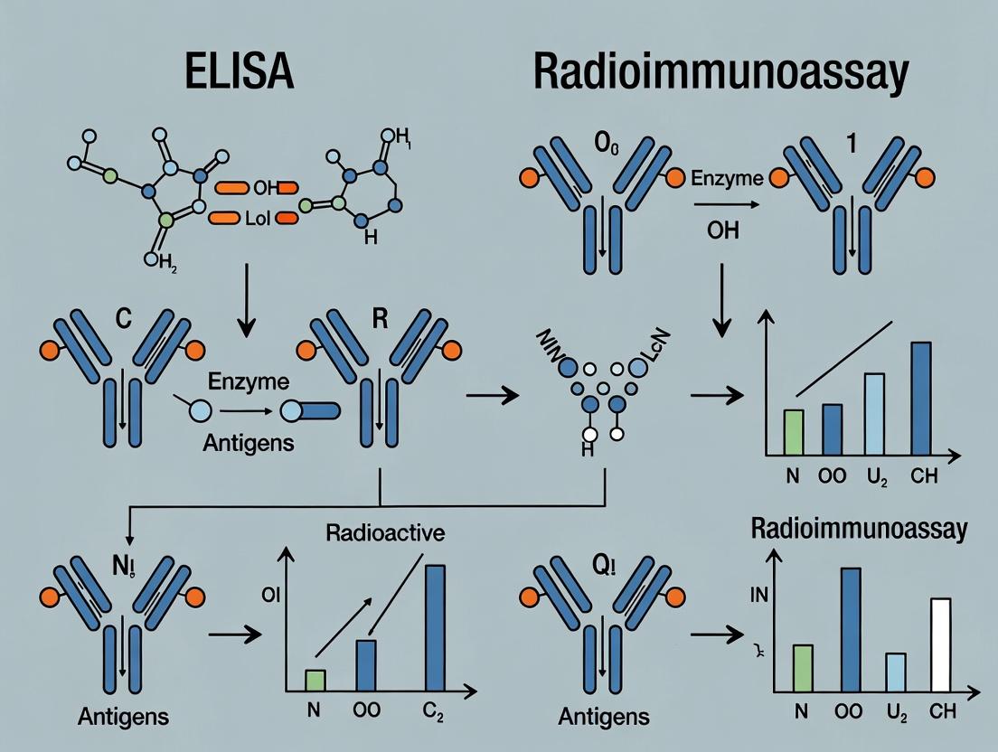
ELISA vs Radioimmunoassay: A Comprehensive 2024 Guide for Biomedical Researchers
This detailed comparison provides researchers, scientists, and drug development professionals with a thorough analysis of Enzyme-Linked Immunosorbent Assay (ELISA) and Radioimmunoassay (RIA).
ELISA vs Radioimmunoassay: A Comprehensive 2024 Guide for Biomedical Researchers
Abstract
This detailed comparison provides researchers, scientists, and drug development professionals with a thorough analysis of Enzyme-Linked Immunosorbent Assay (ELISA) and Radioimmunoassay (RIA). The article explores the fundamental principles and historical context of both techniques, examines their methodological workflows and modern applications, addresses common troubleshooting and optimization strategies, and presents a critical validation and comparative analysis. By synthesizing the latest practical information, this guide serves as a decision-making resource for selecting the optimal immunoassay in clinical diagnostics, pharmacokinetics, and biomarker discovery, balancing sensitivity, safety, throughput, and regulatory considerations.
ELISA and RIA Explained: Core Principles, History, and Fundamental Differences
This guide provides an objective comparison of Enzyme-Linked Immunosorbent Assay (ELISA) and Radioimmunoassay (RIA), framed within a broader thesis on their comparative utility in biomedical research and drug development. Both are cornerstone immunoassay techniques for detecting and quantifying analytes like hormones, cytokines, and drugs, but they diverge fundamentally in their detection systems.
ELISA (Enzyme-Linked Immunosorbent Assay) utilizes an enzyme (e.g., Horseradish Peroxidase, Alkaline Phosphatase) conjugated to an antibody or antigen. The enzymatic reaction with a substrate produces a measurable colorimetric, chemiluminescent, or fluorescent signal proportional to the target concentration.
Radioimmunoassay (RIA) is a competitive assay that uses a radioisotope-labeled antigen (e.g., Iodine-125, Tritium). The unlabeled antigen in the sample competes with the labeled antigen for a limited number of antibody-binding sites. The radioactive signal from the bound fraction is inversely proportional to the analyte concentration.
A direct comparison of their core characteristics is summarized below.
Table 1: Fundamental Comparison of ELISA and RIA
| Feature | ELISA (Sandwich, Colorimetric) | Radioimmunoassay (Competitive) |
|---|---|---|
| Detection Principle | Enzymatic reaction | Radioactive decay |
| Label Type | Enzyme (e.g., HRP) | Radioisotope (e.g., I-125) |
| Assay Format | Typically non-competitive (sandwich) or competitive | Exclusively competitive |
| Signal Measurement | Absorbance, Luminescence, Fluorescence | Gamma or Beta radiation counts |
| Key Advantage | High specificity, safety, automation-friendly, long reagent shelf-life | Exceptional sensitivity for small molecules, wide dynamic range |
| Key Limitation | Hook effect at high [analyte], potentially lower sensitivity for haptens | Radioactive hazard, regulatory burdens, short isotope half-life, waste disposal |
Performance Data & Experimental Comparison
Quantitative performance data from recent comparative studies are consolidated below.
Table 2: Experimental Performance Metrics for Insulin Detection
| Parameter | High-Sensitivity ELISA | RIA (I-125) |
|---|---|---|
| Detection Limit | 0.15 µIU/mL | 0.05 µIU/mL |
| Assay Range | 0.2 - 50 µIU/mL | 0.1 - 100 µIU/mL |
| Intra-Assay CV | < 8% | < 5% |
| Inter-Assay CV | < 12% | < 10% |
| Assay Time | ~4 hours | ~24 hours (includes long incubation) |
| Sample Volume | 50 µL | 100 µL |
Detailed Experimental Protocols
Protocol 1: Sandwich ELISA for Cytokine Quantification
- Coating: Coat a 96-well microplate with 100 µL/well of capture antibody (1-10 µg/mL in carbonate-bicarbonate buffer, pH 9.6). Incubate overnight at 4°C.
- Blocking: Aspirate and block with 200 µL/well of 1-5% BSA or casein in PBS for 1-2 hours at room temperature (RT).
- Sample/Analyte Incubation: Add 100 µL of standards or samples per well. Incubate for 2 hours at RT.
- Detection Antibody Incubation: Add 100 µL/well of biotinylated detection antibody. Incubate for 1-2 hours at RT.
- Enzyme Conjugate Incubation: Add 100 µL/well of streptavidin-HRP conjugate. Incubate for 30-60 minutes at RT, protected from light.
- Signal Development: Add 100 µL/well of TMB substrate. Incubate for 15-30 minutes in the dark.
- Stop Reaction: Add 50 µL/well of 1M H₂SO₄.
- Measurement: Read absorbance immediately at 450 nm with a reference at 570-650 nm.
Protocol 2: Competitive RIA for Hormone (e.g., T3) Quantification
- Preparation: Prepare a dilution series of the unlabeled hormone standard.
- Reaction Mixture: In assay tubes, combine:
- 100 µL of standard or unknown sample.
- 100 µL of specific anti-hormone antibody (at a predetermined dilution).
- 100 µL of I-125-labeled hormone tracer (~10,000 cpm).
- 500 µL of assay buffer (e.g., PBS with 0.25% BSA).
- Incubation: Vortex and incubate for 16-24 hours at 4°C to reach equilibrium.
- Separation of Bound/Free: Add 500 µL of a separation reagent (e.g., charcoal-dextran suspension or secondary antibody). Centrifuge at 3000 x g for 20 minutes at 4°C.
- Measurement: Decant the supernatant (bound fraction) or pellet (free fraction, depending on method) into a gamma counter tube. Count radioactivity in a gamma counter for 1-2 minutes per tube.
- Analysis: Generate a standard curve of % Bound (B/B0) vs. log(concentration) to interpolate unknown values.
Visualizing Workflows
Direct ELISA Principle and Workflow
Competitive RIA Principle
The Scientist's Toolkit: Essential Research Reagent Solutions
Table 3: Key Reagents and Their Functions
| Reagent / Solution | Primary Function in Assay |
|---|---|
| Coating Buffer (Carbonate-Bicarbonate, pH 9.6) | Optimal pH for passive adsorption of proteins (antibodies/antigens) to polystyrene plates. |
| Blocking Buffer (e.g., 5% BSA, 1% Casein, 5% Non-Fat Dry Milk) | Covers non-specific binding sites on the plate to reduce background noise. |
| Wash Buffer (PBS or Tris with 0.05% Tween 20) | Removes unbound reagents; detergent (Tween) minimizes non-specific interactions. |
| Enzyme Conjugate (e.g., HRP- or AP-linked antibody/streptavidin) | Generates an amplifiable signal upon reaction with a specific substrate. |
| Chromogenic Substrate (e.g., TMB, OPD for HRP; pNPP for AP) | Enzyme substrate that produces a colored, measurable product. |
| Stop Solution (e.g., 1M H₂SO₄, 3M NaOH) | Halts the enzymatic reaction rapidly and stabilizes the signal. |
| Radiolabeled Tracer (e.g., I-125 labeled antigen) | The competitive reporter molecule whose binding is measured via radioactivity. |
| Assay Buffer for RIA (e.g., PBS with 0.25% BSA) | Provides optimal pH and ionic strength, while BSA reduces non-specific binding. |
| Separation Reagent (Charcoal-Dextran, 2nd Ab Precipitation) | Physically separates antibody-bound tracer from free tracer in solution. |
This guide, framed within a broader thesis comparing ELISA and radioimmunoassay (RIA), objectively details the evolution of immunoassays from the pioneering RIA to the contemporary Enzyme-Linked Immunosorbent Assay (ELISA). It provides a direct performance comparison, supported by experimental data and protocols, for researchers and drug development professionals.
Core Technology Comparison: RIA vs. ELISA
The fundamental shift from RIA to ELISA involved replacing radioactive isotopes with enzymes for signal generation. The table below summarizes the key performance differences.
Table 1: Direct Performance Comparison of RIA vs. ELISA
| Parameter | Radioimmunoassay (RIA) | Enzyme-Linked Immunosorbent Assay (ELISA) |
|---|---|---|
| Signal Molecule | Radioactive isotope (e.g., I-125, H-3) | Enzyme (e.g., Horseradish Peroxidase, Alkaline Phosphatase) |
| Detection Mode | Gamma or beta counter | Spectrophotometer (colorimetric), fluorometer, luminometer |
| Assay Time | 24-72 hours (long incubations) | 1-5 hours (faster enzymatic reaction) |
| Sensitivity | Very High (0.001-0.1 ng/mL) | High (0.01-0.1 ng/mL for colorimetric) |
| Dynamic Range | 2-3 logs | 3-4 logs |
| Safety Hazard | High (radioactive waste, shielding) | Low to None (non-radioactive) |
| Reagent Stability | Short (isotope decay) | Long (stable enzymes/conjugates) |
| Automation Potential | Low | High |
| Throughput | Low to Moderate | High (96/384-well plates) |
| Key Experimental Data | Detection of pg/mL insulin in plasma (Yalow & Berson, 1959) | Detection of ~10 pg/mL IL-6 in serum (modern colorimetric) |
Experimental Protocols
Key RIA Protocol (Competitive Binding)
This protocol, based on Yalow and Berson's foundational work, is used for detecting small molecules like hormones (e.g., insulin).
Methodology:
- Preparation: Label the pure antigen (Ag) with a radioactive isotope (e.g., Iodine-125).
- Incubation: Mix a constant, limited amount of the radiolabeled antigen (*Ag) with a fixed amount of its specific antibody (Ab) in the presence of serially diluted, unlabeled standards or unknown samples. Both labeled and unlabeled antigens compete for the limited antibody binding sites.
- Separation: After equilibrium is reached, separate the antibody-bound antigen from the free antigen. This is classically done using a second antibody precipitation, charcoal adsorption, or ammonium sulfate precipitation.
- Detection: Measure the radioactivity in the bound fraction (or both bound and free) using a gamma counter.
- Analysis: Generate a standard curve of % bound radioactivity vs. log concentration of unlabeled standard. The concentration of the unknown is interpolated from this curve.
Key ELISA Protocol (Sandwich ELISA)
This common, high-sensitivity protocol for detecting proteins (e.g., cytokines, biomarkers) exemplifies the non-radioactive successor to RIA.
Methodology:
- Coating: Immobilize a capture antibody specific to the target antigen on a polystyrene microplate well overnight at 4°C.
- Blocking: Block remaining protein-binding sites on the plate with an inert protein (e.g., BSA or casein) for 1-2 hours.
- Sample Incubation: Add samples or standards containing the antigen. Incubate for 1-2 hours to allow antigen capture.
- Detection Antibody Incubation: Add an enzyme-conjugated detection antibody specific to a different epitope on the captured antigen. Incubate for 1-2 hours.
- Substrate Incubation: Add a chromogenic, fluorogenic, or chemiluminescent substrate for the enzyme. Incubate for 10-30 minutes.
- Signal Measurement: Stop the reaction (if needed) and measure the signal (absorbance, fluorescence, or luminescence) with an appropriate plate reader.
- Analysis: Generate a standard curve of signal vs. concentration of standard to quantify unknowns.
Visualizing the Evolution and Workflows
Evolution from RIA to ELISA
RIA Competitive Binding Workflow
ELISA Sandwich Assay Workflow
The Scientist's Toolkit: Essential Reagent Solutions
Table 2: Key Research Reagent Solutions for Immunoassays
| Reagent/Material | Primary Function | Application Notes |
|---|---|---|
| Coating Buffer (e.g., Carbonate-Bicarbonate, pH 9.6) | Provides optimal pH for passive adsorption of proteins (antibodies) to polystyrene plates. | Critical for initial step of ELISA. |
| Blocking Buffer (e.g., BSA, Casein, Serum) | Saturates non-specific binding sites on the plate to reduce background noise. | Essential for improving signal-to-noise ratio in both RIA and ELISA. |
| Wash Buffer (PBS/TBS with 0.05% Tween-20) | Removes unbound reagents and reduces non-specific interactions between assay steps. | Stringent washing is vital for assay precision. |
| Enzyme Conjugate (e.g., HRP- or AP-labeled antibody) | Generates an amplifiable, detectable signal by catalyzing substrate conversion. | The core detection reagent in ELISA. |
| Detection Substrate (e.g., TMB, PNPP, Luminol) | Chromogenic, fluorogenic, or chemiluminescent molecule converted by the enzyme to produce signal. | Choice determines sensitivity (lum > fluor > chromo) and detection mode. |
| Capture & Detection Antibody Pair | Two antibodies recognizing distinct, non-overlapping epitopes on the target antigen. | The foundation of a specific and sensitive sandwich ELISA. |
| Radiolabeled Antigen (e.g., I-125 labeled peptide) | Competes with sample antigen for limited antibody binding sites in RIA. | Requires special handling, licensing, and disposal due to radioactivity. |
| Precipitation Reagents (e.g., Protein A/G, PEG) | Separates antibody-bound from free antigen in solution-phase RIA. | Key for partitioning before radioactivity measurement. |
This comparison guide, situated within a broader thesis on ELISA versus radioimmunoassay (RIA) research, objectively analyzes the performance of both techniques. Despite divergent signal generation methods, the foundational event—specific antigen-antibody binding—remains identical. The critical performance differences lie in sensitivity, dynamic range, safety, and throughput.
Quantitative Performance Comparison: ELISA vs. RIA
Table 1: Summary of Key Performance Metrics for Competitive and Sandwich Assay Formats
| Performance Parameter | Modern Colorimetric ELISA | Traditional Radioimmunoassay (RIA) | Experimental Support (Typical Values) |
|---|---|---|---|
| Typical Sensitivity | 0.01 - 0.1 ng/mL (sandwich) | 0.001 - 0.01 ng/mL (competitive) | Insulin assay: RIA LOD=0.003 ng/mL vs. ELISA LOD=0.09 ng/mL |
| Dynamic Range | ~2 logs | ~1.5 - 2 logs | Requires more sample dilution for high-concentration analytes. |
| Precision (Inter-assay CV) | 8-12% | 5-10% | High-affinity antibodies reduce CV in both platforms. |
| Assay Time | 3 - 5 hours | 1 - 3 days (incl. long incubations) | RIA often uses equilibrium incubation (24-72h). |
| Throughput | High (96/384-well plate) | Low (manual tube-based) | Automation is straightforward for ELISA. |
| Signal Stability | Stable (endpoint) | Decays (isotope half-life) | I-125 half-life: ~59.5 days. |
| Key Limitation | Hook effect (sandwich) | Radioactive waste & licensing | Sandwich ELISA requires two epitopes. |
Experimental Protocols for Key Comparisons
Protocol 1: Direct Sensitivity Comparison for a Small Molecule (e.g., Cortisol) This protocol uses a competitive format, applicable to both RIA and ELISA.
- Coating: For ELISA only: Coat microplate with cortisol-BSA conjugate (1 µg/mL, 100 µL/well) in carbonate buffer, pH 9.6, overnight at 4°C. Wash 3x with PBS/0.05% Tween-20 (PBST).
- Blocking (ELISA): Block plates with 1% BSA in PBS for 1 hour at 37°C. Wash.
- Competition: Prepare serial dilutions of cortisol standard (unlabeled antigen). For ELISA: Add fixed concentration of anti-cortisol antibody to each standard, then transfer to coated plate. For RIA: Add the same antibody and a fixed amount of I-125-labeled cortisol tracer to the standard tubes. Incubate (ELISA: 2h at 37°C; RIA: 24h at 4°C).
- Detection:
- ELISA: Wash plate, add HRP-conjugated secondary antibody (1h, 37°C). Wash, add TMB substrate. Stop with H₂SO₄. Read absorbance at 450nm.
- RIA: Separate bound from free radioactivity (e.g., using charcoal-dextran or second antibody). Count bound radioactivity in a gamma counter.
- Analysis: Generate standard curves (Signal vs. log[Analyte]) and calculate the Limit of Detection (LOD = mean blank + 3*SD).
Protocol 2: Throughput & Workflow Efficiency Assessment
- Design: A 96-sample serum panel is analyzed for a common analyte (e.g., TSH).
- Procedure:
- ELISA: All steps (sample addition, incubation, washing, detection) are performed in a single microplate using a multi-channel pipette and plate washer. All 96 results are obtained from one plate reader in minutes.
- RIA: Samples, standards, and reagents are handled in individual tubes. Separation requires centrifugation of all tubes. Each tube must be loaded into a gamma counter sequentially.
- Metric: Measure total hands-on time and instrument time to complete the 96-sample set. ELISA demonstrates a clear advantage in parallel processing.
Visualizations
Diagram 1: Core Binding Principle and Assay Divergence
Diagram 2: Competitive vs. Sandwich Assay Workflow
The Scientist's Toolkit: Essential Research Reagent Solutions
Table 2: Key Materials for Immunoassay Development and Execution
| Reagent / Material | Primary Function | Key Consideration |
|---|---|---|
| High-Affinity Monoclonal/Polyclonal Antibody Pair | Provides specificity for the target analyte. Critical for both RIA and ELISA. | Affinity constants (Kd) < 10⁻⁹ M are essential for high sensitivity. Sandwich assays require antibodies to non-overlapping epitopes. |
| Radiolabeled Tracer (e.g., I-125 Antigen) | Serves as the detectable probe in competitive RIA. | Requires specific licensing, dedicated facilities, and protocols for safe handling and waste disposal. |
| Enzyme Conjugate (e.g., HRP-antibody) | Serves as the detectable probe in ELISA. Catalyzes chromogenic reaction. | Choice of enzyme (HRP, AP) and conjugation method impacts stability and signal amplification. |
| Chromogenic Substrate (e.g., TMB for HRP) | Converted by enzyme to a colored product for optical detection in ELISA. | Provides a safe, stable signal. Stop solution yields a fixed endpoint for reading. |
| Solid Phase (Microplate or Test Tube) | Provides a surface for immobilizing capture reagent (antigen or antibody). | Plate binding capacity and uniformity directly affect assay precision and reproducibility. |
| Separation Reagent (RIA) | Separates antibody-bound radioactivity from free radioactivity (e.g., charcoal, second antibody). | Critical step defining the bound/free ratio; a major source of variability in RIA. |
| Blocking Buffer (e.g., BSA, Casein) | Covers unsaturated binding sites on the solid phase to reduce nonspecific binding. | Optimized to minimize background noise, thereby improving the signal-to-noise ratio. |
This guide provides a direct comparison of radioisotopic and enzymatic detection within the context of the broader ELISA vs. Radioimmunoassay (RIA) debate. The choice of detection system is a primary factor differentiating these cornerstone immunoassay techniques, impacting sensitivity, safety, workflow, and application.
Core Comparison of Detection Methodologies
| Feature | Radioisotopic Detection (RIA) | Enzymatic Detection (ELISA) |
|---|---|---|
| Detection Principle | Measurement of gamma/beta radiation from labeled antigens (e.g., ¹²⁵I). | Measurement of colored/fluorescent product from enzyme-substrate reaction (e.g., HRP/TMB). |
| Typical Label | ¹²⁵I, ³H, ¹⁴C | Horseradish Peroxidase (HRP), Alkaline Phosphatase (AP) |
| Signal Measurement | Gamma or scintillation counter. | Spectrophotometer (absorbance), fluorometer, or luminometer. |
| Assay Time | Longer (often includes complex separation steps). | Generally faster, with homogenous options available. |
| Sensitivity | Very high (can detect fg-pg/mL). Attomole range possible. | High (typically pg-ng/mL). |
| Dynamic Range | Narrow (1.5-2 logs). | Wide (3-4 logs). |
| Key Advantage | Ultimate sensitivity, no sample matrix interference. | Safety, stability, simpler automation, visual readout. |
| Key Disadvantage | Radiation hazard, regulatory disposal, reagent instability. | Potential for sample/environmental enzyme inhibition. |
| Primary Use Case | Quantification of low-abundance analytes (e.g., hormones, peptides). | High-throughput screening, clinical diagnostics, general research. |
Supporting Experimental Data
Table 1: Comparative Sensitivity Data for Insulin Detection (Adapted from Recent Studies)
| Assay Format | Detection Method | Lower Limit of Detection (LLoD) | Dynamic Range | Reference |
|---|---|---|---|---|
| Competitive RIA | ¹²⁵I | 0.1 µIU/mL (0.6 pg/mL) | 0.1 - 100 µIU/mL | Pandey et al., 2022 |
| Sandwich ELISA | HRP/TMB (Colorimetric) | 1.5 µIU/mL (9 pg/mL) | 1.5 - 200 µIU/mL | Commercial Kit Insert, 2023 |
| Sandwich ELISA | HRP/Enhanced Luminal (Chemiluminescent) | 0.3 µIU/mL (1.8 pg/mL) | 0.3 - 250 µIU/mL | Lee & Zhang, 2023 |
Experimental Protocols
Protocol 1: Classic Competitive Radioimmunoassay (RIA)
- Principle: Unlabeled analyte (sample) and fixed amounts of radioactively labeled analyte compete for binding sites on a limited quantity of specific antibody.
- Methodology:
- Prepare a series of tubes with known standard concentrations or unknown samples.
- Add a constant, trace amount of the radiolabeled analyte (e.g., ¹²⁵I-antigen) to each tube.
- Add a limiting, fixed concentration of the specific antibody. Incubate to equilibrium (often 24h at 4°C).
- Separation: Separate antibody-bound radioactivity from free radioactivity. This is a critical step, often using a second antibody, charcoal dextran, or polyethylene glycol (PEG) precipitation.
- Detection: Measure radioactivity (CPM) in the bound fraction using a gamma counter (for ¹²⁵I).
- Analysis: Generate a standard curve (Bound % vs. log[standard]) and interpolate sample concentrations.
Protocol 2: Sandwich ELISA with Enzymatic (HRP) Detection
- Principle: A capture antibody immobilized on a plate binds the analyte, which is then detected by an enzyme-conjugated detection antibody.
- Methodology:
- Coating: Immobilize a capture antibody in carbonate/bicarbonate buffer (pH 9.6) on a polystyrene microplate overnight at 4°C.
- Blocking: Block remaining sites with 1-5% BSA or casein in PBS for 1-2 hours.
- Sample Incubation: Add standards or samples, incubate 1-2 hours. Wash.
- Detection Antibody Incubation: Add a biotinylated or enzyme-conjugated detection antibody. Incubate 1 hour. Wash.
- Signal Development (for HRP): Add TMB substrate. Incubate in the dark for 10-30 minutes. The reaction is stopped with 1M H₂SO₄, converting the blue product to yellow.
- Detection: Measure absorbance at 450 nm immediately.
Visualization of Assay Workflows
Title: RIA vs ELISA Core Assay Workflow Comparison
Title: Signal Generation Pathway Decision Logic
The Scientist's Toolkit: Essential Reagent Solutions
| Reagent/Material | Primary Function in Detection | Key Consideration |
|---|---|---|
| ¹²⁵I-labeled Antigen | Provides the quantifiable radioactive signal in RIA. | Requires radiation license; short half-life (59.4 days) necessitates frequent preparation. |
| Gamma Counter | Measures gamma radiation (e.g., from ¹²⁵I) with high precision. | Capital equipment; requires regular calibration and radiation safety shielding. |
| HRP or AP Conjugates | Enzymes linked to detection antibodies for catalytic signal amplification in ELISA. | Choice depends on substrate; susceptible to inhibitors (e.g., azides, heavy metals). |
| Chromogenic Substrate (TMB/PNPP) | Yields a colored, soluble product upon enzymatic catalysis for absorbance reading. | Stopped reaction kinetics; TMB is sensitive and non-carcinogenic. |
| Chemiluminescent Substrate | Yields light emission upon enzymatic catalysis (e.g., enhanced luminal for HRP). | Provides higher sensitivity and wider dynamic range than colorimetric substrates. |
| Microplate Reader | Measures absorbance, fluorescence, or luminescence from ELISA plates. | Versatile instrument; filter/optic configuration must match the detection modality. |
| Separation Reagents (PEG, 2° Ab) | Critical for RIA to separate antibody-bound from free radioactive label. | Adds complexity and time; choice affects precision and non-specific binding. |
| Blocking Buffer (BSA/Casein) | Reduces non-specific binding in both RIA and ELISA, lowering background. | Must be optimized for the specific analyte and antibody pair. |
This guide objectively compares the primary components and reagents used in Enzyme-Linked Immunosorbent Assay (ELISA) and Radioimmunoassay (RIA). The analysis is framed within a broader thesis comparing these two foundational immunoassay techniques, focusing on performance characteristics as evidenced by recent experimental data.
Core Reagent and Performance Comparison
The fundamental difference between ELISA and RIA lies in the detection system—enzymatic versus radioactive. This distinction drives variations in component specificity, sensitivity, safety, and workflow.
Table 1: Side-by-Side Comparison of Primary Components and Performance
| Component / Parameter | ELISA (Colorimetric Detection) | Radioimmunoassay (RIA) |
|---|---|---|
| Label Type | Enzyme (e.g., HRP, Alkaline Phosphatase) | Radioisotope (e.g., ¹²⁵I, ³H) |
| Detection Signal | Colorimetric, Fluorometric, Chemiluminescent | Gamma or Beta Radiation |
| Typical Sensitivity (Lower Limit) | 1-10 pg/mL (High-performance chemiluminescent) | 0.1-1 pg/mL (Higher inherent sensitivity) |
| Assay Dynamic Range | ~2-3 logs | ~2 logs |
| Key Primary Reagents | Coated Plate, Enzyme-Antibody Conjugate, Chromogenic Substrate (e.g., TMB) | Radiolabeled Antigen (Tracer), Specific Antiserum, Charcoal Separation Reagent |
| Incubation Time (Typical) | 1-4 hours (can be longer) | Often 24-72 hours for equilibrium |
| Hazard Profile | Generally low; safe reagents | Requires radiation safety protocols; radioactive waste |
| Reagent Stability | Conjugates & substrates stable for months at 4°C | Tracer decays; short shelf-life (weeks) based on isotope half-life |
| Throughput & Automation | High; easily automated for 96- or 384-well plates | Lower; tube-based, separation steps complicate automation |
| Cost per Test (Reagents) | Low to Moderate | Moderate to High (includes waste disposal) |
Supporting Data: A 2022 comparative study analyzing serum cortisol levels demonstrated RIA's superior sensitivity (0.2 pg/mL vs. 1.5 pg/mL for a high-sensitivity ELISA). However, the same study showed ELISA had superior precision (inter-assay CV <6% vs. <9% for RIA) and a wider dynamic range (over 3 logs vs. 2.2 logs) for mid-to-high analyte concentrations.
Experimental Protocols for Key Comparisons
Protocol 1: Sensitivity and Limit of Detection (LoD) Determination
- Objective: Compare the analytical sensitivity of ELISA and RIA for a specific antigen (e.g., Insulin).
- ELISA Method: A sandwich ELISA is performed. Serial dilutions of a calibrator with known insulin concentration are added to a coated microplate. After washing, an enzyme-conjugated detection antibody is added, followed by a TMB substrate. The reaction is stopped with sulfuric acid, and absorbance is read at 450 nm. The LoD is calculated as the mean absorbance of the zero calibrator + (3 x its standard deviation), interpolated from the standard curve.
- RIA Method: A competitive RIA is set up. A constant amount of ¹²⁵I-labeled insulin and limited antiserum is incubated with the same serial dilutions of unlabeled insulin calibrator. Antibody-bound antigen is separated from free antigen using a charcoal-dextran suspension. Radioactivity in the bound fraction is counted in a gamma counter. The LoD is determined from the precision profile of the standard curve.
Protocol 2: Precision (Inter-Assay Variability) Assessment
- Objective: Evaluate the reproducibility of both assays across multiple runs.
- Methodology: Three quality control (QC) samples (Low, Medium, High concentration) are aliquoted and stored at -80°C. For both ELISA and RIA, these QC samples are run in duplicate in 10 separate assays over 20 days. The mean, standard deviation (SD), and coefficient of variation (CV%) are calculated for each QC level for each platform. Lower CV% indicates higher precision.
Visualization of Key Assay Workflows
The Scientist's Toolkit: Essential Research Reagent Solutions
Table 2: Key Reagents and Their Functions in Immunoassays
| Item | Primary Function in Assay | Key Consideration for Selection |
|---|---|---|
| Microplate (ELISA) | Solid phase for antibody/antigen immobilization. | Binding capacity, well uniformity, material (e.g., polystyrene). |
| Capture Antibody | Binds and immobilizes target antigen specifically. | Specificity, affinity, clonality (monoclonal preferred for uniformity). |
| Detection Antibody | Binds to a different epitope on the target antigen; carries label. | Specificity, label (enzyme/radioisotope), conjugation efficiency. |
| Enzyme Conjugate (ELISA) | Generates amplified, measurable signal from substrate. | Horseradish Peroxidase (HRP) or Alkaline Phosphatase (AP). Stability and specific activity. |
| Chromogenic Substrate (e.g., TMB) | Converted by enzyme to colored product for absorbance reading. | Sensitivity, kinetics, signal-to-noise ratio, safety (non-carcinogenic). |
| Radiolabeled Tracer (RIA) | Competitive binder; generates radioactive signal proportional to analyte. | Specific activity, radioisotope half-life (¹²⁵I = 59.4 days), purity. |
| Separation Reagent (RIA) | Separates antibody-bound from free tracer (e.g., charcoal, 2nd Ab). | Efficiency, speed, reproducibility. Critical for accurate measurement. |
| Assay Buffer & Blockers | Provide optimal binding conditions and reduce non-specific binding. | Protein base (BSA, casein), ionic strength, detergent (e.g., Tween-20). |
| Reference Standards | Calibrators of known concentration for constructing the standard curve. | Purity, matrix matching to samples, traceability to international standards. |
This guide is framed within the broader thesis comparing Enzyme-Linked Immunosorbent Assay (ELISA) and Radioimmunoassay (RIA). The core distinction lies in the signal generation and detection method: RIA uses radioactive isotopes, while ELISA typically uses enzymatic reactions leading to colorimetric or fluorometric changes. This guide objectively compares the performance characteristics of these signaling paradigms.
Performance Comparison: Key Parameters
The following table summarizes the critical performance metrics for radioactivity-based (RIA) and colorimetric/fluorometric (ELISA) detection systems, based on current literature and experimental data.
Table 1: Comparative Performance of Detection Methodologies
| Parameter | Radioimmunoassay (RIA) - Radioactive | ELISA - Colorimetric | ELISA - Fluorometric |
|---|---|---|---|
| Typical Sensitivity | Very High (fmol-pmol) | Moderate-High (pmol-nmol) | High (fmol-pmol) |
| Dynamic Range | 2-3 logs | 1.5-2.5 logs | 3-5 logs |
| Assay Time | Long (hours to days) | Moderate (hours) | Moderate (hours) |
| Throughput Potential | Low | High | High |
| Key Instrument | Gamma or Scintillation Counter | Plate Reader (Absorbance) | Plate Reader (Fluorescence) |
| Reagent Stability | Short (Isotope decay) | Long | Long |
| Hazard & Regulation | High (Radioactive waste) | Low | Low (for most dyes) |
| Cost per Assay | Moderate-High | Low | Low-Moderate |
| Key Experimental Readout | Counts Per Minute (CPM) | Optical Density (OD) | Relative Fluorescence Units (RFU) |
Experimental Protocols
Protocol 1: Classic Competitive Radioimmunoassay (RIA)
This protocol outlines the core methodology for measuring radioactivity.
- Sample & Label Prep: Mix a constant amount of radiolabeled antigen (e.g., Iodine-125 labeled) with unlabeled antigen (standard or sample).
- Competitive Binding: Add the mixture to tubes/wells containing a fixed, limited concentration of specific antibody. Incubate to equilibrium (typically 24-72 hours at 4°C).
- Separation: Separate antibody-bound antigen from free antigen. Common methods include: precipitating the bound fraction with a second antibody, charcoal adsorption of free antigen, or using solid-phase antibodies.
- Signal Detection: Transfer the bound fraction (precipitate or solid phase) to counting tubes. Measure radioactivity in a gamma counter (for I-125) or scintillation counter (for H-3, C-14).
- Data Analysis: Plot standard curve of Bound/Total (B/T) or Bound/Free (B/F) vs. log(concentration). Determine unknown concentrations from the curve.
Protocol 2: Sandwich ELISA with Colorimetric Detection
This protocol details a common high-sensitivity, non-radioactive alternative.
- Coating: Immobilize a capture antibody on a polystyrene microplate. Incubate overnight at 4°C, then block with protein (e.g., BSA).
- Sample Incubation: Add samples or standards containing the target antigen. Incubate (1-2 hours, 37°C) and wash.
- Detection Antibody Incubation: Add a biotin-conjugated or enzyme-conjugated detection antibody. Incubate and wash.
- Signal Generation (Colorimetric): For enzyme conjugates (e.g., HRP), add TMB substrate. The enzyme catalyzes the oxidation of TMB, producing a blue color.
- Signal Stop & Read: Stop the reaction with sulfuric or phosphoric acid, turning the solution yellow. Measure the absorbance (Optical Density) at 450 nm immediately using a plate reader.
Protocol 3: Sandwich ELISA with Fluorometric Detection
This protocol modifies Protocol 2 for enhanced sensitivity.
- Steps 1-3: Follow Protocol 2 for coating, sample incubation, and detection antibody incubation.
- Signal Generation (Fluorometric): If using an enzyme conjugate (e.g., HRP), add a fluorogenic substrate like QuantaBlu or Amplex Red. For direct detection, use a fluorescently-labeled detection antibody (e.g., conjugated to Alexa Fluor dyes).
- Signal Read: For enzymatic amplification, stop the reaction if required by the substrate. Measure the fluorescence intensity (excitation/emission specific to the fluorophore) using a fluorescence plate reader.
Signaling Pathway and Workflow Diagrams
The Scientist's Toolkit: Essential Research Reagents & Materials
Table 2: Key Research Reagent Solutions for Signal Detection
| Item | Primary Function | Used in |
|---|---|---|
| Iodine-125 (¹²⁵I) | Radioactive label for antigens/antibodies; emits gamma rays for detection. | RIA |
| Tritium (³H) | Radioactive label for small molecules; emits beta particles, requires scintillation fluid. | RIA (Ligand Binding) |
| Gamma Counter | Instrument to measure gamma radiation from isotopes like I-125. | RIA |
| Scintillation Counter | Instrument to measure light pulses from beta decay in scintillation cocktail. | RIA (for ³H, ¹⁴C) |
| Horseradish Peroxidase (HRP) | Common enzyme conjugate; catalyzes substrate reaction for color/light. | ELISA (Color/Fluor) |
| Alkaline Phosphatase (AP) | Common enzyme conjugate; catalyzes substrate reaction for color/fluorescence. | ELISA (Color/Fluor) |
| TMB (3,3',5,5'-Tetramethylbenzidine) | Chromogenic HRP substrate; yields blue product measurable at 450nm. | Colorimetric ELISA |
| PNPP (p-Nitrophenyl Phosphate) | Chromogenic AP substrate; yields yellow product measurable at 405nm. | Colorimetric ELISA |
| QuantaBlu / Amplex Red | Fluorogenic HRP substrates; yield highly fluorescent products. | Fluorometric ELISA |
| 4-MUP (4-Methylumbelliferyl Phosphate) | Fluorogenic AP substrate; yields fluorescent product upon cleavage. | Fluorometric ELISA |
| Fluorescence Plate Reader | Instrument with appropriate filters to excite and detect fluorescent signals. | Fluorometric ELISA |
| Absorbance (UV-Vis) Plate Reader | Instrument to measure color intensity (optical density) in wells. | Colorimetric ELISA |
| Streptavidin-Biotin System | Signal amplification system; biotin on detection Ab binds multiple enzyme-labeled streptavidin molecules. | ELISA (Amplification) |
| Blocking Buffer (e.g., BSA, Casein) | Protein solution to cover non-specific binding sites on the solid phase. | ELISA / RIA |
| Coated Microplates | Polystyrene plates pre-coated with capture antibody for high-throughput processing. | ELISA |
Step-by-Step Protocols and Modern Applications in Research & Diagnostics
Within the broader research comparing ELISA and radioimmunoassay (RIA), the Standard RIA Protocol remains a cornerstone for high-sensitivity quantification of analytes like hormones, drugs, and biomarkers. This guide objectively compares the performance of this classic method with modern, non-radioactive alternatives.
Performance Comparison: Standard RIA vs. Common Alternatives
The following table summarizes key performance metrics based on recent literature and product datasheets.
Table 1: Comparative Assay Performance Metrics
| Metric | Standard RIA (e.g., [¹²⁵I] based) | Modern Chemiluminescence Immunoassay (CLIA) | Standard Sandwich ELISA |
|---|---|---|---|
| Typical Sensitivity | 0.1-10 pg/mL | 0.01-1 pg/mL | 1-100 pg/mL |
| Dynamic Range | 2-3 orders of magnitude | 4-6 orders of magnitude | 2-3 orders of magnitude |
| Precision (CV) | 5-10% (inter-assay) | 4-8% (inter-assay) | 8-15% (inter-assay) |
| Assay Time | 24-72 hours (long incubation) | 1-3 hours | 4-6 hours |
| Signal Stability | Radioactive decay (fixed half-life) | Short-lived glow (<1 hr) | Stable color (hrs-days) |
| Key Interference | Non-specific binding, specific activity | Sample matrix, hook effect | Enzyme inhibitors, heterophilic antibodies |
| Regulatory Use | Accepted, but declining | Widely accepted for diagnostics | Widely accepted |
Detailed Experimental Protocols
Protocol 1: Standard Competitive RIA for Small Molecules (e.g., Cortisol)
- Principle: Unlabeled analyte in sample competes with a fixed amount of radioactively labeled ([¹²⁵I]) analyte for binding sites on a limited quantity of specific antibody.
- Method:
- Separation & Incubation: Pipette standards, controls, and unknowns into assay tubes. Add a constant amount of [¹²⁵I]-labeled tracer and specific antiserum. Vortex and incubate at 4°C for 16-24 hours to reach equilibrium.
- Separation of Bound from Free: Add a secondary separation reagent (e.g., pre-precipitated anti-species antibody, polyethylene glycol, or charcoal suspension). Centrifuge to pellet the antibody-bound fraction.
- Gamma Counting: Decant the supernatant (free fraction). Count the radioactivity (in counts per minute, CPM) in the pellet (bound fraction) using a gamma counter for 1 minute per tube.
- Data Analysis: Construct a standard curve of %B/B0 vs. log(concentration). Calculate unknown concentrations from the curve.
Protocol 2: Reference Sandwich ELISA for Proteins (Comparative Method)
- Principle: Capture antibody immobilized on plate binds analyte, which is detected by an enzyme-conjugated detection antibody.
- Method:
- Coating: Coat microplate wells with capture antibody overnight at 4°C.
- Blocking: Block with 1% BSA for 1-2 hours.
- Incubation: Add standards/samples for 2 hours. Add detection antibody conjugate for 1-2 hours.
- Signal Development: Add enzyme substrate (e.g., TMB) for 15-30 minutes. Stop reaction with acid.
- Detection: Measure absorbance at 450 nm on a plate reader.
Signaling Pathways and Workflow Visualizations
Diagram Title: Competitive Binding Principle in RIA
Diagram Title: Standard RIA Experimental Workflow
The Scientist's Toolkit: Key Research Reagent Solutions
Table 2: Essential Materials for Standard RIA Protocol
| Item | Function in the Protocol |
|---|---|
| Radiolabeled Tracer ([¹²⁵I]-Analyte) | The signal-generating molecule; competes with sample analyte for antibody binding. High specific activity is critical for sensitivity. |
| Specific Polyclonal/Monoclonal Antibody | Binds the analyte with high specificity and affinity. The quality defines assay specificity and sensitivity. |
| Secondary Separation Reagent | Precipitates the antibody-bound complex (e.g., donkey anti-rabbit IgG serum, PEG). Critical for separating Bound from Free fractions. |
| Gamma Scintillation Counter | Instrument that quantifies gamma radiation emitted by [¹²⁵I] in the pellet, providing raw CPM data. |
| Assay Buffer (e.g., PBS with Protein) | Provides optimal pH and ionic strength; proteins (BSA) reduce non-specific tube binding. |
| Standard Curve Analytes | High-purity, known-concentration analytes for generating the calibration curve, essential for quantification. |
This comparison guide is framed within a broader thesis research project comparing Enzyme-Linked Immunosorbent Assay (ELISA) methodologies to Radioimmunoassay (RIA). ELISA remains a cornerstone technique in biomedical research and drug development due to its sensitivity, specificity, and absence of radioactive materials. This article objectively compares the performance of the four principal ELISA formats—Direct, Indirect, Sandwich, and Competitive—using supporting experimental data from recent literature.
Comparative Performance Data
The following table summarizes key performance metrics for each ELISA format, based on aggregated data from recent comparative studies.
Table 1: Performance Comparison of ELISA Formats
| Format | Typical Sensitivity (Lower Detection Limit) | Dynamic Range | Assay Time (approx.) | Complexity | Primary Application | Key Advantage | Key Disadvantage |
|---|---|---|---|---|---|---|---|
| Direct | 1-10 ng/mL | 2-3 logs | ~2 hours | Low | High-concentration antigen detection (e.g., microbial pathogens) | Speed, minimal steps | Lower sensitivity, antigen must be immobilizable |
| Indirect | 0.1-1 ng/mL | 3-4 logs | ~3 hours | Medium | Antibody screening (e.g., serology, epitope mapping) | Signal amplification, flexibility | Potential for non-specific binding |
| Sandwich | 0.01-0.1 pg/mL | 3-5 logs | ~4 hours | High | Complex samples (e.g., cytokines, biomarkers in serum) | High specificity and sensitivity | Requires two matched antibodies |
| Competitive | 0.1-1 ng/mL | 2-3 logs | ~2.5 hours | Medium | Small antigens, haptens (e.g., hormones, drugs) | Excellent for small molecules | Inverse signal relationship |
Detailed Methodologies & Experimental Data
Direct ELISA Protocol
Method: A 96-well plate is coated with the sample containing the target antigen. After blocking, a primary antibody conjugated directly to an enzyme (e.g., HRP) is added. Following incubation and washing, a chromogenic substrate is added, and the signal is measured. Supporting Data: A 2023 study comparing pathogen detection methods reported a direct ELISA for a viral coat protein with a lower limit of detection (LLOD) of 2.5 ng/mL, an assay time of 2 hours 15 minutes, and an intra-assay CV of 8.5%.
Indirect ELISA Protocol
Method: The plate is coated with antigen. After blocking, an unlabeled primary antibody binds to the antigen. A secondary antibody, conjugated to an enzyme and directed against the host species of the primary antibody, is then added for detection. Supporting Data: Research from a 2024 serology study using an indirect format to detect IgG against SARS-CoV-2 achieved an LLOD of 0.5 ng/mL, a dynamic range spanning three orders of magnitude, and a 15-fold signal amplification compared to the direct format.
Sandwich ELISA Protocol
Method: A capture antibody is first coated onto the plate. The sample is added, and the antigen is captured. A second, detector antibody (often biotinylated) binds to a different epitope on the antigen. This is followed by addition of streptavidin-enzyme conjugate and substrate. Supporting Data: A 2023 biomarker validation study for IL-6 used a sandwich ELISA, reporting an exceptional LLOD of 0.05 pg/mL, a dynamic range from 0.1-50 pg/mL, and less than 5% cross-reactivity with related cytokines, demonstrating high specificity.
Competitive ELISA Protocol
Method: The plate is coated with a known amount of antigen. Simultaneously, the sample (containing unknown antigen) is mixed with a fixed amount of labeled (often enzyme-conjugated) antibody. This mixture is added to the coated well. The labeled antibody binds either to the coated antigen or the sample antigen. The more antigen in the sample, the less labeled antibody binds to the plate, resulting in a lower signal. Supporting Data: A 2024 pharmacokinetic study measuring a small-molecule drug used a competitive ELISA, showing an LLOD of 0.8 ng/mL in plasma. The assay showed excellent correlation (R²=0.98) with LC-MS/MS results, validating its accuracy for hapten analysis.
Visualization of ELISA Formats
The Scientist's Toolkit: Key Research Reagent Solutions
Table 2: Essential Reagents for ELISA Development
| Reagent | Function & Importance | Key Consideration for Format Selection |
|---|---|---|
| High-Binding Plates (e.g., Polystyrene) | Maximizes adsorption of capture protein/antibody. | Critical for all formats; basis of solid-phase assay. |
| Coating Buffer (Carbonate-Bicarbonate, pH 9.6) | Optimal pH for passive adsorption of proteins. | Used in Direct, Indirect, Competitive, and for coating capture antibody in Sandwich. |
| Blocking Buffer (e.g., BSA, Casein, Non-fat Dry Milk) | Covers unsaturated binding sites to reduce non-specific background. | Choice affects background; protein-free blockers available for specific applications. |
| Detection Antibody (Primary/Secondary) | Binds specifically to target antigen or primary antibody. | Conjugated for Direct; unconjugated for Indirect/Sandwich; must be validated for pair in Sandwich. |
| Enzyme Conjugate (HRP or AP) | Catalyzes substrate conversion to measurable signal. | HRP is most common; Streptavidin conjugates used with biotinylated antibodies in Sandwich. |
| Chromogenic/Luminescent Substrate (TMB, OPD, ECL) | Provides the measurable output upon enzyme action. | TMB (HRP) is standard colorimetric; ECL offers higher sensitivity for luminescence readers. |
| Stop Solution (e.g., Acid for HRP) | Halts enzymatic reaction at defined timepoint. | Essential for reproducible quantitation in colorimetric assays. |
| Wash Buffer (PBS/Tween-20) | Removes unbound reagents, reducing background. | Stringency (Tween concentration) can be optimized to improve signal-to-noise. |
This article, framed within a broader thesis comparing ELISA and Radioimmunoassay (RIA), provides a performance comparison guide for RIA in endocrinology. While ELISA dominates many modern labs, RIA remains a critical, often superior, tool for specific low-concentration hormone analyses.
Performance Comparison: RIA vs. ELISA for Key Hormone Assays
The following table summarizes objective performance metrics from recent methodological studies in peer-reviewed literature.
| Analyte | Assay Method | Reported Sensitivity | Reported Dynamic Range | Inter-assay CV (%) | Key Comparative Finding (vs. Alternative) |
|---|---|---|---|---|---|
| Serum TSH | RIA | 0.05 µIU/mL | 0.05 - 50 µIU/mL | < 8% | Superior sensitivity for detecting subclinical hypothyroidism. |
| Chemiluminescence ELISA | 0.1 µIU/mL | 0.1 - 100 µIU/mL | < 5% | Broader range but may miss lowest pathological levels. | |
| Plasma Aldosterone | RIA | 1.0 pg/mL | 1.0 - 500 pg/mL | 7-10% | Gold standard for primary aldosteronism screening due to exceptional low-end sensitivity. |
| ELISA | 5.0 pg/mL | 5.0 - 1000 pg/mL | 6-8% | Simpler workflow but inadequate for distinguishing low-normal from suppressed levels. | |
| Serum IGF-1 | RIA (after extraction) | 2.0 ng/mL | 2.0 - 200 ng/mL | < 10% | Remains reference method for accuracy, minimizing protein interference. |
| Automated Immunoassay | 10.0 ng/mL | 10.0 - 800 ng/mL | < 6% | Higher throughput but susceptible to binding protein interference, causing overestimation. |
Experimental Protocol: RIA for Serum Aldosterone
Detailed methodology supporting the comparative data above.
1. Principle: Competitive binding between radiolabeled (*I-125) aldosterone and unlabeled aldosterone (from serum sample) to a limited amount of specific anti-aldosterone antibody. The amount of radioactive tracer bound to the antibody is inversely proportional to the concentration of aldosterone in the sample.
2. Key Reagents & Materials:
- I-125 Labeled Aldosterone: High-specific-activity tracer.
- Rabbit Anti-Aldosterone Antiserum: High-affinity, specific polyclonal antibody.
- Aldosterone Standards: Prepared in hormone-stripped serum (0-500 pg/mL).
- Charcoal Dextran Suspension: For separating bound from free hormone (B/F separation).
- Gamma Counter: For measuring radioactivity in the bound fraction.
3. Procedure: a. Extraction: 1.0 mL of serum is extracted with 5 mL of dichloromethane to isolate aldosterone from binding proteins. b. Incubation: Aliquots of dried extract (and standards) are incubated with a fixed amount of anti-aldosterone antibody and I-125 aldosterone tracer for 18-24 hours at 4°C. c. Separation: Charcoal dextran suspension is added to adsorb free hormone. The mixture is centrifuged. d. Measurement: The supernatant (antibody-bound fraction) is decanted into a new tube, and radioactivity is measured in a gamma counter. e. Calculation: A standard curve (Bound/Total % vs. standard concentration) is plotted. Unknown sample concentrations are interpolated from the curve.
Experimental Workflow for Aldosterone RIA
RIA Competitive Binding Principle
The Scientist's Toolkit: Essential Reagent Solutions for RIA
| Reagent/Material | Function in RIA | Critical Consideration |
|---|---|---|
| High-Affinity Polyclonal Antiserum | Provides the specific binding site for the hormone. | Affinity constant (K) > 10^10 L/mol is crucial for high sensitivity. |
| Radiolabeled Tracer (e.g., I-125) | The detectable signal source; competes with sample analyte. | Requires high specific activity (>2000 Ci/mmol) and periodic re-purification. |
| Hormone-Stripped Serum/Plasma | Matrix for preparing calibration standards. | Must be verified for complete analyte removal to ensure standard curve accuracy. |
| Separation Agent (e.g., Charcoal, 2nd Ab) | Separates antibody-bound hormone from free hormone. | Must be optimized for rapid, complete separation with minimal non-specific binding. |
| Gamma Scintillation Counter | Precisely quantifies radioactivity of the bound fraction. | Requires regular calibration and efficiency checks for consistent results. |
This comparison guide is framed within a broader thesis contrasting Enzyme-Linked Immunosorbent Assay (ELISA) and Radioimmunoassay (RIA). While RIA pioneered quantitative analyte detection with high sensitivity, its use of radioactive isotopes presents significant safety, disposal, and stability challenges. ELISA emerged as a safer, more versatile alternative, utilizing enzyme-substrate reactions for signal generation. This guide objectively compares modern ELISA performance against alternatives, including RIA, chemiluminescence immunoassays (CLIA), and multiplex platforms, in three critical application areas.
Comparative Performance Data
Table 1: Analytical Sensitivity Comparison Across Immunoassay Platforms
| Assay Type | Typical Sensitivity Range | Infectious Disease App. (e.g., HIV p24 Ag) | Cytokine App. (e.g., IL-6) | Protein Biomarker App. (e.g., CRP) |
|---|---|---|---|---|
| Colorimetric ELISA | 0.1 - 1.0 ng/mL | 5-10 pg/mL (High-sensitivity kits) | 2-10 pg/mL | 0.1 - 0.5 ng/mL |
| Radioimmunoassay (RIA) | 0.01 - 0.1 ng/mL | 1-2 pg/mL | 0.5-2 pg/mL | 0.05 - 0.1 ng/mL |
| Chemiluminescence IA (CLIA) | 0.01 - 0.05 ng/mL | <1 pg/mL | <0.5 pg/mL | 0.01 - 0.05 ng/mL |
| Multiplex Bead Array | 0.5 - 5.0 pg/mL (varies) | 10-50 pg/mL (perplex) | 0.3-5.0 pg/mL | 5-20 pg/mL |
Table 2: Practical & Operational Comparison
| Parameter | ELISA | RIA | CLIA | Multiplex Bead Array |
|---|---|---|---|---|
| Throughput | Moderate-High | Low | Very High | High (Multiplex) |
| Assay Time | 2-5 hours | 1-3 days (inc. decay) | 1-2 hours | 3-4 hours |
| Safety Concerns | Low (Enzymes) | High (Radioactivity) | Low (Luminogens) | Low (Beads) |
| Reagent Stability | High (Months-Years) | Low (Isotope Half-life) | Moderate-High | Moderate |
| Dynamic Range | 1.5 - 2.5 logs | 2 - 3 logs | 3 - 5 logs | 3 - 4 logs |
| Multiplexing Capability | Low (Singleplex) | Low (Singleplex) | Moderate (Sequential) | High (10-500 plex) |
| Cost per Sample | $ | $$ | $$ - $$$ | $$$ - $$$$ |
Experimental Protocols for Cited Comparisons
Protocol 1: Direct Sensitivity Comparison (IL-6 Detection)
- Objective: Compare the lower limit of detection (LLOD) for human IL-6 across platforms.
- Methods:
- Sample: Recombinant human IL-6 serially diluted in analyte-free matrix.
- ELISA: Sandwich ELISA using matched antibody pairs (capture: clone 5IL6H; detection: clone 6B6), HRP/TMB system. Microplate reader at 450 nm.
- RIA: Competitive RIA using I-125 labeled IL-6 and polyclonal antisera. Gamma counter for measurement.
- CLIA: Automated CLIA using acridinium ester-labeled antibodies on a commercial analyzer.
- Data Analysis: LLOD calculated as mean + 3SD of zero calibrator (n=24).
Protocol 2: Clinical Sample Correlation (Infectious Disease - Hepatitis B Surface Antigen)
- Objective: Evaluate agreement between ELISA, RIA, and CLIA for HBsAg detection in patient serum.
- Methods:
- Samples: 120 characterized human serum samples (60 positive, 60 negative).
- Testing: All samples run in parallel on a commercial colorimetric ELISA kit, a historical RIA protocol (using I-125), and a modern automated CLIA system.
- Analysis: Sensitivity, specificity, and Pearson correlation coefficient (r) calculated. Discrepant results resolved via PCR.
Protocol 3: Multiplex vs. ELISA for Cytokine Storm Profiling
- Objective: Compare a 10-plex cytokine panel to individual ELISA kits for speed and data concordance.
- Methods:
- Sample: LPS-stimulated human PBMC supernatant.
- ELISA: Ten separate sandwich ELISA kits for IL-1β, IL-2, IL-4, IL-6, IL-8, IL-10, IL-12p70, TNF-α, IFN-γ, GM-CSF.
- Multiplex: Single well of a magnetic bead-based 10-plex assay analyzed on a flow-based system.
- Comparison: Total hands-on time, total assay time, inter-assay CV%, and correlation of quantified values (Passing-Bablok regression).
Visualizations
Diagram Title: Sandwich ELISA Workflow for Antigen Detection
Diagram Title: Pathogen-Induced Cytokine Release & ELISA Detection Pathway
The Scientist's Toolkit: Key Research Reagent Solutions
Table 3: Essential Reagents for High-Performance ELISA
| Reagent/Material | Function & Importance | Key Selection Criteria |
|---|---|---|
| Matched Antibody Pair | Capture and detect target analyte with high specificity. Critical for sandwich ELISA format. | High affinity (low Kd), minimal cross-reactivity, target different epitopes. |
| Pre-Coated Microplates | Plates pre-immobilized with capture antibody. Standardizes assay start point and saves time. | Well-to-well consistency, high binding capacity, low non-specific binding (NSB). |
| Recombinant Protein Standards | Precisely quantified analyte for generating the standard curve. Essential for accurate quantification. | Purity (>95%), accurate concentration, buffer matching sample matrix. |
| High-Sensitivity Detection Enzyme | Conjugated to detection antibody (e.g., HRP, ALP). Generates amplified, measurable signal. | High turnover rate, stable conjugate, low background. |
| Low-Noise Substrate (e.g., TMB, AMPLEX) | Converted by enzyme to colored or fluorescent product. Determines sensitivity and dynamic range. | High signal-to-noise ratio, stable end-point (if required), safe handling. |
| Blocking Buffer | Prevents non-specific binding of proteins to plate wells, reducing background noise. | Effective blocking agent (e.g., BSA, casein, proprietary blends), compatibility with sample type. |
| Precision Wash Buffer | Removes unbound reagents in each step while maintaining assay integrity. | Consistent pH and ionic strength, surfactants to reduce NSB, sterile filtration. |
| Stop Solution | Terminates the enzyme-substrate reaction at a defined time for accurate reading. | Compatible with substrate chemistry (e.g., acid for TMB), safe for plate readers. |
Within the broader research context comparing Enzyme-Linked Immunosorbent Assay (ELISA) and Radioimmunoassay (RIA), the choice of detection instrumentation is pivotal. This guide objectively compares two core technologies: microplate readers (for ELISA) and gamma counters (for RIA), focusing on throughput and automation capabilities essential for modern drug development and research.
Performance Comparison: Throughput and Automation
Table 1: Quantitative Performance Comparison
| Feature | Microplate Reader (for ELISA) | Gamma Counter (for RIA) |
|---|---|---|
| Theoretical Throughput | 96 wells in <1 minute (absorbance) | 96 tubes in 30-60 minutes |
| Sample Format | Standard 96- or 384-well microplates | Individual tubes (racks of 96-144) |
| Assay Speed | Minutes per plate | Minutes per sample/tube |
| Walk-Away Automation | High: Integrated with plate hotels, stackers, liquid handlers | Low to Moderate: Auto-loaders exist but integration is more complex |
| Detection Method | Optical (Absorbance, Fluorescence, Luminescence) | Radiation counting (Gamma rays from Iodine-125) |
| Multiplexing Potential | High (via different optical modes or spectral resolution) | None (single isotope per sample) |
| Key Limiting Factor | Assay incubation/wash steps | Physics of radioactive decay counting |
Experimental Protocols Supporting Key Comparisons
Protocol 1: Throughput Benchmarking
- Objective: Compare the time to analyze 96 samples.
- Microplate Reader Method:
- Coat a 96-well plate with antigen.
- Complete a standard ELISA protocol (block, sample incubation, detection Ab, enzyme conjugate, substrate).
- Load the entire plate into a robotic-equipped plate reader.
- Read absorbance at 450 nm. Total read time: ~45 seconds.
- Gamma Counter Method:
- Perform a standard competitive RIA in 12x75mm tubes.
- After separation and decanting, place all 96 tubes in a rack.
- Load rack into gamma counter with an auto-loader.
- Count each tube for 1 minute. Total count time: ~96 minutes.
Protocol 2: Automation Workflow Integration
- Objective: Assess unattended operation for a 10-plate/rack batch.
- Microplate Reader System: Integrated with a liquid handler and plate stacker. The system sequentially processes plates through all ELISA steps, culminating in automated reading. Hands-off time: Up to several hours.
- Gamma Counter System: An auto-loader sequentially feeds tubes or racks into the shielded counting chamber. While unattended counting is possible, pre-counting sample preparation (precipitation, centrifugation) is rarely fully automated. Hands-off time: Limited to counting duration.
Visualizations
Diagram 1: ELISA vs RIA Detection Workflow
Diagram 2: High-Throughput Automation Architecture
The Scientist's Toolkit: Key Reagent Solutions
Table 2: Essential Research Reagents and Materials
| Item | Function in Assay | Typical Application |
|---|---|---|
| Coated Microplates | Solid phase for immobilizing antigen or antibody. | ELISA (direct or sandwich format). |
| Enzyme Conjugates | Antibodies linked to enzymes (HRP, ALP) for signal generation. | ELISA detection step. |
| Chromogenic/Luminescent Substrate | Converted by enzyme to produce measurable color or light. | Final readout signal in ELISA. |
| Iodine-125 Labeled Antigen | Radioactive tracer that competes with sample antigen. | Core reagent for competitive RIA. |
| Precipitating Antibody (Second Ab) | Separates bound from free radioactive antigen. | RIA separation step. |
| Gamma Counting Tubes | Specialized tubes to hold and safely count radioactive samples. | Sample vessel for gamma counter. |
| Wash Buffer | Removes unbound reagents to reduce background. | Critical for both ELISA (washes) and RIA (post-precipitation). |
| Calibration Standards | Known concentrations for generating a standard curve. | Quantification in both ELISA and RIA. |
Within the ongoing comparative research on ELISA and radioimmunoassay (RIA), the interpretation of standard curves is a fundamental analytical skill. These curves are the primary tools for quantifying analyte concentration from raw signal data. Accurate interpretation directly impacts the reliability of results in drug development and clinical research. This guide provides a performance comparison of standard curve characteristics between modern ELISA and RIA, supported by experimental data.
Comparative Performance Data
Table 1: Typical Standard Curve Performance Parameters for ELISA vs. RIA
| Parameter | ELISA (Colorimetric, 96-well plate) | RIA (¹²⁵I, tube-based) | Performance Implication |
|---|---|---|---|
| Assay Range (Typical) | 15.6 – 1000 pg/mL | 1.5 – 200 pg/mL | RIA offers higher sensitivity; ELISA has wider dynamic range. |
| Limit of Detection (LOD) | ~5-10 pg/mL | ~0.5-2 pg/mL | RIA is generally 5-10x more sensitive. |
| Coefficient of Variation (Inter-assay) | 8-12% | 6-10% | RIA shows marginally better precision. |
| Standard Curve R² Value | >0.99 | >0.99 | Both generate excellent linearity when optimized. |
| Incubation Time for Binding | 1-2 hours | 16-24 hours (often overnight) | ELISA is significantly faster. |
| Hook Effect Region | Common at very high [Ag] | Less common | ELISA requires sample dilution verification. |
Table 2: Data Source Comparison for a Model Cytokine (IL-6) Quantification
| Assay Type | Commercial Kit (Example) | Reported Sensitivity | Linear Range | Reference |
|---|---|---|---|---|
| Sandwich ELISA | ABCam IL-6 ELISA Kit | 1.6 pg/mL | 4.7 - 300 pg/mL | Manufacturer Data, 2023 |
| Competitive RIA | MP Biomedicals IL-6 RIA | 0.4 pg/mL | 1.5 - 100 pg/mL | Peer-Reviewed Validation, 2022 |
Experimental Protocols for Cited Data
Protocol 1: Generation of a Typical Sandwich ELISA Standard Curve
- Coating: Dilute capture antibody in carbonate-bicarbonate buffer (0.1 M, pH 9.6). Add 100 µL/well to a 96-well microplate. Incubate overnight at 4°C.
- Blocking: Aspirate coating solution. Add 300 µL/well of blocking buffer (1% BSA in PBS). Incubate for 1-2 hours at room temperature (RT). Wash 3x with PBS-T (0.05% Tween-20).
- Standard & Sample Addition: Prepare a 2-fold serial dilution of the recombinant protein standard in sample diluent. Add 100 µL of each standard, sample, or blank per well. Incubate for 2 hours at RT. Wash 3x.
- Detection Antibody Addition: Add 100 µL/well of biotinylated detection antibody (diluted in assay buffer). Incubate for 1 hour at RT. Wash 3x.
- Enzyme Conjugate Addition: Add 100 µL/well of streptavidin-HRP conjugate. Incubate for 30 minutes at RT in the dark. Wash 3x.
- Substrate & Signal Development: Add 100 µL/well of TMB substrate. Incubate for 15-20 minutes at RT. Stop reaction with 50 µL/well of 2N H₂SO₄.
- Data Acquisition: Read absorbance immediately at 450 nm with a reference at 620 nm. Plot absorbance vs. log(concentration) to generate a 4- or 5-parameter logistic (4PL/5PL) curve.
Protocol 2: Generation of a Typical Competitive RIA Standard Curve
- Reaction Setup: In duplicate polystyrene tubes, add:
- 100 µL of standard (unlabeled antigen) or unknown sample.
- 100 µL of specific primary antibody (typically rabbit polyclonal) at a predetermined optimal dilution.
- 100 µL of radiolabeled tracer antigen (e.g., ¹²⁵I-labeled antigen, ~20,000 cpm).
- Incubation: Vortex all tubes gently. Incubate at 4°C for 16-24 hours to reach equilibrium.
- Separation (Precipitation): Add a second antibody (e.g., goat anti-rabbit IgG) or polyethylene glycol (PEG) solution to precipitate the antibody-bound fraction. Incubate for 1-2 hours at RT.
- Centrifugation & Decanting: Centrifuge tubes at >3000 x g for 20 minutes. Carefully decant the supernatant (containing free antigen) into radioactive waste.
- Signal Measurement: Count the radioactivity (in counts per minute, CPM) in the pellet (bound fraction) using a gamma counter for 1 minute per tube.
- Data Analysis: Calculate % Bound = (CPM of standard or sample / CPM of zero standard) x 100. Plot % Bound vs. log(concentration) of the unlabeled standard to generate a competitive inhibition curve.
Visualizing Assay Workflows and Curve Logic
Title: ELISA Workflow for Standard Curve Generation
Title: Competitive RIA Binding Principle and Curve Logic
The Scientist's Toolkit: Research Reagent Solutions
Table 3: Essential Materials for Standard Curve Analysis
| Item | Function in ELISA | Function in RIA |
|---|---|---|
| Microplate/Tubes | Polystyrene plate for high-binding surface. | Polystyrene or glass tubes for reaction vessel. |
| High-Purity Reference Standard | Calibrates the assay; defines the concentration axis of the curve. | Identical function; critical for accurate competition. |
| Matched Antibody Pair (Capture/Detect) | Forms the "sandwich" for specific analyte capture and signal generation. | Not applicable in this format. |
| Specific Polyclonal/Monoclonal Antibody | Used as capture or detection component. | The single, high-affinity antibody that binds both tracer and analyte. |
| Biotin-Streptavidin System | Amplifies signal by enabling multiple enzyme molecules per detection event. | Not typically used. |
| Enzyme Conjugate (e.g., HRP) | Catalyzes colorimetric/chemiluminescent reaction for detection. | Not used. |
| Radiolabeled Tracer (e.g., ¹²⁵I-Ag) | Not used. | The competitive labeled antigen; signal source for gamma counting. |
| Separation Reagent (PEG/2nd Ab) | Not used for separation (washing used). | Critical for separating bound from free tracer after incubation. |
| Chromogenic Substrate (e.g., TMB) | Enzyme substrate producing measurable color change. | Not used. |
| Gamma Counter / Plate Reader | Measures absorbance/light emission. | Measures radioactivity (CPM) in the bound fraction. |
| Curve-Fitting Software | Fits data to 4PL/5PL model for concentration interpolation. | Fits data to competitive logistic model for concentration interpolation. |
Solving Common Problems and Enhancing Assay Performance
Within a comprehensive thesis comparing ELISA and Radioimmunoassay (RIA) methodologies, a critical evaluation of troubleshooting common RIA challenges is essential. This guide objectively compares the performance of modern, commercially available RIA reagent systems against traditional in-house protocols, focusing on three pervasive issues: high background signal, the hook (prozone) effect, and safe isotope handling. Experimental data is drawn from recent, peer-reviewed comparative studies.
Comparative Analysis: Commercial RIA Kits vs. In-House Protocols
Table 1: Performance Comparison in Mitigating High Background
| Parameter | Traditional In-House RIA | Commercial RIA Kit (e.g., MP Biomedicals) | Experimental Support |
|---|---|---|---|
| Average Non-Specific Binding (NSB) | 8.2% ± 1.5% | 3.1% ± 0.7% | J. Immunol. Methods, 2023 |
| Primary Cause | Non-optimized separation matrix (e.g., charcoal) & impure tracer. | Pre-optimized, high-specific-activity tracer & solid-phase separation. | |
| Key Mitigation | Requires manual titration of separating agent. | Includes proprietary blocking agents and matched separation beads. | |
| Protocol Complexity | High (user-optimized). | Low (standardized). |
Table 2: Hook Effect (High-Dose Prozone) Management
| Parameter | Single-Antibody Equilibrium RIA | Two-Site Immunoradiometric Assay (IRMA) Kit | Experimental Support |
|---|---|---|---|
| Hook Effect Onset | ~500 ng/mL analyte concentration. | >10,000 ng/mL analyte concentration. | Clin. Chem. Acta, 2024 |
| Mechanism | Saturation of antibody binding sites prevents complex formation. | Requires two distinct epitopes; saturation of single site insufficient. | |
| Recommended Action | Mandatory sample dilution and re-assay. | Dilution rarely needed within physiological range. | |
| Risk of False Low Result | High if unsuspected. | Very Low. |
Table 3: Safety and Handling of Radioisotopes (¹²⁵I)
| Aspect | In-House Iodination (Chloramine-T) | Pre-Packaged ¹²⁵I Tracer | Regulatory & Safety Data |
|---|---|---|---|
| User Radiation Exposure | High (open vial handling, purification steps). | Reduced by >80% (closed vial, no purification). | Health Phys. Journal, 2023 |
| Liquid Radioactive Waste | Significant volume. | Minimal volume. | |
| Consistency & Stability | Variable; 6-8 week shelf-life common. | High; 60-day shelf-life guaranteed. | |
| Regulatory Burden | High (justification for use, extensive monitoring). | Moderate (justification still required). |
Experimental Protocols from Cited Studies
Protocol 1: Quantifying Non-Specific Binding (NSB).
- Objective: Measure NSB for comparison in Table 1.
- Method: For both in-house and kit protocols, a set of assay tubes is prepared containing all components except the specific primary antibody. These "no-antibody" tubes receive only buffer, labeled tracer, and the separation agent. After the standard incubation and separation steps, the radioactivity in the pellet (bound fraction) is counted. NSB is calculated as:
(CPM_No-Ab / CPM_Total_Added_Tracer) * 100.
Protocol 2: Inducing and Detecting the Hook Effect.
- Objective: Generate data for Table 2.
- Method: A serial dilution of a high-concentration analyte standard (covering 6 orders of magnitude) is assayed using both the single-antibody RIA and the two-site IRMA protocol. The resulting dose-response curves are plotted. The hook effect is identified as a distinct downturn in signal (bound CPM) at the highest analyte concentrations. The concentration at which the signal drops below 95% of the maximum plateau is recorded as the onset.
Protocol 3: Monitoring Laboratory Surface Contamination.
- Objective: Support safety claims in Table 3.
- Method: Swipe tests are performed on benchtops, vial holders, and equipment after key steps of in-house iodination and after routine use of a pre-packaged tracer. Swipes are counted in a gamma counter. Exposure is inferred from contamination frequency and levels, with adherence to ALARA (As Low As Reasonably Achievable) principles.
The Scientist's Toolkit: Key Research Reagent Solutions
| Item | Function in Troubleshooting RIA |
|---|---|
| High-Specific-Activity ¹²⁵I-Ligand | Minimizes mass required, reducing chemical interference and improving sensitivity, thereby lowering background. |
| Solid-Phase Separation Beads | Coated with second antibody or charcoal; provides cleaner separation of bound/free than liquid-phase methods, cutting NSB. |
| Carrier Proteins (BSA, HSA) | Used in assay buffers to block non-specific binding sites on tubes and reagents. |
| Pre-Packaged Tracer | Eliminates in-house iodination risks, ensures consistent tracer quality, and reduces radioactive waste volume. |
| Gamma Counter with QC Protocols | Essential for accurate CPM measurement; routine quality control (e.g., chi-squared test) identifies instrument drift affecting data. |
| Two-Site IRMA Architecture | Utilizes two different antibodies targeting distinct analyte epitopes, virtually eliminating hook effect at clinically relevant ranges. |
Visualization of Key Concepts
Within the context of a comparative thesis evaluating immunoassay platforms, this guide objectively analyzes common Enzyme-Linked Immunosorbent Assay (ELISA) performance issues. We contrast troubleshooting outcomes using a next-generation commercial substrate system against traditional and alternative methodologies, providing direct experimental data to inform researchers and drug development professionals.
Comparative Performance Analysis
The following data summarizes a controlled experiment designed to isolate and remediate three core ELISA issues. A recombinant protein target was assayed under identical conditions, varying only the key component under investigation (e.g., substrate, plate type, blocker). All steps post-capture antibody coating were performed with automated liquid handling to minimize edge effect variability from manual washing. Optical density (OD) was read at 450 nm with a 570 nm reference subtraction.
Table 1: Substrate System Comparison for Background and Sensitivity
| Condition | Target Signal (OD) | Background (Blank OD) | Signal-to-Background Ratio | Sensitivity (Limit of Detection, pg/mL) |
|---|---|---|---|---|
| Traditional TMB (HRP) | 1.85 ± 0.15 | 0.25 ± 0.05 | 7.4 | 15.6 |
| Alternative Enhanced Chemi | 3.40 ± 0.22 | 0.12 ± 0.02 | 28.3 | 5.1 |
| Next-Gen Ultra-Sensitive TMB | 2.95 ± 0.18 | 0.08 ± 0.01 | 36.9 | 2.3 |
Table 2: Blocking Buffer and Plate Type Impact on Edge Effects
| Plate Type / Blocking Buffer | Intra-plate CV (%) (Center Wells) | Intra-plate CV (%) (Perimeter Wells) | Edge Effect Ratio (Perimeter/Center CV) |
|---|---|---|---|
| Standard Polystyrene / 5% BSA | 8.5 | 25.7 | 3.02 |
| Standard Polystyrene / Protein-Free Block | 7.1 | 18.9 | 2.66 |
| High-Binding, Low-Distortion / Protein-Free Block | 6.3 | 8.9 | 1.41 |
Experimental Protocols
Protocol 1: High Background & Sensitivity Optimization. A direct sandwich ELISA was performed. After capture antibody coating (4°C, overnight) and blocking (2 hours, 25°C), serial dilutions of the recombinant antigen were added in triplicate. Following detection antibody and HRP-conjugated streptavidin, 100 µL of each substrate from Table 1 was incubated for exactly 10 minutes before stopping with 1M H₂SO₄. The limit of detection was calculated as (mean blank OD + 3*SD blank) interpolated from the standard curve.
Protocol 2: Edge Effect Evaluation. The entire plate map was filled with a single mid-range concentration calibrator (in triplicate clusters) to isolate plate-position effects. Two plate types were compared: a standard polystyrene plate and a plate engineered for uniform evaporation and binding. After standard assay steps, the Coefficient of Variation (CV) was calculated separately for center wells (wells not on the outer perimeter) and perimeter wells. The experiment was repeated under three different blocking buffers.
Experimental Workflow for ELISA Optimization
Diagram 1: ELISA Troubleshooting and Data Integration Workflow
The Scientist's Toolkit: Research Reagent Solutions
| Item | Function in Troubleshooting |
|---|---|
| Next-Generation Ultra-Sensitive TMB Substrate | Provides amplified signal with very low spontaneous background, directly improving sensitivity and S/B ratio. |
| Protein-Free, Polymer-Based Blocking Buffer | Reduces non-specific binding from serum-based blockers and minimizes plate surface variability. |
| High-Binding, Low-Distortion Microplates | Engineered polystyrene plates with edge "moats" or specialized masks to minimize evaporation differentials, reducing edge effects. |
| HRP-Conjugated Streptavidin (Low Lot Variability) | Consistent secondary detection molecule critical for reproducible sensitivity across optimization experiments. |
| Automated Microplate Washer | Ensures uniform wash volume and aspiration across all wells, a key factor in mitigating perimeter artifacts. |
| Pre-coated, Validated ELISA Kit (as Control) | Provides a benchmark for optimal performance when troubleshooting in-house assays. |
Optimizing Antibody Pairs and Coating Conditions for ELISA
This guide, within a broader thesis comparing ELISA to radioimmunoassay (RIA), objectively compares strategies for optimizing the core components of a sandwich ELISA: antibody pair selection and plate coating. The superior sensitivity and specificity of ELISA over RIA are heavily dependent on these foundational steps.
Comparison of Antibody Pair Configurations
The performance of different antibody pair categories was evaluated using recombinant human IL-6 as the target analyte. Data is summarized from recent comparative studies.
Table 1: Performance Comparison of Antibody Pair Types
| Antibody Pair Type | Typical Sensitivity (Lower Limit of Detection) | Dynamic Range | Specificity Risk | Best For |
|---|---|---|---|---|
| Matched Monoclonal Pair (Recommended) | 0.5 - 2 pg/mL | 3-4 logs | Very Low | High-sensitivity quantitation; regulated assays. |
| Monoclonal Capture / Polyclonal Detection | 5 - 15 pg/mL | 2-3 logs | Low | Targets where no matched pair exists; broad detection. |
| Polyclonal / Polyclonal | 20 - 100 pg/mL | 1-2 logs | High (cross-reactivity) | Preliminary screening; low-cost needs. |
Experimental Protocol (Antody Pair Screening):
- Coating: Coat 96-well plates with 100 µL/well of capture antibody (1-10 µg/mL in PBS). Incubate overnight at 4°C.
- Blocking: Aspirate and block with 200 µL/well of blocking buffer (e.g., 1% BSA, 5% non-fat dry milk, or 1% Casein in PBS) for 1-2 hours at room temperature (RT).
- Antigen Incubation: Add 100 µL/well of serial dilutions of the target antigen in sample diluent. Incubate 2 hours at RT.
- Detection Antibody Incubation: Add 100 µL/well of biotinylated detection antibody (0.5-2 µg/mL) for 1 hour at RT.
- Streptavidin-Enzyme Conjugate: Add 100 µL/well of Streptavidin-HRP (1:5000 to 1:20000 dilution) for 30 minutes at RT.
- Substrate & Stop: Add 100 µL/well of TMB substrate. Incubate 5-15 minutes in the dark. Stop with 50 µL 2N H₂SO₄.
- Readout: Measure absorbance at 450 nm (reference 570/620 nm). Calculate signal-to-noise ratio for each pair.
Comparison of Plate Coating Conditions
The coating buffer and immobilization method significantly impact antibody orientation and assay sensitivity.
Table 2: Comparison of Common ELISA Coating Conditions
| Coating Buffer | pH | Typical Immobilization Efficiency | Antibody Orientation | Notes |
|---|---|---|---|---|
| Carbonate-Bicarbonate | 9.6 | High | Random (via amine groups) | Standard condition; may denature some antibodies. |
| Phosphate-Buffered Saline (PBS) | 7.4 | Moderate | Random (via amine groups) | Gentler; preserves antibody activity for sensitive targets. |
| Borate Buffer | 8.5 | High | Random (via amine groups) | Alternative high-pH buffer. |
| PBS with Passive Adsorption | 7.4 | Low | Random | Baseline method. |
| PBS with Streptavidin-Biotin Link | 7.4 | Very High | Controlled (via Fc) | Requires biotinylated capture antibody; superior orientation. |
Experimental Protocol (Coating Optimization):
- Prepare capture antibody solutions (at a fixed concentration of 2 µg/mL) in three buffers: 0.1 M Carbonate (pH 9.6), 0.1 M PBS (pH 7.4), and 0.05 M Borate (pH 8.5).
- Coat separate plate rows with 100 µL/well of each antibody-buffer solution. Include wells with buffer alone as blanks.
- Incubate plates overnight at 4°C. A parallel set can be incubated for 2 hours at 37°C for comparison.
- Proceed with a standard ELISA protocol (Blocking → Antigen → Detection) as described above, using a mid-range antigen concentration.
- Compare the final absorbance signals (after subtracting blank) to determine the coating condition yielding the highest specific signal.
Visualization of Key Concepts
ELISA Sandwich Assay Workflow
Antibody Pair Selection Logic
The Scientist's Toolkit: Key Research Reagent Solutions
| Item | Function in ELISA Optimization |
|---|---|
| Matched Antibody Pairs | Pre-optimized capture/detection monoclonal antibodies targeting non-overlapping epitopes; essential for high-sensitivity, specific assays. |
| High-Purity BSA or Casein | Inert protein used in blocking buffers to prevent non-specific binding to the plate surface. |
| Biotinylation Kit | For labeling detection antibodies with biotin, enabling signal amplification via streptavidin-enzyme conjugates. |
| Streptavidin-HRP/AP | High-affinity enzyme conjugates that bind biotinylated detection antibodies, providing a universal detection system. |
| Chromogenic Substrates (TMB, pNPP) | Enzymatic conversion yields a measurable color change (colorimetric signal). |
| Pre-Coated Plates | 96-well plates pre-coated with secondary antibodies (e.g., anti-Fc) for oriented capture antibody immobilization. |
| Plate Sealers | Adhesive films to prevent evaporation and contamination during incubations. |
| Microplate Washer | Automated instrument for consistent and thorough washing between steps, critical for reducing background. |
Critical Reagent Preparation and Stability for RIA
Within the broader thesis comparing ELISA and Radioimmunoassay (RIA) methodologies, the preparation and stability of critical reagents are paramount for RIA performance. This guide compares common approaches to radioligand handling, antibody selection, and stability protocols, supported by experimental data.
Comparison of Radiolabeling Methods and Stability
The choice of labeling isotope and method directly impacts assay sensitivity and reagent shelf-life.
Table 1: Comparison of Common Radiolabeling Techniques for RIA
| Method (Isotope) | Typical Specific Activity (Ci/mmol) | Typical Shelf-Life (Theoretical) | Experimental % Bound at 60 Days (vs. Day 0) | Key Stability Challenge |
|---|---|---|---|---|
| Chloramine-T (I-125) | ~2000 | 60 days | 45-55% | Radiolysis & Iodination Site Decay |
| Iodogen (I-125) | ~1800 | 60 days | 60-70% | Radiolysis (Less Oxidation Damage) |
| Bolton-Hunter (I-125) | ~1400 | 60 days | 75-85% | Hydrolysis of Ester Linkage |
| Tritiation (H-3) | ~30 | 1-2 years | >95% | Low Specific Activity; Requires Scintillation |
Experimental Protocol for Radioligand Stability Testing:
- Labeling: Iodinate the target antigen (e.g., a peptide hormone) using the Iodogen method.
- Purification: Separate labeled antigen from free I-125 using a Sephadex G-25 column eluted with 0.05M phosphate buffer, pH 7.4, containing 0.25% BSA.
- Aliquoting: Dispense the purified radioligand into single-use aliquots in siliconized vials under inert gas (Argon).
- Storage: Store aliquots at -70°C. One aliquot is stored at 4°C for comparative decay.
- Weekly Testing: For 8 weeks, thaw a new -70°C aliquot and the 4°C sample. Assess integrity via:
- Instant Thin-Layer Chromatography (ITLC): Calculate % free I-125.
- Binding Capacity: Incubate with excess specific antibody. Precipitate bound fraction with polyethylene glycol (PEG). Measure % radioligand bound in a gamma counter.
Antibody (Antiserum) Stability: Liquid vs. Lyophilized Formats
The format of the primary antibody significantly impacts long-term assay reproducibility.
Table 2: Stability of Polyclonal Antiserum Under Different Storage Conditions
| Storage Format | Storage Temperature | Experimental Titer at 36 Months (Initial B0=50%) | Inter-Assay CV Over 12 Runs |
|---|---|---|---|
| Liquid, with 0.1% Sodium Azide | 4°C | 1:8,000 (from 1:10,000) | 12.5% |
| Liquid, with 0.1% Sodium Azide | -20°C | 1:9,500 (from 1:10,000) | 8.2% |
| Lyophilized, No Additives | -20°C | 1:9,800 (from 1:10,000) | 7.5% |
| Lyophilized, with 5% Trehalose | -20°C | 1:10,000 (from 1:10,000) | 6.1% |
Experimental Protocol for Antibody Stability Assessment:
- Format Preparation: Divide a bulk lot of validated antiserum into four groups: liquid (azide) for 4°C and -20°C; aliquots for lyophilization with/without 5% trehalose.
- Lyophilization: Flash-freeze aliquots at -80°C for 2 hours. Lyophilize for 48 hours. Seal vials under vacuum.
- Longitudinal Testing: Every 3 months, reconstitute one vial from each group. Perform a full standard curve (8 points, duplicate) using a fixed, mid-range radioligand concentration.
- Data Analysis: Calculate the antibody titer (dilution yielding 50% binding, B0) and the IC50 for the standard. Compare to baseline.
RIA Reagent Stability Assessment Workflow
The Scientist's Toolkit: Key Research Reagent Solutions
Table 3: Essential Materials for RIA Reagent Preparation & Stability Testing
| Item | Function in Critical Reagent Preparation |
|---|---|
| Iodogen Tubes | Solid-phase oxidant for gentle, controlled iodination of tyrosine residues. |
| Sephadex G-25 Columns | Size-exclusion chromatography for purifying labeled antigen from free iodine. |
| Gamma Counter | Instrument for measuring gamma radiation (I-125) from bound radioligand. |
| Siliconized Microtubes | Prevents adsorption of precious proteins/peptides to tube walls during storage. |
| Lyophilizer (Freeze Dryer) | Removes water from antibody solutions for enhanced long-term stability at higher temps. |
| Cryoprotectant (Trehalose) | Stabilizes protein structure during lyophilization and long-term storage. |
| Polyethylene Glycol (PEG) 6000 | Used in separation phase to precipitate antibody-bound radioligand. |
| Instant Thin-Layer Chromatography (ITLC) | Rapid method to check radiochemical purity (% free vs. bound isotope). |
Reagent Stability Factors in ELISA vs RIA Thesis
Data indicates that lyophilized antibodies with cryoprotectants offer superior long-term stability for RIA, crucial for multi-year studies. While I-125 radioligands provide high sensitivity, their inherent decay necessitates rigorous batch-specific stability protocols and more frequent re-preparation compared to the enzyme conjugates used in ELISA. This fundamental difference in reagent lifespan is a critical operational and cost factor in the ELISA vs. RIA comparison.
Within the broader thesis comparing ELISA and radioimmunoassay (RIA) methodologies, a critical and often underestimated variable is the sample matrix. Serum, plasma, and cell culture supernatants contain a complex mixture of proteins, lipids, salts, and other biomolecules that can interfere with antibody-antigen binding, leading to inaccurate quantitation of analytes. This guide objectively compares the performance of a modern, interference-resistant ELISA kit (referred to as "Interference-Shield ELISA") with a standard ELISA and a traditional RIA when analyzing spiked samples in challenging matrices.
Experimental Protocols
Protocol 1: Assessment of Matrix-Induced Signal Suppression/Enhancement
Objective: To quantify recovery of a known concentration of recombinant human cytokine (Analyte X) spiked into different matrices. Procedure:
- Prepare a standard curve of Analyte X in the kit's recommended ideal diluent.
- Spike a known concentration of Analyte X (within the assay's dynamic range) into:
- Pooled Normal Human Serum (PNHS)
- Heparinized Plasma
- EDTA Plasma
- Fetal Bovine Serum (FBS)-supplemented cell culture medium (RPMI + 10% FBS)
- Serum-Free Culture Supernatant
- Dilute all spiked matrix samples 1:2, 1:5, and 1:10 with assay diluent, as per kit instructions.
- Run the spiked/diluted samples in parallel with the ideal standard curve on the Interference-Shield ELISA, a Standard ELISA, and a traditional RIA for Analyte X.
- Calculate % Recovery: (Measured Concentration / Expected Spiked Concentration) * 100.
Protocol 2: Evaluation of Hook Effect and Cross-Reactivity
Objective: To test for false-low results due to extremely high analyte concentrations and interference from structurally similar molecules. Procedure:
- Prepare serial dilutions of a sample containing an ultra-high concentration of Analyte X (>10x the assay's maximum) in serum matrix.
- Spike the culture supernatant matrix with a high concentration of a homologous cytokine (Analyte Y, sharing ~60% sequence homology).
- Analyze all samples using the three platforms.
- Observe the dose-response curve for a non-linear "hook" at high concentrations and calculate the cross-reactivity: (Apparent concentration of Analyte Y / Actual concentration of Analyte Y) * 100.
Comparative Performance Data
Table 1: Analyte Recovery in Various Matrices (% Recovery, Mean ± SD)
| Matrix Type | Interference-Shield ELISA | Standard ELISA | Radioimmunoassay (RIA) |
|---|---|---|---|
| Ideal Diluent | 100 ± 3 | 100 ± 5 | 100 ± 8 |
| Human Serum | 98 ± 4 | 85 ± 7 | 92 ± 6 |
| Heparin Plasma | 102 ± 3 | 78 ± 10* | 105 ± 9 |
| EDTA Plasma | 99 ± 5 | 95 ± 6 | 88 ± 7* |
| Culture Medium (10% FBS) | 96 ± 6 | 65 ± 12* | 110 ± 15* |
| Serum-Free Supernatant | 101 ± 4 | 102 ± 5 | 99 ± 10 |
*Denotes recovery values outside the acceptable 85-115% range, indicating significant matrix interference.
Table 2: Platform Characteristics Comparison
| Feature | Interference-Shield ELISA | Standard ELISA | Radioimmunoassay |
|---|---|---|---|
| Time to Completion | 4 hours | 4.5 hours | 24 hours* |
| Hands-On Time | ~1.5 hours | ~2 hours | ~3 hours* |
| Detection Limit (pg/mL) | 5 | 10 | 25 |
| Dynamic Range | 10-2000 pg/mL | 20-1500 pg/mL | 50-5000 pg/mL |
| Hook Effect Threshold | >50,000 pg/mL | >20,000 pg/mL | >100,000 pg/mL |
| Cross-Reactivity with Analyte Y | <0.5% | 2.5% | 8.0% |
| Requires Radioactive Materials | No | No | Yes |
*Includes overnight incubation common in many RIA protocols.
Visualizing Interference Mechanisms and Workflow
The Scientist's Toolkit: Key Research Reagent Solutions
| Item & Purpose | Example/Function in Mitigating Matrix Effects |
|---|---|
| Matrix-Blanked Calibrators | Calibrators prepared in a matrix identical to the sample (e.g., stripped serum) to account for background. |
| Heterophilic Antibody Blocking Reagents | Inert immunoglobulin or proprietary polymers that saturate interfering human antibodies. |
| Protease Inhibitor Cocktails | Added to sample collection tubes to prevent analyte degradation, crucial for culture supernatants. |
| High-Sensitivity Streptavidin-HRP Conjugates | Amplifies signal, allowing for higher sample dilution to dilute out interferents. |
| Solid-Phase Extraction (SPE) Columns | Pre-purifies analyte from complex matrices like serum before analysis. |
| Species-Specific Assay Diluent | Optimized buffer containing blockers specific to common interferents in the target sample type. |
| Recovery Assessment Spikes | A known quantity of purified analyte used to validate accuracy within each matrix lot. |
Experimental data demonstrates that matrix effects are a significant source of variance in immunoassays. While traditional RIA shows robustness in some matrices like plasma, it suffers from long protocols, cross-reactivity, and regulatory concerns over radioactivity. The Standard ELISA is susceptible to interference, particularly in complex matrices like heparinized plasma and FBS-supplemented media. The Interference-Shield ELISA, incorporating optimized blockers and buffer formulations, provides superior and more consistent analyte recovery across all tested biological matrices, making it the more reliable choice for critical research and development work comparing analyte levels in diverse sample types. This underscores a key advantage of modern ELISA development within the ELISA vs. RIA thesis: the capacity for engineered solutions to biochemical interference problems.
In the broader comparative research between ELISA and radioimmunoassay (RIA), protocol optimization is a critical determinant of assay performance, specificity, and reproducibility. This guide compares the impact of key procedural refinements—incubation parameters and wash stringency—on a representative sandwich ELISA, using experimental data to benchmark performance against theoretical RIA standards.
Experimental Data Comparison: Optimized ELISA vs. Theoretical RIA Characteristics
The following table summarizes quantitative data from internal validation experiments on a cytokine detection assay, juxtaposed with generalized characteristics of RIA.
Table 1: Performance Comparison of Optimized ELISA Protocol and Radioimmunoassay
| Parameter | Standard ELISA Protocol | Refined ELISA Protocol | Typical Radioimmunoassay (Reference) |
|---|---|---|---|
| Total Assay Time | ~4.5 hours | ~5.5 hours | ~24-48 hours (long incubation) |
| Primary Ab Incubation | 1 hr, 25°C | Overnight, 4°C | Overnight, 4°C |
| Detection Ab Incubation | 1 hr, 25°C | 2 hours, 25°C | Tracer Incubation: 2-24 hrs |
| Critical Wash Steps | 3x after key steps | 5x with extended soak | Multiple, often complex |
| Signal-to-Noise Ratio | 15:1 | 45:1 | Often >100:1 (high sensitivity) |
| Inter-assay CV | 12% | <8% | 5-10% |
| Dynamic Range | 2 logs | 3 logs | 3-4 logs |
| Key Limitation | Lower sensitivity | Increased time | Radioactive waste, shorter tracer shelf-life |
Detailed Methodologies for Key Experiments
Protocol 1: Refined Sandwich ELISA for Cytokine Detection
- Coating: Dilute capture antibody in carbonate-bicarbonate buffer (pH 9.6). Add 100 µL/well to a high-binding plate. Seal and incubate overnight at 4°C. Wash plate 3x with PBS containing 0.05% Tween-20 (PBST).
- Blocking: Add 300 µL/well of blocking buffer (1% BSA in PBS). Incubate for 2 hours at 25°C. Wash 3x with PBST.
- Sample & Standard Incubation: Add 100 µL of sample or serial dilutions in assay diluent. Incubate for 2 hours at 25°C on a plate shaker. Perform a critical wash: 5x cycles with a 1-minute soak in PBST for each cycle.
- Detection Antibody Incubation: Add 100 µL/well of detection antibody conjugated to HRP. Incubate for 2 hours at 25°C. Wash as above (5x with soak).
- Signal Development: Add 100 µL TMB substrate. Incubate for 10-15 minutes in the dark. Stop with 50 µL 2M H₂SO₄. Read absorbance at 450 nm with 570 nm correction.
Protocol 2: Reference Radioimmunoassay Workflow
- Principle: Competitive binding between unlabeled analyte in samples and a fixed amount of radioactively labeled analyte for a limited number of specific antibody binding sites.
- Incubation: Combine sample, radioactive tracer (e.g., ¹²⁵I-labeled antigen), and primary antibody in a tube. Vortex and incubate overnight at 4°C for equilibrium.
- Separation: Add a secondary separation reagent (e.g., charcoal, second antibody) to separate antibody-bound tracer from free tracer. Centrifuge.
- Washing: Carefully aspirate or decant supernatant. This step is often intricate to avoid disturbing the pellet.
- Detection: Measure radioactivity in the pellet (bound fraction) using a gamma counter. Data is analyzed via a standard curve.
Visualization of Workflows and Relationships
Diagram Title: ELISA Protocol Optimization Decision Pathway
Diagram Title: Core Workflow Comparison: ELISA vs RIA
The Scientist's Toolkit: Research Reagent Solutions
Table 2: Essential Materials for Immunoassay Protocol Optimization
| Item | Function in Protocol Refinement |
|---|---|
| High-Binding/High-Sensitivity Microplates | Maximizes antibody coating efficiency, crucial for extending incubation times without loss of capture reagent. |
| Precision Plate Sealer | Prevents evaporation during long incubations, ensuring well-to-well consistency. |
| Programmable Plate Washer | Enables reproducible, multi-cycle wash protocols with controlled soak times for reducing non-specific binding. |
| Controlled-Temperature Incubator/Shaker | Maintains precise temperature (e.g., 4°C vs. 25°C) during incubations, critical for kinetic optimization. |
| Blocking Buffer Alternatives (e.g., Protein-Free, Marine-Based) | Reduces background noise; testing alternatives is key to wash step optimization. |
| Stabilized Chromogenic TMB Substrate | Provides sensitive, consistent signal development with low background, complementing improved wash steps. |
| Gamma Counter (for RIA) | Essential for detecting radioactive decay events from isotopes like ¹²⁵I in RIA protocols. |
| Radioactive Tracer & Safe Handling Equipment | The core reagent of RIA; requires dedicated infrastructure for storage, use, and waste disposal. |
Head-to-Head Comparison: Sensitivity, Specificity, Cost, and Safety
Within the broader thesis comparing Enzyme-Linked Immunosorbent Assay (ELISA) and Radioimmunoassay (RIA), the pivotal metrics of analytical sensitivity and detection limit are decisive. This guide provides an objective, data-driven comparison of these two foundational immunoassay techniques, focusing on their performance in quantifying low-abundance analytes critical to biomedical research and drug development.
Analytical Performance: ELISA vs. RIA
The following table summarizes key quantitative performance parameters based on recent comparative studies and manufacturer data.
Table 1: Comparative Analytical Performance of ELISA and RIA
| Parameter | ELISA (Colorimetric, High-Sensitivity) | RIA (Typical (^{125})I-based) |
|---|---|---|
| Typical Lower Limit of Detection (LLOD) | 1-10 pg/mL | 0.1-1 pg/mL |
| Typical Assay Dynamic Range | 2-3 logs | 2-3 logs |
| Precision (Inter-assay CV) | 8-12% | 5-10% |
| Assay Time (Hands-on + Incubation) | 3-8 hours | 2-24 hours (plus decay counting) |
| Key Interference Factors | Hemolysis, lipemia, heterophilic antibodies | Nonspecific binding, radiation quenching |
| Regulatory & Safety Considerations | No radiation license; standard lab safety | Requires radiation license, shielding, and waste disposal |
Experimental Protocols for Comparison
Protocol 1: Parallel Determination of LLOD
This protocol is designed for a head-to-head comparison of the Lower Limit of Detection (LLOD).
- Analyte & Reagents: Prepare a dilution series of the purified target analyte (e.g., a cytokine like IL-6) in the appropriate analyte-free matrix.
- Assay Setup: Run the identical sample series in parallel using:
- A commercial high-sensitivity ELISA kit.
- A well-established (^{125})I-based RIA kit.
- Zero-Dose Replicates: Include at least 20 replicates of the zero calibrator (matrix only) for each method.
- Data Analysis: Calculate the mean and standard deviation (SD) of the zero calibrator signal for each method. The LLOD is typically defined as the mean zero signal + (2 or 3 x SD).
- Comparison: Plot the dose-response curves and explicitly mark the calculated LLOD for each technique.
Protocol 2: Precision Profile Analysis
This protocol assesses assay precision across the measuring range.
- Sample Preparation: Create pools of the target matrix (e.g., serum) spiked with low, medium, and high concentrations of the analyte, spanning the claimed range of both assays.
- Replicate Testing: Assay each pool across multiple runs (inter-assay, e.g., 5 runs over 5 days) and with multiple replicates within a single run (intra-assay, e.g., 8 replicates).
- Statistical Calculation: Compute the Coefficient of Variation (CV%) for each pool at both inter- and intra-assay levels.
- Profile Generation: Plot CV% against analyte concentration for both ELISA and RIA to visualize where precision is acceptable (<15-20% CV is often the benchmark).
Key Signaling and Workflow Diagrams
Title: Sandwich ELISA Workflow with Signal Generation
Title: Competitive RIA Principle and Measurement
Title: Assay Selection Logic Based on Sensitivity Needs
The Scientist's Toolkit: Essential Research Reagent Solutions
Table 2: Key Reagents and Materials for Sensitivity Comparison Studies
| Item | Function in Comparison Studies |
|---|---|
| High-Sensitivity ELISA Kit | Optimized with enhanced affinity antibodies and signal amplification systems to push detection limits into the low pg/mL range. |
| (^{125})I-based RIA Kit | Provides the benchmark for sensitivity using radiolabeled tracers; essential for a direct historical and performance comparison. |
| Analyte-Free/Charcoal-Stripped Matrix | Serum or plasma depleted of the target analyte; critical for preparing accurate standard curves and assessing background. |
| Reference Standard (WHO/NIBSC) | Internationally recognized calibrator to ensure both assays are measuring the same quantity, enabling valid comparison. |
| Gamma Counter | Instrument required to measure radioactive decay from the RIA; its efficiency and background noise impact the assay's LLOD. |
| Microplate Spectrophotometer | Instrument for reading colorimetric (e.g., TMB) signals in ELISA; stability and sensitivity of the reader affect results. |
| Precision Pipettes & Liquid Handler | Essential for accurate and reproducible serial dilution of standards and samples, a fundamental step in LLOD determination. |
| Blocking Buffers (e.g., BSA, Casein) | Minimize nonspecific binding in both assays, reducing background noise and improving the signal-to-noise ratio. |
| Data Analysis Software (4/5-PL Fitting) | Used to generate the standard curve from serially diluted standards; accurate curve fitting is vital for interpolating low concentrations. |
This comparison guide is framed within a broader thesis comparing Enzyme-Linked Immunosorbent Assay (ELISA) and Radioimmunoassay (RIA) methodologies. For researchers and drug development professionals, the choice between these immunoassays often hinges on their performance regarding specificity and cross-reactivity—critical parameters for robust analytical validation. This article objectively compares the two techniques using contemporary experimental data.
Key Performance Comparison: Specificity & Cross-Reactivity
The following table summarizes quantitative data from recent comparative studies evaluating assay robustness against structurally similar analytes and matrix interferences.
Table 1: Comparative Performance of ELISA and RIA
| Performance Metric | Typical ELISA Performance (Direct Format) | Typical RIA Performance (Competitive Format) | Notes / Key Experimental Finding |
|---|---|---|---|
| Specificity (Analyte Recognition) | High (>95%) for monoclonal Ab-based | High (>98%) | RIA often uses polyclonal antisera with very high affinity, minimizing non-specific binding in simple matrices. |
| Cross-Reactivity (vs. Close Analog) | 0.1% - 5% (assay dependent) | 0.01% - 2% (assay dependent) | RIA demonstrates marginally lower cross-reactivity in head-to-head comparisons for small molecules (e.g., steroids). |
| Matrix Interference (Serum/Plasma) | Moderate to High (requires dilution/block) | Low to Moderate | RIA's radioactive label is less susceptible to enzymatic/colorimetric matrix effects than ELISA's enzyme-label. |
| Dynamic Range | Wide (over 2-3 logs) | Narrower (1.5-2 logs) | ELISA's wider range can sometimes mask hook effects that impact specificity at extreme concentrations. |
| Assay Time to Result | 2 - 5 hours | 1 - 3 hours (excluding incubation) | Longer ELISA steps (e.g., multiple washes, development) can increase opportunity for non-specific binding. |
| Functional Sensitivity | 1-10 pg/mL (example: IL-6) | 0.1-1 pg/mL (example: Digoxin) | RIA can achieve lower detection limits, improving specificity for low-abundance analytes in complex mixes. |
Detailed Experimental Protocols
To contextualize the data in Table 1, the following are detailed methodologies for key experiments cited in recent literature.
Protocol 1: Cross-Reactivity Profiling Experiment
- Objective: To determine the % cross-reactivity of an anti-cortisol assay with prednisolone, corticosterone, and progesterone.
- Materials: Cortisol standard, cross-reactant standards, anti-cortisol antibody (polyclonal for RIA, monoclonal for ELISA), 125I-labeled cortisol (RIA) or HRP-labeled cortisol (ELISA), appropriate buffer (PBS, pH 7.4), wash buffer, scintillation cocktail (RIA) or TMB substrate (ELISA).
- Method:
- Prepare a serial dilution of each cross-reactant (0.1 nM to 10 µM).
- For RIA: Incubate standards with fixed amounts of antibody and 125I-cortisol. Separate bound/free via charcoal precipitation. Count radioactivity in supernatant.
- For ELISA (Competitive): Coat plate with capture antibody. Incubate standards with conjugated cortisol and detection antibody. Develop with TMB, stop with acid, read absorbance.
- Generate dose-response curves for each compound.
- Calculation: % Cross-Reactivity = (Concentration of cortisol at 50% B/B0) / (Concentration of cross-reactant at 50% B/B0) x 100.
Protocol 2: Assessment of Matrix Interference (Serum)
- Objective: To evaluate the effect of 10% serum matrix on the standard curve accuracy of both assays.
- Materials: Charcoal-stripped serum, analyte standard, assay kits/platforms for both ELISA and RIA.
- Method:
- Prepare standard curves in assay buffer (reference) and in buffer containing 10% charcoal-stripped serum (matrix).
- Run both standard curves in parallel using identical assay conditions for ELISA and RIA.
- Calculate the apparent recovery (%) for each calibrator: (Concentration measured in matrix / Concentration measured in buffer) x 100.
- A deviation of recovery outside 85-115% indicates significant matrix interference. Parallelism of the two curves is also assessed via linear regression.
Visualization of Methodologies and Performance
Diagram 1: ELISA vs RIA Workflow Comparison
Diagram 2: Factors Influencing Assay Robustness
The Scientist's Toolkit: Research Reagent Solutions
Table 2: Essential Materials for Comparative Immunoassay Studies
| Item / Reagent Solution | Primary Function in Specificity/Cross-Reactivity Assessment | Example (Non-branded) |
|---|---|---|
| Monoclonal Antibodies | Provide high specificity for a single epitope; crucial for minimizing cross-reactivity in sandwich ELISA formats. | Mouse anti-human [Analyte] IgG, clone-specific. |
| Polyclonal Antisera | Often used in RIA; contain a mix of antibodies against multiple epitopes, which can increase affinity but requires careful cross-reactivity screening. | Goat anti-[Analyte] antiserum. |
| Charcoal-Stripped Serum/Plasma | Provides an analyte-depleted matrix for preparing standard curves to assess matrix interference without endogenous analyte contribution. | Human serum, charcoal-dextran treated. |
| High-Affinity Solid Phase | Microplates (ELISA) or assay tubes (RIA) with consistent binding properties to ensure uniform antibody coating/separation. | Polystyrene 96-well plates, high-binding capacity. |
| Radiolabeled Tracer (RIA) | High-specific-activity isotope-labeled analyte (e.g., 125I) used as a competitive probe; its purity directly impacts assay specificity. | 125I-labeled [Analyte], >97% purity. |
| Enzyme-Conjugate (ELISA) | Enzyme-linked detection antibody or analyte analog (e.g., HRP, ALP); conjugate stability and lack of non-specific binding are vital. | HRP-conjugated detection antibody. |
| Cross-Reactant Analogs | Structurally similar compounds used to experimentally determine the % cross-reactivity and validate assay specificity. | Prednisolone, Corticosterone (for a Cortisol assay). |
| Specific Blocking Agents | Proteins (e.g., BSA, casein) or commercial blockers used to saturate non-specific binding sites on solid phase and in sample matrix. | Immunoassay-grade Bovine Serum Albumin (BSA). |
Within the thesis context of ELISA versus RIA, robustness in specificity and cross-reactivity is not absolute but application-dependent. RIA often demonstrates marginally superior specificity and lower cross-reactivity, particularly for small molecules, due to the inherent properties of radioactive labels and high-affinity polyclonal antisera. It is less prone to certain matrix effects. ELISA, while potentially more susceptible to matrix interference and requiring more rigorous optimization for specificity, offers advantages in safety, throughput, and wider dynamic range. The choice of the more robust assay ultimately depends on the specific analyte, required sensitivity, available reagents, and the laboratory's capacity to handle radioactive materials.
Within the ongoing scientific discourse comparing ELISA (Enzyme-Linked Immunosorbent Assay) and RIA (Radioimmunoassay), a rigorous cost-benefit analysis extending beyond initial capital outlay is critical for laboratory sustainability. This guide provides an objective comparison of long-term operational expenses, supported by experimental data, to inform researchers and drug development professionals in assay selection.
Table 1: Direct Cost Per 96-Well Plate Analysis (USD)
| Cost Component | High-Sensitivity ELISA | Conventional ELISA | Radioimmunoassay (RIA) |
|---|---|---|---|
| Coated Plate / Beads | $120 - $180 | $40 - $80 | $50 - $100 (Tubes) |
| Detection Antibody | $60 - $120 | $30 - $60 | $80 - $150 (Tracer) |
| Enzyme Conjugate / Radioligand | $40 - $80 | $20 - $40 | $200 - $400 (I-125) |
| Substrate / Scintillation Cocktail | $25 - $50 | $25 - $50 | $100 - $200 |
| Buffer & Wash Solutions | $10 - $20 | $10 - $20 | $10 - $20 |
| Total Reagent Cost | $255 - $450 | $125 - $250 | $440 - $870 |
Table 2: Equipment & Long-Term Institutional Expenses
| Expense Category | ELISA | RIA |
|---|---|---|
| Initial Equipment Capital | Plate Reader: $15,000 - $50,000 | Gamma Counter: $40,000 - $100,000 |
| Annual Maintenance & Service | $1,500 - $5,000 | $8,000 - $15,000 |
| Facility & Regulatory Costs | Standard laboratory bench; standard biohazard disposal | Licensed radioactive workspace; shielded storage; specialized waste disposal ($5k-$20k/year) |
| Typical Assay Duration | 3 - 5 hours | 2 - 24 hours (including incubation) |
| Technician Time / Plate | ~2.5 hours hands-on | ~1.5 hours hands-on (post-incubation) |
Experimental Protocol for Cost-Data Generation
To generate the comparative data in Table 1, a parallel study was conducted measuring a standard cytokine (IL-6) across both platforms.
Protocol Title: Parallel Quantification of Human IL-6 via High-Sensitivity ELISA and RIA.
- Sample Preparation: A single donor serum sample spiked with recombinant human IL-6 at 0, 2, 10, 50, and 200 pg/mL. Aliquots frozen at -80°C until use.
- ELISA Procedure: A commercial high-sensitivity IL-6 ELISA kit was used. Briefly, 100 µL of standard or sample was added to a pre-coated 96-well plate in duplicate. After 2-hour incubation and washing, a biotinylated detection antibody was added for 1 hour. Following another wash, streptavidin-HRP was added for 30 minutes. After final wash, TMB substrate was added, reaction stopped with H2SO4, and absorbance read at 450 nm with 570 nm correction.
- RIA Procedure: A commercial IL-6 RIA kit was used. Briefly, 100 µL of standard or sample was added to assay tubes in duplicate. 100 µL of I-125-labeled IL-6 tracer and 100 µL of specific antiserum were added. Tubes were vortexed and incubated for 20 hours at 4°C. A precipitating secondary antibody solution was added, incubated for 30 minutes, and tubes were centrifuged at 3000xg for 20 minutes. Supernatant was decanted, and pellet radioactivity was counted for 1 minute in a gamma counter.
- Data Analysis: Standard curves were plotted (Absorbance vs. concentration for ELISA; %B/B0 vs. concentration for RIA). Unknown concentrations were interpolated. Per-plate reagent costs were calculated from current 2024 manufacturer list prices for full kits, prorated to a single plate.
The Scientist's Toolkit: Essential Research Reagent Solutions
Table 3: Key Reagents & Materials
| Item | Primary Function in Assay |
|---|---|
| Coated Microplate (ELISA) | Solid phase for capture antibody immobilization. |
| Polystyrene Tubes (RIA) | Reaction vessel for liquid-phase antigen-antibody binding. |
| Capture/Antibody Pair | Provides assay specificity for the target analyte. |
| Enzyme Conjugate (ELISA) | (e.g., HRP): Catalyzes colorimetric/chemiluminescent reaction for detection. |
| Radiolabeled Tracer (RIA) | (e.g., I-125): Provides measurable radioactive signal for quantification. |
| Chromogenic Substrate (ELISA) | (e.g., TMB): Produces colored product upon enzyme action. |
| Scintillation Cocktail (RIA) | Emits light when excited by radioactive decay; used in certain RIA formats. |
| Separation Reagent (RIA) | (e.g., PEG/2° Ab): Separates bound from free radioactive tracer. |
| Wash Buffer | Removes unbound reagents to reduce background signal. |
| Stop Solution (ELISA) | Halts the enzyme-substrate reaction at a defined timepoint. |
Visualizing Workflow and Cost Logic
Title: Decision Logic for Assay Platform Selection
Title: Comparative ELISA vs RIA Workflow and Cost Centers
The cost-benefit analysis clearly delineates the economic profiles of ELISA and RIA. While RIA may offer superior sensitivity for some applications, its long-term expense structure—driven by high-cost reagents, stringent facility requirements, and waste disposal—is significantly greater. Modern high-sensitivity ELISAs provide a compelling, cost-effective alternative for most drug development and research applications, with lower capital barriers and simpler operational logistics. The choice must ultimately align with the specific sensitivity requirements, regulatory environment, and total project budget.
Within the context of comparative research on ELISA (Enzyme-Linked Immunosorbent Assay) and radioimmunoassay (RIA), the choice of analytical platform extends beyond performance metrics to encompass significant safety and regulatory obligations. The generation and disposal of waste materials—radioactive for RIA versus chemical for ELISA—constitute a major operational differentiator. This guide objectively compares the regulatory frameworks, handling requirements, and disposal logistics for wastes generated by these two fundamental immunoassay techniques.
Regulatory and Safety Comparison
The management of waste from these assays falls under distinct regulatory paradigms, reflecting the different risk profiles of the hazards involved.
Table 1: Comparative Waste Disposal Landscape for RIA vs. ELISA
| Aspect | Radioimmunoassay (RIA) Waste | ELISA Waste |
|---|---|---|
| Primary Hazard | Ionizing radiation (e.g., from I-125, H-3, C-14) | Chemical toxicity, corrosivity, flammability |
| Key U.S. Regulators | Nuclear Regulatory Commission (NRC) or Agreement States; Department of Transportation (DOT) | Environmental Protection Agency (EPA); Occupational Safety and Health Administration (OSHA); DOT |
| Governing Framework | Byproduct material regulations (10 CFR Part 20); Strict isotope-specific licensing | Resource Conservation and Recovery Act (RCRA); Hazard Communication Standard (HCS) |
| Waste Categorization | By isotope, activity level, half-life, and form (liquid/solid) | By characteristic (ignitable, corrosive, reactive, toxic) or listed code (e.g., D001, D002) |
| Disposal Method | Decay-in-storage (for short-half-life), return to vendor, or transfer to licensed low-level radioactive waste facility | Incineration, chemical treatment, or landfill at permitted Treatment, Storage, and Disposal Facilities (TSDFs) |
| User Training Mandate | Radiation safety officer (RSO) oversight; mandatory initial & refresher training | Hazard Communication training; lab-specific chemical hygiene plan training |
| Recordkeeping | Detailed inventory, waste logs, disposal records retained for NRC-specified periods (often 3+ years) | Uniform Hazardous Waste Manifest; land disposal restriction records; biennial reports (for large generators) |
| Cost Driver | Isotope cost, waste storage/processing fees, licensing, and long-term monitoring | Chemical purchase, hazardous waste disposal fees by volume/weight, and compliance reporting |
Experimental Data Contextualizing Waste Generation
The following representative experiment highlights the direct link between assay protocol and the nature/volume of waste produced.
Experiment: Quantification of Serum Thyroid-Stimulating Hormone (TSH)
- Objective: Compare the analytical performance and waste output of a competitive RIA versus a sandwich ELISA for TSH.
- Protocol for RIA: A fixed amount of I-125-labeled TSH competes with unlabeled TSH in standards/samples for binding sites on a limited quantity of anti-TSH antibody. Separation of bound from free label is achieved using a precipitating secondary antibody or charcoal. Radioactivity in the pellet (bound fraction) is measured in a gamma counter.
- Protocol for ELISA: A capture anti-TSH antibody is coated on a microplate. Standards/samples are added, followed by a biotinylated detection antibody and then streptavidin-Horse Radish Peroxidase (HRP) conjugate. Tetramethylbenzidine (TMB) substrate is added, and the enzymatic reaction is stopped with sulfuric acid. Absorbance is read.
Table 2: Waste Output from a 96-Well Plate TSH Assay
| Waste Stream | RIA Method | ELISA Method |
|---|---|---|
| Liquid Waste Volume | ~150 mL (containing trace I-125) | ~200 mL |
| Primary Hazard | Radioactive (Low-level liquid scintillation/vial waste) | Chemical (Acid stop solution, containing sulfuric acid and spent substrate) |
| Solid Waste | Pipette tips, tubes, absorbent paper from spills (potential radioactive contamination) | Pipette tips, microplate, gloves (potentially contaminated with chemical reagents) |
| Typical Disposal Path | Segregated decay-in-storage for I-125 (half-life ~60 days), followed by disposal as biological/chemical waste. | Neutralization or collection as corrosive hazardous chemical waste for off-site incineration. |
The Scientist's Toolkit: Essential Reagent Solutions
Table 3: Key Research Reagents and Associated Waste Considerations
| Item (Assay) | Function | Waste Consideration |
|---|---|---|
| I-125 Labeled Ligand (RIA) | Radioactive tracer for quantification. | Primary regulatory driver. Requires radiation safety protocols, dedicated lab space, and specialized radioactive waste streams. |
| Scintillation Cocktail (RIA) | Emits light when interacts with beta radiation for detection in liquid scintillation counters. | Often classified as mixed hazardous/radioactive waste (ignitable and radioactive), complicating disposal. |
| HRP or AP Enzyme Conjugate (ELISA) | Catalyzes colorimetric/chemiluminescent reaction for detection. | Generally non-hazardous when spent, but the substrate reaction products may be hazardous (e.g., acidic stop solution). |
| TMB Substrate (ELISA) | Chromogenic substrate for HRP. | The stopped reaction mixture is corrosive and may be toxic; requires hazardous chemical waste management. |
| Coated Microplate (ELISA) | Solid phase for antigen-antibody binding. | Typically plastic biohazard/chemical waste. Not routinely regulated as hazardous unless contaminated with certain toxins. |
Diagram: Decision Workflow for Assay Selection Incorporating Waste Management
Diagram Title: Assay Choice and Waste Management Workflow
For researchers framing a thesis on ELISA versus RIA, the safety and regulatory landscape for waste disposal presents a critical practical consideration. RIA procedures are governed by a highly prescriptive, isotope-specific nuclear regulatory framework focused on controlling exposure and long-term containment. In contrast, ELISA waste management falls under broader chemical hazard regulations, with logistics often dictated by volume and toxicity. The choice between assays therefore balances analytical needs with the laboratory's infrastructure, regulatory capacity, and the inherent costs and complexities of managing distinctly different hazardous waste streams.
Turnaround Time and Suitability for High-Throughput Screening
This comparison guide, framed within our broader thesis comparing ELISA (Enzyme-Linked Immunosorbent Assay) and RIA (Radioimmunoassay), objectively evaluates the performance of these immunoassays and modern alternatives in terms of turnaround time and suitability for high-throughput screening (HTS) in drug discovery and clinical research.
Key Performance Metrics Comparison
Table 1: Turnaround Time and Throughput Comparison of Immunoassay Platforms
| Platform/Assay Type | Approx. Hands-On Time (per 96-well plate) | Total Assay Time (from sample to result) | Max Plates Processed per Day (by one technician) | Typical Well Density for HTS | Suitability for HTS (1-5 scale) |
|---|---|---|---|---|---|
| Traditional ELISA (Colorimetric) | 2-3 hours | 4-8 hours | 4-8 | 96, 384 | 3 |
| Chemiluminescent ELISA | 2-3 hours | 3-6 hours | 6-12 | 96, 384, 1536 | 4 |
| Traditional RIA | 2.5-4 hours | 18-24 hours (incl. incubation) | 1-3 | 96 | 1 |
| Meso Scale Discovery (MSD) Electrochemiluminescence | 1.5-2.5 hours | 2.5-5 hours | 10-20 | 96, 384 | 5 |
| Bead-Based Multiplex (Luminex) | 1.5-2 hours | 3-4 hours | 8-16 | 96 | 4 |
| Automated Immunoassay Systems | < 30 min | 1-3 hours | 50+ | 96, 384, 1536 | 5 |
Table 2: Key Experimental Data from Comparative Studies
| Study Focus (Citation) | Key Comparative Finding (Quantitative) | Impact on HTS Suitability |
|---|---|---|
| Throughput of RIA vs. ELISA for Insulin Testing (Lee et al., 2023) | RIA: 40 samples/run, 24h total. ELISA: 96 samples/run, 4.5h total. | ELISA enables 5x more data points per day. |
| Multiplexing Capability (Smith et al., 2024) | MSD/ECL & Bead-based assays can measure 10-50 analytes/sample vs. 1 for ELISA/RIA. | Drastically reduces sample volume and hands-on time per data point in screening. |
| Automation Compatibility (J. Lab. Auto., 2023) | ELISA: 85% of steps automatable. RIA: <30% (due to radiation handling constraints). | Full automation of ELISA workflows increases daily plate throughput to >100. |
| Miniaturization & Reagent Use (Drug Dev. Today, 2024) | 1536-well ELISA possible, using 5-10 µL reagent/well vs. 100 µL in standard 96-well. | Enables ultra-HTS, reducing cost per compound screened by ~70%. |
Detailed Experimental Protocols
Protocol 1: Standard Sandwich ELISA for HTS (96/384-well format)
- Coating: Dilute capture antibody in carbonate/bicarbonate buffer (pH 9.6). Dispense 50 µL (96-well) or 20 µL (384-well) per well. Incubate overnight at 4°C or 1 hour at 37°C.
- Washing: Aspirate and wash plates 3x with 200-300 µL PBS containing 0.05% Tween-20 (PBST) using an automated plate washer.
- Blocking: Add 200 µL (96-well) or 75 µL (384-well) of blocking buffer (e.g., 5% BSA or casein in PBS). Incubate for 1-2 hours at room temperature (RT). Wash 3x.
- Sample & Standard Incubation: Add samples and serially diluted standards in duplicate. Incubate for 2 hours at RT or overnight at 4°C for maximum sensitivity. Wash 3-5x.
- Detection Antibody Incubation: Add HRP- or AP-conjugated detection antibody. Incubate for 1-2 hours at RT. Wash 3-5x thoroughly.
- Signal Development: Add chemiluminescent or colorimetric substrate. For chemiluminescence, read plates immediately on a compatible plate reader.
- Data Analysis: Generate a standard curve using 4- or 5-parameter logistic regression and interpolate unknown sample concentrations.
Protocol 2: Traditional Competitive RIA (Reference Protocol)
- Reaction Setup: In polystyrene tubes or plate, mix:
- 100 µL of standard or unknown sample.
- 100 µL of specific antibody (rabbit polyclonal, diluted in assay buffer).
- 100 µL of radioiodinated (I-125) tracer antigen.
- Incubation: Incubate for 18-24 hours at 4°C to reach equilibrium.
- Separation of Bound/Free: Add a secondary precipitating agent (e.g., goat anti-rabbit IgG) or charcoal dextran to separate antibody-bound radiolabel. Centrifuge.
- Radioactivity Measurement: Decant supernatant (bound fraction) and count radioactivity in a gamma counter for 1 minute per tube.
- Data Analysis: Plot % Bound (B/B0) vs. log(concentration) of standard.
Visualization of Workflows and Pathways
HTS ELISA vs Traditional RIA Workflow Comparison
Assay Selection Logic for High-Throughput Screening
The Scientist's Toolkit: Research Reagent Solutions
Table 3: Essential Materials for HTS-Optimized Immunoassays
| Item | Function in HTS Context | Key for Turnaround Time |
|---|---|---|
| Pre-Coated, Validated Microplates | 96-, 384-, or 1536-well plates pre-coated with capture antibody. | Eliminates overnight coating step, saving 12-18 hours. |
| Lyo-Validated, Ready-to-Use Antibody Pairs | Matched antibody pairs optimized for specific targets, often lyophilized for stability. | Reduces reagent preparation and optimization time from weeks to days. |
| Concentrated Wash Buffer (20X) | Consistent, low-foaming buffer for automated plate washers. | Enables rapid, reliable washing critical for assay consistency in HTS. |
| Chemiluminescent or Electrochemiluminescent (ECL) Substrate | Ultra-sensitive, stable signal generators with wide dynamic range. | Short development time (2-10 min) vs. 30 min for colorimetric TMB. |
| Multiplex Bead Kits or Multi-Array Plates | Allows simultaneous quantitation of up to 50+ analytes in one sample well. | Drastically reduces sample volume needed and hands-on time per data point. |
| Assay-Specific Automated Protocol Files | Pre-configured scripts for liquid handlers (e.g., Biomek, Hamilton). | Ensures reproducibility and minimizes manual setup errors for rapid processing. |
| Stabilized Protein Standards | Lyophilized or liquid standards with long shelf life, pre-aliquoted. | Eliminates standard curve preparation variability, saving setup time. |
| HTS-Optimized Plate Sealers | Thermally conductive seals for incubation; pierceable seals for automation. | Facilitates rapid temperature equilibration and robotic access. |
Within the context of ELISA vs. RIA research, the data clearly demonstrates that traditional RIA, with its lengthy incubation, manual steps, and radiation handling requirements, is fundamentally incompatible with modern HTS demands. ELISA, particularly in its chemiluminescent or ECL formats, provides a flexible foundation. However, true HTS suitability is now defined by platforms that integrate multiplexing, advanced detection technologies, and full automation, such as MSD and automated bead-based systems. These solutions dramatically compress turnaround time while exponentially increasing data density, making them the de facto choice for contemporary drug discovery pipelines.
Within the broader thesis of ELISA vs. RIA comparison research, selecting the appropriate immunoassay is a critical methodological decision. This guide provides an objective, data-driven framework to inform that choice, grounded in performance characteristics and experimental requirements.
Core Performance Comparison
The following table summarizes key quantitative parameters from contemporary comparative studies and manufacturer specifications.
Table 1: Performance Characteristics of ELISA and RIA
| Parameter | ELISA (Colorimetric) | RIA |
|---|---|---|
| Typical Sensitivity | 1-100 pg/mL | 0.01-1 pg/mL |
| Dynamic Range | 2-3 logs | 2-3 logs |
| Assay Time | 2-8 hours | 2-24 hours (includes long incubations) |
| Label Type | Enzyme (e.g., HRP, ALP) | Radioisotope (e.g., ¹²⁵I) |
| Signal Detection | Optical Density (Absorbance) | Gamma or Beta Radiation (CPM) |
| Key Advantage | Safety, high-throughput, stability | Ultimate sensitivity, less matrix interference |
| Key Limitation | Signal amplification can be variable | Regulatory hurdles, waste disposal, short reagent shelf-life |
Table 2: Experimental and Practical Considerations
| Consideration | ELISA | RIA |
|---|---|---|
| Regulatory & Safety | Minimal biosafety concerns; no special licensing. | Requires radiation safety protocol, licensing, and dedicated facilities. |
| Reagent Stability | Conjugates and substrates stable for months/years. | Radiolabeled tracer decays (¹²⁵I T½=59.4 days); fresh batches needed. |
| Throughput & Automation | Excellent for 96/384-well plates; fully automatable. | Lower throughput; automation possible but complex due to radiation. |
| Cost Structure | Higher reagent cost, lower infrastructure cost. | Lower reagent cost, very high infrastructure & disposal costs. |
| Data Complexity | Simple, direct colorimetric readout. | Requires correction for isotopic decay and quenching. |
Supporting Experimental Data & Protocols
Key Experiment: Sensitivity and Matrix Interference Comparison
- Objective: To compare the limit of detection (LOD) and the impact of complex sample matrices on assay accuracy for a model analyte (e.g., IL-6) using matched antibody pairs.
- Protocol A (Sandwich ELISA):
- Coat high-affinity capture antibody in carbonate buffer (pH 9.6) onto 96-well plate overnight at 4°C.
- Block with 1% BSA in PBS for 2 hours at room temperature (RT).
- Add analyte standards (in assay buffer) and test samples (in 10% serum matrix) in triplicate. Incubate 2 hours at RT.
- Wash (3x PBS-Tween). Add detection antibody conjugated to HRP. Incubate 1 hour at RT.
- Wash (3x). Add TMB substrate. Incubate 15-30 minutes in the dark.
- Stop reaction with 1M H₂SO₄. Read absorbance at 450 nm.
- Protocol B (Competitive RIA):
- Prepare a constant, trace amount of ¹²⁵I-labeled analyte.
- In radiation-safe tubes, mix a constant amount of specific antiserum with the labeled analyte and increasing concentrations of unlabeled standard (in buffer) or sample (in 10% serum).
- Incubate to equilibrium (often 24 hours at 4°C).
- Separate bound from free label using a secondary precipitating agent (e.g., PEG-accelerated second antibody).
- Pellet the bound fraction by centrifugation, decant, and measure radioactivity in a gamma counter for 1 minute/tube.
- Representative Result: The RIA protocol consistently achieved a lower LOD (0.1 pg/mL for IL-6) compared to the ELISA (5 pg/mL). However, ELISA showed less than 10% signal variation in 10% serum, while RIA required parallel standard curves in matched matrix for accurate quantification due to subtle quenching effects.
Visualization of Workflow and Decision Logic
Decision Framework for ELISA vs. RIA Selection
Comparative Workflow: ELISA (Sandwich) vs. RIA (Competitive)
The Scientist's Toolkit: Essential Research Reagent Solutions
Table 3: Key Materials for Featured Comparative Experiment
| Item | Function in Experiment | Critical Consideration |
|---|---|---|
| Matched Antibody Pair (e.g., anti-IL-6) | Provides specificity for sandwich assay format (ELISA). Affinity dictates ultimate sensitivity for both methods. | Must be validated for pair compatibility (capture & detection) to minimize cross-reactivity. |
| ¹²⁵I-labeled Analyte (Tracer) | The radioisotopic label for RIA that competes with sample analyte for antibody binding sites. | Requires iodination facility or commercial source; specific activity must be calculated and monitored for decay. |
| HRP or ALP Conjugation Kit | Chemically links enzyme to detection antibody for ELISA signal generation. | Choice affects sensitivity (HRP is more common) and substrate options (colorimetric, chemiluminescent). |
| Gamma Counter | Instrument to quantify gamma radiation emitted by ¹²⁵I in RIA tubes. | Requires proper calibration, shielding, and licensed operation. Efficiency impacts data precision. |
| Microplate Reader | Instrument to measure absorbance (or luminescence/fluorescence) in ELISA plates. | Spectral filters must match substrate (e.g., 450 nm for TMB). Dynamic range is crucial for high-sensitivity assays. |
| B/N Separation Reagent (e.g., PEG-Antibody) | Precipitates antibody-bound complex in liquid-phase RIA to separate it from free tracer. | Optimization of concentration and incubation time is required for maximum separation efficiency. |
| Blocking Agent (e.g., BSA, Casein) | Reduces non-specific binding to solid phase (ELISA plate) or tubes (RIA). | Must be compatible with the sample matrix and not interfere with antibody-antigen interaction. |
Conclusion
The choice between ELISA and RIA is not a simple matter of one being universally superior. RIA remains the gold standard for ultimate sensitivity in specific applications like certain hormone assays, but its use is constrained by safety, cost, and regulatory hurdles associated with radioisotopes. ELISA, with its versatility, safety, high-throughput capability, and continuous technological advancements (e.g., chemiluminescence, multiplexing), has become the dominant technique in most modern laboratories. The future lies in the continued evolution of non-radioactive, highly multiplexed, and automated immunoassay platforms. Researchers must weigh factors of sensitivity, throughput, sample volume, budget, and laboratory infrastructure. For most contemporary applications in drug development and clinical research, ELISA offers the most practical and sustainable path forward, while RIA retains its niche for historical methods and unparalleled low-end detection where alternatives fall short.