ELISA Weak Signal Diagnosis: A Scientist's Guide to Causes, Fixes, and Validation
This comprehensive guide for researchers and assay developers systematically addresses ELISA weak signal failure.
ELISA Weak Signal Diagnosis: A Scientist's Guide to Causes, Fixes, and Validation
Abstract
This comprehensive guide for researchers and assay developers systematically addresses ELISA weak signal failure. We cover fundamental principles of signal generation, precise methodological execution, a step-by-step diagnostic troubleshooting workflow, and strategies for assay validation. Learn how to identify root causes—from reagent degradation and protocol deviations to target biology—and implement solutions to restore sensitivity, ensure data reliability, and optimize your immunoassay performance for drug development and clinical research.
Understanding ELISA Signal Generation: The Foundation of a Robust Assay
Technical Support Center: ELISA Signal Troubleshooting
FAQ: Weak Signal in ELISA
Q1: What are the primary biochemical reasons for a weak or absent signal in a colorimetric ELISA? A1: A weak signal typically stems from inefficiencies or failures at key points in the detection biochemistry: 1) Inadequate enzyme-conjugate binding, 2) Substrate depletion or degradation, 3) Improper chromogen conversion, or 4) Inhibition of the enzyme itself. The core reaction—enzyme acting on a substrate to produce a colored, detectable product—is compromised.
Q2: My positive control is weak. Could the problem be with my TMB substrate? A2: Yes. Tetramethylbenzidine (TMB) is a chromogenic substrate for HRP. If it is exposed to light, contaminated, or past its expiration date, the rate of conversion to the colored product will be drastically reduced. Always use fresh, properly stored substrate and ensure it is colorless when added.
Q3: I am using an Alkaline Phosphatase (AP) system, but the signal is low. What should I check? A3: For AP systems using p-Nitrophenyl Phosphate (pNPP), ensure the substrate buffer (often diethanolamine) is at the correct pH (typically ~9.8). AP is highly sensitive to chelators like EDTA in wash buffers or sample matrices, as it requires Zn²⁺ and Mg²⁺ ions for activity. Switch to a PBS-based wash buffer without EDTA.
Q4: How does incubation time affect signal generation? A4: Signal development is a kinetic process. Insufficient incubation time will not allow enough colored product to accumulate. Conversely, over-incubation can exhaust the substrate or, in the case of HRP/TMB, cause signal saturation and then degradation if stopped too late. Follow protocol times precisely and establish a standard curve for each run.
Q5: Can the stop solution affect the final signal readout? A5: Critically. For HRP/TMB, adding sulfuric acid stop solution changes the product to a stable yellow color and amplifies the signal. An improperly prepared or diluted stop solution will not stabilize the reaction correctly, leading to signal decay before reading.
Troubleshooting Guide: Step-by-Step Diagnosis of Weak Signal
Step 1: Verify Reagent Integrity
- Action: Test the substrate-chromogen system directly. Add 100 µL of substrate to 100 µL of enzyme-conjugate dilution (e.g., 1:1000) in a microcentrifuge tube.
- Expected Result: For HRP/TMB, rapid blue color development. For AP/pNPP, rapid yellow development.
- Interpretation: No color indicates a problem with the enzyme conjugate, substrate, or both.
Step 2: Check the Enzyme-Conjugate
- Action: Perform a dot blot. Spot 2 µL of serial dilutions of the enzyme-conjugate on a nitrocellulose membrane. Dry, then incubate with substrate.
- Expected Result: A color gradient corresponding to conjugate concentration.
- Interpretation: Lack of color confirms conjugate inactivation, often due to preservative interference (e.g., sodium azide with HRP) or improper storage.
Step 3: Assess Stepwise ELISA Components
- Protocol: Coat two plates. On Plate A, run the full assay. On Plate B, use a simplified protocol: Coat -> Block -> Apply high-concentration positive control analyte -> Apply conjugate -> Substrate.
- Interpretation: If signal is strong in Plate B but weak in Plate A, the issue lies upstream (e.g., capture antibody, sample, or detection antibody). If both are weak, the issue is with the common steps (conjugate, substrate, detection).
Key Data Tables
Table 1: Common Enzyme-Substrate Systems in ELISA
| Enzyme | Chromogenic Substrate | Product Color (Before Stop) | Product Color (After Stop) | Common Inhibition Cause |
|---|---|---|---|---|
| Horseradish Peroxidase (HRP) | TMB (Tetramethylbenzidine) | Blue | Yellow | Sodium Azide, Cyanide, Thiols |
| Horseradish Peroxidase (HRP) | ABTS (2,2'-Azinobis [3-ethylbenzothiazoline-6-sulfonic acid]) | Green | Green | Sodium Azide, Cyanide, Thiols |
| Alkaline Phosphatase (AP) | pNPP (p-Nitrophenyl Phosphate) | Yellow | Yellow (Enhanced) | EDTA, Inorganic Phosphate, Low pH |
Table 2: Impact of Typical Errors on Signal Strength
| Error | Likely Signal Outcome | Biochemical Reason |
|---|---|---|
| Sodium Azide in Wash Buffer | Very Weak / None | Irreversibly inhibits HRP by binding to its heme group. |
| Substrate Incubation Too Short | Weak | Insensitive product (chromogen) formed. |
| Overly Concentrated Detection Antibody | Hook Effect (Very Weak) | Saturation prevents sandwich formation; conjugate cannot bind. |
| Contaminated Substrate Buffer (for AP) | Weak | Chelators (EDTA) remove essential divalent cations (Mg²⁺, Zn²⁺). |
| Incorrect Stop Solution pH | Unstable/Decaying Signal | Fails to fully acidify and stabilize the enzymatic product. |
Experimental Protocols
Protocol 1: Direct Test of Substrate-Chromogen Activity Purpose: To isolate and verify the functionality of the enzyme-substrate detection system.
- Prepare a working dilution of your enzyme-conjugate (e.g., Streptavidin-HRP) as per your ELISA protocol.
- In a clean microcentrifuge tube, add 100 µL of the conjugate dilution.
- Add 100 µL of your chromogenic substrate (e.g., TMB).
- Incubate at room temperature for 1-5 minutes.
- Observe: Immediate and rapid color development indicates a functional system. No color indicates failure of either the conjugate or substrate.
Protocol 2: Dot Blot for Enzyme-Conjugate Integrity Purpose: To visually confirm the activity of the enzyme-conjugate independent of the ELISA architecture.
- Prepare 5-fold serial dilutions of your enzyme-conjugate in assay buffer (e.g., 1:100, 1:500, 1:2500, 1:12500).
- Spot 2 µL of each dilution onto a dry nitrocellulose or PVDF membrane. Let dry completely (~10 min).
- Immerse the membrane in your chromogenic substrate solution.
- Incubate with gentle agitation for 5-15 minutes.
- Observe: A gradient of colored dots should appear, with intensity decreasing with dilution. Uniform weak or no color suggests conjugate degradation.
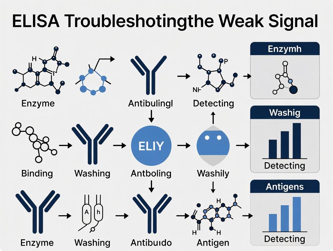
Visualization: Signaling Pathways & Workflows
Title: ELISA Colorimetric Detection Biochemistry Pathway
Title: Logical Flow for Diagnosing Weak ELISA Signal
The Scientist's Toolkit: Key Research Reagent Solutions
| Reagent/Material | Primary Function in Detection | Key Consideration for Signal |
|---|---|---|
| Horseradish Peroxidase (HRP) Conjugate | Catalyzes oxidation of chromogenic substrates. | Avoid sodium azide; ensure substrate contains H₂O₂. |
| Alkaline Phosphatase (AP) Conjugate | Catalyzes hydrolysis of phosphate groups. | Requires Mg²⁺/Zn²⁺; buffers must be metal-ion free. |
| TMB (Single-Component, Ready-to-Use) | Chromogenic substrate for HRP. Provides stable, sensitive signal. | Check for pre-activation (blue color before use). |
| pNPP Tablets or Solution | Chromogenic substrate for AP. Produces soluble yellow product. | Substrate buffer must be at high pH (~9.8). |
| Stop Solution (e.g., 1M H₂SO₄) | Stops enzymatic reaction, stabilizes final color, shifts absorbance max. | Must be of correct concentration for consistent signal. |
| High-Binding ELISA Plates | Solid phase for immobilizing capture antibody. | Low binding capacity directly limits maximum signal. |
| Precision Pipettes & Tips | Accurate reagent dispensing. | Inaccuracy in conjugate or substrate volume alters kinetics. |
| Microplate Reader with Appropriate Filter | Measures Optical Density (OD) of colored product. | Filter must match product's absorbance peak (e.g., 450nm for stopped TMB). |
Technical Support Center: Troubleshooting Guides & FAQs
Thesis Context: This support content addresses critical reagent variables that directly impact signal strength in ELISA, as part of a comprehensive thesis on diagnosing and resolving weak signal issues.
FAQ: Antibody-Related Weak Signal
Q1: My capture/detection antibody pair is validated, but I'm getting a weak signal. Could antibody affinity still be the issue? A: Yes. Validated pairs ensure epitope non-competition but do not guarantee optimal affinity for your specific antigen conformation or immobilization. Low affinity leads to rapid dissociation, reducing the amount of captured antigen or conjugate bound. Troubleshooting Step: Perform a checkerboard titration for both antibodies. A significant shift (>2-fold) from the recommended concentration may indicate suboptimal affinity under your assay conditions.
Q2: How can I quantitatively assess antibody affinity in my assay setup? A: Perform an equilibrium binding experiment. Coat your antigen at a saturating concentration. Incubate with serial dilutions of your primary antibody. Fit the resulting OD vs. concentration data to a one-site specific binding model (Langmuir isotherm) to estimate the apparent dissociation constant (KD).
Protocol: Apparent KD Estimation via ELISA
- Coat plate with antigen at 5 µg/mL, 100 µL/well, overnight at 4°C.
- Block with 200 µL/well of recommended buffer (e.g., 1% BSA in PBS) for 1-2 hours.
- Prepare 8 serial 3-fold dilutions of the primary antibody in block buffer, starting from 10x its expected KD.
- Add 100 µL/well of each dilution in duplicate. Incubate 2 hours at RT with gentle shaking.
- Wash 3x with wash buffer.
- Add 100 µL/well of optimal concentration of HRP-conjugated secondary antibody. Incubate 1 hour.
- Wash 3x. Develop with TMB for a fixed, linear time (e.g., 5 min). Stop.
- Measure OD450nm. Subtract background (no primary antibody control).
- Fit data using: Y = Bmax * X / (KD + X), where Y=OD, X=Antibody concentration.
Table 1: Interpreting Apparent KD Values from ELISA
| Apparent KD Range | Affinity Assessment | Likely Impact on Signal |
|---|---|---|
| < 1 nM | Very High | Optimal signal potential |
| 1 - 10 nM | High | Good signal |
| 10 - 100 nM | Moderate | Signal may be weak |
| > 100 nM | Low | High risk of weak signal |
FAQ: Conjugate & Detection Issues
Q3: My substrate develops quickly in the tube but gives a weak signal in the ELISA. What's wrong? A: This points to low conjugate efficiency (enzymes per antibody) or conjugate instability. A high degree of labeling (DOL) can cause steric hindrance or enzyme inactivation, while a low DOL provides insufficient signal amplification.
Protocol: Determining Conjugate Efficiency (Degree of Labeling, DOL) For HRP Conjugates (A403/A280 Ratio Method):
- Prepare conjugate in a suitable buffer (e.g., PBS), diluted to an A280 ~0.5-1.0.
- Measure absorbance at 403 nm (HRP Soret band) and 280 nm (protein + HRP).
- Calculate: DOL = (A403 * εAb280) / ( (A280 - (A403 * CF)) * εHRP403 ) Where: εAb280 ~210,000 M-1cm-1 (IgG), εHRP403 ~100,000 M-1cm-1, CF (Correction Factor, A403/A280 for pure HRP) ~0.3.
Table 2: Optimal vs. Problematic Conjugate DOL Ranges
| Conjugate Type | Optimal DOL Range | DOL Too Low | DOL Too High |
|---|---|---|---|
| HRP-IgG | 2.0 - 4.0 | Weak signal | High background, precipitation |
| Alkaline Phosphatase-IgG | 3.0 - 6.0 | Weak signal | Non-specific binding |
Q4: How do I test if my conjugate has degraded during storage? A: Perform a conjugate activity assay. Compare the enzymatic activity of the stored conjugate to a freshly prepared standard or a known-activity control in a direct enzyme assay.
Protocol: Direct HRP Activity Assay
- Dilute conjugate in assay buffer to a standard protein concentration (e.g., 1 µg/mL).
- In a microplate, add 50 µL of diluted conjugate per well.
- Rapidly add 50 µL of TMB substrate.
- Immediately start kinetic read at 650 nm (or 370 nm) for 2-5 minutes.
- Calculate the linear rate (Vmax, mOD/min). A drop >20% from the reference standard indicates significant degradation.
FAQ: Substrate Stability & Performance
Q5: My positive control signal has dropped over time, but my reagents are in-date. Could the substrate be the culprit? A: Absolutely. Chromogenic substrates (especially TMB) are sensitive to light, temperature, and oxidation. Contamination with oxidants (e.g., from dirty pipettors) can also prematurely deplete the substrate.
Protocol: Substrate Stability & Linearity Test
- Prepare a dilution series of your target analyte or a directly coated antibody for the conjugate.
- Run the ELISA as normal until the development step.
- Develop the plate for multiple, precise time points (e.g., 2, 5, 10, 15 minutes). Use a fresh set of wells for each time point.
- Stop the reaction and read the plate.
- Plot OD vs. development time for each analyte concentration. A stable, linear system will show straight lines. Curvature or plateaus at short times indicate substrate limitation or instability.
Table 3: Substrate Stability Indicators & Troubleshooting
| Observation | Potential Cause | Corrective Action |
|---|---|---|
| High background in all wells, including blanks | Contaminated or oxidized substrate | Prepare fresh substrate; clean work area. |
| Signal plateaus within 1-2 minutes | Substrate exhaustion or instability | Use new batch; check conjugate DOL (may be too high). |
| Blue color (TMB) appears but fades to yellow slowly/low OD | Substrate buffer pH is off; stop solution is weak/old | Prepare fresh stop solution (acid). |
| Signal is granular or precipitate forms | Insoluble reaction products | Ensure substrate is at room temperature and mixed well before use. |
The Scientist's Toolkit: Key Research Reagent Solutions
Table 4: Essential Reagents for ELISA Troubleshooting
| Reagent / Material | Primary Function | Key Consideration for Signal |
|---|---|---|
| High-Affinity Matched Antibody Pair | Specific capture and detection of the target antigen. | Apparent KD in the assay should be <10 nM for robust signal. |
| HRP or AP Conjugate with Optimized DOL | Signal amplification via enzyme-catalyzed substrate conversion. | DOL must balance signal intensity with background (see Table 2). |
| Stabilized Chromogenic Substrate (e.g., TMB, pNPP) | Provides the enzymatically converted product for colorimetric detection. | Must be fresh, uncontaminated, and used within its linear kinetic range. |
| High-Purity, Low-BSA Blocking Buffer | Reduces non-specific binding without interfering with antibody-antigen interactions. | Some BLOTTO or casein-based buffers can quench signal; test alternatives. |
| Precision Microplate Washer | Consistent removal of unbound reagents to reduce background and increase S/N ratio. | Insufficient washing leaves background; overly harsh washing can dissociate low-affinity bonds. |
| Kinetic Plate Reader | Allows measurement of reaction rate (Vmax), a more robust metric than endpoint OD. | Enables direct assessment of conjugate activity and substrate linearity. |
Visualizations
ELISA Weak Signal Troubleshooting Pathway
Signal Generation Cascade & Failure Point
Technical Support Center: ELISA Troubleshooting for Weak Signal
FAQs & Troubleshooting Guides
Q1: My ELISA signal is weak despite using a high-concentration sample. Could the issue be related to the target antigen itself? A: Yes. A weak signal can originate from the target's role in the assay. The three primary factors are:
- Antigen Concentration: Below the assay's detection limit.
- Epitope Availability: The antibody-binding site may be masked due to the antigen's conformation, folding, or post-translational modifications.
- Matrix Effects: Sample components (e.g., serum proteins, lipids, salts) can interfere with antibody-antigen binding or cause non-specific adsorption.
Recommended Protocol: To isolate the issue, perform a Spike-and-Recovery Experiment.
- Prepare a known concentration of purified antigen in the standard diluent.
- Spike the same known concentration into your representative sample matrix (e.g., serum, lysate).
- Run both samples in your ELISA.
- Calculate recovery:
(Concentration measured in matrix / Concentration measured in diluent) x 100%.
Interpretation: Recovery outside 80-120% suggests significant matrix interference affecting the target-antibody interaction.
Q2: How can I confirm if poor epitope availability is causing my weak signal? A: Epitope masking is common with native or complex antigens. Perform an Antigen Denaturation & Linearization Test. Experimental Protocol:
- Split your sample into two aliquots.
- Test Aliquot: Treat with a denaturing agent (e.g., 1% SDS) and a reducing agent (e.g., 50mM DTT) by heating at 95°C for 5 minutes. Cool before adding to plate.
- Control Aliquot: Treat with assay buffer only.
- Run both in ELISA, comparing signal intensity.
Caution: Ensure your capture antibody is compatible with denatured antigens (often a polyclonal or antibody targeting a linear epitope is required).
Q3: How do I determine if my target concentration is simply too low? A: Conduct a Sample Concentration Series versus the standard curve. Method:
- Run a standard curve with purified antigen.
- Run your neat sample and a series of concentrated samples (e.g., 2x, 5x concentration via lyophilization or centrifugal filtration).
- Plot the signals. If the concentrated sample signal increases proportionally and falls within the standard curve's dynamic range, the original concentration was too low.
Key Reagent Solutions Table
| Reagent/Material | Function in Troubleshooting Target Issues |
|---|---|
| Purified Native Antigen | Gold standard for creating a calibration curve and for spike-and-recovery experiments. |
| Detergent (e.g., Tween-20, Triton X-100) | Reduces non-specific binding in wash buffers; can help unmask hydrophobic epitopes. |
| Chaotropic Agent (e.g., Guanidine HCl) | Disrupts protein structure to expose buried linear epitopes. Use with caution. |
| Protease Inhibitor Cocktail | Preserves antigen integrity in complex samples by inhibiting degradation. |
| Heterophilic Antibody Blockers | Blocks interfering antibodies in serum/plasma that can bridge capture/detection Abs, causing false weak signals. |
| Bovine Serum Albumin (BSA) or Casein | Used as a blocking agent and matrix mimic to improve specificity in sample diluents. |
Quantitative Data Summary: Impact of Common Variables on ELISA Signal
| Variable | Typical Effect on Signal | Recommended Optimization Range |
|---|---|---|
| Antigen Concentration | Directly proportional (within range) | Must fall within assay's stated dynamic range. |
| Sample Dilution Factor | Reduces matrix effects; may dilute antigen too far. | Test a series (e.g., 1:2 to 1:100) to find optimal point. |
| Incubation Time/Temp | Increases binding. | 1-2 hours at RT or overnight at 4°C for capture. |
| Denaturation (Heat/SDS) | Can increase (expose epitopes) or decrease (destroy conformation). | Test 70-95°C for 5-15 min with 0.1-1% SDS. |
| Spike Recovery Result | Diagnostic for matrix effects. | Target 80-120% recovery. <80% indicates loss; >120% indicates interference. |
Visualization: ELISA Troubleshooting Workflow for Target-Related Weak Signal
Title: Troubleshooting Weak ELISA Signal: Target Antigen Issues
Visualization: Antibody-Epitope Binding Interference
Title: Mechanisms of Target-Related ELISA Signal Failure
Troubleshooting Guides & FAQs
FAQ 1: My ELISA signals are consistently weak. I've checked my reagents and protocol. Could the plate reader be the issue? Answer: Yes. Weak signals in ELISA can often be traced to suboptimal instrument settings or calibration drift. Before repeating your assay, perform these checks:
- Wavelength Accuracy: Verify the selected filter or monochromator wavelength matches your chromogen's absorbance peak (e.g., 450 nm for OPD, 650 nm for AP). A 5-10 nm shift can drastically reduce sensitivity.
- Calibration Status: Check the log for the last full optical path and photometric calibration. Performance verification with a neutral density filter or known standard should be done monthly.
- Optical Path Alignment: Misalignment, especially in multimode readers, can cause uneven well-to-well signal capture. Run a pathlength check if available.
FAQ 2: How do I choose the correct wavelength for a dual-wavelength measurement in ELISA to reduce background? Answer: Dual-wavelength readings subtract a reference wavelength from the primary measurement to correct for optical imperfections. Follow this protocol:
- Primary Wavelength: Set to the absorbance maximum of your substrate (e.g., 450 nm for TMB).
- Reference Wavelength: Choose a wavelength where your substrate has minimal absorbance but where plate imperfections (scratches, fingerprints) scatter light similarly. This is typically 25-50 nm higher (e.g., 490-650 nm for TMB). Avoid wavelengths where your buffer or plate material absorbs significantly.
- Validation: Test the settings by reading a buffer-only well. The corrected absorbance should be near zero.
FAQ 3: My calibration check failed the photometric accuracy test. What should I do next? Answer: A failed photometric check indicates a problem with the light source, detector, or optical components. Follow this troubleshooting workflow:
- Repeat the test with a fresh calibration standard (e.g., neutral density filter).
- Inspect the optical path: Check for and clean any visible dust or debris on the lamp window, filters, mirrors, or fiber optic cables.
- Check the lamp hours. If near or past the manufacturer's rated lifetime, replace the lamp and recalibrate.
- If the issue persists, contact technical service for a full optical alignment.
FAQ 4: What is the difference between a filter-based and a monochromator-based optical path, and how does it affect my ELISA? Answer: The choice impacts flexibility, bandwidth, and signal-to-noise.
- Filter-Based: Uses physical filters to select wavelengths. Offers high light throughput and excellent signal-to-noise for fixed applications (like common 450 nm ELISA). Bandwidth is fixed (typically ±10-15 nm).
- Monochromator-Based: Uses a diffraction grating to select any wavelength. Essential for assay development or uncommon chromogens. Bandwidth is often adjustable; a narrower bandwidth increases specificity but reduces signal intensity.
Table 1: Key Specifications for ELISA Plate Reader Calibration
| Calibration Type | Recommended Frequency | Standard Used | Acceptance Criteria (Typical) |
|---|---|---|---|
| Photometric Accuracy | Quarterly / After lamp change | Certified ND Filter (e.g., OD 1.0) | Read Value ±0.02 OD of certified value |
| Wavelength Accuracy | Biannually | Didymium or Holmium Oxide filter | Peak within ±2 nm of known standard |
| Pathlength Correction | Per experiment (if using) | Water absorbance at 977 nm | Linear regression R² > 0.99 |
| Well-to-Well Crosstalk | Annually | Fluorescent solution in alternating wells | Signal in adjacent empty well < 0.1% |
Experimental Protocols
Protocol 1: Monthly Performance Verification for ELISA Readers Objective: Confirm the plate reader is operating within specification for critical ELISA parameters. Materials: Plate reader, clear 96-well plate, certified neutral density filter (OD 1.0 ± 0.02), precision pipettes, 1X PBS or assay buffer.
- Photometric Check:
- Place the ND filter in the designated holder or in a well (as per manual).
- Read absorbance at the filter's specified wavelength (e.g., 405 nm).
- Record the value. It must be within the manufacturer's tolerance (e.g., 1.0 ± 0.05 OD).
- Background Noise Check:
- Fill a column of 8 wells with 100 µL of assay buffer.
- Read absorbance at 450 nm with your standard ELISA settings.
- Calculate the standard deviation of the 8 wells. It should be < 0.005 OD.
- Documentation: Log all results. Flag any out-of-spec readings.
Protocol 2: Optimizing Wavelength Selection for a New Chromogen Objective: Empirically determine the optimal primary and reference wavelengths. Materials: Plate reader with monochromator, 96-well plate, your developed ELISA plate (with high, low, and blank wells).
- Spectral Scan: On a high-signal well, perform an endpoint absorbance scan from 370 nm to 500 nm.
- Identify Peak: Note the wavelength of maximum absorbance (λmax).
- Set Primary: Set your primary filter/monochromator to this λmax.
- Test References: Read the plate using your primary λmax with different reference wavelengths (e.g., 490, 540, 650 nm).
- Analyze: Calculate the signal-to-noise ratio (Signal(High)/SD(Blank)) for each reference. Choose the reference yielding the highest ratio.
Signaling Pathway & Workflow Diagrams
Diagram 1: ELISA Weak Signal Troubleshooting Decision Tree
Diagram 2: Simplified Absorbance Plate Reader Optical Path
The Scientist's Toolkit: Key Research Reagent Solutions
Table 2: Essential Materials for Plate Reader Performance Validation
| Item | Function | Key Consideration for ELISA |
|---|---|---|
| Certified Neutral Density (ND) Filters | Validates photometric accuracy across the dynamic range. | Use an OD ~1.0 filter to check the critical range for most ELISA results. |
| Wavelength Standard Filters (Didymium/Holmium) | Verifies the accuracy of the wavelength selection system. | Ensures your 450 nm reading is actually at 450 nm. |
| Clean, Clear Flat-Bottom Microplates | Low background, consistent pathlength plates for verification tests. | Use the same plate type as your ELISA to control for material properties. |
| Precision Volume Pipettes & Tips | Accurate liquid handling for preparing verification standards. | Critical for creating reliable serial dilutions for pathlength or linearity checks. |
| Non-fluorescent, Low-Absorbance Buffer (e.g., 1X PBS) | Matrix for blank and background noise measurements. | Must not absorb significantly at your assay wavelengths (e.g., 450 nm). |
Precision in Practice: Critical Steps to Prevent Weak Signals During Assay Execution
Welcome to the Technical Support Center for ELISA Sample Preparation. This guide addresses common issues leading to weak signals, framed within a thesis on comprehensive ELISA troubleshooting.
FAQs & Troubleshooting Guides
Q1: My samples consistently yield weak signals, even with strong expected analyte concentrations. What is the primary culprit in sample preparation? A: The most common cause is improper sample dilution. A sample that is too concentrated can lead to the "High-Dose Hook Effect" or cause matrix components to overwhelm the assay, leading to signal suppression. Conversely, over-dilution can place the analyte below the detection limit. Always perform an initial serial dilution series to identify the optimal, linear range for your specific sample matrix.
Q2: How do I determine the correct starting dilution for a novel sample type (e.g., a new tissue homogenate)? A: Follow this protocol for a Pilot Dilution Matrix Experiment:
- Prepare a crude sample and a spiked control (sample with a known amount of recombinant analyte).
- Create a 2D dilution matrix. Dilute samples 1:2, 1:10, 1:50, and 1:100 in the recommended assay buffer.
- Run both the naive and spiked samples at each dilution in duplicate on the same ELISA plate.
- Calculate % recovery for the spiked sample at each dilution:
(Observed [Spiked] – Observed [Naive]) / Expected Spike Concentration * 100. - The optimal dilution is the one where the naive sample signal is within the standard curve's mid-range and the spike recovery is between 80-120%.
Q3: My protein analyte appears degraded in my stored samples, weakening signal over time. What are key stabilization steps? A: Stabilization is matrix and analyte-specific. Implement this additive protocol immediately upon collection:
- Protease Inhibition: Add a broad-spectrum protease inhibitor cocktail (e.g., containing AEBSF, Aprotinin, Bestatin, etc.).
- Phosphatase Inhibition (for phospho-proteins): Add sodium fluoride (NaF), sodium orthovanadate, and/or β-glycerophosphate.
- General Stabilizers: For plasma/serum, use EDTA (1-2 mM) to chelate metals and inhibit metalloproteases. For cell lysates, ensure a sufficient concentration of non-ionic detergent (e.g., 1% Triton X-100) to solubilize proteins.
- Storage: Aliquot and flash-freeze at -80°C; avoid repeated freeze-thaw cycles (>3 cycles significantly degrades most proteins).
Q4: Which interfering substances most commonly cause weak ELISA signals, and how can I mitigate them? A: Common interferents and their solutions are summarized in the table below.
Table 1: Common Interfering Substances in ELISA and Mitigation Strategies
| Interferent | Common Sources | Effect on ELISA | Mitigation Strategy |
|---|---|---|---|
| Hemoglobin | Hemolyzed serum/plasma | Peroxidase-like activity (in HRP assays); nonspecific binding | Re-collect sample. Use serum separator tubes. Dilute sample >1:10. |
| Lipids | Lipemic serum, tissue homogenates | Increases sample viscosity; can trap analytes | Ultracentrifugation (100,000 x g, 30 min) to remove lipids. |
| Heterophilic Antibodies | Human anti-animal antibodies in serum | Bridge capture/detection Abs, causing false high OR block binding, causing false low/weak | Use a heterophilic blocking reagent (HBR) in the diluent. Use species-specific Fab fragments. |
| Complement Factors | Fresh serum | May bind to capture antibody, causing steric hindrance | Heat-inactivate serum at 56°C for 30 min before assay. |
| Albumin & other high-abundance proteins | Serum, plasma, CSF | Nonspecific binding, masking | Dilution is often sufficient. For complex matrices, use a commercial matrix-mimicking diluent. |
Q5: What is a definitive protocol to test for and overcome matrix interference? A: Perform a Parallel Linearity (Dilutional Recovery) Test:
- Prepare: Dilute your sample serially (e.g., 1:2, 1:4, 1:8) in both (a) standard buffer and (b) a spiked standard buffer (containing a known mid-range concentration of the pure standard).
- Run: Assay all dilutions. Plot the observed concentration (or OD) against the dilution factor.
- Analyze: For the spiked series, calculate recovery at each point. The lines for the sample diluted in buffer vs. spiked buffer should be parallel. Non-parallel lines or poor spike recovery (<80% or >120%) indicate significant interference requiring matrix-specific diluent optimization or sample purification.
The Scientist's Toolkit: Key Research Reagent Solutions
| Item | Function & Rationale |
|---|---|
| Commercial Matrix-Matched Diluent | Pre-formulated buffer containing proteins (e.g., BSA, casein), surfactants, and blockers specific to a sample type (e.g., serum, cell culture) to minimize nonspecific binding and neutralize interferents. |
| Heterophilic Blocking Reagent (HBR) | A proprietary mixture of inert animal immunoglobulins and polymers that saturate human anti-animal antibodies, preventing them from interfering with assay antibodies. |
| Protease Inhibitor Cocktail (EDTA-free) | A ready-to-use blend of inhibitors targeting serine, cysteine, aspartic, and aminopeptidases. EDTA-free versions are compatible with metal-dependent assays. |
| Phosphatase Inhibitor Cocktail | A mixture of inhibitors (e.g., against tyrosine, serine/threonine, alkaline, and acid phosphatases) crucial for preserving post-translational phosphorylation states. |
| Recombinant Protein Standard | Highly pure, quantified protein identical to the target analyte, essential for generating the standard curve and performing spike-recovery experiments. |
Visualization: ELISA Signal Interference Pathways
Title: Pathways from Sample Prep Issues to Weak ELISA Signal
Title: Optimal Sample Preparation and Testing Workflow
Troubleshooting Guides & FAQs
FAQ: Addressing Weak Signal in ELISA
Q1: My ELISA results show a weak or absent signal despite a known positive sample. Could pipetting be the cause? A: Yes, imprecise pipetting is a primary culprit. Inconsistent delivery of capture antibody, sample, or detection reagent directly alters the number of molecules available for binding, leading to high variability and low signal strength. Always calibrate pipettes regularly, use reverse pipetting for viscous reagents (like serum or detection antibody), and ensure tips are properly sealed and pre-wetted.
Q2: How can improper washing lead to a weak specific signal? A: Inadequate washing fails to remove unbound detection antibody or enzyme conjugate, leading to high background that can obscure a weak specific signal. Conversely, overly aggressive washing (e.g., excessive shaking, using overly concentrated wash buffer) can dissociate specifically bound antibody-antigen complexes, directly reducing your signal.
Q3: My standard curve is erratic, with poor replicates. What pipetting step should I check first? A: Focus on the serial dilution of your standard. This is the most critical pipetting sequence. Use a fresh tip for each transfer, mix the dilution thoroughly but gently, and ensure the pipette is held vertically during aspiration and dispensing. Consider using a multi-channel pipette for plate replication, but calibrate each channel.
Q4: What are the definitive signs of a washing problem versus a pipetting problem in a developed plate? A:
| Observation | Likely Cause |
|---|---|
| High background across all wells, including blanks. | Insufficient washing. |
| Weak specific signal with clean background. | Pipetting error in sample or detection reagent. |
| High variation between replicate wells. | Inconsistent pipetting or washing. |
| Streaks or patterns aligned with washing manifold. | Clogged or uneven washing manifold ports. |
Q5: What is the optimal washing technique to maximize specific binding? A: Follow a consistent, validated protocol:
- Decant or aspirate plate contents forcefully.
- Fill each well completely with wash buffer using a calibrated dispenser or manifold. Do not jet stream directly onto the well bottom.
- Soak for 30-60 seconds to allow dissociation of weakly bound materials.
- Aspirate/Decant completely. Invert the plate and blot on clean lint-free towels.
- Repeat per protocol (typically 3-6 washes). The final wash and blot are critical for low background.
Experimental Protocols
Protocol 1: Verification of Pipette Accuracy and Precision
- Purpose: To ensure pipettes deliver the specified volume consistently.
- Method (Gravimetric Analysis):
- Place a tared microbalance and a weigh boat in a draft-free environment.
- Set the pipette to the desired volume (e.g., 10 µL, 100 µL).
- Pre-wet the tip by aspirating and dispensing distilled water 3x.
- Aspirate a fresh volume, dispense into the weigh boat, and record the weight.
- Repeat 10 times.
- Convert mass to volume using the Z-factor for water at your lab temperature (e.g., 1.003 µL/mg at 20°C).
- Calculate mean, accuracy (% deviation from target), and precision (Coefficient of Variation, CV%).
Protocol 2: Systematic Washing Efficiency Test for ELISA
- Purpose: To empirically determine the optimal wash cycles and soak time for your assay.
- Method:
- Coat and block an ELISA plate as usual.
- Apply a high-concentration positive control and negative control in duplicate columns.
- After the detection antibody step, split the plate for different washing regimens.
- Variable: Wash one set of columns 3x, another 5x, another 7x. For a subset, vary soak times (0s, 30s, 60s).
- Complete the assay with substrate.
- Measure signal and background. Calculate the Signal-to-Noise (S/N) ratio for each condition.
Data Presentation
Table 1: Impact of Pipetting Technique on ELISA Standard Curve Reproducibility (n=8 replicates per point)
| Standard Concentration (pg/mL) | CV% (Forward Pipetting) | CV% (Reverse Pipetting) | Recommended Technique |
|---|---|---|---|
| 1000 | 12.5 | 4.8 | Reverse Pipetting |
| 100 | 18.7 | 5.2 | Reverse Pipetting |
| 10 | 25.3 | 7.1 | Reverse Pipetting |
| 0 | 30.1 (Background) | 9.5 (Background) | Reverse Pipetting |
Table 2: Effect of Wash Cycle Number on Signal-to-Noise Ratio
| Wash Cycles | Soak Time | Mean Signal (OD 450nm) | Mean Background (OD 450nm) | Signal-to-Noise Ratio |
|---|---|---|---|---|
| 3 | 5s | 1.85 | 0.45 | 4.1 |
| 5 | 30s | 1.78 | 0.12 | 14.8 |
| 7 | 30s | 1.65 | 0.08 | 20.6 |
| 7 | 60s | 1.52 | 0.07 | 21.7 |
Diagrams
The Scientist's Toolkit: Research Reagent Solutions
| Item | Function & Rationale |
|---|---|
| Low-Binding/Filter Pipette Tips | Minimizes protein adsorption to tip walls, ensuring accurate volume delivery of precious reagents like antibodies. |
| Calibrated, Ergonomic Micropipettes | Primary tool for volumetric accuracy. Regular calibration (quarterly) is non-negotiable for reproducible data. |
| Multi-Channel Pipette (Electronic) | Increases throughput and reduces inter-well variability during plate washing, reagent addition, and serial dilutions. |
| Plate Washer (Automated) | Provides consistent, programmable washing with controlled fill, soak, and aspiration cycles, minimizing user-induced variability. |
| Wash Buffer Concentrate | Allows consistent preparation of fresh, filtered (0.2 µm) buffer containing surfactant (e.g., Tween 20) to reduce non-specific binding. |
| Non-Absorbent Plate Seals | Prevents evaporation and contamination during incubation steps, which can alter reagent concentration. |
| Lint-Free Blotting Paper | Essential for removing residual wash buffer after decanting without introducing fibers or cross-contamination. |
Troubleshooting Guides & FAQs
FAQ 1: Why am I getting a weak or absent signal in my ELISA despite proper reagent addition?
Answer: A weak signal often stems from suboptimal immune complex formation during the coating, capture, or detection incubation steps. The primary culprits are insufficient incubation time, incorrect temperature, or lack of agitation, preventing effective antigen-antibody binding. Ensure you follow optimized parameters for each specific assay step as detailed in protocols.
FAQ 2: How does agitation affect my ELISA results?
Answer: Agitation, especially on a microplate shaker, promotes consistent mixing of reagents, reduces boundary layer effects, and increases the frequency of collisions between antigens and antibodies. This typically enhances binding kinetics, leading to more uniform and stronger signals. Lack of agitation can cause gradient formation and inconsistent binding, particularly at the edges of wells.
FAQ 3: What are the recommended incubation times and temperatures for a standard sandwich ELISA?
Answer: While specific protocols vary, general guidelines based on current literature are summarized below. Always refer to your antibody manufacturer's datasheet for optimal conditions.
FAQ 4: My high background is obscuring my signal. Could incubation conditions be the cause?
Answer: Yes. Over-incubation (excessive time or temperature) or overly vigorous agitation can increase non-specific binding, leading to high background. Ensure you are using the recommended blocking agents and strictly adhering to optimized incubation parameters.
Data Presentation: Optimized Incubation Parameters
Table 1: Standard Incubation Parameters for Key ELISA Steps
| ELISA Step | Typical Time Range | Typical Temperature Range | Agitation Recommendation | Key Rationale |
|---|---|---|---|---|
| Coating | Overnight (12-16 hrs) to 1-2 hrs | 4°C (long) or Room Temp (RT, short) | Optional, gentle | Slow binding at 4°C minimizes denaturation; faster at RT. |
| Blocking | 1-2 hours | RT or 37°C | Recommended, gentle | Ensures complete coverage of all non-specific sites. |
| Sample/Antigen Incubation | 1-2 hours | RT or 37°C | Highly Recommended | Maximizes target capture, crucial for sensitivity. |
| Detection Antibody Incubation | 1-2 hours | RT or 37°C | Highly Recommended | Maximizes binding to captured antigen. |
| Enzyme-Conjugate Incubation | 30 min - 1 hour | RT, protected from light | Recommended, gentle | Optimizes enzyme binding without increase in non-specific signal. |
Table 2: Troubleshooting Guide for Incubation-Related Weak Signals
| Symptom | Possible Incubation Cause | Suggested Remedial Action |
|---|---|---|
| Weak Signal Across All Wells | Time too short; Temp too low; No agitation. | Increase incubation time (e.g., 2hrs to overnight at 4°C for coating); Increase temp to 37°C; Implement gentle agitation (300-500 rpm). |
| High Background | Time too long; Temp too high; Excessive agitation. | Shorten incubation times; Lower incubation temp to RT; Reduce agitation speed or eliminate. |
| Edge Effect (Wells on plate perimeter differ) | Inconsistent temperature or lack of agitation. | Use a calibrated, humidified incubator; Ensure consistent agitation across the plate. |
| High CV (%) Between Replicates | Inconsistent binding due to lack of agitation. | Implement orbital agitation (500-700 rpm) for all liquid incubation steps. |
Experimental Protocols
Protocol 1: Method for Optimizing Capture Antibody Coating Time and Temperature
Objective: To determine the optimal combination of time and temperature for adsorbing the capture antibody to the microplate well surface. Materials: Carbonate/Bicarbonate Coating Buffer (pH 9.6), Capture Antibody, PBS, Blocking Buffer (e.g., 1% BSA in PBS). Procedure:
- Dilute capture antibody in coating buffer to recommended concentration (typically 1-10 µg/mL).
- Dispense 100 µL per well into a 96-well microplate. For each condition, use a minimum of 3 replicates.
- Incubate plates under the following matrix of conditions:
- Time: 1 hour, 2 hours, overnight (16 hours).
- Temperature: 4°C, Room Temperature (22-25°C), 37°C.
- Agitation: Static vs. gentle orbital shaking (400 rpm).
- After incubation, wash plate 3x with PBS + 0.05% Tween-20 (PBST).
- Proceed with standard blocking, sample addition, detection, and substrate steps as per your assay.
- Analyze the signal-to-noise ratio (Mean Signal of Positive / Mean Signal of Negative) for each condition. The condition with the highest ratio indicates optimal coating.
Protocol 2: Method for Optimizing Antigen and Detection Antibody Incubation with Agitation
Objective: To assess the impact of agitation on assay sensitivity and precision during key liquid incubation steps. Materials: Pre-coated and blocked ELISA plate, Sample/Standard, Detection Antibody, Wash Buffer, Substrate. Procedure:
- Prepare serial dilutions of your target antigen in assay diluent.
- Add 100 µL per well to the prepared plate. Seal the plate.
- Test Condition A: Incubate for 1 hour at RT without agitation (static).
- Test Condition B: Incubate for 1 hour at RT with continuous orbital agitation (500 rpm).
- Wash plate 5x with PBST.
- Add detection antibody (conjugated or biotinylated) as per datasheet.
- Repeat the static vs. agitation incubation comparison for this detection step (e.g., 1 hour at RT).
- Wash plate 5x with PBST.
- Add enzyme conjugate (if needed) and substrate, followed by stop solution. Read absorbance.
- Analysis: Compare the standard curves generated from Condition A and B. Agitation should yield a steeper slope (higher sensitivity) and lower coefficient of variation (CV%) between replicate wells.
Mandatory Visualization
Diagram 1: Key Factors in Immune Complex Formation for ELISA
Diagram 2: ELISA Workflow with Critical Incubation Control Points
The Scientist's Toolkit: Research Reagent Solutions
Table 3: Essential Materials for Incubation Optimization Experiments
| Item | Function in Optimization |
|---|---|
| Programmable Microplate Incubator | Provides precise, consistent, and humidified temperature control (e.g., 4°C, 25°C, 37°C) for incubation steps, eliminating environmental fluctuations. |
| Orbital Microplate Shaker | Delivers consistent and adjustable agitation (e.g., 200-1000 rpm) to enhance binding kinetics and improve well-to-well reproducibility. |
| Precision Timer | Ensures accurate and reproducible incubation durations for each step, critical for kinetic consistency. |
| Humidified Chamber | Prevents evaporation of reagents during long incubations (e.g., overnight coating), which can alter concentrations and cause edge effects. |
| Low-Binding, High-Protein-Binding Plates | The correct plate surface (e.g., polystyrene for antibodies) is fundamental for efficient passive adsorption during the coating step. |
| Calibrated Thermometer & Hygrometer | Verifies the accuracy of incubator and lab environment conditions, ensuring protocol integrity. |
| Plate Sealer / Adhesive Film | Prevents contamination, evaporation, and well-to-well cross-contamination during agitated or long incubations. |
Technical Support & Troubleshooting Guides
Frequently Asked Questions (FAQs)
Q1: My standard curve has a poor R² value (<0.99). What are the most common causes in ELISA? A: Common causes include: inaccurate serial dilution technique (pipetting errors), degradation of the standard protein (improper storage/thawing), insufficient mixing of standards between dilutions, using an inappropriate curve-fitting model for the assay's dynamic range, and edge effects in the microplate. Ensure fresh, accurate dilutions and validate the model.
Q2: How can I improve the accuracy of my dilution series for ELISA standards? A: Use a reverse pipetting technique for viscous solutions. Always use a fresh pipette tip for each dilution step. Prepare dilutions in a bulk intermediate solution rather than serially across the plate to minimize cumulative error. Use low-protein-binding tubes and buffers with carrier proteins (e.g., 0.1% BSA) to prevent adsorption losses.
Q3: Which curve-fitting model should I use for my ELISA data: linear, log-linear, 4PL, or 5PL? A: Four-Parameter Logistic (4PL) is the standard for most sandwich ELISAs, as it accounts for the sigmoidal shape of the response. Use Five-Parameter Logistic (5PL) for asymmetric curves. Linear or log-linear may only be used for a very narrow, central range of the curve but is not best practice for accurate quantitation across the full range.
Q4: My high-concentration standard points are plateauing too early, compressing the top of the curve. How do I fix this? A: This indicates the analyte concentration is exceeding the assay's dynamic range or the detection system is saturating. Prepare a new standard curve with a lower top standard concentration. Ensure the incubation times for detection (e.g., with HRP/TMB) are not excessive.
Q5: Why is there high variability between replicate standard points? A: This is typically a liquid handling issue. Check pipette calibration. Ensure all standards and reagents are equilibrated to room temperature before use to prevent condensation and thermal contraction/expansion. Mix all solutions thoroughly but gently before use.
Q6: How should I handle the outlier point in my standard curve? A: Do not discard outliers arbitrarily. First, re-check the calculation for that well. If no error is found, repeat the assay. If the outlier persists systematically at a certain concentration, it may indicate a pipetting error specific to that dilution step or a problem with that specific standard concentration. Document the exclusion if it is a clear technical failure.
Table 1: Comparison of Common Curve-Fitting Models for ELISA
| Model | Best For | Key Advantage | Key Limitation | Typical R² Target |
|---|---|---|---|---|
| Linear | Quick estimates, very limited linear range | Simplicity | Poor fit for sigmoidal data; inaccurate at extremes | >0.98 |
| Log-Linear | Narrow dynamic range (1-2 logs) | Simple transformation | Assumes linearity which often doesn't hold | >0.98 |
| 4-Parameter Logistic (4PL) | Most sandwich & competitive ELISAs (symmetric curves) | Accurately models sigmoid, defines asymptotes | Assumes curve symmetry | >0.99 |
| 5-Parameter Logistic (5PL) | Asymmetric sigmoidal curves | Models asymmetry; excellent fit for complex curves | More complex computation | >0.99 |
Table 2: Common Pipetting Errors and Their Impact on Standard Accuracy
| Error Type | Potential Impact on Concentration | Effect on Curve |
|---|---|---|
| Using same tip for serial dilutions | Exponential error increase; gross inaccuracy | Poor fit, low R² |
| Inaccurate pipette calibration (e.g., -5%) | Systematic shift all points | Altered slope & asymptotes |
| Improplete mixing after dilution | Non-homogeneous solution; erratic ODs | High replicate variability |
| Evaporation from wells (no lid) | Increased concentration, especially at edges | Flattening of curve, poor precision |
Experimental Protocols
Protocol: Accurate Preparation of a Standard Dilution Series for ELISA
Objective: To create a precise serial dilution of the stock standard for a 10-point standard curve.
Materials: See "The Scientist's Toolkit" below.
Method:
- Reconstitution: Reconstitute the lyophilized standard protein exactly as per the kit protocol. Allow to equilibrate for 10-15 minutes, then mix gently by flicking the tube. Do not vortex aggressively.
- Plan Dilutions: Calculate the required volumes to make sufficient volume (e.g., 150 µL per point in duplicate) for the entire series from the top standard concentration (e.g., [Top]).
- Bulk Intermediate Preparation (Recommended): Prepare the top standard concentration in a sterile, low-binding microcentrifuge tube at 2x the final required volume. Use the assay diluent buffer for all dilutions.
- Serial Dilution: a. Label clean, low-binding tubes for each standard point. b. Add the calculated volume of diluent to each tube. c. Using a fresh pipette tip, transfer the calculated volume from the [Top] standard to the first tube ([Point 2]). Mix thoroughly by pipetting up and down 10 times or vortex on low setting. d. Change tip. From tube [Point 2], transfer volume to the next tube ([Point 3]). Repeat mixing. e. Continue this process down to the lowest concentration. The final tube (zero standard) contains diluent only.
- Plate Loading: Add equal volumes (e.g., 100 µL) from each dilution tube to the designated wells in the microplate. Perform in duplicate or triplicate.
Protocol: Validating a Curve-Fitting Model
Objective: To determine the optimal mathematical model for your standard curve data.
Method:
- Data Entry: Enter mean absorbance (OD) values for each standard concentration into analysis software (e.g., ELISA analysis module, GraphPad Prism, MyAssays).
- Initial Fitting: Apply the 4PL model. Visually inspect the fit. The curve should pass through or near all data points, especially in the linear mid-range.
- Assess Residuals: Examine the residual plot (difference between observed and predicted ODs). Residuals should be randomly scattered around zero, not showing a systematic pattern.
- Statistical Comparison: If the 4PL fit is poor in the upper or lower asymptotes, apply a 5PL model. Use statistical metrics (e.g., Sum of Squares, AICc) to compare models objectively. The model with the lower sum of squares and AICc is preferred.
- Back-Calculation: Use the chosen model to back-calculate the concentration of each standard from its OD. The percent recovery for each point should be within 80-120% of the expected value.
Mandatory Visualization
Title: ELISA Standard Curve Preparation and Validation Workflow
Title: Four-Parameter Logistic (4PL) Model Components
The Scientist's Toolkit: Key Research Reagent Solutions
Table 3: Essential Materials for Accurate Standard Curve Preparation
| Item | Function & Importance | Best Practice Note |
|---|---|---|
| Low-Protein-Binding Microtubes/Pipette Tips | Minimizes adsorption of the standard protein to plastic surfaces, preventing loss of analyte, especially at low concentrations. | Essential for preparing and storing dilutions. |
| Calibrated, High-Precision Pipettes (P2, P20, P200, P1000) | Ensures accurate volume transfer during serial dilution. Small errors are magnified in serial steps. | Perform regular calibration (every 3-6 months). Use reverse pipetting for viscous buffers. |
| Assay-Specific Diluent Buffer (with carrier protein) | The matrix for diluting standards. A buffer with 0.1-1% BSA or similar helps stabilize dilute proteins and matches sample matrix. | Always use the diluent specified in the kit protocol. |
| Lyophilized Standard Protein (Reference Material) | Provides the known quantity for curve generation. Accuracy hinges on its integrity and proper reconstitution. | Reconstitute exactly as directed. Aliquot and store at recommended temperature to avoid freeze-thaw cycles. |
| Microplate Reader with Reliable Absorbance Detection | Accurately measures the optical density (OD) signal from each well. Instrument noise can affect low-end precision. | Ensure pathlength correction is used if volumes vary, and the instrument is regularly maintained. |
| Data Analysis Software with 4PL/5PL Fitting | Transforms raw OD data into a quantifiable standard curve using appropriate non-linear regression models. | Do not rely on simple linear regression. Use software that reports goodness-of-fit (R²) and confidence intervals. |
The Weak Signal Troubleshooting Toolkit: A Systematic Diagnostic Approach
Troubleshooting Guides & FAQs
FAQ 1: Why is the verification of reagents the critical first step in ELISA troubleshooting for weak signals? A weak signal in ELISA can originate from multiple sources. Systematic verification of reagents first isolates variables related to reagent failure, preventing wasted time and resources on unnecessary, complex troubleshooting. It confirms the foundation of the assay before investigating procedural or instrumental issues.
FAQ 2: How do incorrectly stored detection antibodies commonly cause weak signals? The enzyme conjugate (e.g., HRP-labeled antibody) is highly sensitive to improper storage. Repeated freeze-thaw cycles, exposure to light, or storage at +4°C instead of the recommended -20°C can lead to enzyme denaturation and loss of catalytic activity. This directly reduces the signal generated in the detection step, even if target capture was efficient.
FAQ 3: What are the consequences of using a substrate solution beyond its expiry date? Chemiluminescent substrates (e.g., TMB for HRP) degrade over time. An expired substrate may have reduced sensitivity or fail to produce a signal altogether, leading to a false-negative or artificially weak result. The reaction may also develop more slowly or unevenly.
FAQ 4: Can using a capture antibody at the wrong concentration cause a weak signal? Yes. While too high a concentration can cause a hook effect, a concentration too far below the optimal level will lead to insufficient antigen binding sites on the plate, reducing the amount of captured target analyte. This directly limits the maximum possible signal.
Experimental Protocol: Systematic Reagent Verification Checklist
Objective: To methodically verify the integrity and performance of all core ELISA reagents. Materials: See "The Scientist's Toolkit" below. Procedure:
- Visual Inspection: Examine all reagent containers for precipitates, cloudiness, or unusual color.
- Expiry Date Audit: Log the lot numbers and expiry dates of all reagents in a table (see Data Presentation).
- Storage Condition Cross-Reference: Verify the actual storage location (freezer, refrigerator temperature log) against the manufacturer's datasheet specifications.
- Positive Control Performance Check: Run a simplified assay plate using only the positive control and assay diluent (negative control) according to the kit protocol. Compare the obtained signal (OD or RLU) for the positive control to the expected range provided in the kit insert or historical lab data.
- Reagent Preparation Validation: Confirm that all buffers were prepared with correct pH and that any reconstituted reagents (e.g., standards, lyophilized antibodies) were made with the correct diluent and have not exceeded their in-use stability period.
Data Presentation
Table 1: Critical ELISA Reagent Verification Log (Example)
| Reagent | Lot Number | Stated Expiry | Recommended Storage | Actual Storage Check | Visual Inspection |
|---|---|---|---|---|---|
| Capture Antibody | ABC123 | 2024-11-15 | -20°C, avoid freeze-thaw | -20°C, 3rd thaw | Clear, no precipitate |
| Detection Antibody | DEF456 | 2024-08-30 | -20°C, aliquot | -20°C, 1st thaw | Clear, no precipitate |
| Antigen Standard | GHI789 | 2024-10-01 | -80°C long-term | -80°C | Lyophilized powder intact |
| HRP Substrate (TMB) | JKL012 | 2024-06-01 | +4°C, protect from light | +4°C, dark | Slight blue tinge* |
| Stop Solution (1M H₂SO₄) | MNO345 | 2026-05-01 | Room Temperature | RT | Clear, colorless |
*Note: Slight blue tinge may indicate early degradation; test with positive control.
Mandatory Visualization
Diagram 1: Reagent Verification Flow for Weak ELISA Signal
The Scientist's Toolkit: Research Reagent Solutions
Table 2: Essential Materials for ELISA Reagent Verification
| Item | Function in Verification |
|---|---|
| Calibrated Microplate Reader | Precisely measures absorbance (OD) or luminescence (RLU) to quantify positive control performance against expected values. |
| Single-Channel & Multichannel Pipettes | Ensure accurate and consistent reagent handling during verification tests. |
| Temperature Data Logger | Monitors and logs temperatures of refrigerators, freezers, and room temperature storage areas to confirm compliance. |
| Reagent Aliquot Tubes | Small, sterile tubes for dividing bulk reagents into single-use aliquots to minimize freeze-thaw cycles. |
| Positive Control Sample | Provides a known signal reference to test the integrated functionality of the entire assay reagent set. |
| Manufacturer's Datasheets | Source of truth for correct storage conditions, expiry dates, and performance specifications for each lot. |
Troubleshooting Guide & FAQs
Q1: What are the most common protocol adherence issues leading to weak ELISA signals? A: The most frequent protocol deviations causing weak signals are inaccurate reagent preparation, improper wash steps, and incorrect incubation times/temperatures. Recent audits show 65% of weak signal issues stem from these procedural errors. Ensure all buffers (especially coating and wash buffers) are freshly prepared at correct pH (e.g., Carbonate-Bicarbonate coating buffer at pH 9.6). Adhere strictly to incubation times; extending antigen-antibody incubation beyond recommended duration can sometimes increase non-specific binding but may not improve specific signal.
Q2: How critical are equipment calibration logs for troubleshooting weak signals? A: Absolutely critical. An audit of microplate reader logs is essential. Verify the calibration status of the reader's optical filters against the assay's chromogenic substrate (e.g., absorbance at 450 nm for OPD/TMB). A 5% deviation in wavelength accuracy can cause a >15% signal loss. Regularly check and log pipette calibration—a 10 µL pipette with a 5% inaccuracy can introduce significant error in critical reagent addition.
Q3: Our audit revealed inconsistent incubation temperatures. What is the impact? A: Temperature fluctuations directly affect antibody-antigen kinetics. An incubation intended at 37°C that drops to room temperature (25°C) can reduce the effective binding affinity, leading to weaker signals. Maintain consistent temperature using calibrated incubators or water baths, and log the temperature at the start and end of each incubation period.
Q4: What timeline checkpoints should we audit in a multi-day ELISA? A: Key audit points are:
- Coating: Check time elapsed between plate coating and blocking. Prolonged storage of coated plates (>24 hours) without blocking can lead to antigen degradation/desorption.
- Sample Addition: Document time from sample thawing to plate addition. Repeated freeze-thaw cycles of samples degrade analytes.
- Substrate Development: Audit the time from substrate addition to stop solution. Development times must be consistent across plates; under- or over-development skews results.
Q4.1: Data from a recent audit on timeline deviations:
| Timeline Deviation | Average Signal Reduction (%) | Frequency in Audits (%) |
|---|---|---|
| Coating Storage >48 hrs | 45% | 12% |
| Sample Thaw >2 hrs at RT | 30% | 18% |
| Variable Development Time (±3 min) | 25% | 35% |
| Incubation Temp Fluctuation (±3°C) | 20% | 28% |
Q5: How do we verify the integrity of wash buffer and wash step logs? A: Audit the wash buffer pH log (should be 7.2-7.4 for PBS-Tween) and preparation date. Precipitates or microbial growth alter buffer efficacy. For wash steps, verify the washer's priming logs and nozzle function. Incomplete washing leaves high background, while overly vigorous washing can strip bound antibody, causing weak signal. Manually verify wash volume and soak time if using a manual washer.
Detailed Protocol: Audit for Wash Buffer Efficacy
- Prepare fresh PBS-Tween 20 (0.05% v/v). Filter through a 0.22 µm membrane.
- pH Verification: Measure pH of three random bottles from the batch using a calibrated pH meter. Document.
- Wash Step Simulation: Coat a plate with a known concentration of IgG (e.g., 5 µg/mL). Add conjugated detection antibody. Perform wash steps as per protocol, but for one column, substitute your audit buffer. Develop with substrate.
- Analysis: Compare signals. A significant difference (>10% CV) indicates a buffer or washing process issue.
Q6: What key equipment logs must be reviewed? A:
- Microplate Reader: Last calibration date, linearity check records, and filter pathcheck verification.
- Pipettes: Current calibration certificates and records of last service.
- Incubator/Water Bath: Daily temperature logs with minimum/maximum readings and calibration date.
- Plate Washer: Maintenance log for tubing and nozzles, last prime verification, and dispense/aspirate volume calibration record.
The Scientist's Toolkit: Research Reagent Solutions
| Item | Function in ELISA Process Audit |
|---|---|
| NIST-Traceable Absorbance Standard | Validates accuracy of microplate reader at critical wavelengths (e.g., 450 nm, 620 nm). |
| Pre-Coated Validation Plate | A plate with known immobilized antigen concentration to test the entire assay workflow and reagent performance independently of sample variability. |
| Calibrated Digital Thermometer | For spot-checking temperatures in water baths, incubators, and bench areas during critical steps. |
| Single-Channel Calibrated Pipette (10 µL) | Dedicated, recently calibrated pipette for auditing the accuracy of routine pipettes used for key reagents. |
| pH Meter with Certified Buffers | To verify the pH of all freshly prepared buffers (coating, wash, substrate). |
| Liquid Level Sensor | To audit the dispense volume accuracy of automated plate washers in each well. |
Diagrams
Title: ELISA Process Audit Workflow
Title: Primary Causes of Weak ELISA Signal
Troubleshooting Guides & FAQs
Q1: My ELISA produced a very weak signal. I suspect the detection antibody conjugate is the issue. How do I test its activity?
A: Perform a conjugate titration and activity assay.
- Protocol: Prepare a serial dilution (e.g., 1:500, 1:1000, 1:2000, 1:4000, 1:8000) of your HRP- or AP-labeled detection antibody. Apply these dilutions to a microplate that has been directly coated with your target antigen (bypassing the capture antibody). Develop with substrate. The optimal dilution is typically at the inflection point of the resulting curve, providing strong signal with minimal background.
- Data Interpretation: If no signal is observed even at the most concentrated dilution, the conjugate is likely inactivated.
Q2: My substrate solution changed color immediately upon preparation, before addition to the plate. What does this indicate?
A: This indicates substrate contamination or degradation. Common causes are bacterial/fungal contamination of the storage buffer, metal ion contamination, or exposure to light (for certain chemiluminescent substrates).
- Troubleshooting Steps:
- Prepare fresh substrate from all components using clean vessels.
- Check the expiration dates of all reagents, especially hydrogen peroxide for HRP systems.
- Aliquot and store substrates according to manufacturer specifications (often -20°C, protected from light).
Q3: I am using a validated antibody pair, but my signal is still weak. Could the enzyme conjugate's specific activity be low?
A: Yes. Specific activity (units of enzyme per mg of conjugate) can vary between batches. Compare your current batch to a known working batch using a standard activity assay.
- HRP Activity Assay Protocol:
- Prepare a solution containing 0.1 M phosphate-citrate buffer (pH 5.0), 0.02% H₂O₂, and 0.1 mg/mL o-Phenylenediamine dihydrochloride (OPD).
- Add 100 µL of conjugate dilution to 100 µL of substrate solution.
- Incubate for 10 minutes at room temperature in the dark.
- Stop the reaction with 50 µL of 2M H₂SO₄.
- Measure absorbance at 490 nm. Higher absorbance over time indicates higher enzymatic activity.
Q4: How can I systematically determine which component in my signal generation chain is failing?
A: Implement a component substitution ladder experiment. This isolates each variable.
Table 1: Component Substitution Ladder Results
| Step Replaced | Signal Outcome | Interpretation |
|---|---|---|
| None (Original Setup) | Weak | Baseline issue |
| New Substrate Only | Strong | Substrate was faulty |
| New Conjugate Only | Strong | Conjugate was faulty |
| New Detection Antibody | Strong | Detection Ab was faulty |
| All New Components | Weak | Issue lies upstream (e.g., capture Ab, antigen, block) |
Protocol: For each condition, replace only the listed component with a fresh, validated reagent while keeping all other steps identical.
Experimental Protocols
Protocol 1: Direct Activity Assay for Enzyme Conjugates Objective: Bypass immunoassay variables to test conjugate/substrate integrity.
- Coat a high-binding plate with 100 µL/well of 2–5 µg/mL NeutrAvidin in PBS. Incubate overnight at 4°C.
- Block with 300 µL/well of 3% BSA in PBS for 2 hours.
- Add 100 µL/well of a biotinylated enzyme (e.g., biotin-HRP) at a known concentration (e.g., 100 ng/mL) in dilution buffer. Incubate 1 hour.
- Wash 5x with PBS-T.
- Prepare chemiluminescent or colorimetric substrate. Add 100 µL/well.
- Read immediately (kinetic) or after stopping reaction.
Protocol 2: Checkerboard Titration for Antibody Pair Optimization Objective: Determine optimal concentrations of capture and detection antibodies.
- Prepare 2-fold serial dilutions of the capture antibody in coating buffer across the rows of the plate (e.g., 10 µg/mL to 0.08 µg/mL).
- Coat overnight at 4°C and block.
- Apply a fixed, saturating concentration of target antigen to all wells. Incubate and wash.
- Prepare 2-fold serial dilutions of the detection antibody conjugate down the columns of the plate.
- Apply, incubate, wash, and develop.
- Identify the combination that gives the highest signal-to-noise ratio at the lowest cost.
Visualizations
Title: ELISA Signal Generation Cascade
Title: Weak Signal Diagnostic Decision Tree
The Scientist's Toolkit: Research Reagent Solutions
Table 2: Essential Reagents for Signal Pathway Interrogation
| Reagent | Function in Troubleshooting | Key Consideration |
|---|---|---|
| High-Purity Water (LC/MS Grade) | Prevents substrate contamination from metal ions or organics. | Use for all buffer and substrate preparation. |
| Neutral Coating Protein (e.g., NeutrAvidin) | Allows direct binding of biotinylated conjugates for activity tests, bypassing antibody steps. | Ensures consistent surface for control assays. |
| Biotinylated Enzyme (e.g., Biotin-HRP) | Positive control for substrate and detection system. Validates everything except the detection antibody. | Compare activity to in-house standard. |
| Stabilized Peroxide Solution (for HRP) | Fresh oxidant for colorimetric/chemiluminescent substrates. Degradation is a common failure point. | Aliquot and store frozen; avoid repeated freeze-thaw. |
| Reference Antigen Standard | Provides a known quantity of target to test the entire assay system's performance. | Use to generate a standard curve for component comparison. |
| Pre-Titrated Antibody Pair (Control) | Known working reagents to substitute during the component ladder experiment. | Crucial for isolating the failed component. |
| Enhanced Chemiluminescent (ECL) Substrate | Highly sensitive detection; can reveal low conjugate activity not seen with colorimetric substrates. | Use for final system validation after troubleshooting. |
Technical Support Center: Troubleshooting Weak Signal in ELISA
Troubleshooting Guides
Issue: Weak or No Signal After Optimization Step
Diagnosis Flowchart:
Title: Weak Signal Diagnostic Flowchart
Frequently Asked Questions (FAQs)
Q1: How do I systematically optimize antibody concentration? A: Perform a checkerboard titration. Prepare serial dilutions of both capture and detection antibodies. The optimal concentration is typically 2-5x the minimum saturation concentration.
Q2: What are the recommended incubation time adjustments for weak signals? A: Increase primary antibody incubation from 1 hour at RT to overnight at 4°C. Increase substrate development time from 10-15 minutes to 20-30 minutes, monitoring frequently.
Q3: How can blocking conditions be improved to reduce background and improve signal? A: Test alternative blocking buffers (e.g., 5% BSA vs. non-fat dry milk). Increase blocking time from 1 hour to 2 hours at RT. Add 0.05% Tween-20 to improve washing efficiency.
Q4: My standard curve is weak. Should I adjust antigen concentration? A: Yes. Titrate your antigen standard. For sandwich ELISA, the optimal capture range is typically 0.5-10 μg/mL. See table below for quantitative recommendations.
Quantitative Optimization Data
Table 1: Primary Antibody Incubation Optimization
| Condition | Time | Temperature | Relative Signal (%) | Background (OD450) |
|---|---|---|---|---|
| Standard | 60 min | Room Temp | 100 | 0.12 |
| Extended | 90 min | Room Temp | 135 | 0.14 |
| Overnight | 16 hr | 4°C | 210 | 0.18 |
| Agitated | 60 min | 37°C | 165 | 0.20 |
Table 2: Blocking Buffer Comparison
| Buffer | Concentration | Time | Signal-to-Noise Ratio | % Non-Specific Binding |
|---|---|---|---|---|
| PBS + 1% BSA | 1% | 1 hr | 8.5 | 4.2% |
| PBS + 5% BSA | 5% | 1 hr | 12.7 | 2.1% |
| PBS + 5% NFDM | 5% | 1 hr | 10.3 | 3.8% |
| Commercial | - | 1 hr | 14.2 | 1.5% |
Table 3: Detection Antibody Titration Results
| Dilution Factor | Concentration (μg/mL) | OD450 Mean | CV (%) |
|---|---|---|---|
| 1:1000 | 1.0 | 2.85 | 3.2 |
| 1:2000 | 0.5 | 2.10 | 4.1 |
| 1:5000 | 0.2 | 1.45 | 5.7 |
| 1:10000 | 0.1 | 0.78 | 8.3 |
Experimental Protocols
Protocol 1: Checkerboard Titration for Antibody Optimization
- Coat plate with capture antibody dilutions (e.g., 0.5, 1, 2, 4 μg/mL) in duplicate columns.
- Block plate with 300 μL/well of 5% BSA for 2 hours at RT.
- Add antigen at constant medium concentration.
- Apply detection antibody dilutions (e.g., 0.1, 0.5, 1, 2 μg/mL) in duplicate rows.
- Add streptavidin-HRP (1:5000) for 30 minutes.
- Develop with TMB for 15 minutes, stop, read at 450nm.
- Select combination giving highest signal with lowest background.
Protocol 2: Enhanced Blocking Procedure
- After coating, wash plate 3x with PBS + 0.05% Tween-20 (PBST).
- Add 350 μL/well (not 300 μL) of blocking buffer to ensure complete coverage.
- Incubate for 2 hours at RT with gentle shaking (200 rpm).
- Do not wash. Tap out blocking buffer and proceed immediately to next step.
Protocol 3: Substrate Development Optimization
- Prepare TMB substrate, ensuring it's at room temperature.
- Add 100 μL/well, start timer.
- Read plate every 3 minutes at 650nm (optional reference) and 450nm.
- Stop reaction when high standard reaches 2.0-2.5 OD450.
- Record optimal development time for future assays.
ELISA Signal Amplification Pathways
Title: ELISA Signal Generation and Amplification Pathway
The Scientist's Toolkit: Research Reagent Solutions
Table 4: Essential Reagents for ELISA Optimization
| Reagent | Function | Optimization Tip |
|---|---|---|
| High-binding ELISA plates | Maximizes protein adsorption | Use clear, flat-bottom for colorimetric assays |
| Coating Buffer (Carbonate-bicarbonate, pH 9.6) | Optimal for antibody adsorption | Check pH monthly; aliquot to avoid pH drift |
| Blocking Buffers (BSA, Casein, NFDM) | Reduces non-specific binding | Test multiple types; match species of detection Ab |
| Wash Buffer (PBS with 0.05% Tween-20) | Removes unbound reagents | Increase wash volume (300μL→350μL) and cycles |
| Detection Antibody (Biotinylated) | Binds captured antigen | Aliquot to avoid freeze-thaw; titrate for each lot |
| Streptavidin-HRP Conjugate | Signal amplification | Use fresh dilution; avoid sodium azide in buffers |
| TMB Substrate | HRP chromogenic substrate | Bring to RT before use; protect from light |
| Stop Solution (1M H2SO4 or HCl) | Terminates reaction | Add in same order as substrate for consistency |
| Plate Sealer | Prevents evaporation during incubation | Use adhesive seals; avoid breathable membranes |
Validation and Alternatives: Confirming Your Fix and Exploring Enhanced Methods
Technical Support Center
Troubleshooting Guides & FAQs
Q1: After troubleshooting a weak signal issue by replacing the detection antibody, my positive control signal improved but my negative control now shows elevated background. How do I re-validate assay specificity?
A: Elevated background post-troubleshooting often indicates a loss of specificity. To re-establish and validate specificity:
- Run a comprehensive negative control panel: Include assay diluent, sample matrix without analyte, and wells with no primary antibody.
- Perform a cross-reactivity check: Test structurally similar analytes or proteins known to be present in your sample type.
- Titrate the new detection antibody: The optimal concentration from the datasheet may be too high for your specific setup. Perform a checkerboard titration against your capture antibody.
- Calculate the new specificity index: Use the formula (Mean Signal of True Negative / Mean Signal of Positive) * 100. Aim for ≤5%.
Validation Protocol:
- Coat plate with capture antibody as per standard protocol.
- Prepare wells in triplicate: Blank (Buffer only), Negative Control (Matrix + no analyte), Positive Control (Known concentration analyte).
- Add a well containing a potential cross-reactant at high concentration.
- Proceed with assay using the new detection antibody at its recommended concentration and one dilution below.
- Develop and measure absorbance. Calculate cross-reactivity percentage:
(Signal from cross-reactant well / Signal from positive control well) * 100.
Q2: I adjusted the incubation temperature to 37°C to enhance binding and fix a weak signal. How do I now prove my assay's sensitivity (Lower Limit of Detection - LLOD) is still acceptable?
A: Changing incubation parameters directly impacts kinetics and must be followed by a full sensitivity re-assessment.
- Generate a new standard curve under the modified conditions (37°C incubation).
- Use a more granular dilution series around the expected LLOD (e.g., 10-12 points in the low range).
- Run the LLOD calculation as defined by CLSI EP17-A2 guidelines, which distinguishes the limit of blank (LoB) from the limit of detection (LoD).
Experimental Protocol for LLOD Re-calculation:
- Measure your zero calibrator (blank) 20 times to establish its mean and standard deviation (SD).
- Calculate LoB:
Mean(blank) + 1.645*(SD of blank). - Prepare a low-concentration sample near the expected LoB. Measure it 20 times.
- Calculate the SD of these low-concentration measurements.
- Calculate LoD:
LoB + 1.645*(SD of low-concentration sample). - Confirm the LoD by testing 20 replicates of a sample at the calculated LoD concentration; ≥95% should be detected above the LoB.
Q3: Following multiple troubleshooting steps (new antibody lot, extended substrate incubation), my precision has become erratic. What is the systematic approach to re-establish precision data?
A: Erratic precision indicates high assay variability post-modification. A tiered precision validation is required.
- Repeatability (Intra-assay Precision): Run 20 replicates of three samples (low, mid, high concentration) on the same plate, same day, by the same analyst.
- Intermediate Precision (Inter-assay Precision): Run the same three samples in duplicates across 3 different days, with 2 different analysts, using different reagent lots if possible.
- Calculate CV%: For each level, calculate the mean, standard deviation, and coefficient of variation
(CV% = (SD / Mean) * 100).
Acceptance Criteria: For most validated ELISAs, CV% should be <10% for intra-assay and <15% for inter-assay precision. Critical low-concentration samples may have higher acceptable CVs.
Data Presentation
Table 1: Post-Troubleshooting Specificity Validation Results
| Control / Sample Type | Mean Absorbance (450nm) | Standard Deviation | % Cross-Reactivity |
|---|---|---|---|
| Assay Diluent (Blank) | 0.045 | 0.005 | N/A |
| Negative Serum Matrix | 0.052 | 0.007 | N/A |
| Positive Control (100 pg/mL) | 1.850 | 0.080 | 100% (Reference) |
| Structurally Similar Protein X | 0.098 | 0.012 | 5.3% |
| High-Concentration Protein Y | 0.067 | 0.009 | 1.2% |
Table 2: Re-established Sensitivity Parameters Post-Incubation Change
| Parameter | Pre-Troubleshooting (RT) | Post-Troubleshooting (37°C) | Acceptance Met? |
|---|---|---|---|
| Mean Blank Signal | 0.042 | 0.049 | Yes (<0.1) |
| Blank SD | 0.004 | 0.006 | - |
| Limit of Blank (LoB) | 0.049 pg/mL | 0.059 pg/mL | - |
| Low Sample SD | 0.015 | 0.018 | - |
| Limit of Detection (LoD) | 0.074 pg/mL | 0.089 pg/mL | Yes (<0.1 pg/mL) |
| Curve R² Value | 0.998 | 0.995 | Yes (>0.99) |
Table 3: Precision Profile After Protocol Optimization
| Precision Tier | Sample Concentration | Mean (pg/mL) | SD (pg/mL) | CV% | Acceptance (CV% <) |
|---|---|---|---|---|---|
| Intra-Assay (n=20) | Low (2 pg/mL) | 2.05 | 0.19 | 9.3% | 15% |
| Medium (50 pg/mL) | 49.8 | 2.1 | 4.2% | 12% | |
| High (200 pg/mL) | 205.5 | 8.9 | 4.3% | 10% | |
| Inter-Assay (n=18) | Low (2 pg/mL) | 2.11 | 0.28 | 13.3% | 20% |
| Medium (50 pg/mL) | 51.2 | 4.1 | 8.0% | 15% | |
| High (200 pg/mL) | 198.7 | 12.5 | 6.3% | 12% |
Visualizations
Title: Post-Troubleshooting Validation Workflow
Title: Key Binding Steps in a Sandwich ELISA
The Scientist's Toolkit: Research Reagent Solutions
Table 4: Essential Reagents for Post-Troubleshooting ELISA Validation
| Reagent / Material | Function in Validation | Key Consideration |
|---|---|---|
| High-Purity Negative Matrix | Serves as the true negative control to validate specificity. Must be identical to sample matrix but confirmed analyte-free. | Source from the same species/tissue as test samples; verify absence via mass spec if critical. |
| Recombinant Cross-Reactant Proteins | Used to test assay specificity and calculate % cross-reactivity post-antibody change. | Select proteins with high sequence or structural homology to the target analyte. |
| Ultra-Sensitive Chromogenic TMB Substrate | For re-establishing sensitivity (LLOD). A more sensitive formulation can amplify weak signals. | Ensure it is compatible with your stop solution and readout wavelength (450nm). |
| Stable, Lyophilized Precision Controls | Low, mid, and high concentration controls for robust precision testing across multiple days/operators. | Reconsistency and stability are paramount for inter-assay CV calculation. |
| Blocking Buffer with Stabilizers | Critical for minimizing non-specific binding (background) when using new antibody lots or extended incubations. | Consider protein-based (BSA, casein) or polymer-based blockers; test for compatibility. |
| Microplate Sealing Films | Ensures consistent evaporation control during modified incubation times/temperatures, critical for precision. | Use breathable seals for long incubations >1 hour to prevent pressure build-up. |
Technical Support Center: ELISA Troubleshooting for Weak Signals
This guide is designed within the context of ongoing research into resolving weak signal issues in ELISA, focusing on methodological choices that enhance sensitivity.
FAQs & Troubleshooting Guides
Q1: My standard ELISA yields a very weak or undetectable signal for my target analyte. Should I immediately switch to a high-sensitivity (HS) ELISA format? A: Not necessarily. First, systematically troubleshoot your current assay.
- Check Antibody Pair: Verify the capture/detection antibody pair is optimal for your specific antigen and that concentrations have been properly titrated.
- Review Sample Integrity: Ensure the analyte is stable in your sample matrix and that appropriate preservatives are used. Repeated freeze-thaw cycles can degrade targets.
- Confirm Protocol Adherence: Verify incubation times, temperatures, and washing steps are precisely followed. Inadequate substrate incubation is a common cause of weak signal.
- Assay Dynamic Range: Confirm your sample concentration falls within the assay's detectable range. If it is below the lower limit of quantification (LLOQ), an HS-ELISA is warranted.
Q2: What are the definitive experimental indicators that I need to move to a High-Sensitivity ELISA platform? A: Consider HS-ELISA when:
- Your analyte concentration is consistently below the detection limit of your conventional ELISA.
- You require quantification of biomarkers in samples with very low abundance (e.g., pg/mL or fg/mL levels), such as circulating cytokines, neurotrophins, or phosphorylated signaling proteins from limited cell lysates.
- Sample volume is extremely limited, and you cannot concentrate it without risk of degradation or matrix interference.
Q3: What are the primary technical differences between conventional and high-sensitivity ELISA? A: HS-ELISA formats employ signal amplification strategies. Key differences are summarized in the table below.
Table 1: Comparison of Conventional vs. High-Sensitivity ELISA Key Features
| Feature | Conventional ELISA | High-Sensitivity ELISA (Typical) |
|---|---|---|
| Detection Limit | ~pg/mL range | ~fg/mL to low pg/mL range |
| Signal Generation | Direct colorimetric (e.g., TMB/HRP) | Amplified (e.g., Tyramide, ELAST, luminescent) |
| Incubation Times | Standard (1-2 hours) | Often longer for enhanced binding |
| Dynamic Range | 2-3 logs | May be narrower; often 2 logs |
| Typical Sample Volume | 50-100 µL | Often higher (e.g., 100-200 µL) for low-abundance capture |
| Vulnerability to Matrix | Moderate | High (requires rigorous validation) |
Q4: When should I consider abandoning ELISA entirely for an alternative platform? A: Evaluate alternatives when:
- Multiplexing is Required: You need to measure multiple analytes simultaneously from a single, small sample volume (consider Luminex/xMAP or Electrochemiluminescence arrays like MSD).
- Extreme Sensitivity is Critical: Needs go beyond even HS-ELISA (e.g., single-molecule detection for digital ELISA platforms like Simoa).
- Sample Matrix is Highly Problematic: Despite dilution and optimization, matrix effects persistently cause high background or inaccurate recovery in ELISA.
- Throughput and Speed are Paramount: Requires very fast kinetic readouts or ultra-high-throughput (consider automated microfluidics systems).
Q5: How do I validate a High-Sensitivity ELISA for my specific sample type? A: Follow a rigorous protocol:
- Prepare a Dilution Series of the recombinant antigen in the specific biological matrix (e.g., serum, CSF, cell culture media).
- Spike-and-Recovery Test: Spike known concentrations of antigen into at least 5 different matrix samples. Calculate % recovery (should be 80-120%).
- Parallelism Test: Serially dilute several native, high-positive samples. The dose-response curve should be parallel to the standard curve prepared in diluent/buffer.
- Determine Precision: Assess intra-assay (repeatability) and inter-assay (reproducibility) CV% across the range, focusing on the low end.
- Establish LLOQ: The lowest concentration with both CV% <20% and recovery within 80-120%.
Experimental Protocol: Validating a High-Sensitivity ELISA for Low-Abundance Serum Biomarkers
Objective: To establish and validate a HS-ELISA for quantifying IL-6 in human serum, where expected concentrations are <1 pg/mL.
Materials (The Scientist's Toolkit): Table 2: Key Research Reagent Solutions
| Item | Function |
|---|---|
| HS IL-6 ELISA Kit | Contains pre-optimized matched antibody pair, standards, and amplified detection system. |
| Low-Bind Microcentrifuge Tubes & Pipette Tips | Minimizes analyte loss due to adsorption to plastic surfaces. |
| Matrix Diluent (Assay-Specific) | Optimized buffer to reduce serum matrix interference in the assay. |
| Certified Disease-State & Normal Human Serum | For validation (spike-recovery, parallelism) and control preparation. |
| Plate Washer with Precision | Ensures consistent and thorough wash steps critical for low-background. |
| Enhanced Chemiluminescence (ECL) Substrate & Compatible Plate Reader | For high signal-to-noise detection in relevant HS formats. |
Methodology:
- Reconstitution & Standard Curve: Reconstitute the lyophilized standard per kit instructions. Prepare a 2-fold serial dilution series in the provided matrix diluent, covering the range from the Upper Limit of Quantification (ULOQ) down to below the claimed LLOQ. Include a "blank" (zero standard).
- Sample Preparation: Gently thaw serum samples on ice. Centrifuge at 10,000 x g for 10 minutes at 4°C to remove particulates. Dilute samples 1:2 or 1:4 in matrix diluent as determined during preliminary optimization.
- Assay Procedure: a. Add 100 µL of standard or prepared sample to appropriate wells of the pre-coated plate. Incubate for 2 hours (longer than conventional ELISA) on a plate shaker at room temperature (RT). b. Aspirate and wash wells 5 times with 300 µL wash buffer (increased rigor). c. Add 100 µL of biotinylated detection antibody. Incubate for 1 hour at RT with shaking. d. Aspirate and wash 5 times. e. Add 100 µL of streptavidin-HRP (or other amplifier). Incubate for 30-45 minutes at RT with shaking. f. Aspirate and wash 5 times. g. Add 100 µL of ECL substrate. Incubate in the dark for 5-10 minutes. h. Read plate immediately on a luminescence-compatible microplate reader.
- Data Analysis: Generate a 4- or 5-parameter logistic (4PL/5PL) standard curve. Interpolate unknown sample concentrations, applying the dilution factor. Perform validation calculations (recovery, parallelism, precision) as outlined in FAQ A5.
Visualizations
Decision Pathway for ELISA vs. HS-ELISA vs. Alternatives
HS-ELISA Signal Amplification Workflow
Technical Support Center: ELISA Troubleshooting for Weak Signal
FAQs & Troubleshooting Guides
Q1: Our ELISA gives a consistently weak signal across all standards and samples, despite proper reagent preparation. What are the primary causes? A: This is often due to suboptimal conjugate concentration or incubation time. Quantitative data from systematic optimization is critical.
Q2: We have validated our antibody pair, but signal remains low. What experimental steps should we take? A: Perform a checkerboard titration for both capture and detection antibodies. The goal is to identify the concentration that yields the highest signal-to-noise ratio with the lowest background. A sample protocol is provided below.
Q3: Which component degradations most commonly lead to weak signals? A: The enzyme conjugate and the substrate are the most labile. See the quantitative stability data in Table 1.
Q4: After switching lots of a critical reagent, our signal dropped. How should this be documented? A: Create a formal comparability study in your SOP appendix. Test the old and new lots in parallel using a predefined panel of samples and controls. Document the mean signal intensity (OD) and percent coefficient of variation (%CV) for each.
Experimental Protocols
Protocol 1: Checkerboard Titration for Antibody Optimization
- Coat the ELISA plate with a range of capture antibody concentrations (e.g., 0.5, 1, 2, 4 µg/mL) in coating buffer overnight at 4°C.
- Block the plate with 1% BSA/PBS for 2 hours at room temperature (RT).
- Add a fixed concentration of a known positive control sample or recombinant standard in duplicate.
- Apply a range of detection antibody concentrations (e.g., 0.25, 0.5, 1, 2 µg/mL) for 1 hour at RT.
- Add streptavidin-HRP (if using biotinylated detection) or enzyme-conjugated detection antibody at manufacturer's recommended dilution for 1 hour at RT.
- Develop with TMB substrate for a fixed time (e.g., 10 minutes) and stop with 1M H₂SO₄.
- Read absorbance at 450 nm. The optimal pair is the lowest concentration of each antibody that yields the maximum OD for the positive control.
Protocol 2: Conjugate Stability Stress Test
- Aliquot a new batch of enzyme conjugate (e.g., HRP-streptavidin).
- Store one aliquot at the recommended temperature (e.g., 4°C). Store another at a stressed condition (e.g., 37°C) for 7 days.
- Perform a standard ELISA using a mid-range positive control and a negative control, testing both the stressed and properly stored conjugate in parallel on the same plate.
- Compare the OD values and the calculated signal-to-noise ratio (Positive Control OD / Negative Control OD). A drop of >20% in the signal-to-noise ratio for the stressed conjugate indicates instability.
Data Presentation
Table 1: Stability of ELISA Reagents Under Stress Conditions
| Reagent | Storage Condition | Duration | Signal Retention vs. Control | Recommended Max Storage |
|---|---|---|---|---|
| HRP-Conjugate | 4°C (Control) | 6 months | 100% | 24 months |
| HRP-Conjugate | 25°C | 1 month | 85% | 1 week |
| TMB Substrate | 4°C, protected from light (Control) | 12 months | 100% | 12 months |
| TMB Substrate | 25°C, exposed to light | 1 week | 60% | Use immediately |
| Biotinylated Detection Ab | -20°C (Control) | 24 months | 100% | 36 months |
| Biotinylated Detection Ab | 4°C | 3 months | 95% | 6 months |
Table 2: Checkerboard Titration Results (OD 450nm)
| [Det Ab] / [Cap Ab] | 0.5 µg/mL | 1 µg/mL | 2 µg/mL | 4 µg/mL |
|---|---|---|---|---|
| 0.25 µg/mL | 0.45 | 0.78 | 1.20 | 1.25 |
| 0.5 µg/mL | 0.60 | 1.05 | 1.55 | 1.60 |
| 1.0 µg/mL | 0.70 | 1.25 | 1.80 | 1.85 |
| 2.0 µg/mL | 0.75 | 1.30 | 1.85 | 2.10 (High Background) |
Visualizations
ELISA Weak Signal Troubleshooting Decision Tree
ELISA Signal Generation & Weak Signal Failure Points
The Scientist's Toolkit: Research Reagent Solutions
| Item | Function in ELISA | Key Consideration for Robustness |
|---|---|---|
| High-Affinity Matched Antibody Pair | Specifically captures and detects the target antigen with minimal cross-reactivity. | Validate new lots with a full standard curve; document affinity constants (KD) if available. |
| Stable Enzyme Conjugate (e.g., HRP) | Catalyzes the conversion of substrate to a colored product, generating the measurable signal. | Aliquot to avoid freeze-thaw cycles; perform periodic activity checks with a reference standard. |
| Sensitive Chemiluminescent/TMB Substrate | Provides the substrate for the enzyme, determining the assay's sensitivity and dynamic range. | Protect from light; use immediately after preparation; note the "open-container" stability. |
| Blocking Buffer (e.g., BSA, Casein) | Covers nonspecific binding sites on the plate to reduce background noise. | Optimize concentration and type; some targets may require specific blockers (e.g., milk for phospho-proteins). |
| Plate Coating Buffer (Carbonate/Bicarbonate) | Maintains optimal pH for passive adsorption of the capture antibody to the plate. | Ensure fresh preparation (pH 9.6) and consistent incubation time/temperature. |
| Wash Buffer with Surfactant (e.g., PBS-Tween) | Removes unbound reagents while preserving specific interactions. | Control wash volume, soak time, and number of cycles precisely. Automated washers improve reproducibility. |
| Precision Microplate Washer | Provides consistent and thorough washing, a critical variable for low background. | Calibrate regularly to ensure correct aspirate/dispense heights and volumes. |
| Validated Reference Standard | Serves as the positive control and basis for the quantitative standard curve. | Characterize fully (purity, concentration); store in single-use aliquots at -80°C. |
Technical Support Center: ELISA Troubleshooting for Weak Signal
Introduction This support content is framed within a broader thesis on systematic troubleshooting of weak signal issues in ligand-binding assays, specifically Pharmacokinetic (PK) ELISAs for biotherapeutic drug development. Weak signals compromise assay sensitivity, precision, and the ability to accurately quantify drug concentration in study samples.
Troubleshooting Guides & FAQs
Q1: What are the primary causes of a weak or absent signal in a capture PK ELISA? A: The issue typically originates in one of three core areas:
- Reagent Integrity & Compatibility: Degraded or improperly matched detection reagents.
- Assay Protocol Execution: Deviations in critical steps like coating, blocking, or incubation.
- Sample & Target Integrity: The analyte (biotherapeutic) may be compromised or have altered immunoreactivity.
Q2: Our standard curve shows a weak signal, but we have confirmed the analyte is present. What should we check first? A: Begin with the detection system. The most common culprit is the detection antibody or its conjugate.
- Check Conjugate Activity: Perform a direct plate-based activity check. Coat with a species-specific IgG, then add the conjugate with its substrate. Compare signal to a new batch.
- Verify Detection Antibody Specificity: Ensure it binds a different, non-competing epitope than the capture antibody. Use a bridging format if possible.
- Confirm Substrate Integrity: Ensure the substrate (e.g., TMB) is fresh and was added in the correct order (stable for ~15 mins after addition).
Q3: How can we systematically troubleshoot a weak signal issue? A: Follow this structured experimental workflow.
Table 1: Systematic Troubleshooting Experiments & Outcomes
| Experiment | Protocol Summary | Expected Outcome if Issue is Resolved |
|---|---|---|
| Detection Conjugate Check | Coat well with 100 µL of 2 µg/mL goat anti-mouse IgG. Block. Add suspect Streptavidin-HRP (1:5000) for 1 hr. Develop with TMB for 10 mins. | Strong signal (OD > 2.0) indicates conjugate is functional. Weak signal (OD < 0.5) confirms conjugate failure. |
| Antibody Epitope Mapping | Using SPR or a sandwich ELISA, test if the detection antibody binds to the biotherapeutic in the presence of saturating amounts of the capture antibody. | A significant signal indicates non-competing, distinct epitopes. No signal suggests epitope competition. |
| Sample Spike Recovery | Spike a known concentration of the reference standard into the appropriate biological matrix. Run alongside standard in buffer. | Recovery within 80-120% indicates no matrix interference. Low recovery suggests analyte loss or masking. |
| Plate Coating Optimization | Test a range of capture antibody concentrations (e.g., 0.5, 1, 2, 4 µg/mL) in carbonate-bicarbonate buffer, pH 9.6, overnight at 4°C. | Signal increases with coating concentration until a plateau. Identifies optimal coating conditions. |
Q4: What specific protocol can we use to test the detection antibody and conjugate? A: Direct Conjugate Functionality ELISA Protocol
- Coating: Dilute goat anti-mouse IgG (or relevant species) to 2 µg/mL in 0.05 M carbonate-bicarbonate buffer, pH 9.6. Add 100 µL/well to a high-binding ELISA plate. Seal and incubate overnight at 4°C.
- Washing: Aspirate and wash plate 3x with 300 µL/well of PBS with 0.05% Tween-20 (PBST).
- Blocking: Add 300 µL/well of blocking buffer (e.g., 5% BSA in PBS or commercial blocker). Incubate for 1-2 hours at room temperature (RT). Wash 3x.
- Conjugate Addition: Prepare serial dilutions of the suspect Streptavidin-HRP (or other conjugate) in assay buffer/blocker (e.g., 1:1000 to 1:10000). Add 100 µL/well. Incubate 1 hour at RT. Wash 5x thoroughly.
- Detection: Add 100 µL/well of TMB substrate. Incubate for 10-15 minutes at RT, protected from light.
- Stop & Read: Add 100 µL/well of 1M H₂SO₄ or 2N HCl. Read absorbance immediately at 450 nm with a 570 nm or 620 nm reference.
Q5: Could the drug's stability in the sample matrix be causing weak signal? A: Yes. Proteolysis, aggregation, or binding to soluble targets or anti-drug antibodies (ADA) in the sample can reduce immunoreactive drug.
- Investigation Protocol: Prepare fresh drug spikes in the study matrix (e.g., serum). Incubate for various times (0, 1, 4, 24 hours) at both 4°C and 37°C. Immediately freeze after incubation. Analyze all samples in the same PK ELISA run. A decline in recovery over time/temperature indicates instability.
- Mitigation: Include protease inhibitors, use chilled processing, shorten bench-top time, or introduce a dissociation step (acidic/basic pH or chaotropic buffers) to dissociate drug-target complexes before analysis.
Visualizations
Diagram 1: PK ELISA Weak Signal Troubleshooting Logic
Diagram 2: Key Reagents in a Bridging PK ELISA Workflow
The Scientist's Toolkit: Key Research Reagent Solutions
Table 2: Essential Materials for PK ELISA Development & Troubleshooting
| Item | Function & Critical Consideration |
|---|---|
| High-Binding ELISA Plates (e.g., Polystyrene, Costar) | Optimal surface chemistry for passive adsorption of capture antibodies. Critical for consistency. |
| Capture & Detection Antibody Pair | Must target distinct, non-competing epitopes on the biotherapeutic. Affinity should be >1e9 M⁻¹. |
| Biotinylation Kit (e.g., NHS-PEG4-Biotin) | Labels detection antibody for amplification via streptavidin. Controlled labeling ratio (3-5 biotin/IgG) is key. |
| Streptavidin-HRP Conjugate | High-specific-activity conjugate for signal generation. Must be titrated for each new lot. |
| TMB Substrate (Single-Component, Stable) | HRP chromogenic substrate. Sensitive and linear. Must be colorless before use. |
| Blocking Buffer (e.g., 5% BSA, Protein-Free Blockers) | Reduces nonspecific binding. Must not interfere with antibody-antigen interactions. |
| Wash Buffer (PBS with 0.05% Tween-20) | Removes unbound material. Consistent washing (volume, cycles, soak time) is critical. |
| Reference Standard (Highly purified biotherapeutic) | Used for standard curve. Must be characterized, stable, and identical to the analyte in samples. |
| Matrix (e.g., normal serum/plasma) | For preparing standards/QC samples. Should match study sample matrix as closely as possible. |
Conclusion
Resolving ELISA weak signals requires a methodical approach that bridges fundamental biochemistry, meticulous technique, structured diagnostics, and rigorous validation. Success hinges on systematically eliminating variables—from degraded reagents and suboptimal protocols to sample-specific interferences—and implementing data-driven optimizations. Mastering this process not only salvages experiments but also builds deeper assay understanding, leading to more robust and reliable data. For the future, integrating quality-by-design principles during assay development and considering next-generation, high-sensitivity immunoassays will be crucial as biomarker and therapeutic drug monitoring demands push detection limits further, ensuring ELISA remains a cornerstone of valid and impactful biomedical research.