Enzyme Immobilization Techniques: A Strategic Guide for Industrial Biocatalysis and Drug Development
This article provides a comprehensive analysis of enzyme immobilization techniques, tailored for researchers, scientists, and professionals in drug development.
Enzyme Immobilization Techniques: A Strategic Guide for Industrial Biocatalysis and Drug Development
Abstract
This article provides a comprehensive analysis of enzyme immobilization techniques, tailored for researchers, scientists, and professionals in drug development. It bridges foundational principles with advanced methodological applications, offering a clear pathway from selecting support materials to implementing techniques like covalent binding, entrapment, and carrier-free systems in industrial and pharmaceutical contexts. The content delivers practical troubleshooting strategies and comparative validation frameworks to optimize biocatalyst performance, enhance process sustainability, and reduce operational costs, directly addressing the core needs of high-value industrial biocatalysis.
Enzyme Immobilization Fundamentals: Principles, Benefits, and Industrial Drivers
Enzyme immobilization refers to the process of confining enzyme molecules to a distinct solid phase or support, separate from the reaction mixture, while retaining their catalytic activity [1]. This technology has evolved into a powerful tool for biocatalyst engineering, enabling researchers to overcome inherent limitations of free enzymes, such as poor stability under industrial conditions, limited reusability, and difficulties in separation from reaction products [1] [2]. The fundamental concept was first observed by Nelson and Griffin in 1916, who discovered that invertase could hydrolyze sucrose even after being adsorbed onto charcoal [3]. Since the 1960s, enzyme immobilization has developed into a sophisticated field integrating biotechnology, materials science, and process engineering [2].
Immobilized enzymes possess significantly enhanced resistance to environmental changes and can be easily recovered and recycled compared to their free counterparts [3]. The primary benefits include protection from harsh process conditions, continuous processing capability, and minimized enzyme contamination in final products [3] [4]. These advantages position immobilized enzymes as critical components in sustainable industrial applications across pharmaceutical manufacturing, food processing, biomedical diagnostics, and environmental biotechnology [1] [4].
Core Immobilization Techniques and Mechanisms
Classical Immobilization Methods
Immobilization techniques are broadly categorized into carrier-bound and carrier-free methods, each with distinct mechanisms and applications [1].
Table 1: Classical Enzyme Immobilization Techniques
| Technique | Mechanism | Binding Force | Reversibility | Key Advantages | Major Limitations |
|---|---|---|---|---|---|
| Adsorption | Enzyme attached to support surface | Hydrophobic, ionic, van der Waals forces [2] | Reversible [2] | Simple, inexpensive, high activity retention [2] | Enzyme leakage, sensitive to pH/ionic strength [2] |
| Covalent Binding | Formation of covalent bonds with support | Covalent bonds [3] [2] | Irreversible [3] | No enzyme leakage, high stability, reusable [2] | Possible activity loss, expensive supports, complex process [2] |
| Entrapment | Enzyme confined within porous matrix | Physical confinement [1] | Irreversible | Protection from denaturation, high loading capacity [1] | Mass transfer limitations, enzyme leakage possible [1] |
| Encapsulation | Enzyme enclosed in semi-permeable membranes | Physical confinement [1] | Irreversible | Suitable for sensitive enzymes/cells [1] | Diffusion barriers, limited substrate size [1] |
| Cross-linking | Enzyme molecules linked without support | Covalent bonds between enzymes [2] | Irreversible | High enzyme concentration, no support needed | Reduced activity, mass transfer issues [2] |
Advanced and Site-Specific Methods
Modern immobilization strategies integrate protein engineering with bio-orthogonal chemistry to achieve precise control over enzyme orientation and interaction with carriers [1]. These advanced techniques include:
- Immobilization by Metal Affinity: Utilizes recombinant production of enzymes with histidine tags (His-tag) attached to N- or C-terminus, enabling specific binding to metal-functionalized supports [1].
- Site-Specific Immobilization: Incorporation of unique unnatural amino acid residues or specific tags into enzyme sequences to control orientation during immobilization [1].
- Carrier-Free Immobilization: Cross-linked enzyme aggregates (CLEAs) and crystals (CLECs) that eliminate the need for solid supports, resulting in high volumetric activity [1].
These sophisticated approaches optimize catalytic performance by preserving active site accessibility and maintaining enzyme conformation, thereby maximizing activity retention and operational stability [1].
Experimental Protocols for Enzyme Immobilization
Protocol 1: Adsorption Immobilization
Principle: Immobilization through weak physical interactions between enzyme and support surface [2].
Materials:
- Enzyme solution (1-10 mg/mL in appropriate buffer)
- Adsorbent support (e.g., silica, chitosan, mesoporous materials)
- Incubation buffer (pH optimized for enzyme stability)
- Centrifuge tubes and laboratory centrifuge
- Shaking incubator or orbital shaker
Procedure:
- Support Preparation: Weigh 100 mg of adsorbent material and wash with incubation buffer (3×) to equilibrate.
- Enzyme Loading: Add 5 mL of enzyme solution to the support and incubate at 25°C with gentle shaking (100-150 rpm) for 2-4 hours.
- Washing: Centrifuge at 5000 × g for 10 minutes and discard supernatant. Wash immobilized enzyme with fresh buffer (3×) to remove unbound enzyme.
- Storage: Resuspend in storage buffer and store at 4°C until use.
Validation: Measure protein content in wash fractions to calculate immobilization yield. Assess activity retention using standard enzyme assays.
Protocol 2: Covalent Immobilization
Principle: Formation of stable covalent bonds between enzyme functional groups and activated support [2].
Materials:
- Enzyme solution (1-10 mg/mL in coupling buffer)
- Functionalized support (agarose, Eupergit C, chitosan)
- Cross-linker (glutaraldehyde or carbodiimide)
- Coupling buffer (avoiding amines for glutaraldehyde method)
- Blocking solution (e.g., 1M ethanolamine for glutaraldehyde method)
- Centrifuge equipment and vacuum filtration setup
Procedure:
- Support Activation: Incubate 100 mg of functionalized support with 2% (v/v) glutaraldehyde in coupling buffer for 2 hours at 25°C with shaking.
- Washing: Wash activated support extensively with coupling buffer to remove excess cross-linker.
- Enzyme Coupling: Add 5 mL enzyme solution to activated support and incubate for 12-24 hours at 4°C with continuous mixing.
- Blocking: Wash immobilized enzyme and incubate with blocking solution for 1-2 hours to quench unreacted groups.
- Final Wash: Wash with storage buffer (3×) and store at 4°C.
Validation: Determine immobilization yield by measuring protein concentration in initial and final solutions. Test enzyme activity and compare to free enzyme.
Research Reagent Solutions and Essential Materials
Table 2: Essential Research Reagents for Enzyme Immobilization
| Reagent/ Material | Function/Application | Examples/Alternatives |
|---|---|---|
| Inorganic Supports | High surface area, mechanical stability | Silica, titania, hydroxyapatite, porous glass [2] |
| Natural Polymer Supports | Biocompatibility, functional groups | Chitosan, chitin, alginate, cellulose, agarose [2] |
| Synthetic Polymer Supports | Tunable properties, chemical resistance | Polyacrylamide, Eupergit C, polysulfone membranes [1] [2] |
| Cross-linking Reagents | Form covalent bonds between enzyme and support | Glutaraldehyde, carbodiimide [2] |
| Activation Reagents | Create reactive groups on support surface | Cyanogen bromide, N-hydroxysuccinimide [2] |
| Eco-friendly Carriers | Sustainable, cost-effective options | Coconut fibers, microcrystalline cellulose, kaolin [2] |
| Nanoparticle Supports | Enhanced surface area, unique properties | Mesoporous silica nanoparticles (MSNs) [2] |
Industrial Applications and Performance Metrics
Immobilized enzymes demonstrate superior performance across multiple industrial sectors, enabling sustainable manufacturing processes with reduced environmental impact [4].
Table 3: Industrial Applications and Performance of Immobilized Enzymes
| Industry Sector | Key Enzymes | Application Examples | Performance Metrics |
|---|---|---|---|
| Pharmaceutical | Proteases, lipases | Drug synthesis, chiral resolution, intermediate production | High stereoselectivity, reduced production steps, >60% cost reduction [1] [4] |
| Food Processing | Proteases, amylases, lactases | Dairy processing (cheese, yogurt), starch conversion, baking | 50% lower energy input, continuous processing, extended shelf-life [3] [4] |
| Bioenergy | Cellulases, ligninases | Biomass conversion, biofuel production, platform chemicals | 85% sugar yields, 35% lower energy demands, 40-60% water usage reduction [4] |
| Environmental | Laccases, peroxidases | Wastewater treatment, dye degradation, pollutant removal | Enhanced stability under harsh conditions, reusable for multiple cycles [1] |
| Detergents | Proteases, amylases | Stain removal, fabric care, color brightening | Stability in alkaline conditions, surfactant tolerance, cold water activity [3] |
Visualization of Immobilization Workflows
Figure 1: Generalized Workflow for Enzyme Immobilization
Figure 2: Covalent Immobilization Protocol Detail
Technical Considerations and Optimization Strategies
Successful implementation of immobilized enzyme systems requires careful consideration of multiple technical parameters to balance activity, stability, and cost-effectiveness.
Support Material Selection Criteria
The ideal carrier material should possess excellent mechanical stability, substantial porous structure, large surface area, ease of modification, low cost, abundance, and environmental compatibility [3] [2]. Recent advances focus on developing high-quality, inexpensive carriers to address cost limitations of traditional materials like agaroses and Eupergit C [2].
Mass Transfer Considerations
Diffusion limitations represent a significant challenge in immobilized enzyme systems, particularly for entrapment and encapsulation methods [1]. Strategies to mitigate mass transfer constraints include:
- Utilizing supports with optimized pore size distribution
- Implementing nanoscale carriers to increase surface-to-volume ratio
- Designing hierarchical pore structures to facilitate substrate access
- Employing computational modeling to predict diffusion pathways
Integration with Enzyme Engineering
The combination of protein engineering and immobilization technologies represents the cutting edge of biocatalyst development [1]. Site-directed mutagenesis and directed evolution can enhance enzyme properties before immobilization, while specific tags or unnatural amino acids enable controlled orientation during immobilization [1]. This integrated approach maximizes stability and performance across diverse industrial applications.
Enzyme immobilization technology has evolved from basic adsorption methods to sophisticated systems integrating biotechnology, nanotechnology, and materials science. The continued advancement of immobilization strategies, particularly through integration with enzyme engineering and artificial intelligence-driven design, promises to further enhance biocatalyst performance and expand industrial applications [4]. As sustainable manufacturing becomes increasingly imperative, immobilized enzymes are positioned as key enabling technologies for circular bioeconomy models, reducing environmental impacts while maintaining economic viability across pharmaceutical, food, energy, and chemical sectors [1] [4].
Enzyme immobilization has emerged as a cornerstone technology for enabling efficient and sustainable biocatalysis in industrial processes. By fixing enzymes onto solid supports or within functional matrices, this technique directly addresses critical limitations of free enzymes, including poor operational stability, inability to recycle, and challenges in product separation [3] [1]. These advancements are particularly valuable for industries requiring high-purity products, such as pharmaceuticals, food processing, and fine chemicals [5]. This document details the core advantages of enzyme immobilization—enhanced reusability, improved stability, and superior product purity—within the broader context of developing robust industrial biocatalysts. Supported by quantitative data and detailed protocols, this analysis provides researchers and drug development professionals with practical insights for implementing immobilization technologies.
Core Advantages and Quantitative Data
The immobilization of enzymes significantly enhances their practical utility in industrial settings. The table below summarizes key performance metrics demonstrating improvements in reusability, stability, and purity across different immobilized enzyme systems.
Table 1: Quantitative Performance Metrics of Immobilized Enzymes in Industrial Applications
| Enzyme | Support/Method | Reusability | Stability Enhancement | Key Application |
|---|---|---|---|---|
| Chitinase A (SmChiA) [6] | Sodium Alginate-modified Rice Husk Beads (Covalent) | >22 cycles with full activity retention | Superior pH, temperature, and storage stability vs. free enzyme | Dye decolorization in wastewater |
| Lipase [3] | Various (e.g., adsorption, covalent binding) | Easily recovered and recycled multiple times | Higher resistance to elevated temperatures and extreme pH | Biodiesel production, food processing |
| Alkaline Protease [1] | Mesoporous Silica/Zeolite (Entrapment) | Not specified | Immobilization yield of 63.5% and 79.77% | Milk coagulation in dairy production |
| Horseradish Peroxidase [1] | Alginate Beads (Entrapment) | Not specified | Protected from denaturation and environmental stressors | Dye removal from water |
Detailed Experimental Protocols
Protocol 1: Covalent Immobilization on Functionalized Beads
This protocol describes the covalent attachment of recombinant chitinase A (SmChiA) to sodium alginate-modified rice husk beads, a method that demonstrated exceptional reusability and stability [6].
- Research Reagent Solutions:
- Sodium Alginate (SA): A natural polysaccharide that forms the gel matrix of the bead [6].
- Modified Rice Husk Powder (mRHP): An eco-friendly, low-cost filler material that increases surface area and active sites for binding after modification with citric acid [6].
- Calcium Chloride (CaCl₂): A cross-linking agent that gelates sodium alginate to form solid beads [6].
- 1-ethyl-3-(3-dimethylaminopropyl) carbodiimide (EDAC): A cross-linker that catalyzes the formation of amide bonds between carboxyl groups on the beads and amino groups on the enzyme [6].
- Enzyme Solution (SmChiA): The recombinant chitinase A to be immobilized, expressed and purified from E. coli [6].
Procedure:
- Carrier Preparation: Mix 5g of rice husk powder (RHP) with a solution of citric acid to create modified RHP (mRHP). Dry the mixture at 60°C for 2 hours, followed by incubation at 120°C for 12 hours. After cooling, wash and vacuum-filter the mRHP to remove excess citric acid [6].
- Bead Formation: Combine sodium alginate (SA) with mRHP (at 50% weight of SA) in distilled water. Drop this mixture into a 0.1 M CaCl₂ solution using a syringe to form spherical beads. Allow the beads to cure in the CaCl₂ solution for 1 hour to ensure complete gelation, then wash with distilled water [6].
- Enzyme Immobilization: Activate the carboxyl groups on the SA-mRHP beads by incubating with EDAC solution. After activation, add the purified SmChiA enzyme solution (1.75 U/mL) to the beads and incubate for 5 hours under gentle agitation to facilitate covalent bonding [6].
- Washing and Storage: Recover the immobilized enzyme beads by filtration and wash thoroughly with buffer to remove any unbound enzyme. The prepared biocatalyst can be stored in a suitable buffer at 4°C until use [6].
Protocol 2: One-Pot Co-Precipitation in Metal-Organic Frameworks (MOFs)
This protocol outlines a one-pot method to encapsulate enzymes within Zeolitic Imidazolate Framework-8 (ZIF-8), a metal-organic framework, under mild, aqueous conditions [7].
- Research Reagent Solutions:
- Zinc Acetate (Zn(OAc)₂): Provides the metal ion (Zn²⁺) nodes for MOF construction [7].
- 2-Methylimidazole (2-MIM): The organic linker that coordinates with zinc ions to form the ZIF-8 crystal structure [7].
- Target Enzyme: The enzyme to be immobilized, which must be stable under the synthesis conditions [7].
- Polyvinylpyrrolidone (PVP - optional): An additive used to form a protective layer around the enzyme, helping to preserve its activity during encapsulation [7].
Procedure:
- Solution Preparation: Prepare separate aqueous solutions of zinc acetate (e.g., 25 mM) and 2-methylimidazole (e.g., 0.5-1.0 M). The 2-MIM solution should be clear [7].
- Enzyme Mixture: Mix the target enzyme solution with the zinc acetate solution. For sensitive enzymes, include a macromolecular protectorate like PVP at this stage [7].
- One-Pot Synthesis: Rapidly combine the enzyme-zinc mixture with the 2-methylimidazole solution under vigorous stirring. The formation of the ZIF-8 matrix around the enzyme molecules will begin immediately, observable by the solution turning cloudy [7].
- Incubation and Harvesting: Allow the reaction to proceed for a predetermined time (e.g., 1 hour) at room temperature. Recover the solid Enzyme@ZIF-8 biocomposite by centrifugation, then wash several times with a mild buffer (e.g., MOPS or HEPES) to remove unreacted precursors and any superficially adsorbed enzyme [7].
- Characterization and Storage: The final biocomposite can be characterized (e.g., by SEM, FTIR) and stored in a buffer at 4°C [7].
Visualization of Workflows and Relationships
Immobilization Technique Selection Logic
This diagram outlines the decision-making process for selecting an appropriate enzyme immobilization technique based on the application's primary requirements, such as cost, stability, and need for enzyme retention.
Immobilization Impact on Industrial Biocatalysis
This workflow illustrates how the core advantages of enzyme immobilization collectively contribute to more efficient and sustainable industrial biocatalytic processes.
The Scientist's Toolkit: Essential Research Reagents
The following table catalogs key reagents and materials used in enzyme immobilization, along with their primary functions, as demonstrated in the featured protocols.
Table 2: Key Reagent Solutions for Enzyme Immobilization Research
| Reagent/Material | Primary Function in Immobilization | Example Use Case |
|---|---|---|
| Sodium Alginate | Biocompatible polymer for gel bead formation; provides carboxyl groups for covalent attachment [6]. | Entrapment and covalent immobilization [6]. |
| Glutaraldehyde | Homobifunctional cross-linker for creating covalent bonds between enzyme amino groups and support [8] [7]. | Covalent immobilization on animated supports [8]. |
| Carbodiimide (e.g., EDAC) | Promotes amide bond formation between carboxyl and amino groups without being incorporated [6]. | Covalent binding to alginate-based beads [6]. |
| Metal-Organic Frameworks (e.g., ZIF-8) | Micro-/mesoporous crystalline support for enzyme encapsulation via one-pot synthesis [7]. | One-pot enzyme encapsulation under mild conditions [7]. |
| Mesoporous Silica | Inorganic support with high surface area and tunable pores for adsorption or covalent binding [8] [1]. | Adsorptive immobilization of proteases [1]. |
| Calcium Chloride | Divalent cation for ionic cross-linking and gelation of alginate solutions [6]. | Formation of solid alginate beads [6]. |
| Chitosan | Cationic natural polymer used as a support; offers amino groups for functionalization [8]. | Ionic or covalent enzyme immobilization [8]. |
Enzyme immobilization refers to the process of confining or localizing enzyme molecules onto a solid support or within a specific space, with retention of their catalytic activities, allowing for their repeated and continuous use [9]. In the context of industrial applications, this technology provides transformative economic and environmental benefits by enhancing enzyme stability under process conditions, facilitating easy separation from reaction mixtures, and enabling catalyst reuse [9] [2]. These attributes directly translate to reduced operational costs and diminished environmental impact, aligning with the principles of green chemistry by minimizing waste and energy consumption [4].
The driving forces for immobilization are multifaceted. Principally, it confers greater operational stability to enzymes against denaturation from temperature, pH extremes, solvents, and impurities [9] [2]. Furthermore, it simplifies downstream processing and biocatalyst recycling, which significantly reduces the overall cost of enzymatic products [9] [2]. By providing a heterogeneous catalyst system, immobilization allows for continuous fixed-bed operations in bioreactors, a key factor for scalable industrial processes [9].
Economic and Environmental Drivers
The Cost-Benefit Analysis of Immobilization
The economic rationale for enzyme immobilization is compelling. The initial costs associated with the immobilization procedure and support materials are offset by the dramatic extension of the enzyme's functional lifespan and its reusability. Immobilization can reduce biocatalyst costs by over 60% through enhanced durability, making enzymatic processes economically viable on an industrial scale [4]. The table below summarizes the key economic drivers.
Table 1: Economic Benefits of Enzyme Immobilization
| Economic Factor | Impact of Immobilization | Quantitative Outcome |
|---|---|---|
| Enzyme Reusability | Enzyme can be recovered and used for multiple batches or in continuous processes. | Drastic reduction in enzyme consumption per unit of product. |
| Process Efficiency | Facilitates continuous fixed-bed operation; easy arrest of reaction. | Increased productivity; reduced processing time and labor [9]. |
| Downstream Processing | Simplifies separation of enzyme from products and reaction mixtures. | Lower purification costs; minimized protein contamination of products [9] [2]. |
| Operational Stability | Enhanced resistance to temperature, pH, and solvents reduces inactivation. | Less frequent enzyme replacement; more consistent operation under harsh conditions [9] [2]. |
Enhancing Green Chemistry Credentials
Immobilized enzymes are pillars of sustainable industrial processes. They operate under milder conditions (e.g., lower temperatures and near-neutral pH) compared to traditional chemical catalysts, leading to lower energy consumption [4]. Their high specificity minimizes the formation of undesirable by-products, reducing waste and simplifying purification. Lifecycle assessments of processes using immobilized enzymes demonstrate 40–60% reductions in water usage and 35% lower energy demands compared to conventional methods [4]. The integration of immobilized enzymes in biorefineries for converting lignocellulosic biomass into biofuels and chemicals is a prime example of a circular economy approach, transforming renewable waste into marketable products [4].
Core Immobilization Techniques: A Comparative Analysis
Several well-established techniques exist for enzyme immobilization, each with distinct advantages, drawbacks, and suitability for specific applications. The choice of method is critical as it directly influences the activity, stability, and cost-effectiveness of the final biocatalyst [9] [2].
Table 2: Comparison of Core Enzyme Immobilization Techniques
| Technique | Mechanism of Attachment | Advantages | Disadvantages | Best Suited For |
|---|---|---|---|---|
| Adsorption [2] | Weak forces (van der Waals, ionic, hydrophobic, hydrogen bonds). | Simple, reversible, low-cost, high activity retention. | Enzyme leakage due to desorption. | Rapid, low-cost setups; lab-scale screening. |
| Covalent Binding [9] [2] | Stable covalent bonds (e.g., amide, ether) via enzyme functional groups (-NH₂, -COOH). | No enzyme leakage; high stability; reusable carrier. | Harsher conditions; potential activity loss; higher cost. | Industrial processes requiring high stability and continuous use. |
| Entrapment/ Encapsulation [2] | Physical confinement within a porous polymer gel or matrix. | Enzyme protected from harsh external environment. | Diffusion limitations can reduce reaction rate. | Enzymes with small substrates; sensitive enzymes. |
| Cross-Linking [2] | Enzyme molecules linked to each other via bifunctional reagents (e.g., glutaraldehyde). | Carrier-free; high enzyme concentration. | Can be rigid; potential for significant activity loss. | Generating robust, carrier-free biocatalyst aggregates. |
The following workflow diagram illustrates the decision-making process for selecting an appropriate immobilization technique based on enzyme properties and application goals.
Application Notes: Protocol for Covalent Immobilization on Functionalized Supports
This protocol details the covalent immobilization of an enzyme onto an epoxy-functionalized methacrylate support (e.g., ECR8204M), a method known for creating highly stable biocatalysts suitable for continuous industrial processes [10].
Materials and Reagents
Table 3: Research Reagent Solutions for Covalent Immobilization
| Item | Function / Description | Example / Specification |
|---|---|---|
| Enzyme | The biological catalyst to be immobilized. | Purified Lipase CalB (33 kDa) or other target enzyme. |
| Epoxy-Activated Support | Carrier for immobilization. Provides functional groups for stable covalent attachment. | ECR8204M resin (hydrophilic methacrylate with epoxide groups) [10]. |
| Buffer Solution | Provides optimal pH environment for enzyme activity and immobilization. | 10-100 mM Phosphate or Carbonate buffer, pH 7.0-8.5. |
| Glutaraldehyde (Optional) | A bifunctional cross-linker used for pre-activation of supports or additional cross-linking. | 2-5% (v/v) solution in immobilization buffer [2]. |
| Washing Solutions | Removes unbound enzyme and reaction by-products. | Immobilization buffer, followed by buffer with 1M NaCl. |
| Blocking Agent | Quenches unreacted functional groups on the support after immobilization. | 1M Ethanolamine, pH 8.0, or 1% (w/v) Bovine Serum Albumin (BSA). |
Experimental Protocol
Step 1: Support Preparation
Weigh out an appropriate amount of dry epoxy-functionalized support (e.g., 1 g). Hydrate and wash the support with 3 volumes of deionized water, followed by 3 volumes of the chosen immobilization buffer (e.g., 50 mM phosphate buffer, pH 7.5) to equilibrate the matrix.
Step 2: Enzyme Binding
- Dissolve the purified enzyme in the immobilization buffer to a known concentration (e.g., 5-20 mg/mL).
- Combine the enzyme solution with the pre-equilibrated support in a ratio of 10:1 to 20:1 (v/w) solution to support.
- Incubate the mixture with gentle agitation (e.g., on a rotary shaker) at 25°C for a defined period, typically 4-24 hours, to allow for covalent coupling between the enzyme's amino groups (e.g., lysine residues) and the support's epoxy groups [2].
Step 3: Washing and Blocking
- After incubation, separate the immobilized enzyme from the solution by filtration or mild centrifugation.
- Wash the solid biocatalyst thoroughly with immobilization buffer to remove physically adsorbed enzyme. A subsequent wash with 1M NaCl in buffer can remove ionically bound enzyme.
- To block any remaining unreacted epoxy groups on the support, incubate the immobilized enzyme with 1M ethanolamine (pH 8.0) for 2-4 hours. This step prevents non-specific binding during subsequent use.
Step 4: Storage
Wash the final immobilized enzyme preparation with storage buffer (e.g., 50 mM Tris-HCl, pH 8.0) and store at 4°C until use.
Advanced Visualization and Characterization Protocol
Understanding the enzyme's distribution within the carrier bead is crucial for diagnosing performance issues, such as diffusion limitations. The following protocol uses Fourier Transform Infrared (FT-IR) microscopy to visualize enzyme interactions with hydrophobic and hydrophilic carriers [10].
Materials and Reagents
- Immobilized enzyme beads (e.g., CalB on ECR1030M and ECR8204M) [10].
- Paraffin wax (histology-grade).
- Rotary microtome.
- 3D-printed microscopy slides or standard IR-transparent windows (e.g., BaF₂).
- FT-IR microscope with a benchtop IR light source.
Experimental Workflow for FT-IR Microscopy
Step 1: Sample Embedding
Embed the immobilized enzyme beads in histology-grade paraffin wax to provide structural integrity for sectioning.
Step 2: Sectioning
Use a rotary microtome to slice the embedded beads into thin sections (e.g., 10 µm thickness).
Step 3: Mounting
Mount the resulting sections on 3D-printed microscopy slides or standard IR-transparent windows.
Step 4: IR Spectral Acquisition
Using the FT-IR microscope, acquire full transmission IR spectra (spectral range 4000–600 cm⁻¹) across the diameter of the carrier bead section. Record spectra at sequential steps (e.g., 10 µm intervals). The presence of the enzyme is monitored via the absorbance of the amide I band at approximately 1658 cm⁻¹ [10].
Step 5: Data Analysis
Plot the background-corrected absorbance at 1658 cm⁻¹ as a function of the location along the bead's diameter. This generates an enzyme distribution profile, revealing whether the enzyme is confined to the outer surface or has penetrated uniformly throughout the carrier.
Expected Results and Interpretation
As demonstrated in the study, the interaction between the enzyme and carrier is heavily influenced by hydrophobicity [10]:
- Hydrophobic Carriers (e.g., ECR1030M): Immobilization occurs rapidly, forming a dense enzyme layer (~50-70 µm thick) on the external surface of the bead, with minimal penetration into the core. This can lead to higher specific activity but may be prone to surface inactivation or shedding.
- Hydrophilic/Covalent Carriers (e.g., ECR8204M): The enzyme penetrates and binds uniformly throughout the entire bead, resulting in a more robust and stable preparation with a lower risk of enzyme loss, though potentially with higher diffusion resistance.
This characterization is essential for diagnosing the kinetic performance and long-term stability of the immobilized enzyme, enabling researchers to rationally select and optimize the carrier-immobilization system for their specific industrial application.
Historical Evolution and Key Milestones in Immobilization Technology
Immobilization technology represents a cornerstone of modern biocatalysis, enabling the transformation of enzymes from soluble, single-use catalysts into robust, reusable systems integral to industrial and pharmaceutical applications. This technology confines enzymes to a defined space, preserving their catalytic activity while granting the operational advantages of a heterogeneous catalyst, such as easy separation, reusability, and enhanced stability [11]. The drive for sustainable and economically viable industrial processes has propelled the evolution of immobilization from simple adsorption techniques to sophisticated methods leveraging nanotechnology and artificial intelligence [12] [13]. This article details the historical progression, key methodologies, and practical protocols of enzyme immobilization, providing a structured resource for researchers and drug development professionals working within the broader context of industrial enzyme applications.
Historical Evolution and Key Milestones
The development of immobilization technology spans over a century, marked by significant methodological breakthroughs and the advent of novel support materials.
Table 1: Historical Milestones in Enzyme Immobilization Technology
| Time Period | Key Milestones and Paradigm Shifts | Representative Supports and Enzymes |
|---|---|---|
| Mid-20th Century | Initial development of basic techniques: adsorption and covalent binding [14]. | Inert polymers, inorganic materials, glass, polysaccharide derivatives [14]. |
| 1960s-1970s | Technology popularization; expansion of covalent binding methods and introduction of entrapment techniques [12] [11]. | CNBr-activated Sepharose, collagen, (\kappa)-carrageenan [14]. |
| 1980s-1990s | Optimization of existing methods; focus on enzyme stability and carrier functionalization [15]. | Agarose, porous silica, synthetic polymers [1]. |
| 2000s-2010s | Rise of nanomaterials as supports; development of Cross-Linked Enzyme Aggregates (CLEAs) [16] [13]. | Nanofibers, magnetic nanoparticles, mesoporous silica [14] [13]. |
| 2020s-Present | Integration of advanced frameworks and AI-driven design; focus on multifunctional systems [12] [17]. | Metal-Organic Frameworks (MOFs), Covalent Organic Frameworks (COFs), 3D-printed scaffolds [12] [17] [13]. |
The following diagram illustrates the logical relationship and evolution of the primary immobilization strategies:
Established Immobilization Techniques: Mechanisms and Protocols
Physical Adsorption
Principle: This simplest and oldest method relies on weak physical forces—such as hydrophobic interactions, van der Waals forces, hydrogen bonding, and electrostatic ionic bonding—to attach enzymes to the surface of a solid support [16] [11]. Its key advantage is the absence of harsh chemicals, which typically preserves high enzyme activity. However, the binding is weak, often leading to enzyme leakage from the support under changing operational conditions like pH or ionic strength [16] [11].
Protocol: Adsorption of Lipase on Polypropylene-Based Granules (e.g., Accurel EP-100) [14]
- Step 1: Support Preparation. Weigh 1 gram of the hydrophobic polymer granules. Wash the support with 20 mL of ethanol, followed by rinsing with 50 mL of distilled water. Dry the granules at room temperature.
- Step 2: Enzyme Loading. Prepare 10 mL of lipase solution in a 10 mM phosphate buffer (pH 7.0). Add the pre-washed granules to the enzyme solution. Incubate the mixture with gentle shaking (120 rpm) at 4°C for 2-4 hours to allow for physical adsorption.
- Step 3: Washing and Recovery. Separate the immobilized enzyme by filtration. Wash thoroughly with the same phosphate buffer to remove any unbound enzyme. The immobilized lipase can be stored at 4°C or used directly for biocatalysis in organic synthesis. This protocol can achieve over 94% residual activity and reusability for up to 12 cycles with certain supports [14].
Covalent Binding
Principle: This technique involves forming stable covalent bonds between functional groups on the enzyme surface (e.g., amino, carboxyl, thiol, hydroxyl) and reactive groups on a functionalized support [1] [11]. It provides a very stable conjugate, minimizing enzyme leakage. A potential drawback is the risk of enzyme inactivation if the covalent modification occurs at or near the active site [11].
Protocol: Covalent Immobilization on Epoxy-Activated Supports [1]
- Step 1: Support Activation. (If the support is not pre-activated) The chosen support (e.g., sepharose) is often activated with cyanogen bromide (CNBr) or other agents to create reactive epoxy or aldehyde groups [14].
- Step 2: Enzyme Coupling. Dissolve the enzyme in a 0.1 M carbonate buffer (pH 8.5–9.0). Mix the enzyme solution with the activated support. Incubate the mixture for 12-24 hours at room temperature with gentle agitation. The high pH facilitates the nucleophilic attack of the enzyme's amino groups on the epoxy rings.
- Step 3: Blocking and Washing. After coupling, block any remaining reactive groups on the support by adding 1 M Tris-HCl buffer (pH 8.0) and incubating for 4-6 hours. Wash the immobilized enzyme sequentially with the coupling buffer, a high-salt buffer (e.g., 1 M NaCl), and finally with the storage or reaction buffer to remove any adsorbed, non-covalently bound enzyme.
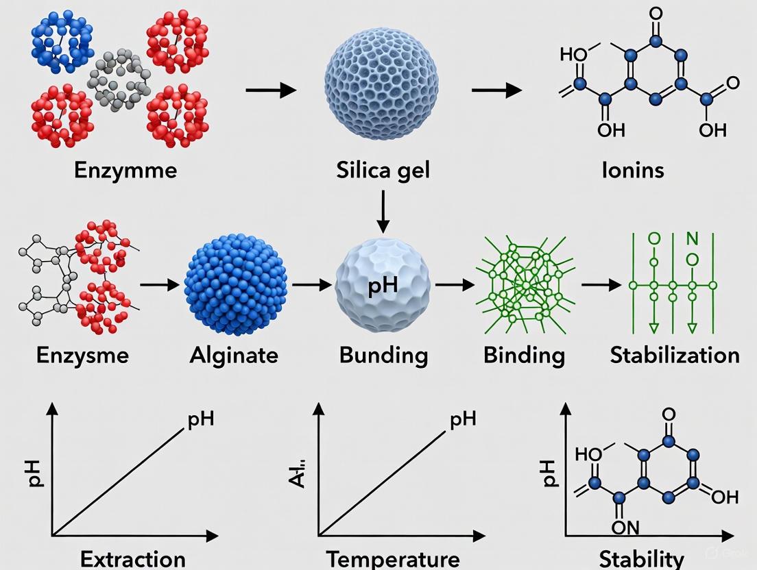
Entrapment and Encapsulation
Principle: This method physically confines enzymes within the interstices of a porous polymer matrix (entrapment) or within a semi-permeable membrane (encapsulation) [1] [16]. The pore size allows substrates and products to diffuse freely while retaining the enzyme. While it generally causes minimal conformational change, it can introduce mass transfer limitations [16].
Protocol: Entrapment in Alginate-Calcium Beads [1]
- Step 1: Polymer-Enzyme Mixture. Prepare a 2-4% (w/v) sodium alginate solution in a suitable buffer. Gently mix this solution with an equal volume of enzyme solution to form a homogeneous suspension.
- Step 2: Gel Bead Formation. Using a syringe with a needle, drop the polymer-enzyme mixture into a 0.1 M calcium chloride (CaCl₂) solution. The divalent calcium ions cross-link the alginate polymer chains instantaneously, forming stable gel beads with the enzyme trapped inside.
- Step 3: Curing and Washing. Allow the beads to cure in the CaCl₂ solution for 30 minutes to ensure complete gelation. Harvest the beads by filtration and wash them with buffer to remove excess Ca²⁺ ions and enzyme on the surface.
Cross-Linking
Principle: This carrier-free strategy uses bifunctional reagents (e.g., glutaraldehyde) to cross-link enzyme molecules with each other, forming insoluble aggregates known as Cross-Linked Enzyme Aggregates (CLEAs) [12] [13]. This method offers high enzyme stability and loading but may reduce activity if cross-linking is excessive.
Protocol: Preparation of Cross-Linked Enzyme Aggregates (CLEAs) [13]
- Step 1: Enzyme Precipitation. To a solution of the target enzyme, slowly add a precipitant such as ammonium sulfate or tert-butanol under constant stirring. The enzyme will precipitate out of solution as fine aggregates. Centrifuge the mixture to collect the aggregates.
- Step 2: Cross-Linking. Re-suspend the wet enzyme aggregates in a small volume of buffer. Add a cross-linking agent, typically glutaraldehyde, to a final concentration of 1-5 mM. Stir the suspension gently for 2-4 hours at 4°C.
- Step 3: Product Recovery. Stop the reaction by centrifugation and wash the resulting CLEAs thoroughly with buffer to remove any unreacted cross-linker. The CLEAs can be stored as a suspension or in a lyophilized form.
Table 2: Comparison of Classic Immobilization Techniques
| Technique | Binding Force | Advantages | Disadvantages | Best Suited For |
|---|---|---|---|---|
| Adsorption | Weak physical interactions (Hydrophobic, ionic) [11] | Simple, inexpensive, high activity retention [16] | Enzyme leakage, non-specific binding [11] | Rapid screening, single-batch processes |
| Covalent Binding | Strong covalent bonds [1] | Very stable, no leakage, reusable [18] | Potential activity loss, complex protocol, expensive [11] | Continuous processes in harsh environments |
| Entrapment/Encapsulation | Physical confinement in a lattice [1] | No chemical modification, protects enzyme [1] | Mass transfer limitations, enzyme leaching from large pores [16] | Biosensors, food processing with sensitive enzymes |
| Cross-Linking (CLEAs) | Covalent bonds between enzyme molecules [13] | High stability & enzyme loading, carrier-free, cost-effective [12] [13] | Potential for over-cross-linking and activity loss [12] | Industrial biocatalysis where support cost is prohibitive |
The Scientist's Toolkit: Key Research Reagent Solutions
Table 3: Essential Materials for Enzyme Immobilization Experiments
| Reagent Category | Specific Examples | Function and Rationale |
|---|---|---|
| Support Materials | Octyl-agarose, Sepabeads [14], Mesoporous Silica (SBA-15) [14], Chitosan [12], Alginate [1], Metal-Organic Frameworks (ZIF-8) [17] | Provides a high-surface-area solid phase for enzyme attachment or entrapment. Choice dictates loading capacity, stability, and mass transfer. |
| Activation Agents/Cross-linkers | Glutaraldehyde [13], Cyanogen Bromide (CNBr) [14], Divinyl sulfone [13], 1-ethyl-3-(3-dimethylaminopropyl) carbodiimide (EDC) [16] | Activates inert support surfaces or creates covalent links between enzyme molecules and the support or between enzymes (in CLEAs). |
| Precipitants (for CLEAs) | Ammonium sulfate, tert-Butanol, Polyethylene glycol (PEG) [13] | Causes the enzyme to aggregate out of solution, forming a physical concentrate ready for cross-linking. |
| Buffers | Phosphate Buffer (for adsorption), Carbonate Buffer (pH 8.5-9.0 for covalent binding) [1] | Maintains optimal pH during the immobilization process to ensure enzyme stability and efficient binding. |
Advanced and Emerging Immobilization Strategies
The frontier of immobilization technology is defined by precision engineering and intelligent design.
- Nanomaterial Carriers: Nanoparticles, nanofibers, and graphene oxide provide exceptionally high surface area-to-volume ratios, drastically increasing enzyme loading and reducing mass transfer barriers [16] [12]. Magnetic nanoparticles, for instance, facilitate easy separation of the biocatalyst using an external magnetic field [12].
- Hybrid Porous Frameworks: Metal-Organic Frameworks (MOFs) and Covalent Organic Frameworks (COFs) are crystalline porous materials with tunable pore environments [17] [13]. They allow for enzyme immobilization via pore adsorption or in-situ encapsulation, offering superior protection against denaturing conditions like organic solvents and high temperatures.
- Carrier-Free Systems: Cross-Linked Enzyme Aggregates (CLEAs) remain a popular carrier-free format due to their high stability and simplicity [13]. Recent innovations include magnetic CLEAs (mCLEAs) for easy separation and combi-CLEAs for multi-enzyme cascade reactions [13].
- AI and Smart Systems: Artificial intelligence and machine learning are now being deployed to predict optimal immobilization conditions, design novel support materials, and model the performance of immobilized enzymes, accelerating research and development cycles [12]. The future points towards "smart" biocatalysts that can dynamically respond to environmental stimuli [13].
Application Notes for Industrial and Pharmaceutical Contexts
The choice of immobilization technique is critically dependent on the final application.
- Pharmaceutical API Synthesis: The synthesis of active pharmaceutical ingredients (APIs), such as sitagliptin, often employs immobilized engineered transaminases via covalent binding. This provides the extreme operational stability and high enantioselectivity (>99.5% e.e.) required for pharmaceutical manufacturing, often in the presence of organic co-solvents like DMSO [18].
- Industrial Biocatalysis: For processes like the production of the herbicide Dimethenamide-P, immobilized lipase B from Candida antarctica (CalB) is used in column reactors. This setup enables continuous operation in organic solvents, high productivity, and excellent stereoselectivity, offering a green alternative to traditional chemical synthesis [18].
- Biosensing and Biofuel Production: Entrapment within polymeric membranes or encapsulation in silica gels is suitable for biosensors and biofuel production, where the enzyme needs to be protected while allowing rapid diffusion of small molecules [1]. CLEAs are also extensively explored for biomass conversion in bioethanol production [13].
The experimental workflow for developing an immobilized enzyme system for industrial use involves multiple critical steps, as visualized below:
The efficacy of immobilized enzyme systems in industrial applications is governed by a fundamental framework: the Enzyme-Support-Mode Interaction Triangle. This paradigm illustrates that optimal biocatalyst performance emerges from the precise interplay between the enzyme (biological catalyst), the support (immobilization matrix), and the mode (method of immobilization). Individually, each component possesses specific characteristics; collectively, they determine critical performance parameters such as activity, stability, specificity, and reusability. Understanding these interactions is paramount for researchers and drug development professionals designing immobilized enzyme systems for manufacturing therapeutics, biosensors, and fine chemicals. The selection of one component directly influences and constrains the choices for the other two, creating a tightly coupled design space that requires systematic optimization [14].
The Support Component: Immobilization Matrices
The support matrix provides the physical foundation for enzyme immobilization, creating a microenvironment that can either stabilize or denature the enzyme structure. An ideal support matrix must be chemically inert, physically robust, stable under operational conditions, and cost-effective [14]. The surface chemistry and morphology of the support directly influence enzyme loading, stability, and accessibility to substrates.
Table 1: Characteristics of Common Support Matrices
| Support Material | Type | Key Properties | Impact on Enzyme | Common Industrial Applications |
|---|---|---|---|---|
| Octyl-Agarose | Natural Polymer | Hydrophobic, macroporous [14] | Enhances affinity for hydrophobic substrates, increases stability [14] | Lipase purification and activation |
| Mesoporous Silica Nanoparticles (MSNs) | Inorganic | Tunable pore size, high surface area, long-term durability [14] | Mitigates diffusion limitation, high activity retention [14] | Biocatalysis in energy applications |
| Chitosan | Natural Polymer | Biocompatible, amenable to chemical modification [14] | Enhances thermal stability and enzyme-binding capacity [14] | Affinity adsorbents for simultaneous purification and immobilization |
| Electrospun Nanofibers | Synthetic/Composite | High surface-area-to-volume ratio, high porosity [14] | Increases residual activity due to greater enzyme loading [14] | High-density enzyme immobilization for biosensing |
| Poly(3-hydroxybutyrate-co-hydroxyvalerate) | Biodegradable Polymer | Eco-friendly, less tough and crystalline [14] | High residual activity and reusability [14] | Environmentally friendly biocatalytic processes |
| Molecular Sieves (Silanized) | Inorganic | Silanols on pore walls facilitate hydrogen bonding [14] | Stable enzyme immobilization, shielded from aggregation [14] | Reduction and oxidation reactions |
The Mode Component: Immobilization Techniques
The immobilization technique defines the nature of the bond between the enzyme and the support. The choice of mode impacts not only the strength of attachment but also the enzyme's conformation, freedom of movement, and accessibility of its active site.
Covalent Binding
Covalent binding involves forming stable chemical bonds between functional groups on the enzyme (e.g., amino, carboxyl, phenolic) and reactive groups on the support [14]. This method often employs cross-linking agents like glutaraldehyde to create stable inter- and intra-subunit bonds, resulting in highly stable, non-leaching preparations with prolonged operational lifetime [14]. The method's drawback is potential loss of activity due to conformational changes or modification of active site residues.
Adsorption
Adsorption relies on weak, non-covalent interactions such as hydrophobic forces, van der Waals forces, and ionic linkages [14]. This simple, cost-effective method preserves high enzyme activity as it causes minimal conformational disruption. However, the binding is weak, leading to enzyme leakage upon changes in pH, ionic strength, or temperature, limiting its industrial applicability.
Affinity Immobilization
This sophisticated technique exploits the specific biological recognition between the enzyme and an affinity ligand pre-attached to the support [14]. Bioaffinity layering can exponentially increase enzyme-binding capacity and reusability. It allows for simultaneous purification and immobilization, yielding highly active and stable preparations, though the supports are often more expensive [14].
Entrapment
Entrapment physically cages enzymes within the interstitial spaces of a polymer network (e.g., alginate, gelatin-calcium hybrids) without direct binding [14]. This protects enzymes from proteolysis and denaturation by creating a sheltered microenvironment. A significant challenge is diffusion limitation, where substrate access and product egress are hindered, reducing observed reaction rates.
Table 2: Comparison of Immobilization Techniques
| Immobilization Mode | Binding Force | Stability | Risk of Activity Loss | Cost | Ideal for Enzymes That Are: |
|---|---|---|---|---|---|
| Covalent Binding | Covalent bonds | Very High | Moderate to High | Moderate | Stable to chemical modification |
| Adsorption | Hydrophobic, Ionic | Low | Low | Low | Sensitive to conformational change |
| Affinity Immobilization | Bio-specific | High | Low | High | Requiring specific orientation/purification |
| Entrapment | Physical barrier | Moderate | Low | Low | Small, and used with small substrates |
Experimental Protocols
Protocol: Covalent Immobilization on Epoxy-Activated Supports
This protocol details the covalent immobilization of an enzyme onto sepabeads, a common epoxy-activated support, ideal for achieving high operational stability [14].
Research Reagent Solutions & Materials
| Item | Function/Brief Explanation |
|---|---|
| Epoxy-Activated Support (e.g., Sepabeads) | Matrix providing stable covalent linkage via epoxy groups. |
| Enzyme Purification Buffer (e.g., 50mM Phosphate, pH 7.0) | Provides a stable chemical environment for the enzyme. |
| Coupling Buffer (e.g., 1M Potassium Phosphate, pH 8.0) | High ionic strength buffer to promote enzyme-support interaction. |
| Blocking Solution (1M Ethanolamine, pH 8.0) | Deactivates unreacted epoxy groups post-immobilization. |
| Washing Buffer (Coupling Buffer + 1M NaCl) | Removes non-covalently adsorbed enzyme. |
Methodology:
- Preparation: Wash 1 gram of epoxy-activated support sequentially with distilled water and coupling buffer.
- Immobilization: Incubate the prepared support with 10-20 mL of enzyme solution (5-10 mg/mL in coupling buffer) under gentle agitation for 24 hours at 25°C.
- Blocking: Recover the immobilized enzyme by filtration and incubate with 10 mL of 1M ethanolamine (pH 8.0) for 4 hours at 25°C to block any remaining epoxy groups.
- Washing: Wash the preparation extensively with coupling buffer, followed by washing buffer (to remove ionically adsorbed enzyme), and finally with the standard assay buffer.
- Activity Assay: Determine the activity of the immobilized enzyme and the supernatant to calculate immobilization yield and expressed activity. The activity should be measured in triplicate to ensure reproducibility [19].
- Storage: Store the final preparation at 4°C in an appropriate buffer.
Protocol: Determining Inhibition Modality via IC₅₀ Replots
Understanding inhibition modality is critical in drug discovery. This protocol uses IC₅₀ shifts at varying substrate concentrations to diagnose the mode of enzyme-inhibitor interaction [20].
Research Reagent Solutions & Materials
| Item | Function/Brief Explanation |
|---|---|
| Target Enzyme | The protein of interest (e.g., kinase, protease). |
| Inhibitor Compound | The small molecule whose mode of action is being characterized. |
| Substrate | The natural molecule converted by the enzyme. |
| Reaction Buffer | Buffered solution at optimal pH for enzyme activity. |
| Detection Reagents | reagents to quantify reaction rate (e.g., NADH, chromogenic substrate). |
Methodology:
- Experimental Setup: For a given inhibitor concentration, measure the initial reaction rate at a minimum of six substrate concentrations, spanning values below and above the known Kₘ. Perform each measurement in triplicate to assess precision [19].
- IC₅₀ Determination: Repeat step 1 for at least six different inhibitor concentrations. For each inhibitor concentration, fit the rate vs. substrate concentration data to the Michaelis-Menten equation (or appropriate inhibition model) using nonlinear regression software to obtain apparent Kₘ and Vₘₐₓ values [19].
- Data Analysis: Plot the initial reaction rate against the logarithm of inhibitor concentration for each substrate level. Fit the data to a sigmoidal dose-response curve to determine the IC₅₀ value at each substrate concentration.
- Diagnostic Replot: Create a secondary plot of the measured IC₅₀ values as a function of the substrate concentration normalized to Kₘ ([S]/Kₘ).
- Mode Identification: Analyze the replot pattern [20]:
- Competitive Inhibition: IC₅₀ increases linearly with increasing [S]/Kₘ.
- Uncompetitive Inhibition: IC₅₀ decreases with increasing [S]/Kₘ.
- Non-competitive/Mixed Inhibition: IC₅₀ changes but does not follow the patterns above.
Workflow Visualization
Figure 1: A systematic workflow for selecting and optimizing the components of the Enzyme-Support-Mode Interaction Triangle for industrial application development.
Figure 2: A detailed experimental workflow for determining enzyme inhibition modality through the analysis of IC₅₀ shifts at varying substrate concentrations, a key technique in drug discovery. [20]
The selection of an appropriate support material is a critical determinant in the success of enzyme immobilization for industrial biocatalysis. An ideal support must concurrently fulfill multiple physicochemical and biological criteria to enhance the immobilized enzyme's stability, activity, reusability, and cost-effectiveness [9] [21]. These properties directly influence the catalytic performance and operational lifespan of the biocatalyst in applications ranging from pharmaceutical synthesis to biofuel production [4] [22]. This document outlines the essential properties of support materials, provides a comparative analysis of common support types, and details standardized protocols for evaluating these key characteristics within a research setting.
Essential Properties of Ideal Support Materials
The performance of an immobilized enzyme system hinges on the properties of its support material. The following properties are considered fundamental for an ideal support [9] [21] [22]:
- Biocompatibility: The support material must be non-toxic and not induce denaturation or conformational changes that impair enzymatic activity. It should provide a favorable microenvironment for the enzyme [23] [22].
- Mechanical Strength: The carrier must exhibit sufficient resistance to compression, shear forces, and abrasion to withstand the hydrodynamic conditions and physical stresses of industrial reactors, especially in continuous fixed-bed operations [9] [21].
- Chemical Stability: The support should be inert and stable under the operational conditions of the process, including extremes of pH, temperature, and the presence of organic solvents or other chemicals [9] [21].
- High Surface Area & Porosity: A large surface area, often provided by a mesoporous structure, permits higher enzyme loading per unit mass. Controlled pore distribution is crucial for optimizing binding capacity, substrate diffusion, and flow properties [9] [24].
- Ease of Functionalization: The surface should possess or be readily modified with functional groups (e.g., amino, carboxyl, epoxy) to facilitate strong and stable attachment of the enzyme via various immobilization chemistries [9] [23].
- Hydrophilicity: A hydrophilic surface is generally preferred as it helps to maintain the essential water layer around the enzyme, preserving its catalytically active tertiary structure [9].
- Cost-Effectiveness and Availability: The material should be readily available, inexpensive, and ideally reusable to ensure the economic viability of the immobilization process on an industrial scale [9] [2].
Table 1: Key Properties and Their Impact on Immobilized Enzyme Performance
| Property | Description | Impact on Immobilized Enzyme |
|---|---|---|
| Biocompatibility | Non-toxic and does not denature the enzyme [23]. | Preserves catalytic activity and prevents enzyme inactivation. |
| Mechanical Strength | Resistance to compression and shear forces [9]. | Ensures long-term structural integrity in industrial bioreactors. |
| Chemical Stability | Inertness across a range of pH, temperatures, and solvents [21]. | Enables application in diverse and harsh industrial processes. |
| High Surface Area & Porosity | Large surface area and controlled pore distribution (e.g., mesoporous) [9] [24]. | Increases enzyme loading capacity and minimizes diffusion limitations. |
| Ease of Functionalization | Availability of reactive functional groups for binding [9]. | Allows for robust and stable enzyme attachment via multiple methods. |
| Hydrophilicity | Hydrophilic surface character [9]. | Maintains the enzyme's hydration shell and native conformation. |
| Cost-Effectiveness | Low cost and ready availability [2]. | Essential for scalable and economically feasible industrial applications. |
Comparative Analysis of Support Material Classes
Support materials can be broadly categorized into organic (natural and synthetic polymers) and inorganic (porous and non-porous) materials. The emergence of nanomaterials and advanced composites has further expanded the options available to researchers [9] [22].
Table 2: Comparison of Common Support Material Classes for Enzyme Immobilization
| Material Class | Examples | Key Advantages | Key Limitations |
|---|---|---|---|
| Natural Polymers | Chitosan, alginate, cellulose, collagen [21] [2]. | Biocompatible, biodegradable, low cost, abundant [2] [22]. | Variable mechanical strength, susceptibility to microbial degradation [9]. |
| Synthetic Polymers | Polyacrylamide, epoxy resins, polyvinyl alcohol (PVA) [21] [23]. | High mechanical/chemical stability, tunable properties [21]. | Potential toxicity of monomers, low biocompatibility, non-biodegradable [9]. |
| Inorganic Porous Materials | Silica, porous glass, zeolites, hydroxyapatite [9] [2]. | High mechanical strength, thermal stability, controlled porosity [9] [23]. | Brittleness, high density, limited functionalization without modification [9]. |
| Carbon-Based Nanomaterials | Carbon nanotubes, graphene [24] [22]. | Very high surface area, excellent electrical/thermal conductivity [24]. | High cost, potential toxicity, complex preparation and functionalization [24]. |
| Magnetic Nanoparticles | Magnetite (Fe₃O₄) [24] [23]. | Easy separation & recovery via magnetic field, high surface area [24] [22]. | Can aggregate, may degrade in acidic/oxidative environments [24]. |
| Metal-Organic Frameworks (MOFs) & Crystalline Porous Organic Frameworks (CPOFs) | ZIF series, COFs, HOFs [4] [25]. | Extremely high surface area, precisely tunable pore size, designable functionality [25]. | Complex synthesis, cost, stability in water/acids/bases can be limited [25]. |
Support Material Selection Workflow
Experimental Protocols for Characterizing Support Materials
Protocol: Determination of Surface Area and Porosity
Principle: The surface area and pore characteristics of a support material are determined from nitrogen adsorption-desorption isotherms measured at 77 K, typically using the Brunauer-Emmett-Teller (BET) method for surface area and the Barrett-Joyner-Halenda (BJH) method for pore size distribution [9].
Materials:
- High-purity nitrogen gas
- Liquid nitrogen
- Degassed sample of the support material
- Surface area and porosity analyzer (e.g., Micromeritics ASAP series, Quantachrome Autosorb series)
Procedure:
- Sample Preparation: Weigh approximately 0.1-0.3 g of the support material. Place it in a sample tube and degas under vacuum at a suitable temperature (e.g., 120°C for silica) for a minimum of 6 hours to remove moisture and adsorbed contaminants.
- Analysis: Transfer the sample tube to the analysis port. The instrument will automatically cool the sample to 77 K using liquid nitrogen and expose it to nitrogen gas at a series of precisely controlled pressures.
- Data Collection: The instrument measures the volume of nitrogen gas adsorbed onto and desorbed from the sample surface at each pressure point, generating an adsorption-desorption isotherm.
- Data Analysis: Use the instrument's software to apply the BET model to the relative pressure (P/P₀) range of 0.05-0.30 to calculate the specific surface area. Apply the BJH model to the desorption branch of the isotherm to determine the pore size distribution and total pore volume.
Reporting: Report the specific surface area in m²/g, the average pore diameter in nm, and the total pore volume in cm³/g. A mesoporous material ideal for enzyme immobilization typically has a pore diameter between 2-50 nm [9].
Protocol: Assessment of Mechanical Stability via Compressibility Testing
Principle: This test evaluates the resistance of support materials to crushing forces, simulating the physical stresses encountered in packed-bed reactors [9].
Materials:
- Universal Testing Machine (UTM) equipped with a flat-plate compression fixture
- Precision balance
- Cylindrical mold (if forming pellets is necessary)
Procedure:
- Sample Preparation: If the support is a powder, it may be pressed into a pellet of defined dimensions (e.g., 5 mm diameter, 3 mm thickness) using a hydraulic press at a standard pressure. Alternatively, a known volume of pre-formed beads can be used.
- Measurement: Place a single pellet or a small, known mass of beads on the lower plate of the UTM. Program the UTM to apply a compressive force at a constant crosshead speed (e.g., 1 mm/min) until the sample fractures or a predefined deformation is reached.
- Data Collection: The instrument software records the applied force (in Newtons, N) and the corresponding displacement (in mm).
Data Analysis: The mechanical strength is reported as the crushing strength, which is the maximum force (N) sustained by the particle before failure. For a more standardized value, the crushing strength can be divided by the particle's diameter to report strength in N/mm.
Protocol: Evaluation of Chemical Stability
Principle: This protocol assesses the structural integrity and mass loss of the support material when exposed to various chemical environments relevant to the intended biocatalytic process [21].
Materials:
- Buffers at different pH values (e.g., pH 4.0, 7.0, 9.0)
- Organic solvents (e.g., methanol, hexane, isopropanol)
- Analytical balance (0.1 mg precision)
- Oven
- Desiccator
Procedure:
- Initial Weighing: Accurately weigh a sample of the dry support material (W₁, ~1.0 g) in a pre-weighed vial.
- Incubation: Add 20 mL of the test solution (buffer or solvent) to the vial. Seal the vial and incubate in a shaking incubator at the operational temperature (e.g., 30°C or 50°C) for 24-72 hours.
- Final Weighing: After incubation, carefully decant the solution. Wash the solid support with a volatile, miscible solvent (e.g., acetone) if needed, and dry to constant weight in an oven. Let the sample cool in a desiccator and record the final dry weight (W₂).
Data Analysis: Calculate the percentage mass loss using the formula: Mass Loss (%) = [(W₁ - W₂) / W₁] × 100 A support with good chemical stability will show minimal mass loss (<5%) under its intended operational conditions.
The Scientist's Toolkit: Essential Research Reagents and Materials
Table 3: Key Reagents and Materials for Support Material Evaluation and Enzyme Immobilization
| Item | Function/Application | Example Specifications |
|---|---|---|
| Mesoporous Silica Nanoparticles | High-surface-area inorganic support for adsorption/covalent binding [23]. | Particle size: 50-200 nm; Pore size: 5-10 nm; Surface area: >500 m²/g. |
| Chitosan | Biocompatible, biodegradable natural polymer support; easily functionalized [21] [2]. | Medium molecular weight; Deacetylation degree ≥75%. |
| Epoxy-Activated Agarose | Robust support for covalent immobilization; stable, hydrophilic [2]. | Bead size: 50-150 μm; Epoxy density: ~20 μmol/mL settled gel. |
| Magnetic Nanoparticles (Fe₃O₄) | Enable easy biocatalyst recovery via magnetic separation [24] [22]. | Core-shell structure (e.g., Fe₃O₄@SiO₂); diameter: 20-50 nm. |
| Glutaraldehyde | Crosslinker for activating amine-bearing supports and creating covalent enzyme bonds [2]. | 25% Aqueous solution, molecular biology grade. |
| Covalent Organic Frameworks (COFs) | Crystalline supports with ultra-high surface area and tunable pores [25]. | Pore size tailored to target enzyme (e.g., 3-8 nm). |
| Eupergit C | Macroporous copolymer beads for stable covalent enzyme immobilization [2]. | Oxirane content: ~0.8 mmol/g. |
| Carbon Nanotubes (CNTs) | Nanoscale support with high conductivity and surface area [9] [24]. | Single or multi-walled; functionalized with -COOH or -NH₂ groups. |
Industrial Immobilization Methods and Their Pharmaceutical Applications
Enzyme immobilization is a cornerstone of industrial biocatalysis, enhancing enzyme reusability, stability, and simplifying downstream processing [16]. Among the various techniques, physical adsorption stands out for its straightforwardness and wide applicability. This method involves the confinement of an enzyme to a solid phase different from that of the substrates and products, utilizing weak physical forces to bind the enzyme to a support matrix [14] [26]. For industrial applications, where cost-effectiveness and operational simplicity are paramount, physical adsorption offers a compelling strategy for developing robust immobilized enzyme systems. This application note provides a detailed overview of the technique, its quantitative performance, and a standardized protocol for implementation.
Mechanism and Characteristics of Physical Adsorption
Physical adsorption relies on non-covalent, intermolecular interactions between the enzyme and the surface of the support material. These interactions primarily include van der Waals forces, hydrophobic effects, and hydrogen bonds [26] [16]. Unlike covalent methods, no chemical linkers or surface modifications are strictly necessary, which helps preserve the native structure and catalytic activity of the enzyme [16].
The following diagram illustrates the core workflow and the key interactions involved in the enzyme immobilization process via physical adsorption.
Comparative Analysis of Immobilization Techniques
Selecting an appropriate immobilization strategy requires balancing factors such as activity retention, stability, and cost. The table below provides a quantitative comparison of physical adsorption with other common techniques, highlighting its characteristic profile of high enzyme loading and simple operation, albeit with a potential risk of enzyme leaching.
Table 1: Quantitative Comparison of Enzyme Immobilization Strategies
| Immobilization Technique | Binding Force | Typical Activity Retention | Operational Stability | Risk of Enzyme Leaching | Relative Cost & Complexity |
|---|---|---|---|---|---|
| Physical Adsorption | Van der Waals, Hydrophobic, Hydrogen bonds [26] [16] | High (Structure not modified) [26] | Moderate (Sensitive to pH, ionic strength) [26] | High [16] | Low [16] |
| Covalent Binding | Covalent bonds [14] | Variable (Can be low due to active site involvement) [16] | High [14] [16] | Very Low [16] | High [16] |
| Entrapment/ Encapsulation | Physical confinement within a polymer network [14] [16] | High to Moderate [16] | High [27] | Low [16] | Moderate [16] |
| Cross-Linking | Covalent bonds between enzyme molecules [14] [16] | Often limited [16] | High [14] | Very Low | Moderate [16] |
Note: The performance metrics are highly dependent on the specific enzyme-support system and immobilization conditions. Data is synthesized from comparative studies. [14] [27] [16]
A specific study comparing glucose oxidase immobilization for biosensing demonstrated that while hydrogel entrapment provided the most stable and sensitive biosensors, adsorption-based biosensors, though functional, showed poor sensitivity and unstable performance [27]. This underscores the need to align the technique with the application's requirements.
Support Materials for Physical Adsorption
The choice of support is critical to the success of the immobilization process. An ideal matrix should be affordable, physically robust, and have a high surface area for enzyme binding [14]. The following table catalogs common and advanced support materials used in adsorption.
Table 2: Overview of Support Materials for Physical Adsorption
| Support Material | Type | Key Characteristics & Examples |
|---|---|---|
| Inorganic Supports | Synthetic/ Natural | Alumina, Silica Gel, Calcium Phosphate Gel, Glass, Kaolin [26]. Mesoporous Silica Nanoparticles (MSNs): Long-term durability, large surface area [14]. Molecular Sieves: Silanols on pore walls facilitate hydrogen bonding [14]. |
| Organic Polymeric Supports | Natural/ Synthetic | Polypropylene-based granules (e.g., Accurel EP-100): Hydrophobic, good for lipases, smaller particle sizes increase reaction rates [14]. Poly(3-hydroxybutyrate-co-hydroxyvalerate): Biodegradable, shown 94% residual activity after 4h at 50°C and reusability for 12 cycles [14]. Starch, Cellulose derivatives (CM-cellulose, DEAE-cellulose), Chitosan [26]. |
| Nanomaterial Supports | Engineered | Electrospun Nanofibers: High surface area and porosity, can lead to greater residual activity [14]. Magnetic Nanoclusters: Enhanced longevity, operational stability, reusability, easy separation [14] [16]. Coconut Fibers: Eco-friendly, good water-holding capacity, high cation exchange [14]. |
Detailed Experimental Protocol: Adsorption of Enzymes on Solid Supports
The Scientist's Toolkit: Essential Research Reagents
Table 3: Key Reagent Solutions and Materials
| Item | Function/Description |
|---|---|
| Solid Support (e.g., Mesoporous Silica, Accurel EP-100, Chitosan) | Provides a high-surface-area matrix for enzyme attachment via physical forces. |
| Enzyme of Interest (Lyophilized powder or pure solution) | The biocatalyst to be immobilized. |
| Buffer Solution (e.g., 0.1 M Sodium Citrate, Phosphate Buffer) | Maintains optimal pH for enzyme stability and binding during immobilization. |
| Orbital Shaker Incubator | Provides agitation to ensure uniform contact between the enzyme and support. |
| Centrifuge or Magnetic Separator (for magnetic supports) | Separates the immobilized enzyme from the free enzyme and washing solutions. |
| Lyophilizer (Freeze Dryer) | For drying and long-term storage of the final immobilized enzyme preparation. |
Step-by-Step Methodology
This protocol describes the adsorption of an enzyme onto a porous solid support, adapted from established procedures in the literature [14] [26].
Step 1: Support Preparation
- Weigh an appropriate amount of dry support material (e.g., 4 mg for small-scale experiments) [26].
- If necessary, pre-wash the support with the immobilization buffer to remove fines and equilibrate it.
Step 2: Enzyme Solution Preparation
- Dissolve the enzyme in a selected buffer (e.g., 0.1 M sodium citrate buffer, pH 4.0 for laccase) to a defined concentration (e.g., 1 mg/mL) [26]. The buffer's pH and ionic strength should be optimized for the specific enzyme to ensure stability.
Step 3: Immobilization Procedure
- Suspend the prepared support in the enzyme solution (e.g., 4 mg support in 10 mL enzyme solution) [26].
- Incubate the mixture with constant agitation (e.g., at 100 rpm) at a controlled temperature (typically 25-30°C) for a predetermined time (e.g., 3 hours) to allow for binding equilibrium [26].
Step 4: Washing and Separation
- Separate the solid support with the immobilized enzyme from the liquid. This can be achieved by centrifugation, filtration, or magnetic separation if using magnetic carriers [14] [26].
- Wash the solid thoroughly with the same buffer to remove any unbound or weakly adsorbed enzyme. The washings can be analyzed for protein content to determine immobilization yield.
Step 5: Storage
- The final immobilized enzyme preparation can be lyophilized and stored at 4°C for future use [26].
Critical Parameters for Optimization
- Enzyme/Support Ratio: A ratio of 0.4 mg enzyme per mL of suspension has been shown to yield 91% activity recovery for laccase on a magnetic carbon composite, beyond which activity may drop due to overcrowding [26].
- pH and Ionic Strength: These parameters dramatically influence the charge and conformation of the enzyme and the support, directly affecting adsorption efficiency and stability. Low ionic strength can enhance adsorption capacity [26].
- Time and Temperature: Sufficient time is required to reach binding equilibrium. While increased temperature can enhance adsorption forces, it must be balanced against the risk of enzyme denaturation [26].
Physical adsorption remains a highly valuable immobilization technique due to its simplicity, cost-effectiveness, and ability to achieve high enzyme loadings with minimal impact on catalytic activity. While challenges such as enzyme leaching under shifting operational conditions persist, the strategic selection of supports—especially modern nanomaterials and renewable agrowaste carriers—and careful optimization of protocols can yield highly effective and stable biocatalysts [14] [16]. For industrial researchers and drug development professionals, this method provides a versatile and accessible starting point for developing immobilized enzyme systems tailored to diverse bioprocessing needs.
In the pursuit of sustainable industrial biocatalysis, enzyme immobilization has emerged as a cornerstone technology. Among the various techniques available, covalent binding stands out for its ability to produce immobilized enzymes with exceptional operational stability and minimal enzyme leakage, which is paramount for cost-effective processes in the pharmaceutical, food, and fine chemicals industries [28] [5]. This method involves the formation of stable covalent bonds between functional groups on the enzyme surface and a complementary solid support [3]. Unlike physical adsorption or entrapment, this robust linkage effectively prevents the enzyme from leaching into the reaction mixture, even under harsh operational conditions or in the presence of solvents, thereby ensuring product purity and enabling catalyst reuse over multiple cycles [3] [5]. This application note delineates standardized protocols and critical considerations for implementing covalent immobilization, framed within a broader research context aimed at developing robust biocatalytic systems for industrial applications.
Key Principles and Rationale
Covalent immobilization enhances enzyme stability by creating strong, irreversible attachments. However, this often comes at the cost of some initial activity, as the chemical reaction can involve amino acid residues critical for catalysis or induce conformational changes [28] [5]. The selection of an appropriate immobilization strategy is therefore a critical compromise, dependent on the specific enzyme characteristics, the support material, and the intended application [28].
The most common covalent techniques leverage carbodiimide chemistry and Schiff base reactions, which target functional groups like –NH2 (from lysine residues) and –COOH (from aspartic or glutamic acid residues) that are abundantly present on the enzyme surface [28] [29]. Achieving optimal enzyme orientation on the support is crucial to minimize unnecessary conformational alterations and steric hindrance of the active site, thereby fostering the development of a stable, highly active, and reproducible biocatalyst [28].
Quantitative Performance of Covalently Immobilized Enzymes
The following table summarizes recent data on the operational stability of various enzymes immobilized via covalent strategies, highlighting their performance in terms of reusability and stability.
Table 1: Performance Metrics of Covalently Immobilized Enzymes
| Enzyme | Support Material | Immobilization Method | Key Performance Outcome | Reference |
|---|---|---|---|---|
| l-Tyrosine Decarboxylase (LbTDC) | Hydroxyapatite (HAP) | APTES/Glutaraldehyde | Retained significant activity over multiple reaction cycles, demonstrating operational stability and reusability. [30] | |
| R-selective Transaminase (TsRTA) | Hydroxyapatite (HAP) | APTES/Glutaraldehyde | Showed promising stability during reusability tests in repeated batch reactions. [30] | |
| Glucose Oxidase (GOx) | Covalent Organic Framework (COF) Microcapsules | Interfacial Assembly & Regrowth | The robust COF shell protected the enzyme from harsh conditions, significantly enhancing stability for biosensing. [31] | |
| Trypsin | Boronate Affinity Monolith | Schiff Base Reaction | Maintained 80% of initial enzyme activity after 28 days of storage at 4°C. [29] |
Standardized Experimental Protocols
Protocol 1: Covalent Immobilization on Hydroxyapatite via APTES-Glutaraldehyde
This widely applicable protocol for covalent immobilization on hydroxyapatite (HAP) uses APTES silanization and glutaraldehyde activation [30].
Research Reagent Solutions
Table 2: Essential Reagents for HAP Covalent Immobilization
| Reagent / Material | Function / Role in Protocol |
|---|---|
| Hydroxyapatite (HAP) | Inorganic support material; provides a biocompatible, high-surface-area matrix for immobilization. |
| (3-Aminopropyl)triethoxysilane (APTES) | Silanizing agent; functionalizes the HAP surface with primary amine groups (–NH₂). |
| Glutaraldehyde | Homobifunctional crosslinker; reacts with amine groups on the support to create an aldehyde-functionalized surface for enzyme coupling. |
| Target Enzyme Solution | The biocatalyst to be immobilized, dissolved in a suitable, non-reactive buffer (e.g., phosphate buffer). |
Sodium Borohydride (NaBH₄) |
Reducing agent; optional, used to reduce unstable Schiff bases to stable secondary amine linkages. |
Step-by-Step Procedure
- Support Functionalization with APTES: Suspend 500 mg of HAP (particle size 180-250 µm) in 2 mL of neat APTES. Mix the suspension for 16 hours at room temperature using an orbital shaker.
- Washing: Dilute the mixture with 2 mL of dichloromethane (DCM), remove and discard the supernatant. Wash the solid with DCM (3 × 2 mL) and allow it to air-dry for 4 hours. This yields amine-functionalized HAP (HAP-NH₂).
- Glutaraldehyde Activation: Activate the HAP-NH₂ by reacting it with a glutaraldehyde solution (e.g., 2.5-5% v/v in a suitable buffer) for 1 hour at room temperature.
- Washing: Wash the activated support thoroughly with buffer to remove any unreacted glutaraldehyde.
- Enzyme Coupling: Incubate the aldehyde-activated HAP with the target enzyme solution (prepared in a weak buffer at a neutral or slightly basic pH to facilitate Schiff base formation) for a predetermined time (e.g., 2-24 hours) at room temperature.
- Washing and Reduction (Optional): Wash the resulting immobilized enzyme preparation extensively with buffer to remove any physically adsorbed enzyme. To enhance the stability of the covalent bond, the preparation may be treated with sodium borohydride to reduce the Schiff bases to alkylamines.
- Storage: The covalently immobilized enzyme can be stored in buffer at 4°C until use [30].
Protocol 2: Encapsulation in Covalent Organic Framework (COF) Microcapsules
This advanced protocol creates a protective microcapsule around the enzyme, combining the stability of covalent bonds with the benefits of a confined microenvironment [31].
Step-by-Step Procedure
- Preparation of COF Dispersion: Disperse 7 mg of pre-synthesized, slightly hydrophobic COF spheres (e.g., COF-1) in 16 mL of mesitylene (oil phase) via sonication (100 W for 5 minutes).
- Emulsion Formation: Add 400 µL of an aqueous solution containing 0.2 mg of your enzyme (e.g., Glucose Oxidase) to the COF dispersion. Homogenize the mixture at 20,000 rpm for 3 minutes to form a stable water-in-oil (w/o) Pickering emulsion, where the COF particles assemble at the water-oil interface.
- Shell Regrowth - Precursor Addition: To reinforce the microcapsule shell, dissolve 2 mg of scandium triflate (
Sc(OTf)₃) and 3.2 mg of 1,3,5-benzenetricarboxaldehyde (BTCA) in 4 mL of chloroform. Add this solution to the emulsion, shake gently, and let it stand for 30 minutes. - Shell Regrowth - Linker Addition: Dissolve 7 mg of 1,3,5-tri(4-aminophenyl)benzene (TAPB) in 4 mL of chloroform. Add this solution to the emulsion to initiate the interfacial polycondensation reaction, which regrows and strengthens the COF shell around the enzyme-containing aqueous droplets. Allow the reaction to proceed for 24 hours.
- Harvesting: Recover the resulting enzyme@MCCOFs (microcapsules) by centrifugation, and wash them thoroughly to remove any unreacted precursors or free enzyme [31].
The workflow for this encapsulation strategy is illustrated below.
Diagram 1: Workflow for Enzyme@MCCOFs Preparation. This diagram illustrates the key stages of creating robust enzyme microcapsules, from initial emulsion formation to final shell regrowth. The process protects the enzyme in a mild aqueous core while constructing a mechanically strong, porous COF shell.
Critical Factors for Success and Troubleshooting
- Support Material Selection: The carrier must possess a high surface area, be insoluble under reaction conditions, and have modifiable functional groups. Materials like hydroxyapatite (HAP) and Covalent Organic Frameworks (COFs) are excellent candidates due to their structural stability and ease of modification [3] [30].
- Enzyme Orientation and Activity Preservation: Uncontrolled immobilization can lead to activity loss by masking the active site or causing unfavorable conformational changes [28] [5]. The use of spacer arms (e.g., PEG) during covalent immobilization can reduce steric hindrance and help maintain activity [29].
- Reaction Condition Optimization: Parameters such as pH, temperature, and ionic strength during the coupling reaction significantly impact immobilization efficiency and the final catalytic properties of the prepared biocatalyst [28]. These must be optimized for each specific enzyme-support system.
Covalent immobilization is a powerful technique to engineer robust biocatalysts for industrial applications. The protocols detailed herein, ranging from the well-established HAP functionalization to the innovative COF microencapsulation, provide researchers with practical tools to achieve high operational stability and prevent enzyme leakage. By carefully selecting the support material and optimizing the immobilization conditions, scientists can develop highly efficient, stable, and reusable enzymatic systems that significantly reduce process costs and advance the principles of green chemistry in the pharmaceutical and fine chemicals sectors.
Enzyme immobilization is a cornerstone of modern industrial biocatalysis, enhancing enzyme stability, enabling reuse, and simplifying product separation [1] [3]. Among various carrier materials, natural hydrogels have emerged as particularly promising matrices due to their high water content, biocompatibility, and structural similarity to the native extracellular environment, which helps preserve enzymatic function [32] [33]. These hydrophilic, cross-linked polymer networks can absorb significant amounts of water while maintaining structural integrity, creating an ideal aqueous microenvironment for entrapped or encapsulated enzymes [33].
The techniques of entrapment and encapsulation within these hydrogels offer distinct advantages for creating robust biocatalytic systems. Entrapment involves incorporating enzymes within the interstitial spaces of a polymer network, while encapsulation confines them within defined, vesicle-like structures surrounded by a semi-permeable membrane [1] [32]. Both approaches physically restrict enzyme mobility without necessarily forming chemical bonds, thereby minimizing conformational changes and activity loss while facilitating easy recovery and reuse of biocatalysts [1]. For industrial and pharmaceutical applications, natural polymer-based hydrogels such as alginate, chitosan, gelatin, and their composites provide additional benefits of biodegradability, low toxicity, and availability from renewable resources [34] [33] [35].
Key Concepts: Entrapment vs. Encapsulation
Although often used interchangeably, entrapment and encapsulation represent distinct immobilization methodologies with different mechanistic bases and practical implications.
Entrapment incorporates enzymes throughout a three-dimensional gel matrix formed by cross-linking hydrophilic polymers. The enzyme solution is typically mixed with polymer precursors before gelation, resulting in random distribution throughout the resulting hydrogel. Substrates and products diffuse through the gel pores, with pore size controlling molecular accessibility [1] [35]. Common examples include calcium alginate gels and polyvinyl alcohol (PVA) beads.
Encapsulation confines enzymes within distinct, microscopic vesicles or capsules surrounded by a continuous hydrogel membrane. This creates a protective barrier between the enzyme core and the external environment, with the membrane controlling molecular transport [1]. The technique is particularly valuable for sensitive enzymes requiring protection from harsh conditions or for controlled release applications [36].
The experimental workflow below illustrates the key steps in preparing immobilized enzymes using these hydrogel-based techniques:
Natural Hydrogel Materials: Properties and Selection
Selecting appropriate hydrogel materials is crucial for optimizing immobilized enzyme performance. Natural polymers offer superior biocompatibility but often require modification to achieve desired mechanical and chemical properties.
Table 1: Characteristics of Common Natural Hydrogel Polymers for Enzyme Immobilization
| Polymer | Source | Key Advantages | Key Limitations | Cross-linking Methods | Common Enzyme Applications |
|---|---|---|---|---|---|
| Alginate | Brown algae | Mild gelation conditions (ionic), biocompatible, low cost | Excessive swelling, low cell adhesion, mechanical weakness | Ionic (Ca²⁺, Ba²⁺) | Laccase, nitrile hydratase, proteases [1] [32] [35] |
| Chitosan | Fungal/crustacean chitin | Antibacterial properties, biocompatible, functional groups for modification | Poor water solubility, high pH sensitivity | Ionic, covalent (glutaraldehyde) | Peroxidases, lipases [34] [35] |
| Gelatin | Animal collagen | Contains RGD cell adhesion sites, promotes cell proliferation | Poor thermal stability, low mechanical strength | Thermal, chemical cross-linking | Various oxidoreductases [34] |
| Hyaluronic Acid | Animal tissues/microbial | Excellent biocompatibility, high hydrophilicity, lubricating | Rapid degradation, poor mechanical properties | Chemical modification, covalent | Drug delivery systems [34] |
| Composite Hydrogels | Multiple sources | Synergistic properties, enhanced functionality | Complex production processes | Multiple mechanisms | Multi-enzyme systems [34] [35] |
Experimental Protocols
Protocol 1: Calcium Alginate Entrapment for Enzyme Immobilization
This protocol describes a standardized method for immobilizing enzymes in calcium alginate hydrogel beads, suitable for various biocatalytic applications [1] [32] [35].
Reagents and Materials
- Sodium alginate (1.5-3.0% w/v aqueous solution)
- Enzyme preparation (purified enzyme or cell lysate)
- Calcium chloride dihydrate (0.1-0.5 M solution)
- Appropriate buffer for enzyme stability (e.g., phosphate, Tris-HCl)
- Syringe with needle (22-26G) or peristaltic pump with droplet generator
- Magnetic stirrer and stir bars
Step-by-Step Procedure
Polymer Solution Preparation: Dissolve sodium alginate in appropriate buffer to achieve 1.5-3.0% (w/v) concentration. Stir continuously (2-4 hours, room temperature) until completely dissolved and homogeneous.
Enzyme Incorporation: Gently mix the enzyme solution with alginate solution at a 1:4 to 1:9 ratio (v/v). Maintain temperature conditions appropriate for enzyme stability (typically 4°C). Avoid vigorous mixing to prevent enzyme denaturation.
Bead Formation: Transfer the alginate-enzyme mixture to a syringe with needle. Slowly drip the solution into gently stirred calcium chloride solution (0.1-0.5 M). Needle diameter (0.2-0.8 mm) and flow rate control bead size (1.0-3.0 mm).
Gelation and Curing: Allow beads to remain in calcium chloride solution for 20-60 minutes with gentle stirring to complete gelation and hardening.
Washing and Storage: Collect beads by filtration or sieving. Rinse thoroughly with appropriate buffer to remove surface-bound enzyme and excess calcium ions. Store in buffer at 4°C until use.
Critical Parameters and Optimization
- Alginate Concentration: Higher concentrations (2.5-3.0%) increase mechanical strength but may hinder substrate diffusion.
- Gelation Time: Extend curing time (>30 minutes) for more uniform and robust beads.
- Bead Size: Smaller beads (<1 mm) reduce mass transfer limitations but require specialized equipment.
Protocol 2: Chitosan-Gelatin Composite Hydrogel for Enhanced Stability
This composite hydrogel system combines the biocompatibility of gelatin with the mechanical and antibacterial properties of chitosan, creating an optimal environment for enzyme stabilization [34] [33] [35].
Reagents and Materials
- Chitosan (medium molecular weight, 1-2% w/v in dilute acetic acid)
- Gelatin (Type A or B, 2-4% w/v aqueous solution)
- Cross-linking agent (e.g., genipin, glutaraldehyde)
- Enzyme preparation
- Phosphate buffered saline (PBS, pH 7.0-7.4)
Step-by-Step Procedure
Polymer Solutions Preparation:
- Dissolve chitosan in 1% (v/v) acetic acid solution to 1-2% (w/v) concentration.
- Dissolve gelatin in buffer or distilled water (40-50°C) to 2-4% (w/v) concentration.
Composite Formation: Mix chitosan and gelatin solutions at 1:1 ratio (v/v) at 37°C with continuous stirring. Adjust pH to 5.5-6.0 if needed.
Enzyme Incorporation: Add enzyme solution to the composite polymer mixture at 37°C with gentle stirring.
Cross-linking: Add cross-linking agent (e.g., 0.1-0.5% genipin) and mix thoroughly. Pour mixture into molds or form droplets.
Gelation and Curing: Maintain at room temperature for initial setting, then at 4°C for 12-24 hours for complete cross-linking.
Post-treatment: Wash extensively with buffer to remove unreacted cross-linker and surface-bound enzyme.
Critical Parameters and Optimization
- Chitosan:Gelatin Ratio: 1:1 ratio typically provides optimal balance between mechanical properties and biocompatibility.
- Cross-linking Density: Vary cross-linker concentration (0.1-0.5%) to control mesh size and enzyme release kinetics.
- Gelation Temperature: Stepwise temperature reduction improves structural homogeneity.
Performance Evaluation and Applications
Quantitative Comparison of Immobilized Enzyme Systems
Table 2: Performance Metrics of Enzymes Immobilized in Natural Hydrogels
| Enzyme | Hydrogel System | Immobilization Efficiency (%) | Retained Activity (%) | Operational Stability (Reuse Cycles) | Key Applications |
|---|---|---|---|---|---|
| Laccase | Calcium alginate beads | 63-80% | 70-85% | 5-10 cycles | Dye degradation, wastewater treatment [1] [35] |
| Alkaline Protease | Alginate/Zeolite | 64-80% | 60-75% | 6-8 cycles | Dairy processing, milk coagulation [1] |
| Horseradish Peroxidase | Tyramine-alginate encapsulation | 75-90% | 80-95% | 10-15 cycles | Biosensing, bioremediation [1] [32] |
| Asparaginase | Peptide hydrogel | >85% | >90% | Sustained release over 72h | Cancer therapy (Erwinase) [36] |
| Nitrile Hydratase | Polyacrylamide encapsulation | 70-88% | 65-80% | >20 batches | Industrial production of acrylamide [1] |
| Lipase | Chitosan-gelatin composite | 75-85% | 70-82% | 8-12 cycles | Biodiesel production, ester synthesis [35] [3] |
Industrial and Biomedical Applications
Pharmaceutical Applications: Sustained Release Formulations
Peptide-based hydrogels have shown remarkable success in creating injectable sustained-release formulations for therapeutic enzymes. In a case study with Erwinase (asparaginase) for acute lymphoblastic leukemia treatment, hexamer peptide hydrogels (H-FEFQFK-NH₂ and H-FQFQFK-NH₂) enabled sustained enzyme release while maintaining full catalytic activity [36]. The hydrogel system provided:
- Extended Release Profile: Controlled release over 72 hours, significantly extending the enzyme's plasma half-life
- Preserved Bioactivity: Full retention of enzymatic function post-encapsulation and release
- Improved Therapeutic Efficacy: Reduced dosing frequency from daily injections to once every 2-3 days
- Minimal Immunogenicity: Natural amino acid composition minimized foreign body response
Industrial Biocatalysis: Wastewater Treatment
Immobilized enzyme systems in hydrogel matrices have demonstrated excellent performance in environmental applications. Laccase encapsulated in calcium-alginate beads effectively degraded crude and weathered oil, with the immobilized enzyme showing enhanced tolerance to temperature and pH variations compared to free enzyme [32] [35]. The hydrogel matrix served as a physical barrier protecting the encapsulated enzyme from harsh environmental conditions while allowing continuous operation in bioreactor systems.
Food Processing Applications
In the food industry, alginate-encapsulated alkaline protease has been successfully employed as a milk coagulant in dairy product manufacturing [1]. The immobilized enzyme system offered:
- Reusability: 6-8 operational cycles without significant activity loss
- Product Purity: Elimination of enzyme contamination in final products
- Process Control: Enhanced predictability of enzymatic reactions
- Cost Reduction: Decreased enzyme consumption per unit product
The Scientist's Toolkit: Essential Research Reagents
Table 3: Key Research Reagents for Hydrogel-Based Enzyme Immobilization
| Reagent/Category | Specific Examples | Function/Purpose | Considerations for Use |
|---|---|---|---|
| Natural Polymers | Sodium alginate, chitosan, gelatin, hyaluronic acid | Form hydrogel matrix backbone | Purity, molecular weight, batch-to-batch variability |
| Cross-linking Agents | CaCl₂ (alginate), genipin, glutaraldehyde | Stabilize 3D network structure | Biocompatibility, potential enzyme toxicity |
| Activity Assays | Substrate-specific colorimetric/fluorometric assays | Quantify immobilized enzyme activity | Substrate diffusion limitations in hydrogel |
| Characterization Tools | FTIR, SEM, rheometry, swelling ratio measurements | Analyze structural and mechanical properties | Sample preparation requirements |
| Microencapsulation Equipment | Microfluidic devices, electrostatic bead generators | Produce uniform hydrogel particles | Capital cost, operational complexity |
Enzyme immobilization is a critical technology for enhancing the stability, reusability, and efficiency of biocatalysts in industrial processes. Among various strategies, carrier-free immobilized enzymes represent a sophisticated approach that eliminates the inert support matrix, leading to highly concentrated catalytic activity within a minimal volume [37]. This category primarily includes Cross-Linked Enzyme Aggregates (CLEAs) and Cross-Linked Enzyme Crystals (CLECs), which are insoluble, robust biocatalytic particles formed via chemical cross-linking [37]. By avoiding the use of a carrier, which typically constitutes up to 95% of the total immobilized enzyme mass, these systems achieve superior productivity and space-time yields compared to carrier-bound methods [38]. The high enzyme concentration and reduced diffusion limitations make CLEAs and CLECs particularly advantageous for continuous flow biocatalysis and industrial applications in pharmaceuticals, fine chemicals, and food processing where cost-effectiveness and operational stability are paramount [39] [18].
The structural integrity of CLEAs and CLECs stems from the formation of covalent bonds between enzyme molecules, creating a stable, cross-linked network. CLEAs are prepared from precipitated enzyme aggregates, making the process applicable to even crude enzyme preparations [37]. In contrast, CLECs utilize highly purified enzymes in crystalline form, offering enhanced structural regularity and often greater stability [37]. Both technologies facilitate efficient catalyst recovery and recycling, significantly reducing operational costs and environmental impact—key considerations for sustainable industrial processes [4] [18].
Cross-Linked Enzyme Aggregates (CLEAs)
Cross-Linked Enzyme Aggregates (CLEAs) are formed through a two-step process involving first the physical aggregation of enzyme molecules followed by chemical cross-linking to create insoluble, stable biocatalyst particles [38]. This methodology combines purification and immobilization into a single operation, as precipitation effectively concentrates the enzyme while simultaneously presenting it in a physically stabilized format amenable to cross-linking [37]. The precipitation step typically employs salts, organic solvents, or polymers to drive enzymes out of solution, forming aggregates that maintain the enzyme's active conformation [38]. Subsequent cross-linking with bifunctional reagents like glutaraldehyde creates covalent bonds between enzyme molecules, permanently stabilizing the aggregate structure while preserving catalytic activity [37] [38].
The versatility of the CLEA platform enables the development of advanced configurations including magnetic CLEAs (m-CLEAs) incorporating iron oxide nanoparticles for simplified magnetic separation, combi-CLEAs co-immobilizing multiple enzymes for cascade reactions, and fusion-protein CLEAs utilizing engineered hybrid enzymes [38]. A notable advancement includes microfluidic approaches for CLEA synthesis, enabling production of highly uniform, nanoscale particles (~100 nm diameter) with significantly enhanced activity recovery (up to 90.5%) compared to conventional batch methods [39].
Experimental Protocol for CLEA Preparation
Materials Required:
- Amine transaminase (ATA) solution (crude or purified)
- Acetone (pre-chilled to 4°C)
- Glutaraldehyde solution (25% w/v)
- Sodium phosphate buffer (0.1 M, pH 7.5)
- Centrifuge tubes and microcentrifuge
- Vortex mixer
- Magnetic stir plate and stir bars
Step-by-Step Procedure:
Enzyme Precipitation:
- Add 1 mL of amine transaminase solution (5-10 mg/mL protein) to a 15 mL centrifuge tube.
- Place the tube on ice and slowly add pre-chilled acetone dropwise under gentle vortexing to a final concentration of 60% (v/v).
- Continue mixing for 15 minutes until a cloudy suspension forms, indicating aggregate formation.
- Centrifuge at 8,000 × g for 10 minutes at 4°C to collect the precipitated enzyme aggregates.
- Carefully decant the supernatant and resuspend the pellet in 1 mL of sodium phosphate buffer (0.1 M, pH 7.5).
Cross-Linking Reaction:
- Add glutaraldehyde to the suspended aggregates to a final concentration of 10 mM.
- Incubate the mixture with gentle stirring for 2 hours at 4°C to facilitate cross-linking.
- Quench the reaction by adding 100 μL of 1 M glycine solution and stirring for an additional 15 minutes.
Washing and Storage:
- Centrifuge the cross-linked aggregates at 10,000 × g for 5 minutes.
- Wash the pellet three times with sodium phosphate buffer to remove unreacted cross-linker.
- Resuspend the final CLEA product in an appropriate storage buffer at 4°C.
Critical Parameters for Optimization:
- Precipitant Selection: Test acetone, ethanol, ammonium sulfate, or polyethylene glycol at varying concentrations.
- Cross-linker Concentration: Optimize glutaraldehyde concentration (typically 5-20 mM) to balance activity recovery and stability.
- Reaction Time: Cross-linking duration affects particle size and mechanical stability.
- pH Control: Maintain optimal pH during cross-linking to preserve enzyme activity.
Performance Data and Industrial Applications
Table 1: Comparative Performance of CLEA Technology in Industrial Applications
| Enzyme | Application | Productivity Gain | Operational Stability | Reference |
|---|---|---|---|---|
| Amine Transaminase (ATA) | Continuous transamination in membrane microreactor | 2.5-fold higher activity recovery vs. batch | 45% higher turnover over 5 days | [39] |
| L-Arabinose Isomerase | Isomerization of D-galactose to D-tagatose | High operational stability | Effective recycling for multiple batches | [38] |
| Cellulase (Trichoderma reesei) | Biomass hydrolysis | Higher activity than free enzyme | Stable for 10 reuse cycles | [38] |
| Chimeric ADH-CHMO | ε-Caprolactone synthesis from cyclohexanol | Comparable to free enzymes | Promising operational and storage stability | [38] |
| Lipase (Candida antarctica) | Chiral compound production (e.g., Dimethenamide-P) | Enantiomeric excess >99% | Stable in organic solvent at <60°C | [18] |
Recent advances demonstrate CLEA integration with membrane microreactors, enabling continuous biotransformation with significantly enhanced productivity. For amine transaminase CLEAs, this approach achieved over 68% immobilization efficiency and 45% higher turnover number during five days of continuous operation compared to non-aggregated counterparts [39]. The microfluidic synthesis platform produced remarkably uniform ATA-CLEAs of approximately 100 nm diameter, representing a substantial improvement over conventional methods that typically yield heterogeneous, micrometer-sized particles with poor reproducibility [39].
Cross-Linked Enzyme Crystals (CLECs)
Cross-Linked Enzyme Crystals (CLECs) represent the most structurally ordered carrier-free immobilization system, comprising enzyme molecules arranged in a regular crystalline lattice stabilized by intermolecular cross-links [37]. The prerequisite for CLEC formation is the availability of highly purified enzyme in crystalline form, making the process more technically demanding than CLEA preparation but often yielding superior mechanical stability and rigidity [37]. The crystalline structure provides exceptional protection against denaturing conditions including organic solvents, proteolytic degradation, and temperature extremes, making CLECs particularly valuable for industrial processes requiring harsh reaction conditions [37].
The cross-linking step in CLEC formation primarily serves to stabilize the crystalline structure against dissolution in aqueous reaction media while maintaining the enzyme's catalytic conformation within the crystal lattice. This stabilization enables CLECs to function effectively across a broad range of pH values, temperatures, and solvent systems where soluble enzymes would rapidly denature or leach from support materials [37]. Although CLECs require more extensive purification and crystallization expertise, their exceptional stability often justifies the additional preparation effort for high-value industrial applications.
Experimental Protocol for CLEC Preparation
Materials Required:
- Purified enzyme (>99% purity)
- Crystallization reagents (precipitants, buffers, additives)
- Glutaraldehyde solution (0.1-0.5% v/v)
- Microbatch plates or hanging drop apparatus
- Inverted microscope for crystal screening
- Centrifugation equipment
Step-by-Step Procedure:
Enzyme Crystallization:
- Concentrate purified enzyme to 10-50 mg/mL in appropriate buffer.
- Screen crystallization conditions using vapor diffusion (hanging/sitting drop) or microbatch methods.
- Identify optimal conditions yielding well-formed crystals of suitable size (typically 10-100 μm).
- Scale up crystallization using optimized parameters.
Cross-Linking Process:
- Harvest crystals by gentle centrifugation (500 × g for 5 minutes).
- Resuspend crystals in cold glutaraldehyde solution (0.1-0.5% in compatible buffer).
- Incubate with gentle agitation for 12-24 hours at 4°C.
- Terminate cross-linking by centrifugation and washing with buffer.
Characterization and Storage:
- Assess crystal integrity by optical microscopy.
- Determine activity recovery using standard enzyme assays.
- Store cross-linked crystals in appropriate buffer at 4°C.
Critical Parameters for Optimization:
- Crystal Quality: Optimize crystal size, morphology, and uniformity.
- Cross-linking Density: Balance between stabilization and activity preservation.
- Crystal Size Control: Smaller crystals reduce diffusion limitations but are more challenging to handle.
Comparative Analysis and Implementation Strategies
Performance Comparison and Selection Criteria
Table 2: Comparative Analysis of CLEA vs. CLEC Technologies
| Parameter | CLEA Technology | CLEC Technology |
|---|---|---|
| Enzyme Purity Requirement | Crude preparations acceptable | High purity essential |
| Particle Uniformity | Variable; improved with microfluidics | Highly uniform |
| Preparation Complexity | Moderate | High (requires crystallization expertise) |
| Development Time | Relatively short | Extended optimization needed |
| Cost Considerations | Lower (minimal purification) | Higher (extensive purification) |
| Activity Recovery | Up to 90.5% reported [39] | Typically high after optimization |
| Stability in Organic Solvents | Excellent | Exceptional |
| Mechanical Stability | Good to excellent | Superior |
| Industrial Scalability | Highly scalable | Moderate scalability |
| Applicable Enzyme Range | Broad range | Limited to crystallizable enzymes |
Implementation in Continuous Flow Systems
The integration of CLEA technology with continuous flow reactors represents a significant advancement for industrial biocatalysis. The implementation involves several critical considerations:
Reactor Configuration:
- Membrane Microreactors: CLEAs immobilized within membrane systems enable continuous substrate feeding and product removal [39].
- Packed-Bed Reactors: CLEAs or CLECs can be packed into columns for continuous processing with minimal pressure drop.
- Fluidized-Bed Systems: Suitable for fragile biocatalyst particles requiring gentle mixing.
Process Optimization Parameters:
- Particle Size Control: Nanoscale CLEAs (~100 nm) reduce mass transfer limitations [39].
- Flow Rate Modulation: Balance between residence time and throughput.
- Operational Stability: CLEAs demonstrate enhanced stability under continuous operation conditions [39].
Recent implementations demonstrate successful continuous transamination of (S)-α-methylbenzylamine with pyruvate using ATA-CLEAs in membrane microreactors, achieving high conversion rates over extended operational periods [39]. This configuration enables one-step purification and immobilization with nearly 100% yield, dramatically simplifying downstream processing [39].
The Scientist's Toolkit: Research Reagent Solutions
Table 3: Essential Reagents and Materials for CLEA/CLEC Development
| Reagent/Material | Function | Application Notes |
|---|---|---|
| Glutaraldehyde | Bifunctional cross-linker | Most common cross-linker; economical but may affect activity [38] |
| Glyceryl Diglycidyl Ether | Epoxy-based cross-linker | Improved mechanical stability; reduced activity loss [38] |
| Polyethylenimine (PEI) | Macromolecular cross-linker | Enhanced stability; functions as cross-linker and stabilizer [38] |
| Acetone | Precipitating agent | Common for CLEA preparation; pre-chill for better results [39] |
| Ammonium Sulfate | Precipitating agent | Mild precipitant; preserves enzyme activity [38] |
| Amino-functionalized Magnetic Particles | Magnetic separation | Enables m-CLEA formation for easy recovery [38] |
| Microfluidic Reactor Chips | Uniform particle synthesis | Produces monodisperse CLEAs with superior properties [39] |
| Chitosan & Derivatives | Natural polymer cross-linker | Biocompatible; used in combi-CLEAs [38] |
Workflow and Technology Integration
The following diagram illustrates the comparative development pathways and advantages of CLEA versus CLEC technologies:
CLEA vs CLEC Development Pathways - This diagram compares the methodological approaches and respective advantages of CLEA and CLEC technologies for carrier-free enzyme immobilization.
Cross-linked carrier-free enzyme systems, particularly CLEAs and CLECs, represent sophisticated biocatalyst design solutions for high-productivity industrial applications. The demonstrated advantages include exceptional stability, simplified downstream processing, and cost-effective implementation across diverse biotransformation processes [39] [18]. Recent technological innovations such as microfluidic synthesis and magnetic recovery systems continue to address earlier limitations related to particle heterogeneity and separation challenges [39] [38].
Future development trajectories suggest increased integration of protein engineering with immobilization strategies, enabling designed cross-linking sites and optimized enzyme orientation [1]. The growing emphasis on continuous flow biocatalysis in pharmaceutical and fine chemical manufacturing将进一步驱动对这些载体固定化系统的需求,它们为可持续的生物催化过程提供了实现高效、经济且环保的解决方案 [39] [4]. 随着材料科学和生物工艺的进步,CLEA和CLEC技术有望在工业生物技术的绿色化和高效化进程中发挥越来越重要的作用。
Application Notes
The integration of advanced nanoparticle platforms is redefining enzyme immobilization for industrial biocatalysis. These nanomaterials provide exceptional surface area, tunable functionalities, and unique magnetic or structural properties, enabling unprecedented control over enzyme stability, activity, and reusability. The table below summarizes the key characteristics and industrial applications of three prominent platforms [13] [40] [24].
Table 1: Advanced Nanoparticle Platforms for Enzyme Immobilization
| Platform | Key Characteristics | Industrial Applications | Performance Advantages |
|---|---|---|---|
| Covalent Organic Frameworks (COFs) | Porous crystalline polymers; tunable pore environments; high surface area; excellent chemical stability [13]. | Biosensing, environmental remediation, selective catalysis, pharmaceutical synthesis [13]. | Prevents enzyme deactivation under hostile conditions; enhances mass transfer; high biocatalyst loading per support mass [13]. |
| Magnetic Nanoparticles (MNPs) | Superparamagnetic properties (easy magnetic separation); high surface-area-to-volume ratio; biocompatible (e.g., Fe₃O₄) [40]. | Biofuel production (biodiesel), wastewater treatment, pharmaceutical manufacturing, repeated-batch and continuous processes [40] [24]. | Enables easy catalyst recovery and reuse; improves operational stability; reduces production costs by simplifying downstream processing [40]. |
| Nanoflowers | Hybrid organic-inorganic structures; flower-like morphology providing high surface area for immobilization [21]. | Biofuel production, bioremediation, biosensors, enhancing thermal and pH stability of enzymes [21]. | Increases enzyme loading and stability; improves catalytic efficiency due to hierarchical structure [21]. |
The selection of an appropriate immobilization technique is crucial for maximizing the benefits of these nanoplatforms. The following workflow outlines the decision-making process for matching an enzyme with a suitable nanoparticle and immobilization strategy.
Experimental Protocols
Protocol: Enzyme Immobilization on Magnetic Nanoparticles (MNPs) via Covalent Binding
This protocol details the immobilization of an enzyme (e.g., lipase) onto amino-functionalized magnetic Fe₃O₄ nanoparticles using glutaraldehyde as a crosslinker, enabling easy magnetic separation and reuse [40] [2] [24].
Research Reagent Solutions
Table 2: Essential Materials for MNP Immobilization
| Reagent/Material | Function/Description | Supplier Example/Specification |
|---|---|---|
| Magnetic Nanoparticles (Fe₃O₄) | Core support; provides superparamagnetism for easy separation. | Sigma-Aldrich, 10-20 nm particle size, >98% purity. |
| (3-Aminopropyl)triethoxysilane (APTES) | Silane agent for functionalizing MNP surface with primary amine groups (-NH₂). | Thermo Scientific, 99% purity. |
| Glutaraldehyde Solution (25%) | Homobifunctional crosslinker; reacts with amine groups on MNP and enzyme. | MilliporeSigma, Grade I for electron microscopy. |
| Target Enzyme (e.g., Lipase) | The biocatalyst to be immobilized. | Recombinant, purified enzyme preparation. |
| Phosphate Buffered Saline (PBS), 0.1 M, pH 7.4 | Provides a stable physiological pH environment for immobilization. | Prepared from PBS buffer tablets, sterile filtered. |
| Sodium Borohydride (NaBH₄) | Stabilizes the Schiff base formed during crosslinking. | Fisher Scientific, powder, reagent grade. |
Step-by-Step Procedure:
Functionalization of MNPs:
- Disperse 100 mg of bare Fe₃O₄ MNPs in 50 mL of anhydrous ethanol.
- Add 2 mL of APTES to the suspension and reflux at 70°C for 6 hours under an inert nitrogen atmosphere with constant stirring [40].
- Separate the amino-functionalized MNPs (NH₂-MNPs) using a permanent magnet and wash thoroughly with ethanol and deionized water to remove unbound APTES. Dry under vacuum at 50°C for 12 hours.
Activation of NH₂-MNPs with Glutaraldehyde:
Enzyme Immobilization:
- Prepare a solution of the target enzyme (e.g., 50 mg of lipase) in 10 mL of PBS (0.1 M, pH 7.4).
- Add the activated MNPs to the enzyme solution and incubate at 4°C for 12-16 hours with continuous mixing.
- To stabilize the Schiff base linkage, add a small amount of solid sodium borohydride (NaBH₄, final concentration ~1 mg/mL) and incubate for 1 hour [2].
Recovery and Washing:
- Separate the immobilized enzyme (Enzyme-MNPs) using a strong magnet.
- Wash the pellet repeatedly with PBS until no protein is detected in the wash supernatant (e.g., using a Bradford assay) to ensure removal of any unbound enzyme.
Storage:
- Store the final Enzyme-MNP biocatalyst in a suitable buffer at 4°C.
Protocol: In-Situ Encapsulation of Enzymes in Covalent Organic Frameworks (COFs)
This protocol describes the synthesis of COFs around enzyme molecules, leading to their in-situ encapsulation and creating a protective microenvironment [13].
Step-by-Step Procedure:
Preparation of Monomer Solutions:
- Dissolve the organic building blocks (e.g., 1,3,5-Triformylphloroglucinol and p-Phenylenediamine) in a mixture of mesitylene and 1,4-dioxane in a sealed vial.
Enzyme Addition:
- Add a concentrated solution of the target enzyme in a mild buffer (e.g., 10 mM phosphate buffer, pH 7.0) to the monomer mixture. The aqueous enzyme solution should be added judiciously to create a heterogeneous system [13].
Synthesis and Encapsulation:
- Add a catalytic amount of acetic acid (e.g., 6 M) to the vial to initiate the polycondensation reaction.
- Seal the vial and maintain the reaction at a controlled temperature (e.g., 80-120°C) for 48-72 hours to allow for the formation of crystalline COF material around the enzyme molecules [13].
Harvesting and Washing:
- After synthesis, collect the resulting Enzyme@COF composite by centrifugation.
- Wash the precipitate thoroughly with water and acetone to remove unreacted monomers and any non-encapsulated enzyme.
Drying and Activation:
- Dry the final composite under vacuum before use. The encapsulated enzyme is now protected within the porous COF structure.
Performance and Validation
The success of the immobilization protocols should be validated by comparing the performance of the immobilized enzyme against its free counterpart. Key metrics are summarized below.
Table 3: Quantitative Performance Metrics of Immobilized Enzymes
| Performance Metric | Free Enzyme | Enzyme immobilized on MNPs | Enzyme encapsulated in COFs |
|---|---|---|---|
| Operational Stability (Half-life) | Several hours | Can be significantly extended [40] | Markedly enhanced under harsh conditions [13] |
| Reusability (Cycles) | Single use | >10 cycles with >70% activity retention [24] | Highly reusable due to strong confinement [13] |
| Thermal Stability | Denatures at moderate temperatures | Increased resistance to thermal denaturation [40] [24] | Superior stability at elevated temperatures [13] |
| pH Stability | Narrow optimal range | Broader pH tolerance [24] | Protected from extreme pH by the COF matrix [13] |
| Catalytic Efficiency (Vmax/Km) | Baseline | May increase or decrease depending on immobilization [24] | Can be maintained or enhanced due to favorable microenvironments [13] |
The relationships between the nanoparticle's properties, the chosen protocol, and the resulting biocatalyst performance are complex. The following diagram maps these critical interactions and their outcomes.
Enzyme immobilization has emerged as a foundational technology for industrial biocatalysis, particularly in the pharmaceutical sector where it enables the production of high-value chiral intermediates and active pharmaceutical ingredients (APIs). This technology enhances enzyme stability, facilitates catalyst recovery and reuse, simplifies downstream processing, and significantly reduces operational costs [2] [18]. The strategic application of immobilized enzymes aligns with green chemistry principles by minimizing waste generation and enabling continuous manufacturing processes [4]. Within this framework, penicillin G acylase (PGA) and trypsin represent two clinically significant biocatalysts with complementary applications in antibiotic synthesis and bioprocessing. This article presents detailed application notes and experimental protocols for these enzymes, providing pharmaceutical scientists with practical methodologies for implementing immobilized biocatalysts in drug synthesis workflows.
Application Notes: Immobilized Penicillin G Acylase
Industrial Context and Significance
Penicillin G acylase (PGA) serves as a cornerstone biocatalyst in the manufacturing of β-lactam antibiotics. This enzyme catalyzes the hydrolysis of penicillin G to produce 6-aminopenicillanic acid (6-APA), the fundamental building block for semi-synthetic penicillins [41] [42]. The immobilization of PGA addresses critical limitations associated with the free enzyme, including operational instability, difficult recovery from reaction mixtures, and limited reusability [42]. Immobilized PGA formats demonstrate enhanced thermal and pH stability, superior longevity, and enable continuous processing, thereby improving the economic viability and sustainability of antibiotic manufacturing processes [41] [18].
Quantitative Performance Data
Recent advances in PGA immobilization have yielded robust biocatalysts with significantly improved operational characteristics. The table below summarizes performance metrics for PGA immobilized on various functionalized magnetic supports.
Table 1: Performance Metrics of Penicillin G Acylase on Various Magnetic Supports
| Immobilization Support | Immobilization Efficiency | Optimal pH | Optimal Temperature (°C) | Kinetic Parameter (Km) | Reusability (Activity Retention after Cycles) | Reference |
|---|---|---|---|---|---|---|
| Ni₀.₄Cu₀.₅Zn₀.₁Fe₂O₄-SiO₂-GA | Not specified | 8.0 | 50 | 0.0436 mol·L⁻¹ | >25% (after 5 cycles) | [41] |
| Ni₀.₃Mg₀.₄Zn₀.₃Fe₂O₄@SiO₂-GA | 7121.00 U/g | 8.0 | 45 | 0.0101 mol·L⁻¹ | Excellent cycling performance reported | [42] |
| α-Fe₂O₃/Fe₃O₄@SiO₂-CHO | 387.03 IU/g | 8.0 | 45 | 0.1082 M | ~66% (after 12 cycles) | [43] |
| Vinyl Sulfone-Agarose | ~55% activity recovery | Optimized at pH 7-10 | Similar stability to glyoxyl-PGA | Not specified | High stability similar to glyoxyl-PGA | [44] |
Magnetic Nanoparticle Immobilization Protocol
The covalent immobilization of PGA onto functionalized magnetic nanoparticles has emerged as a particularly effective strategy, combining excellent catalytic performance with facile magnetic separation [41] [42]. The following protocol details the synthesis and application of glutaraldehyde-activated magnetic nanobiocatalysts.
Support Synthesis and Functionalization
- Preparation of Magnetic Nanoparticles: Synthesize Ni₀.₄Cu₀.₅Zn₀.₁Fe₂O₄ or Ni₀.₃Mg₀.₄Zn₀.₃Fe₂O₄ nanoparticles via rapid combustion method. Combine metal nitrates in stoichiometric ratios (e.g., 0.78 g nickel nitrate hexahydrate, 0.92 g magnesium nitrate hexahydrate, 0.80 g zinc nitrate hexahydrate, and 7.26 g ferric nitrate nonahydrate) in absolute ethanol. Ignite the solution in a well-ventilated area, then calcine the resulting material at 400°C for 2 hours [41] [42].
- Silica Coating: Disperse 1.0 g of magnetic nanoparticles in 200 mL distilled water. Heat the suspension to 80°C with continuous stirring. Slowly add sodium silicate solution while maintaining pH approximately 6.0 using 2.0 M HCl. Recover the resulting Ni₀.₄Cu₀.₅Zn₀.₁Fe₂O₄-SiO₂ or Ni₀.₃Mg₀.₄Zn₀.₃Fe₂O₄@SiO₂ nanoparticles by centrifugation, washing, and drying [41] [42].
- Glutaraldehyde Activation: Suspend 0.1 g of silica-coated nanoparticles in 1 mL of 0.05 M phosphate-buffered saline (PBS, pH 7.0). Add 0.2 mL of 25% glutaraldehyde and stir the mixture for 2 hours at room temperature. Recover the activated support (Ni₀.₄Cu₀.₅Zn₀.₁Fe₂O₄-SiO₂-GA) by centrifugation and wash with 1 mL of 1.0 M NaCl to remove unreacted glutaraldehyde [41].
Enzyme Immobilization
- Preparation of PGA Solution: Dilute commercial PGA enzyme in 0.05 M PBS (pH 8.0) to an appropriate concentration (e.g., 0.1 mL enzyme in 4.5 mL buffer) [43].
- Immobilization Reaction: Add 4.6 mL of diluted PGA solution to 0.1 g of activated magnetic support. Incubate the mixture with shaking for 18 hours at room temperature to facilitate covalent binding [43].
- Recovery and Washing: Separate the immobilized PGA biocatalyst using magnetic separation or centrifugation. Wash thoroughly with PBS to remove unbound protein. Quantify immobilization yield by measuring protein concentration in the wash fractions using Bradford assay or similar methods [41] [43].
Process Optimization Notes
- Thermal Stability: Immobilized PGA typically exhibits a 5°C increase in optimal temperature compared to free enzyme (e.g., 50°C vs. 45°C), broadening the operational range for industrial applications [41].
- Kinetic Considerations: The apparent Km values of immobilized PGA are generally higher than free enzyme (e.g., 0.0101 mol/L vs. 0.00387 mol/L), indicating slightly reduced substrate affinity, which may be compensated by enhanced stability and reusability [42].
- pH Profile: While both free and immobilized PGA show pH optima around 8.0, the immobilized forms demonstrate significantly broader pH stability, maintaining activity over a wider pH range [42] [43].
Application Notes: Immobilized Trypsin
Industrial Context and Significance
Trypsin plays specialized roles in biopharmaceutical manufacturing, including peptide synthesis and processing. However, its application is limited by autolysis and instability under operational conditions [45]. Immobilization mitigates these limitations while enabling enzyme reuse and simplifying product purification. The adsorption of trypsin onto mesoporous silica Santa Barbara Amorphous (SBA)-15 represents a particularly effective approach, enhancing functional stability while maintaining catalytic efficiency [45].
Quantitative Performance Data
Optimization of trypsin immobilization requires careful consideration of multiple parameters to maximize catalytic performance while minimizing autodegradation.
Table 2: Optimization Parameters for Trypsin Immobilized on SBA-15
| Parameter | Optimal Condition | Effect on Specific Activity | Notes |
|---|---|---|---|
| Immobilization pH | 7.0 | Maximum activity | Enhanced electrostatic attraction between support (pI 2.1) and trypsin (pI 10.5) |
| Trypsin:β-cyclodextrin Ratio | 1:1.5 (w/w) | Maximum activity | β-cyclodextrin acts as lyoprotectant during immobilization |
| Immobilization Time | 4 hours | Maximum activity (49.8 μmol/min/g) | Longer times cause multi-layer stacking and increased autolysis |
| Storage Stability | 4°C | Improved retention vs. free enzyme | Immobilized enzyme shows better long-term stability |
Mesoporous Silica Immobilization Protocol
The physical adsorption of trypsin onto SBA-15 mesoporous silica provides a straightforward yet effective immobilization method that preserves enzymatic activity while facilitating catalyst recovery.
Support Preparation
- SBA-15 Synthesis: Prepare mesoporous silica SBA-15 according to established procedures, confirming the characteristic two-dimensional hexagonal P6mm structure with uniform pore diameter of approximately 6.3 nm via X-ray diffraction and nitrogen adsorption-desorption analysis [45].
- Support Activation: Activate SBA-15 by heating at 150°C for 2 hours to remove adsorbed water and contaminants, increasing accessibility of the porous structure for enzyme adsorption [45].
Enzyme Immobilization with β-Cyclodextrin Stabilization
- Preparation of Enzyme/Additive Mixture: Dissolve trypsin and β-cyclodextrin in 0.05 M phosphate buffer (pH 7.0) at a ratio of 1:1.5 (w/w). The final trypsin concentration should be optimized based on the support's adsorption capacity [45].
- Adsorption Process: Add 100 mg of activated SBA-15 to the trypsin/cyclodextrin solution. Incubate the mixture with gentle agitation for 4 hours at 4°C to maximize adsorption while minimizing autolysis [45].
- Recovery and Washing: Separate the immobilized trypsin by centrifugation at 10,000 × g for 5 minutes. Wash the solid residue repeatedly with phosphate buffer (pH 7.0) until no protein is detected in the supernatant (monitored at A280) [45].
- Storage: Store the immobilized trypsin in sealed containers at 4°C. For long-term preservation, lyophilization may be employed with appropriate cryoprotectants [45].
Process Optimization Notes
- Pore Size Considerations: The pore diameter of SBA-15 (∼6.3 nm) must sufficiently accommodate trypsin molecules (∼3.8 nm spherical diameter) while allowing substrate and product diffusion [45].
- Additive Function: β-cyclodextrin serves dual functions as both lyoprotectant during processing and autolysis inhibitor during storage and operation [45].
- Loading Optimization: Excessive immobilization time or enzyme concentration leads to multilayer adsorption, potentially blocking pore access and reducing specific activity due to mass transfer limitations [45].
Experimental Workflows
The following diagrams illustrate key experimental workflows for immobilizing trypsin and penicillin G acylase, highlighting the logical sequence of operations and critical decision points.
PGA Immobilization Workflow
Diagram 1: Workflow for immobilization of Penicillin G Acylase on magnetic nanoparticles. Key steps include support synthesis, surface functionalization, enzyme coupling, and biocatalyst characterization.
Trypsin Immobilization Workflow
Diagram 2: Workflow for immobilization of trypsin on SBA-15 mesoporous silica, highlighting the critical adsorption step with β-cyclodextrin stabilization.
The Scientist's Toolkit: Essential Research Reagents
Successful implementation of immobilized enzyme systems requires careful selection of support materials, activation reagents, and stabilization additives. The following table summarizes key reagents and their functions in biocatalyst development.
Table 3: Essential Research Reagents for Enzyme Immobilization
| Reagent/Material | Function/Application | Examples/Notes |
|---|---|---|
| Magnetic Ferrite Nanoparticles | Core support material enabling magnetic separation | Ni₀.₄Cu₀.₅Zn₀.₁Fe₂O₄, Ni₀.₃Mg₀.₄Zn₀.₃Fe₂O₄; synthesized via rapid combustion [41] [42] |
| Sodium Silicate | Surface modification agent for silica coating | Creates Si-OH rich surface for subsequent functionalization; applied at pH 6.0, 80°C [42] [43] |
| Glutaraldehyde | Homobifunctional crosslinker for covalent immobilization | Links primary amino groups on enzyme to aminated supports; typically used as 25% solution [41] [42] |
| SBA-15 Mesoporous Silica | High-surface-area support for physical adsorption | 2D hexagonal structure with ~6.3 nm pores; requires activation at 150°C before use [45] |
| β-Cyclodextrin | Stabilizing additive for trypsin immobilization | Reduces autolysis during immobilization; optimal trypsin:β-cyclodextrin ratio of 1:1.5 (w/w) [45] |
| Vinyl Sulfone-Agarose | Heterofunctional support for multipoint covalent attachment | Combines hydrophobic adsorption with covalent reactivity; requires ionic strength adjustment for immobilization [44] |
The strategic implementation of immobilized trypsin and penicillin G acylase represents a significant advancement in pharmaceutical biocatalysis, offering enhanced operational stability, reusability, and process efficiency. The protocols and application notes presented herein provide researchers with practical methodologies for developing robust biocatalysts tailored to specific synthetic applications. As immobilization technologies continue to evolve, particularly through innovations in support materials and conjugation chemistry, their impact on sustainable pharmaceutical manufacturing will undoubtedly expand. Future developments will likely focus on further enhancing biocatalyst durability under industrial conditions, optimizing immobilization for continuous flow systems, and expanding the application of immobilized enzymes to novel synthetic transformations in API manufacturing.
The convergence of bioreactors, biosensors, and continuous flow systems represents a transformative advancement in industrial biotechnology. This integration is pivotal for developing more efficient, sustainable, and economically viable bioprocesses. Enzyme immobilization serves as a critical enabling technology, enhancing enzyme stability, facilitating reuse, and allowing for seamless integration into continuous processing setups [2] [4] [18]. These technologies are foundational to modern bio-manufacturing, supporting applications ranging from pharmaceutical production to sustainable biofuel synthesis [46] [47]. This document provides detailed application notes and experimental protocols tailored for researchers and drug development professionals working on scaling immobilized enzyme processes.
The synergistic use of bioreactors, biosensors, and continuous flow systems enables precise control over biological processes, leading to significant gains in productivity, product quality, and process economics.
Table 1: Key Applications and Industrial Impact of Integrated Bioprocessing Platforms
| Application Sector | Specific Use Cases | Key Technology Enablers | Reported Industrial Impact |
|---|---|---|---|
| Pharmaceuticals & Biologics | Monoclonal antibody production [48] [47], Vaccine manufacturing [47], Cell and gene therapy [48] [47] | Perfusion bioreactors [49], Continuous multicolumn chromatography [49], Integrated PAT [49] | 47.5% of continuous flow bioreactor market [48]; 92% reduction in resin use and 105 g/L productivity for mAbs [49] |
| Biofuel & Renewable Chemicals | Lignocellulosic biomass conversion to biofuels [4], Synthesis of platform chemicals [4] | Immobilized cellulases/hemicellulases [4], Packed-bed flow reactors [4] | >60% reduction in biocatalyst costs [4]; 85% sugar yield with 50% lower energy input [4] |
| Food & Nutraceuticals | Cultured meat production [47], Plant-based proteins [47], Chiral compound synthesis [18] | Wave bioreactors [50], Immobilized lipases/transaminases [18] | Production of herbicides (e.g., Dimethenamide-P) with >99% enantiomeric excess [18] |
| Environmental & Waste Valorization | Bioremediation [50], Conversion of industrial emissions to fuels [47] | Airlift bioreactors [50], Whole-cell immobilized systems | Treatment of diverse waste streams (agricultural, industrial, municipal) [4] |
Experimental Protocols
Protocol 1: Covalent Immobilization of Enzymes for Continuous Flow Biocatalysis
This protocol details the covalent attachment of enzymes to solid supports, a method known for high stability and minimal enzyme leakage, making it ideal for continuous processes [2].
Materials:
- Enzyme of interest (e.g., Lipase, Transaminase, Cellulase)
- Solid Support: Epoxy-functionalized agarose beads (e.g., Eupergit C) or amino-functionalized silica [2] [51].
- Coupling Reagent: Glutaraldehyde solution (2-5% v/v) [2].
- Buffers: 0.1 M phosphate buffer (pH 7.0-8.5) for coupling; 0.1 M Tris-HCl buffer (pH 8.0) for blocking; 0.1 M acetate buffer (pH 4.0) for washing.
- Equipment: Lab-scale bioreactor or jacketed column, peristaltic pump, UV-Vis spectrophotometer.
Procedure:
- Support Activation: Suspend 1 gram of epoxy-functionalized support in 10 mL of 0.1 M phosphate buffer (pH 7.5). For non-functionalized supports, incubate with 5% glutaraldehyde solution for 1 hour with gentle agitation, followed by extensive washing with buffer to remove excess reagent [2].
- Enzyme Coupling: Dissolve the purified enzyme (10-50 mg) in 10 mL of the same phosphate buffer. Mix the enzyme solution with the activated support. Incubate the mixture at 25°C for 4-24 hours with gentle agitation to allow for multipoint covalent binding [2].
- Blocking and Washing: After coupling, separate the immobilized enzyme by filtration. To block any remaining active groups, resuspend the solid in 0.1 M Tris-HCl buffer (pH 8.0) for 2 hours. Wash sequentially with 0.1 M acetate buffer (pH 4.0) containing 1 M NaCl and the coupling buffer to remove any physically adsorbed enzyme [2] [51].
- Activity Assay: Determine the activity of the immobilized enzyme and compare it to the free enzyme before immobilization to calculate yield. For a lipase, this can be done using a p-nitrophenyl palmitate assay, monitoring the release of p-nitrophenol at 410 nm [2].
Protocol 2: Establishing a Continuous Flow Bioreactor with Integrated Biosensing
This protocol outlines the setup for a continuous biocatalytic process using an immobilized enzyme in a packed-bed reactor (PBR), integrated with sensors for real-time monitoring [51] [49].
Materials:
- Immobilized Enzyme Biocatalyst (from Protocol 1)
- Reactor System: Glass or PFA column (e.g., 10 mm ID x 150 mm L) equipped with a water jacket for temperature control.
- Pumping & Monitoring: Syringe or peristaltic pumps, inlet and outlet tubing, in-line pH and dissolved oxygen (DO) sensors [50] [49].
- Substrate Solution: Prepared in appropriate buffer.
Procedure:
- Reactor Packing: Slurry the immobilized enzyme preparation in a compatible buffer. Carefully pack the slurry into the column to avoid channeling and air bubbles. Connect the column to the temperature control system to maintain optimal reaction temperature [51].
- System Integration: Connect the substrate reservoir to the column inlet via a pump. Place in-line pH and DO sensors immediately after the column outlet (or in a recirculation loop if available). Connect the column outlet to a fraction collector or product reservoir [49].
- Process Initiation and Optimization: Pump the substrate solution through the PBR at a defined flow rate (e.g., 0.2-1.0 mL/min). Monitor the output signals from the biosensors to ensure process stability.
- Data Collection and Analysis: Collect effluent fractions and analyze for product concentration using HPLC or GC. The conversion efficiency can be calculated based on the substrate consumed and product formed. The operational stability of the immobilized enzyme is assessed by monitoring conversion over time (e.g., 100+ hours) [4] [51].
The Scientist's Toolkit: Essential Research Reagents and Materials
Table 2: Key Reagent Solutions for Enzyme Immobilization and Continuous Bioprocessing
| Item Name | Function/Application | Key Characteristics |
|---|---|---|
| Epoxy-Agarose Beads | Covalent enzyme immobilization [2] | High density of epoxy groups; forms stable covalent bonds; suitable for a wide pH range |
| Chitosan | Carrier for adsorption or covalent binding [2] | Natural polymer; biocompatible; possesses amino and hydroxyl functional groups for activation |
| Glutaraldehyde | Crosslinker for covalent immobilization [2] | Bifunctional reagent; reacts with amino groups on support and enzyme |
| Lipase B from Candida antarctica (CalB) | Model biocatalyst for hydrolysis, esterification [18] | High stability and broad substrate specificity; widely used in industrial processes |
| Lignocellulosic Biomass | Model substrate for biofuel production assays [4] | Complex, renewable feedstock (e.g., corn stover, wheat straw); contains cellulose, hemicellulose, lignin |
| Single-Use Bioreactor | Small-scale upstream process development [47] | Pre-sterilized; eliminates cleaning validation; flexible for multi-product facilities |
| In-line pH/DO Sensor | Real-time monitoring of critical process parameters (CPPs) [50] [49] | Non-destructive; provides real-time data for feedback control; essential for maintaining steady state |
Workflow and System Integration Diagrams
The following diagram illustrates the logical workflow and component relationships for integrating an immobilized enzyme system within a continuous flow bioreactor.
Figure 1: Integrated Continuous Bioprocessing Workflow. This diagram outlines the core operational logic for a continuous bioprocess, from enzyme preparation to product output, highlighting the critical role of real-time monitoring and control loops.
The diagram below details the physical configuration and flow of materials in a continuous packed-bed reactor system, the core unit operation for immobilized enzyme catalysis.
Figure 2: Continuous Packed-Bed Reactor System Schematic. This diagram shows the physical setup for continuous flow biocatalysis, emphasizing the flow path from substrate to product and the integration of sensors for automated process control.
Solving Immobilization Challenges: Activity Loss, Mass Transfer, and Stability Issues
Identifying and Mitigating Catalytic Activity Loss During Immobilization
Enzyme immobilization is a cornerstone of industrial biocatalysis, enabling enzyme reuse, continuous operation, and simpler downstream processing [1] [52]. However, a significant challenge in implementing this technology is the potential for catalytic activity loss during the immobilization process itself. Such activity loss can drastically reduce process efficiency and increase costs, undermining the economic benefits of immobilization [53] [2]. This application note provides a structured analysis of the primary causes of activity loss during enzyme immobilization and offers evidence-based mitigation protocols. Designed for researchers, scientists, and drug development professionals, this document integrates practical methodologies and quantitative data to support the development of robust, high-performance immobilized biocatalysts for industrial applications.
Root Causes of Catalytic Activity Loss
Understanding the mechanisms behind activity loss is the first step toward its mitigation. These losses primarily stem from structural, kinetic, and support-related factors. The table below summarizes the key causes and their impacts on enzyme performance.
Table 1: Primary Causes of Catalytic Activity Loss During Immobilization
| Cause Category | Specific Mechanism | Impact on Enzyme Function |
|---|---|---|
| Structural Changes | Conformational changes or denaturation during binding [1] [2] | Altered active site geometry, leading to reduced substrate affinity or catalytic efficiency. |
| Uncontrolled orientation on the support surface [1] [53] | Obscured or blocked active site, preventing substrate access. | |
| Mass Transfer Limitations | Diffusion resistance through support pores or matrix [1] [52] | Reduced substrate and product flux, lowering observed reaction rate (Vobs). |
| Steric hindrance from the support matrix [54] | Limited access for bulky substrates, altering substrate specificity. | |
| Inappropriate Binding | Covalent attachment involving functional groups critical for catalysis [2] [54] | Direct chemical modification of residues essential for activity, causing inactivation. |
| Strong multi-point adsorption causing rigidification and reduced dynamics [2] | Loss of flexibility required for catalytic cycle, decreasing activity. |
The following diagram illustrates the decision-making workflow for diagnosing the root cause of activity loss based on experimental observations.
Diagram 1: Diagnostic Workflow for Activity Loss
Strategies and Protocols to Mitigate Activity Loss
Selection of Immobilization Strategy
The choice of immobilization chemistry and support material is critical. Different strategies offer distinct trade-offs between activity retention, stability, and binding strength, as quantified in the table below.
Table 2: Comparative Analysis of Immobilization Strategies on Activity and Stability
| Immobilization Method | Typical Activity Yield Range | Key Advantages for Activity | Key Drawbacks for Activity | Best Suited For |
|---|---|---|---|---|
| Physical Adsorption | Variable; can be high [2] | Minimal enzyme conformation change; simple and inexpensive [2]. | Enzyme leakage and desorption [2]. | Preliminary screening, reversible binding. |
| Covalent Binding (Non-specific) | 30-80% [53] [2] | Strong binding, no leakage; high operational stability [28] [2]. | Risk of active site involvement; conformational changes [1] [2]. | Harsh reaction conditions, continuous processes. |
| Covalent Binding (Site-Specific) | 60-90%+ [1] | Controlled orientation prevents active site blockage [1]. | Requires engineered enzymes; more complex [1]. | High-value applications (e.g., pharmaceuticals). |
| Entrapment/ Encapsulation | 60-80% [1] [2] | No direct chemical modification; protects enzyme [1]. | Mass transfer limitations for large substrates [1] [54]. | Sensitive enzymes, small substrate molecules. |
Optimized Immobilization Protocols
Protocol 1: Covalent Immobilization with Controlled Orientation
This protocol leverages site-specific chemistry to minimize uncontrolled binding and preserve the active site.
Support Activation:
- Materials: Porous silica or agarose beads, (3-Aminopropyl)triethoxysilane (APTS), Glutaraldehyde solution (2.5% v/v in PBS, pH 7.0).
- Procedure: Suspend 1 g of support in 20 mL of a 0.043 M APTS alcoholic solution. Agitate at 150 rpm for 40 hours at 28°C [53]. Wash thoroughly with ethanol and water. Incubate the aminated support with 20 mL of 2.5% glutaraldehyde in PBS (pH 7.0) for 1 hour under gentle agitation. Wash extensively with PBS to remove excess cross-linker [2].
Enzyme Coupling:
- Materials: Purified enzyme (e.g., His-tagged lipase or protease), Phosphate-Buffered Saline (PBS, 10 mM, pH 7.2).
- Procedure: Prepare an enzyme solution (30 mg mL⁻¹) in PBS buffer [53]. Add the activated support to the enzyme solution at a ratio of 1:10 (w/v). Incubate the mixture with agitation (150 rpm) for 1-2 hours at room temperature.
Washing and Storage:
- Procedure: Separate the immobilized enzyme using filtration or centrifugation (or a magnetic field for magnetic supports [53]). Wash sequentially with PBS buffer (to remove physically adsorbed enzyme) and a high-ionic-strength buffer (e.g., 1 M NaCl) to remove ionically bound enzyme. The final biocatalyst should be stored in an appropriate buffer at 4°C.
Protocol 2: Evaluation of Immobilization Efficiency and Activity
This protocol outlines how to quantitatively assess the success of the immobilization process.
Determine Immobilization Yield and Efficiency:
- Activity Assay: Measure the activity of the initial enzyme solution and the supernatant after immobilization using a standard assay (e.g., hydrolysis of p-nitrophenyl palmitate (p-NPP) for lipases, monitored at 410 nm [53]).
- Calculation:
- Immobilization Yield (%) = (Activity in initial solution - Activity in supernatant) / Activity in initial solution × 100
- Activity Recovery (%) = Activity of immobilized enzyme / Activity of initial enzyme × 100 [52]
Assess Reusability:
- Procedure: Use the immobilized enzyme for a batch reaction. After each cycle, recover the biocatalyst by filtration/centrifugation/magnetism, wash with buffer, and reassay activity. Plot the residual activity versus the number of cycles to determine operational stability [53].
The Scientist's Toolkit: Essential Reagents and Materials
Table 3: Key Research Reagent Solutions for Immobilization
| Reagent/Material | Function | Example Use Case |
|---|---|---|
| Aminopropyltriethoxysilane (APTS) | Functionalizes inorganic supports (e.g., silica, magnetic nanoparticles) to introduce primary amine groups (-NH₂) for further coupling [53]. | Creating an aminated surface on Fe₃O₄ nanoparticles for subsequent glutaraldehyde activation. |
| Glutaraldehyde | A homobifunctional crosslinker that forms Schiff bases with primary amines, enabling covalent attachment between aminated supports and enzyme surface lysines [2]. | Activating APTS-functionalized supports for covalent enzyme immobilization. |
| Carbodiimide (e.g., EDC) | Activates carboxyl groups (-COOH) on supports or enzymes to form amide bonds with primary amines, facilitating covalent coupling [28]. | Immobilizing enzymes onto carboxy-modified magnetic beads or surface carboxyls on polymers. |
| p-Nitrophenyl Palmitate (p-NPP) | A chromogenic substrate used to assay lipase activity. Hydrolysis releases yellow p-nitrophenol, measurable at 410 nm [53]. | Quantifying free and immobilized lipase activity from Rhizomucor miehei or other sources. |
| Porous Silica / Agarose Beads | Common inorganic and organic support matrices with high surface area and tunable porosity to maximize enzyme loading and minimize diffusion barriers [2]. | Serving as a carrier for both adsorption and covalent immobilization protocols. |
| Magnetic Nanoparticles (Fe₃O₄) | Nanoscale supports that allow for easy recovery of immobilized enzymes using an external magnet, simplifying reuse and downstream processing [53]. | Creating magnetically separable biocatalysts for repeated batch operations. |
Mitigating catalytic activity loss during enzyme immobilization requires a rational, methodical approach. Key strategies include selecting an immobilization method that minimizes structural distortion and mass transfer limitations, employing site-specific techniques to control enzyme orientation, and systematically characterizing the resulting biocatalyst. The protocols and data presented herein provide a framework for developing efficient and stable immobilized enzymes. By applying these principles, researchers can overcome a significant bottleneck in biocatalysis, paving the way for more economical and sustainable industrial processes in sectors ranging from pharmaceutical manufacturing to bioenergy and beyond.
Overcoming Mass Transfer and Diffusional Limitations in Porous Supports
The immobilization of enzymes onto porous supports is a foundational technique in industrial biocatalysis, crucial for enhancing enzyme stability, enabling reuse, and simplifying product separation [1]. However, a significant challenge emerges from mass transfer and diffusional limitations inherent in porous systems. These limitations occur when the rate of substrate diffusion through the support pore network is slower than the enzyme's catalytic conversion rate, creating concentration gradients and ultimately reducing the apparent efficiency of the biocatalyst [55]. For researchers and scientists in drug development and industrial biotechnology, understanding and mitigating these limitations is essential for designing high-performance immobilized enzyme systems. This application note details the core principles, provides quantitative frameworks for evaluation, and outlines proven protocols to overcome these barriers, thereby maximizing the productivity of immobilized biocatalysts in industrial applications.
Core Principles and Quantitative Analysis of Mass Transfer
In immobilized enzyme systems, mass transfer limitations can be broadly categorized into external diffusion (substrate movement from the bulk liquid to the external surface of the support particle) and internal diffusion (substrate movement within the pores of the support to the enzyme's active site). Internal diffusion is often the more significant limiting factor in porous supports.
The kinetic parameters of the enzyme, particularly the Michaelis constant ((KM)), become critically important in these systems. The relationship between the intrinsic (KM) of the free enzyme and the apparent (KM') observed after immobilization is directly influenced by diffusional resistances. Furthermore, in multi-enzyme cascade systems, the relative (KM) values of the involved enzymes ((KM1) and (KM2)) dictate the optimal formulation strategy. Dynamic simulations have demonstrated that co-immobilization provides the greatest kinetic advantage, especially when (KM2 < KM1), as this configuration minimizes the accumulation and diffusion limitation of the intermediate product [55].
The Thiele modulus ((φ)), a dimensionless number, is a key metric for evaluating the relative magnitude of reaction rate to diffusion rate. A modified Thiele modulus is particularly useful for this analysis [55]. The effectiveness factor (η), which is the ratio of the actual reaction rate to the rate without diffusion limitations, is a direct function of the Thiele modulus. An effectiveness factor of less than 1 indicates significant diffusional limitations.
Table 1: Key Dimensionless Numbers for Analyzing Mass Transfer Limitations
| Parameter | Symbol | Definition | Interpretation |
|---|---|---|---|
| Thiele Modulus | (φ) | (φ = L \sqrt{\frac{V{max}}{D{eff} \cdot K_M}}) | Low value ((φ < 1)): Reaction-limited system. High value ((φ > 1)): Diffusion-limited system. |
| Effectiveness Factor | η | (\eta = \frac{\text{Observed Reaction Rate}}{\text{Intrinsic Reaction Rate}}) | η = 1: No limitations. η < 1: Significant diffusional limitations. |
| Modified Thiele Modulus | - | Ratio of mass transport time to reaction time [55] | Useful for evaluating the relative magnitude of mass transport limitations in multi-enzyme systems. |
The physical and chemical properties of the porous support itself are primary levers for controlling mass transfer. The following attributes play a decisive role:
- Pore Size and Architecture: Macropores (>50 nm) generally facilitate better substrate diffusion than mesopores (2-50 nm) or micropores (<2 nm), but offer a lower surface area for enzyme loading [1].
- Support Surface Chemistry: Hydrophilic/hydrophobic character and functional groups can influence substrate and product partitioning between the bulk solution and the support matrix.
- Particle Size and Geometry: Smaller particles reduce the internal diffusion path length but can create challenges in packed-bed reactors due to high pressure drops.
Experimental Protocols for Evaluation and Optimization
Protocol 3.1: Determining the Effectiveness Factor (η)
This protocol allows for the experimental quantification of diffusional limitations by comparing the activity of the immobilized enzyme with its free form.
Research Reagent Solutions & Essential Materials: Table 2: Key Reagents and Materials for Immobilization Studies
| Item | Function/Description |
|---|---|
| Porous Support Material | (e.g., silica gel, porous acrylic resin, chitosan beads). The scaffold for enzyme attachment. Properties dictate mass transfer. |
| Target Enzyme | The biocatalyst to be immobilized (e.g., Lipase B from Candida antarctica, transaminase). |
| Cross-linking Agent | (e.g., Glutaraldehyde). Used in covalent immobilization methods to form stable enzyme-support bonds. |
| Buffer Solutions | For maintaining optimal pH during immobilization and activity assays. |
| Spectrophotometer / HPLC | For quantifying substrate depletion or product formation to measure reaction rates. |
Methodology:
- Immobilize the Enzyme: Using a chosen method (e.g., covalent binding, adsorption) onto the porous support of interest.
- Determine Apparent Activity ((V{obs})): In a well-mixed batch reactor, add a known amount of immobilized enzyme (e.g., 100 mg) to a substrate solution. Use a substrate concentration ([S]) that is saturating (([S] >> KM')) to ensure the measurement of (V{max, app}). Continuously monitor product formation to calculate the observed reaction rate, (V{obs}).
- Determine Free Enzyme Activity ((V_{free})): Under identical reaction conditions (pH, temperature, [S]), measure the reaction rate using an equivalent amount of free enzyme. Crush a portion of the immobilized enzyme particles to liberate the enzymes and confirm the total immobilized activity, if possible.
- Calculate Effectiveness Factor (η): [ \eta = \frac{V{obs}}{V{free}} ] An η value significantly less than 1 indicates pronounced internal diffusional limitations.
Protocol 3.2: Optimizing a Co-immobilized Two-Enzyme Cascade System
For multi-enzyme cascades (e.g., A (\xrightarrow{E1}) B (\xrightarrow{E2}) C), the spatial organization of enzymes is critical. This protocol outlines a strategy for optimizing a co-immobilized system.
Methodology:
- Characterize Kinetic Parameters: First, determine the intrinsic (K_M) values ((KM1), (KM2)) and specific activities for both free enzymes E1 and E2.
- Screen Immobilization Formulations: Prepare three different biocatalyst formulations:
- Individually Immobilized Enzymes (E1 + E2): Immobilize E1 and E2 on separate support particles and use them as a physical mixture.
- Co-immobilized Enzymes (E1&E2): Co-immobilize both enzymes onto the same support particle. Systematically vary the mass ratio of E1 to E2 (e.g., 1:1, 1:2, 2:1) during the immobilization process.
- Evaluate Performance: For each formulation, measure the time to reach a target yield of the final product (C) (e.g., 95%). Use dynamic simulation or experimental data to correlate the performance with the modified Thiele modulus, which considers the ratio of mass transport to reaction times [55].
- Identify Optimal Ratio: The optimal enzyme mass ratio in the co-immobilized system is often different from that derived from initial rate studies of free or individually immobilized enzymes. The parameter "time to reach target yield" is more reliable for design than initial rates [55].
Visualization of Experimental Workflow and System Optimization
The following diagram illustrates the logical workflow for developing an optimized immobilized enzyme system, from initial characterization to final performance validation.
Workflow for Immobilized Enzyme Development
For co-immobilized systems in cascade reactions, the kinetic relationship between the two enzymes is a key design consideration, as summarized below.
Cascade Reaction and Enzyme Kinetics
Discussion and Industrial Outlook
Overcoming mass transfer limitations is not merely an academic exercise but a practical necessity for viable industrial processes. In the pharmaceutical industry, immobilized enzymes are used for the synthesis of chiral intermediates and Active Pharmaceutical Ingredients (APIs) under demanding conditions, such as in organic solvents [18]. For instance, an engineered transaminase immobilized for the production of Sitagliptin successfully operates in the presence of DMSO, converting high concentrations of prositagliptin with exceptional enantiomeric excess [18]. The success of such processes hinges on biocatalyst formulations that balance high enzyme loading with efficient substrate and product diffusion.
The choice between individual immobilization and co-immobilization, as well as the selection of the support material, must be guided by the specific kinetic parameters of the enzymes and the reaction thermodynamics. As demonstrated, co-immobilization is particularly advantageous for cascade reactions where the intermediate (B) has different diffusional properties than the initial substrate (A), and when the (K_M) of the second enzyme is lower than that of the first ((KM2 < KM1)) [55]. Extrapolating optimal enzyme ratios from individually immobilized systems to co-immobilized ones can lead to sub-optimal biocatalyst efficiency, underscoring the need for careful, application-specific design.
By applying the principles and protocols outlined in this document, researchers and drug development professionals can systematically engineer robust, efficient, and productive immobilized enzyme systems, thereby fully leveraging the power of biocatalysis for sustainable and cost-effective industrial manufacturing.
Strategies for Enhancing Thermal and pH Stability in Harsh Process Conditions
For researchers and scientists in drug development and industrial biotechnology, enzyme stability under harsh process conditions remains a significant challenge. The immobilization of enzymes on different carriers is a pivotal strategy in biotechnology to obtain stable and reusable enzymes with enhanced resistance to environmental factors such as elevated temperatures and extreme pH values [56] [3]. This document outlines detailed application notes and protocols for enhancing enzyme stability, framed within a broader thesis on enzyme immobilization techniques for industrial applications. The content provides both foundational methodologies and advanced, data-driven approaches for developing robust biocatalysts.
Core Stabilization Strategies
Enzyme Immobilization Techniques
Immobilization is a primary technique for stabilizing free enzymes, with the main objective being to increase an enzyme's resistance to temperature changes, solvents, pH, contaminants, and impurities [2]. It also simplifies separation from the reaction mixture and allows for multiple reuses, significantly reducing the cost of enzymatic processes [3] [2]. The choice of technique depends on the enzyme's physicochemical properties and the intended application.
The following workflow outlines the strategic selection and primary steps for implementing key immobilization methods:
Table 1: Comparison of Enzyme Immobilization Techniques
| Technique | Mechanism | Advantages | Disadvantages | Best For |
|---|---|---|---|---|
| Adsorption [2] | Weak forces (van der Waals, ionic, hydrophobic) | Simple, reversible, low cost, high activity retention [2] | Enzyme leakage under harsh conditions [2] | Preliminary studies, cost-sensitive applications |
| Covalent Binding [2] | Covalent bonds with functional groups (e.g., -NH₂, -COOH) | No enzyme leakage, high stability, reusable [56] [2] | Potential activity loss, complex optimization, higher cost [2] | Processes requiring extreme stability (organic solvents, high T) |
| Entrapment/Encapsulation [56] [3] | Physical confinement in a porous matrix | Protects from macromolecules & microenvironments [56] | Diffusion limitations, lower activity yield [3] | Sensing, cofactor-dependent reactions [56] |
| Cross-Linking [3] | Enzyme aggregates cross-linked with glutaraldehyde | High enzyme concentration, no inert support needed | Potential loss of activity, mechanical fragility | Enzyme concentration, non-aqueous media |
Advanced and Integrated Strategies
Beyond basic immobilization, integrating chemical modification with machine learning (ML)-guided protein engineering represents the cutting edge for developing super-stable enzymes.
Chemical Modification involves the covalent conjugation of enzymes with polymers (e.g., polysaccharides) to enhance their physicochemical characteristics without a solid support, improving solubility and stability in harsh environments [2].
Machine Learning-Guided Engineering: The stability-activity trade-off presents a significant challenge in enzyme engineering [57]. The iCASE (isothermal compressibility-assisted dynamic squeezing index perturbation engineering) strategy uses structure-based supervised ML to predict enzyme function and fitness, demonstrating robust performance across different datasets and reliable prediction for epistasis [57]. This method constructs hierarchical modular networks to identify key regulatory residues outside the active site based on conformational dynamics, enabling the synergistic improvement of stability and activity [57].
Quantitative Data on Stabilization Efficacy
The effectiveness of stabilization strategies is quantified through parameters such as melting temperature (Tm), half-life, and specific activity under various conditions.
Table 2: Quantitative Stabilization Data from Experimental Studies
| Enzyme | Stabilization Method | Condition Tested | Performance Improvement | Reference |
|---|---|---|---|---|
| Xylanase (XY) [57] | Supersecondary-structure-based iCASE (Triple-point mutant R77F/E145M/T284R) | Thermal stability | 3.39-fold increase in specific activity; ΔTm = +2.4 °C [57] | [57] |
| Protein-glutaminase (PG) [57] | Secondary-structure-based iCASE (Single-point mutant M49L) | Activity & Stability | 1.82-fold increase in specific activity; slightly increased thermal stability [57] | [57] |
| Candida antarctica Lipase B (CalB) [18] | Immobilization on a carrier | Organic solvent, <60 °C | Enabled production of chiral herbicide (Dimethenamide-P) with >99% enantiomeric excess [18] | [18] |
| Transaminase [18] | Immobilization of engineered enzyme | Presence of DMSO cosolvent | Conversion of 200 g/l prositagliptin to API with e.e. >99.5% [18] | [18] |
| β-Lactamase [58] | Substrate stabilization | Temperatures above Tm | Significant stabilization with substrate concentrations above Km [58] | [58] |
Detailed Experimental Protocols
Protocol: Covalent Immobilization of Enzymes using Glutaraldehyde
This protocol provides a detailed methodology for the covalent immobilization of enzymes onto aminated supports using glutaraldehyde as a cross-linker, a widely used and effective method [2].
4.1.1 Research Reagent Solutions
Table 3: Essential Materials for Covalent Immobilization
| Item | Function/Description | Example/Note |
|---|---|---|
| Aminated Support | Carrier material providing amino groups for activation. | Chitosan, porous silica, agarose, or Eupergit C [2]. |
| Glutaraldehyde Solution | Homobifunctional cross-linker; forms Schiff bases with amino groups [2]. | Typically 2-5% (v/v) in a suitable buffer (e.g, 0.1 M phosphate). |
| Enzyme Solution | The target enzyme to be immobilized. | Dissolved in a buffer that maintains enzyme stability (avoiding Tris). |
| Carbonate/Bicarbonate Buffer | For activation step (pH ~10). | |
| Sodium Borohydride (NaBH₄) | For reduction of Schiff bases to stable secondary amines (optional). | |
| Blocking Agent | To quench unreacted aldehyde groups post-immobilization. | Ethanolamine, glycine, or cysteine [2]. |
4.1.2 Step-by-Step Procedure
Support Activation:
- Suspend the aminated support (e.g., chitosan beads) in a 0.1 M carbonate buffer, pH 10.0.
- Add glutaraldehyde to a final concentration of 2-5% (v/v).
- Incubate the mixture for 2-4 hours at room temperature with gentle agitation to form a self-assembled monolayer (SAM) [2].
- Wash the activated support thoroughly with the same buffer and then with the coupling buffer (e.g., 0.1 M phosphate buffer, pH 7.0) to remove excess glutaraldehyde.
Enzyme Coupling:
- Resuspend the activated support in the enzyme solution, prepared in coupling buffer. The protein-to-support ratio should be optimized (e.g., 10-50 mg enzyme per g of support).
- Allow the coupling to proceed for 12-24 hours at 4°C with gentle agitation to minimize denaturation [2].
- Recover the immobilized enzyme by filtration or centrifugation and wash extensively with buffer, followed by a 1 M NaCl solution to remove any physically adsorbed enzyme.
Post-Immobilization Treatment (Optional but Recommended):
- To stabilize the covalent linkage, treat the immobilized enzyme with a freshly prepared sodium borohydride solution (1 mg/mL) for 30 minutes to reduce the Schiff bases.
- Alternatively, or additionally, block any remaining aldehyde groups by incubating with 1 M ethanolamine (pH 8.0) for 2 hours.
Storage:
- Store the final prepared immobilized enzyme in an appropriate storage buffer (often at 4°C) until use.
The workflow for this covalent immobilization protocol is as follows:
Protocol: Assessing Stability via Thermal Shift Assay
This protocol describes a high-throughput method to measure protein thermal stability and the stabilizing effects of ligands or immobilization using a fluorescent dye [59].
4.2.1 Research Reagent Solutions
Table 4: Essential Materials for Thermal Shift Assay
| Item | Function/Description | Example/Note |
|---|---|---|
| SYPRO Orange Dye | Fluorescent dye that binds hydrophobic patches of unfolded proteins [59]. | Commercially available as 5000x concentrate in DMSO. |
| Real-Time PCR Instrument | Precise temperature control and fluorescence detection. | Instruments from Bio-Rad, Applied Biosystems, etc. |
| Multi-well Plate | Sample vessel compatible with the RT-PCR instrument. | 96-well or 384-well optical plates [59]. |
| Protein Buffer | A buffer that does not interfere with fluorescence. | Avoid strong detergents. |
| Test Ligand/Substrate | Compound to test for stabilization effect. | e.g., substrate at concentrations above Km [58]. |
4.2.2 Step-by-Step Procedure
Sample Preparation:
- Dilute the enzyme (free or immobilized) in an appropriate buffer to a final volume of 20-50 µL per reaction. For immobilized enzymes, a suspension of beads can be used.
- Add SYPRO Orange dye to a final recommended concentration (e.g., 1-5X) from the stock solution.
- For stabilization studies, include samples with potential stabilizers (e.g., substrates at concentrations above Km [58]).
Instrument Setup:
- Load the samples into a multi-well plate, seal the plate to prevent evaporation.
- Place the plate in the real-time PCR instrument.
- Set the temperature gradient program. A typical run heats the samples from 25°C to 95°C with a gradual temperature ramp (e.g., 1°C per minute) while continuously monitoring the fluorescence signal (e.g., ROX or SYBR Green filter).
Data Analysis:
- Plot fluorescence intensity against temperature for each sample.
- Determine the melting temperature (Tm) as the midpoint of the protein unfolding transition, typically by identifying the temperature at which the derivative of the fluorescence curve (dF/dT) is at a maximum.
- A positive shift in Tm (ΔTm) in the presence of a ligand or after immobilization indicates stabilization [59]. This data can be used to extract quantitative binding affinities for protein-protein interactions [59].
The Scientist's Toolkit: Research Reagent Solutions
Table 5: Key Research Reagent Solutions for Enzyme Stabilization Studies
| Category | Item | Primary Function in Stabilization Research |
|---|---|---|
| Carriers/Supports | Chitosan & its derivatives [2] | Natural, low-cost, biocompatible, and biodegradable polymer with multiple functional groups for covalent or ionic enzyme attachment. |
| Mesoporous Silica Nanoparticles (MSNs) [2] | Inorganic carriers with high surface area and tunable pore size, ideal for adsorption and covalent immobilization. | |
| Agarose & Eupergit C [2] | High-quality but more expensive supports for covalent immobilization, offering stable linkages. | |
| Cross-linkers / Activators | Glutaraldehyde [2] | Homobifunctional cross-linker for activating aminated supports and forming covalent bonds with enzyme amino groups. |
| Carbodiimide [2] | Activates carboxylic groups on supports or enzymes for covalent coupling with amino groups. | |
| Analytical Tools | SYPRO Orange Dye [59] | Fluorescent dye for monitoring protein thermal unfolding in high-throughput thermal shift assays. |
| Real-Time PCR Instrument [59] | Provides precise thermal control and fluorescence detection for thermal stability assays. | |
| Engineering Tools | Rosetta 3.13 Software Suite [57] | Predicts changes in free energy (ΔΔG) upon mutations to guide stable variant selection. |
| iCASE Strategy (Computational) [57] | Machine learning-based method to identify key mutation sites for synergistic improvement of stability and activity. |
The selection of an appropriate support matrix is a critical determinant in the success of enzyme immobilization for industrial biocatalysis. An ideal support material combines physical robustness, chemical compatibility, and economic feasibility to enhance enzyme performance, reusability, and stability under process conditions [9] [21]. The core parameters of pore size, surface chemistry, and available functional groups directly influence key outcomes, including enzyme loading capacity, activity retention, stability, and the degree of mass transfer limitations [60] [2]. Proper optimization of these parameters ensures the development of efficient, scalable, and cost-effective immobilized enzyme systems, which is central to the broader thesis of advancing enzyme immobilization techniques for industrial applications.
Critical Support Parameters and Their Optimization
The interplay between support characteristics and enzyme properties dictates the efficiency of the immobilization process and the operational performance of the final biocatalyst.
Pore Size and Surface Area
The porous architecture of a support material is paramount, as it must accommodate the enzyme molecule and facilitate substrate and product diffusion.
- Optimal Pore Size: The pore diameter should be sufficiently large to allow the enzyme to diffuse into the interior of the support particle. A general rule is that the pore size should be at least 3-5 times the hydrodynamic diameter of the enzyme to prevent steric hindrance and ensure high loading capacity [9]. Mesoporous materials (pore diameters of 2-50 nm) are often preferred for their high surface area and suitability for most enzymes [9] [2].
- Surface Area: A large surface area is directly correlated with a high enzyme loading capacity per unit mass of support [60] [21]. Nanoparticles and mesoporous materials like mesoporous silica nanoparticles (MSNs) are excellent examples, offering vast surfaces for enzyme attachment [9] [2].
Table 1: Guide for Support Pore Size Selection Relative to Enzyme Size
| Enzyme Molecular Weight (kDa) | Estimated Hydrodynamic Diameter (nm) | Recommended Minimum Pore Diameter (nm) |
|---|---|---|
| < 30 | < 4.5 | 13.5 - 22.5 |
| 30 - 100 | 4.5 - 7.0 | 22.5 - 35.0 |
| 100 - 200 | 7.0 - 9.0 | 35.0 - 45.0 |
| > 200 | > 9.0 | > 45.0 |
Surface Chemistry and Functional Groups
The chemical nature of the support surface determines the mode of interaction with the enzyme, influencing binding strength, enzyme orientation, and conformational stability.
- Hydrophilicity/Hydrophobicity: A support's hydrophilicity should be compatible with the enzyme's surface properties to prevent aggregation or denaturation. Highly hydrophilic matrices are generally preferred for aqueous reactions to maintain the enzyme's native hydrate structure [60].
- Surface Functional Groups: These groups enable specific interactions or covalent attachments with enzyme surface residues. Multipoint covalent bonding, where an enzyme is attached via several amino acid residues, can significantly rigidify the enzyme structure, leading to a dramatic enhancement in stability [2].
Table 2: Common Functional Groups and Their Roles in Enzyme Immobilization
| Functional Group | Immobilization Mechanism | Reacts With Enzyme Group | Key Characteristics |
|---|---|---|---|
| Amino (-NH₂) | Covalent, Ionic | Carboxyl, Aldehyde | Common in chitosan; can be activated with glutaraldehyde for covalent binding [2]. |
| Carboxyl (-COOH) | Covalent, Ionic | Amino | Can be activated with carbodiimide for amide bond formation [2]. |
| Epoxy | Covalent | Amino, Thiol, Hydroxyl | Versatile; reacts under mild conditions without need for pre-activation [1]. |
| Aldehyde (-CHO) | Covalent | Amino | Often generated using glutaraldehyde; forms Schiff's bases [2]. |
| Thiol (-SH) | Covalent, Affinity | Thiol | Enables disulfide bridge formation or specific affinity binding [60] [2]. |
| Hydroxyl (-OH) | Covalent (after activation) | Various | Abundant in polysaccharides (e.g., agarose, cellulose); requires activation [9]. |
Experimental Protocols for Support Characterization
Rigorous characterization of the support matrix is essential prior to immobilization to validate its suitability for the intended enzyme and application.
Protocol: Determining Surface Area and Pore Size Distribution
This protocol outlines the standard method for characterizing the textural properties of porous support materials using nitrogen physisorption.
- Principle: The method is based on the physical adsorption of nitrogen gas molecules onto the surface of a solid at the boiling point of nitrogen (-196 °C). The quantity of gas adsorbed at different relative pressures is used to calculate the specific surface area, pore volume, and pore size distribution.
- Materials and Equipment:
- Surface area and porosity analyzer (e.g., BET analyzer)
- Degassing station
- Sample tube
- High-purity (≥99.998%) N₂ gas
- Liquid N₂ bath
- Support material sample (approx. 50-200 mg)
- Procedure:
- Sample Preparation: Weigh an appropriate amount of support material into a pre-cleaned sample tube.
- Degassing: Place the sample tube on the degassing station and heat under vacuum (or a flow of inert gas) for a specified time (e.g., 3-12 hours) at a temperature sufficient to remove moisture and contaminants (e.g., 150 °C for inorganic materials). The temperature must not degrade the support's structure.
- Analysis:
- Transfer the degassed sample tube to the analysis port of the BET instrument.
- Immerse the sample tube in a liquid N₂ bath.
- The instrument automatically admits controlled doses of N₂ gas and measures the pressure equilibrium.
- Data is collected across a range of relative pressures (P/P₀).
- Data Calculation:
- The Brunauer-Emmett-Teller (BET) theory is applied to the adsorption data in the relative pressure range of 0.05-0.3 P/P₀ to determine the specific surface area.
- The Barrett-Joyner-Halenda (BJH) method is applied to the desorption branch of the isotherm to determine the pore size distribution and total pore volume.
Protocol: Quantifying Surface Functional Groups by Titration
This protocol describes a wet-chemical method for determining the concentration of ionizable functional groups (e.g., amino, carboxyl) on a support surface.
- Principle: The support is exposed to an excess of a strong acid or base, which protonates or deprotonates the surface groups. The amount of acid or base consumed is then determined by back-titration, allowing for the calculation of the group density.
- Materials and Equipment:
- Support material (e.g., 1.0 g of chitosan beads for amino group quantification)
- HCl standard solution (e.g., 0.1 M)
- NaOH standard solution (e.g., 0.1 M)
- Conductivity meter or pH meter
- Thermostatted shaker or stirrer
- Erlenmeyer flasks
- Burette
- Procedure (for Amino Group Quantification on Chitosan):
- Weigh precisely 1.0 g of dry support into an Erlenmeyer flask.
- Add 50 mL of a known concentration of HCl solution (e.g., 0.1 M).
- Seal the flask and agitate continuously for 24 hours at room temperature to reach equilibrium.
- Filter the solution to separate the support material.
- Pipette a 10 mL aliquot of the filtrate and titrate with a standardized NaOH solution (e.g., 0.1 M) using a pH meter to determine the endpoint.
- Perform a blank titration using 50 mL of the same HCl solution without the support.
- Calculation:
- Functional Group Density (mmol/g) = [CₕₕVₕₕ - CₒₕVₒₕ] / m
- Where Cₕₕ and Vₕₕ are the concentration and volume of HCl, Cₒₕ and Vₒₕ are the concentration and volume of NaOH used in the sample titration, and m is the mass of the support.
The following workflow diagrams the logical process of selecting and characterizing an immobilization support, from initial parameter definition to final experimental validation.
The Scientist's Toolkit: Essential Reagents and Materials
Successful immobilization relies on a suite of specialized reagents and materials. The table below details key solutions for adsorption and covalent immobilization protocols.
Table 3: Key Research Reagent Solutions for Enzyme Immobilization
| Reagent/Material | Function/Description | Common Application Example |
|---|---|---|
| Glutaraldehyde | A homobifunctional cross-linker that reacts primarily with lysine residues on the enzyme surface and amino groups on the support, forming Schiff's base linkages [2]. | Activation of aminated supports (e.g., chitosan, aminated silica) for covalent enzyme immobilization. |
| Carbodiimide (e.g., EDC) | A zero-length cross-linker that activates carboxyl groups to form amide bonds with primary amines without becoming part of the final bond [2]. | Coupling enzymes to carboxylated supports or vice versa. |
| Epoxy-Activated Supports | Supports pre-functionalized with epoxy groups that can directly react with amino, thiol, or hydroxyl groups of enzymes under mild alkaline conditions without additional activating agents [1]. | Simple, one-step covalent immobilization with controlled orientation. |
| Mesoporous Silica Nanoparticles (MSNs) | Inorganic support with high surface area, tunable pore size, and excellent mechanical and chemical stability. Surface can be functionalized with various groups [2] [21]. | High-loading support for both adsorption and covalent immobilization, particularly in energy and biocatalysis applications. |
| Chitosan | A natural, cationic polysaccharide derived from chitin. Its high content of amino and hydroxyl groups makes it a versatile, biocompatible, and low-cost support material [2] [21]. | Can be used for ionic adsorption of negatively charged enzymes or cross-linked with glutaraldehyde for covalent binding. |
| Agarose Beads | A porous polysaccharide gel bead with hydroxyl groups. Known for its low non-specific adsorption and hydrophilicity. Often chemically derivatized for specific immobilization [61]. | A classic chromatographic support adapted for enzyme immobilization, available in various activated forms (e.g., epoxy, VS). |
| Vinyl Sulfone (VS) Supports | An activated support that reacts rapidly with nucleophilic amino acids (Cys, His, Lys) on the enzyme surface. The reaction can be controlled by blocking with different nucleophiles [61]. | Site-specific or multi-point covalent immobilization to enhance enzyme stability and activity. |
The strategic selection and optimization of support matrices based on pore size, surface chemistry, and functional groups are foundational to harnessing the full potential of immobilized enzymes in industrial applications. A methodical approach involving careful characterization of these parameters, as outlined in the provided protocols and tables, enables researchers to rationally design biocatalysts with enhanced performance, stability, and cost-efficiency. This systematic framework for support selection directly contributes to the advancement of robust and sustainable bioprocesses in pharmaceuticals, fine chemicals, and beyond.
Enzyme immobilization serves as a cornerstone technology for enabling the widespread industrial application of biocatalysts, offering enhanced stability, reusability, and simplified downstream processing [21] [2]. However, a significant challenge plaguing many immobilized enzyme systems is enzyme leakage—the undesired release of enzymes from their solid supports into the reaction mixture during operation. This phenomenon leads to progressive loss of catalytic activity, product contamination, and increased operational costs, thereby undermining the core advantages of immobilization [1] [5]. Preventing enzyme leakage is therefore paramount for developing robust, durable, and economically viable biocatalytic processes for industries such as pharmaceuticals, biomedicine, and food processing [21] [12]. This Application Note provides a detailed analysis of the mechanisms behind enzyme leakage and offers evidence-based protocols and strategies to achieve superior binding durability, ensuring consistent performance in industrial applications.
Understanding Leakage in Different Immobilization Techniques
The propensity for enzyme leakage is intrinsically linked to the specific immobilization technique employed. Each method relies on distinct interactions between the enzyme and the support material, which in turn dictate its stability under operational conditions.
Table 1: Comparative Analysis of Immobilization Techniques and Leakage Propensity
| Immobilization Technique | Primary Binding Force | Leakage Risk | Main Cause of Leakage | Mitigation Strategy |
|---|---|---|---|---|
| Adsorption [21] [2] | Weak forces (Hydrophobic, Ionic, Van der Waals) | High | Changes in ionic strength, pH, temperature, or substrate presence [21] [2]. | Support surface modification, cross-linking after adsorption [21] [12]. |
| Covalent Binding [21] [2] | Strong, irreversible covalent bonds | Very Low | Support degradation or bond hydrolysis (rare) [2]. | Use of stable support materials and appropriate linkers [2] [12]. |
| Entrapment/Encapsulation [1] [5] | Physical confinement within a matrix | Moderate | Inadequate pore size, matrix erosion, or high pressure [1] [5]. | Optimize polymerization, use composite matrices, cross-linking [1] [6]. |
| Cross-Linking (Carrier-free) [12] | Covalent bonds between enzyme molecules | Low | Incomplete cross-linking or physical abrasion [12]. | Optimization of cross-linker concentration and reaction time [12]. |
| Affinity Binding [1] | Highly specific bio-interactions (e.g., His-Tag) | Low to Moderate | Ligand leaching or competitive elution [1]. | Use of high-affinity, stable ligands and controlled conditions. |
The following framework outlines the strategic decision-making process for selecting an immobilization method focused on preventing leakage.
Figure 1: Strategic Selection of Immobilization Methods to Minimize Leakage.
Advanced Materials and Hybridization Strategies
Innovations in support material engineering and the development of hybrid techniques represent the forefront of preventing enzyme leakage.
Advanced Support Materials
- Nanostructured Materials: Metal-Organic Frameworks (MOFs) and magnetic nanoparticles offer exceptionally high surface areas and tunable surface chemistry, allowing for higher enzyme loading and stronger multipoint attachments that significantly reduce leaching [4] [12].
- Natural Polymer Composites: Chitosan and sodium alginate are biocompatible and possess functional groups (e.g., amine, carboxyl) that facilitate covalent immobilization, overcoming the leaching issues common in simple entrapment methods [2] [12] [6]. Composite beads, such as sodium alginate-modified rice husk powder, create a robust matrix with increased surface area and active sites for stable covalent enzyme attachment [6].
- Synthetic Polymers and Functionalized Carriers: Epoxy-activated resins and other functionalized synthetic polymers provide stable, inert, and mechanically robust platforms for covalent immobilization, widely used in industrial biocatalysis for the production of pharmaceuticals like 6-APA and 7-ACA [21] [62].
Hybrid Immobilization Techniques
- Cross-Linked Enzyme Aggregates (CLEAs): This carrier-free method involves precipitating and cross-linking enzymes, resulting in a stable, leach-resistant catalyst with high volumetric activity [12].
- Adsorption Followed by Cross-Linking: This hybrid approach leverages the simplicity of adsorption to achieve high initial loading, followed by a cross-linking step to permanently "lock" the enzymes onto the support surface, preventing desorption under changing conditions [21] [12].
- Core-Shell Electrospinning: Techniques such as encapsulating enzymes within a core-shell polymer nanofiber (e.g., a PVDF-HFP shell) provide a physical barrier that protects the enzyme and drastically reduces leakage, even in liquid media, while maintaining enzyme activity [63].
Quantitative Comparison of Leakage Prevention Techniques
Selecting the right strategy requires a balance between binding strength, retained activity, and reusability.
Table 2: Quantitative Performance of Leakage-Resistant Strategies
| Strategy | Typical Binding Strength | Relative Activity Retention | Demonstrated Reusability (Cycles) | Key Supporting Evidence |
|---|---|---|---|---|
| Covalent on Modified Alginate [6] | Very High | >90% (after immobilization) | >20 cycles (full activity) | Covalent SmChiA on SA-mRHP beads [6]. |
| Cross-Linked Enzyme Aggregates (CLEAs) [12] | High | Varies (50-90%) | 10-15 cycles | Magnetic CLEAs show enhanced stability and reusability [12]. |
| Adsorption + Cross-linking [21] | High (after cross-linking) | High (if cross-linking is optimized) | Significantly improved vs. adsorption alone | Ionic adsorption followed by cross-linking with glutaraldehyde [21]. |
| Core-Shell Electrospinning [63] | High (Physical) | Stable activity for 4 weeks | N/A (Continuous use) | Lactase in PVDF-HFP/PVP core-shell fibers [63]. |
Detailed Experimental Protocols
Below are two robust protocols designed to minimize enzyme leakage, suitable for research and pilot-scale applications.
Protocol 1: Covalent Immobilization on Sodium Alginate-Modified Rice Husk Beads
This protocol, adapted from a recent study, details a method for creating a highly stable, covalently bound biocatalyst [6].
Research Reagent Solutions
| Reagent/Material | Function in the Protocol |
|---|---|
| Sodium Alginate (SA) | Natural polymer matrix for bead formation. |
| Rice Husk Powder (RHP) | Inexpensive, renewable material to enhance surface area and functionality. |
| Citric Acid (CA) | Modifies RHP, introducing carboxylic groups for enzyme binding. |
| Calcium Chloride (CaCl₂) | Cross-links SA to form stable gel beads. |
| 1-ethyl-3-(3-dimethylaminopropyl) carbodiimide (EDAC) | Carbodiimide cross-linker; activates carboxyl groups for covalent amide bond formation. |
| Target Enzyme (e.g., SmChiA) | The biocatalyst to be immobilized. |
Procedure:
- Carrier Preparation:
- Modify RHP by mixing 5 g with a citric acid solution to form a paste. Dry at 60°C for 2 hours, then incubate at 120°C for 12 hours. Wash thoroughly with water and vacuum-filter to obtain modified RHP (mRHP) [6].
- Create a homogeneous mixture of sodium alginate (e.g., 2% w/v) and mRHP (e.g., 50% of SA weight) in water.
- Using a syringe, drip this mixture into a 0.1 M CaCl₂ solution under gentle stirring to form spherical beads. Allow the beads to harden in the solution for 30-60 minutes. Wash with distilled water.
- Enzyme Immobilization:
- Activate the beads by incubating them in a solution of EDAC (e.g., 0.1 M) in a buffer for 1-2 hours to activate the carboxyl groups on the mRHP and SA.
- Wash the activated beads to remove excess EDAC.
- Incubate the activated beads with the enzyme solution (in a suitable buffer like phosphate buffer, pH 7.0) for a predetermined time (e.g., 5 hours) at room temperature with gentle shaking.
- Post-Immobilization:
- Collect the beads and wash extensively with buffer and a mild detergent solution to remove any physically adsorbed enzyme.
- The immobilized enzyme beads are now ready for use or storage at 4°C.
Protocol 2: Hybrid Immobilization via Adsorption and Cross-Linking
This protocol combines the high loading capacity of adsorption with the stability of covalent cross-linking [21] [12].
Procedure:
- Initial Adsorption:
- Select a suitable adsorptive support (e.g., ion-exchange resin, mesoporous silica, or chitosan beads).
- Incubate the support with the enzyme solution under optimized conditions of pH and ionic strength to maximize initial loading. This typically takes 1-2 hours.
- Recover the support and wash gently with buffer to remove unbound enzyme.
- Cross-Linking:
- Prepare a solution of a bifunctional cross-linker like glutaraldehyde (e.g., 0.5-2.0% v/v) in the same buffer.
- Incubate the enzyme-loaded support in the cross-linking solution for a fixed time (e.g., 30-60 minutes) at room temperature.
- Quench the cross-linking reaction by adding a quenching agent (e.g., sodium borohydride or Tris buffer) to neutralize unreacted aldehyde groups.
- Final Processing:
- Wash the cross-linked, immobilized enzyme thoroughly with buffer to remove any quenching agents or loosely bound material.
- The final biocatalyst is stable and ready for repeated use in batch or continuous reactors.
The workflow for this hybrid method is outlined below.
Figure 2: Workflow for Hybrid Adsorption and Cross-Linking Immobilization.
Preventing enzyme leakage is not a one-size-fits-all endeavor but a deliberate design choice. As evidenced by the protocols and data herein, moving beyond simple adsorption to covalent strategies and hybrid methods like cross-linking and advanced encapsulation is crucial for developing robust industrial biocatalysts. The integration of engineered support materials, such as functionalized natural polymer composites and nanostructured carriers, provides the physical and chemical foundation for durable enzyme binding. By adopting these targeted strategies, researchers and process engineers can create immobilized enzyme systems that maintain high activity, ensure long-term operational stability over dozens of cycles, and ultimately fulfill the economic and sustainability promises of industrial biocatalysis.
The transition of enzyme-catalyzed processes from laboratory-scale bioreactors to industrial production presents a complex set of scientific and engineering challenges. While enzyme immobilization significantly enhances stability and reusability for industrial applications, scale-up processes introduce critical considerations regarding mass transfer limitations, reactor design, and process economics that are negligible at smaller scales [64] [65]. Successful scale-up requires meticulous optimization of both the immobilized enzyme system and the bioreactor operational parameters to maintain catalytic efficiency, stability, and cost-effectiveness in industrial settings [66]. This application note examines the principal challenges encountered during this scale-up process and provides detailed protocols for addressing them, framed within the broader context of enzyme immobilization research for industrial biotechnology.
Fundamental Principles of Enzyme Bioreactor Scale-Up
Types of Industrial Enzyme Bioreactors
The choice of bioreactor configuration is paramount for successful scale-up and is primarily determined by the nature of the immobilized enzyme system and the specific reaction kinetics [64]. The table below summarizes the key industrial bioreactor types and their operational characteristics.
Table 1: Comparison of Industrial Enzyme Bioreactor Configurations
| Bioreactor Type | Optimal Immobilization Method | Advantages | Scale-Up Challenges | Industrial Applications |
|---|---|---|---|---|
| Stirred-Tank Reactor (STR) | Encapsulation, Cross-linked Enzyme Aggregates (CLEAs) | Excellent mixing and temperature control, homogenous reaction conditions | Shear stress on immobilized enzymes, potential particle attrition | Pharmaceutical synthesis, fine chemicals [64] |
| Packed-Bed Reactor (PBR) | Covalent binding to porous supports | High surface-to-volume ratio, continuous operation, no mechanical agitation | Pressure drops, channeling, mass transfer limitations across the bed | High-fructose corn syrup production, continuous hydrolysis [64] [67] |
| Fluidized-Bed Reactor (FBR) | Adsorption on lightweight carriers | Efficient mixing and heat transfer, low pressure drop | Particle size and density uniformity critical, potential enzyme leaching | Wastewater treatment, bioremediation [64] |
| Membrane Reactor | Physical entrapment, covalent attachment | Continuous product removal, reduced inhibition, enzyme retention | Membrane fouling, concentration polarization, membrane stability | Bioconversions with product inhibition, coupled reactions [64] |
Quantitative Scale-Up Parameters and Challenges
Scaling immobilized enzyme processes involves systematic adjustment of key parameters to maintain performance. The following table outlines the primary scaling parameters and associated challenges.
Table 2: Key Scale-Up Parameters and Associated Industrial Challenges
| Scale-Up Parameter | Laboratory Scale Characteristics | Industrial Scale Challenges | Impact on Immobilized Enzyme |
|---|---|---|---|
| Mass Transfer | Minimal internal/external diffusion limitations | Significant concentration gradients, pore diffusion limitations | Reduced apparent activity, altered kinetics [64] [67] |
| Mixing Efficiency | Highly efficient, rapid homogenization | Incomplete mixing, dead zones, feed distribution issues | Variable substrate access, localized pH/temp shifts [66] |
| Shear Stress | Negligible mechanical and hydraulic shear | Significant shear from impellers, pumps, and bubble aeration | Enzyme leaching from support, particle fragmentation, protein denaturation [64] |
| Heat Transfer | Excellent temperature control | Heat buildup in catalyst bed, cooling limitations | Thermal deactivation, reduced operational half-life [66] |
| Process Control | Stable, tightly controlled parameters | Oscillations in flow, temperature, and pH | Progressive deactivation, batch-to-batch variability [64] [66] |
Experimental Protocols for Scale-Up Evaluation
Protocol for Quantifying Enzyme Immobilization Efficiency Using TD-NMR
Principle: Traditional methods for quantifying enzyme loading on porous carriers (e.g., Bradford assay) are indirect and prone to cumulative errors from washing steps [68]. Time-Domain Nuclear Magnetic Resonance (TD-NMR) relaxometry provides a non-invasive, direct quantification of enzyme adsorption within carrier pores by measuring changes in water proton relaxation times (T₂) caused by the presence of enzymes [68].
Applications: This technique is particularly valuable for optimizing immobilization yields and understanding the spatial distribution of enzymes within porous supports during catalyst development for industrial bioreactors.
Reagents and Materials:
- Epoxy methacrylate carriers (e.g., ECR8204M, ECR8215M)
- Purified enzyme solution (e.g., L-threonine aldolase)
- Potassium phosphate buffer (pH 8, 50 mmol/L and 1 mol/L)
- Magritek Spinsolve or similar TD-NMR spectrometer (60 MHz)
Procedure:
- Carrier Preparation: Wash 1 g of epoxy methacrylate carrier with 44 mL of potassium phosphate buffer (1 mol/L, pH 8).
- Immobilization: Incubate the washed carrier with 4 mL of enzyme solution at 4°C for 47 hours with gentle agitation.
- Washing: Remove the supernatant and wash the carrier twice with 1 mL of potassium phosphate buffer (50 mmol/L, pH 8). Retain all supernatants and wash fractions for comparative analysis.
- TD-NMR Measurement:
- Place the immobilized enzyme carrier in the NMR tube.
- Perform T₂ relaxation measurements at ambient temperature.
- Record the transverse magnetization decay curve.
- Data Analysis:
- Fit the decay curve to a multi-exponential model to extract T₂ values.
- Correlate the reduction in T₂ relaxation time with enzyme concentration.
- Calculate the NMR pore-filling ratio to quantify enzyme loading within the carrier pores.
- Validation: Compare the TD-NMR results with enzyme loading quantified from supernatant analysis using the Bradford assay to validate the method.
Notes: This method is highly reproducible and directly probes the enzyme environment within the pores, providing superior insight into immobilization efficiency compared to indirect methods [68].
Protocol for Assessing Immobilized Enzyme Stability in Simulated Industrial Conditions
Principle: Industrial bioreactors expose enzymes to harsh conditions including mechanical shear, temperature fluctuations, and solvent exposure. This protocol evaluates the operational stability of immobilized enzymes under simulated industrial stresses.
Reagents and Materials:
- Immobilized enzyme preparation
- Appropriate substrate solution
- Orbital shaker or stirred-tank reactor
- HPLC system or relevant analytical equipment for activity assay
Procedure:
- Thermal Stability:
- Incubate the immobilized enzyme in buffer at various temperatures (e.g., 40°C, 50°C, 60°C).
- Withdraw samples at regular intervals and measure residual activity under standard assay conditions.
- Plot the natural log of residual activity versus time to determine the deactivation rate constant (k_d) and half-life.
- Shear Stress Resistance:
- Suspend the immobilized enzyme in buffer in a stirred vessel.
- Agitate at different impeller speeds (e.g., 100-500 rpm) for a set duration.
- Measure activity retention and particle integrity (via microscopy or particle size analysis).
- Operational Stability in a Packed-Bed Reactor:
- Pack a column reactor with the immobilized enzyme.
- Continuously pump substrate through the bed at the desired space velocity.
- Monitor product formation over time to determine the operational half-life of the preparation.
- Solvent Tolerance:
- Incubate the immobilized enzyme in buffers containing varying concentrations of organic solvents (e.g., methanol, acetonitrile, DMSO).
- Measure residual activity after incubation to determine solvent tolerance.
Advanced Scale-Up Strategies and Characterization
The Scientist's Toolkit: Key Reagent Solutions
Table 3: Essential Research Reagents for Immobilization and Scale-Up Studies
| Reagent / Material | Function in Scale-Up Research | Application Notes |
|---|---|---|
| Epoxy Methacrylate Carriers | Robust, covalently binding support with tunable pore size (30-180 nm). | ECR8204M (30-60 nm) for smaller enzymes; ECR8215M (120-180 nm) for enzymes or multi-enzyme complexes [68]. |
| Metal-Organic Frameworks (MOFs) | Advanced nano-porous support for enzyme encapsulation. | Enhance stability against organic solvents and high temperatures; useful for specialized biocatalysis [67]. |
| Cross-Linking Agents (e.g., Glutaraldehyde) | Form cross-linked enzyme aggregates (CLEAs) or crystals (CLECs). | Carrier-free immobilization; high volumetric activity; can suffer from mass transfer limitations [65] [67]. |
| Functionalized Magnetic Nanoparticles | Support for easy enzyme recovery and immobilization. | Enable use of fluidized-bed reactors; simplify catalyst separation and reuse [67]. |
| Bradford Reagent | Standard photometric quantification of protein concentration. | Used for indirect measurement of immobilization yield by analyzing supernatant post-immobilization [68]. |
Computational and Modeling Approaches
The integration of Artificial Intelligence (AI) and Machine Learning (ML) represents a paradigm shift in scaling up immobilized enzyme processes [67]. These tools analyze complex, non-linear relationships in experimental data to predict optimal immobilization protocols, reactor parameters, and enzyme performance at scale. Furthermore, Molecular Dynamics (MD) simulations provide atomic-level insights into how enzymes interact with support surfaces, helping to rationally design immobilization strategies that minimize structural deformation and maximize stability [67].
Diagram 1: Integrated Scale-Up Development Workflow
Successfully navigating the journey from laboratory bench to industrial bioreactor for immobilized enzyme systems requires a multidisciplinary approach. Key to this process is the careful selection of appropriate bioreactor configurations based on the specific immobilized enzyme characteristics, coupled with thorough evaluation of stability and performance under simulated industrial conditions. Emerging technologies, particularly non-invasive characterization methods like TD-NMR and computational approaches such as AI and machine learning, are providing powerful new tools to de-risk and accelerate this scale-up process. By systematically addressing the challenges of mass transfer, shear stress, and operational stability, researchers can harness the full potential of immobilized enzymes to develop more efficient and sustainable industrial bioprocesses.
Benchmarking Immobilized Enzymes: Performance Metrics and Comparative Analysis
The industrial application of enzymes as biocatalysts is often constrained by inherent limitations of their free form, including poor stability under process conditions, short shelf life, and economic challenges related to their non-reusability and difficulty in separation from reaction mixtures [8] [5]. Enzyme immobilization has emerged as a powerful strategy to engineer biocatalysts, enhancing their robustness, facilitating recovery and reuse, and ultimately reducing operational costs [3] [21]. However, the success of any immobilization protocol must be quantitatively evaluated using specific Key Performance Indicators (KPIs) that describe the catalytic and economic efficiency of the prepared biocatalyst [8].
This application note focuses on three critical KPIs for assessing immobilized enzymes in industrial contexts: Activity Yield, Reusability, and Operational Half-Life. These metrics are indispensable for researchers and process engineers to screen immobilization methods, optimize biocatalyst production, and forecast the economic viability of large-scale enzymatic processes. We provide standardized protocols for their determination and present comparative data to guide biocatalyst selection and development.
Defining the Key Performance Indicators
Activity Yield
Activity Yield is a fundamental KPI that measures the success of the immobilization process in retaining catalytic function. It is defined as the percentage of initial enzyme activity recovered after the immobilization procedure [5]. A high Activity Yield indicates that the immobilization method and support material have minimally disrupted the enzyme's native structure and active site.
The calculation is as follows: Activity Yield (%) = (Total activity of immobilized enzyme / Total activity of free enzyme used in immobilization) × 100
Reusability
Reusability quantifies the operational stability of an immobilized enzyme by measuring its ability to maintain activity over multiple catalytic cycles. This KPI is crucial for reducing process costs, as it directly determines how many times a single batch of biocatalyst can be used [6] [21]. It is typically evaluated by repeatedly using the immobilized enzyme in batch reactions, followed by separation and assaying of the residual activity.
The stability is commonly expressed as the Residual Activity (%) after a defined number of cycles relative to the initial activity of the immobilized catalyst.
Operational Half-Life
The Operational Half-Life (t₁/₂) is defined as the duration over which an immobilized enzyme retains 50% of its initial activity under continuous operational conditions, such as in a packed-bed reactor [4]. This KPI is essential for predicting the lifespan of the biocatalyst in continuous processes and for scheduling catalyst replacement. A longer half-life signifies a more stable and cost-effective biocatalyst, reducing downtime and material consumption.
Quantitative Comparison of Immobilization Techniques
The following tables summarize performance data for different enzyme-immobilization system combinations, as reported in recent literature. These data illustrate how the choice of enzyme, support material, and immobilization method critically impacts the key performance indicators.
Table 1: Performance of Adsorption and Covalent Immobilization Techniques
| Enzyme | Support Material | Immobilization Method | Activity Yield (%) | Reusability (Cycles with >80% Activity) | Operational Half-Life / Stability | Reference |
|---|---|---|---|---|---|---|
| Oxalate Decarboxylase (Co-OxdC) | Bacterial Ghosts (BGs) | Covalent (Bicistronic Strategy) | Not Specified | Not Specified | Retained ~70% activity after 10 cycles; Enhanced thermal & pH stability | [69] |
| Chitinase (SmChiA) | Sodium Alginate-Modified Rice Husk Beads | Covalent (EDAC crosslinking) | Not Specified | >22 cycles with maintained activity | Superior pH, temperature, and storage stability vs. free enzyme | [6] |
| Inulinase & E. coli Cells | Covalent Organic Framework (COF) | Co-Immobilization (Covalent) | High Efficiency | High Recyclability | >90% initial efficiency after 7 days in continuous-flow reactor | [70] |
Table 2: Performance of Entrapment and Carrier-Free Immobilization Techniques
| Enzyme | Support Material | Immobilization Method | Activity Yield (%) | Reusability (Cycles with >80% Activity) | Operational Half-Life / Stability | Reference |
|---|---|---|---|---|---|---|
| Alkaline Phosphatase | Silica | Entrapment | Not Specified | Multiple uses | Retained 30% activity over two months | [12] |
| α-Glucosidase | pHEMA | Entrapment | Not Specified | Multiple uses | Retained 90% activity after multiple uses | [12] |
| Cellulase | Magnetic Nanoparticles | Covalent | 73% retained activity | Not Specified | Not Specified | [12] |
| Cross-Linked Enzyme Aggregates (CLEAs) | N/A (Carrier-Free) | Cross-Linking | Varies | High | Superior thermal stability; Magnetically separable CLEAs enhance reusability | [12] |
Experimental Protocols for KPI Determination
Protocol: Determination of Activity Yield
This protocol outlines the steps to determine the Activity Yield of an immobilized enzyme preparation.
Research Reagent Solutions
| Reagent/Material | Function in Protocol |
|---|---|
| Free Enzyme Solution | The starting biocatalyst whose activity is the benchmark for yield calculation. |
| Solid Support Material | The matrix (e.g., alginate beads, silica, MOFs) for enzyme attachment. |
| Immobilization Buffer | Provides optimal pH and ionic conditions for the enzyme-support interaction. |
| Substrate Solution | The compound converted by the enzyme to measure catalytic activity. |
| Assay Buffer | Maintains optimal pH for enzyme activity during the assay. |
| Stopping Solution | Halts the enzymatic reaction at a precise time for quantification. |
Procedure:
- Initial Activity Measurement: Assay the activity of a precise volume of the free enzyme solution. For example, for an oxidase, monitor the initial rate of product formation spectrophotometrically. The total activity units (U) used in immobilization are calculated as: Activity_free (U) = Enzyme Volume (mL) × Activity Concentration (U/mL).
- Immobilization: Perform the immobilization (e.g., covalent binding, adsorption) using the chosen protocol and a known total amount of free enzyme.
- Washing: After immobilization, separate the immobilized enzyme from the solution and wash thoroughly with buffer to remove any unbound enzyme. Retain the washings.
- Immobilized Enzyme Activity: Assay the activity of the washed, immobilized enzyme under identical conditions to the free enzyme. Use an appropriate method to stir or mix the suspension to minimize diffusion limitations.
- Unbound Activity Measurement: Assay the combined supernatant and washings from step 3 to determine the activity of the enzyme that failed to bind.
- Calculation: Calculate the Activity Yield using the formula in Section 2.1. The total activity of the immobilized enzyme can be confirmed by summing the immobilized activity and the unbound activity; this sum should approximate the initial activity, accounting for any inactivation.
Protocol: Determination of Reusability
This protocol assesses the stability of an immobilized enzyme through repeated batch reactions.
Research Reagent Solutions
| Reagent/Material | Function in Protocol |
|---|---|
| Immobilized Enzyme | The biocatalyst being tested for operational stability. |
| Substrate Solution | Freshly prepared for each cycle to ensure consistent reaction conditions. |
| Reaction Buffer | Provides consistent pH for each reuse cycle. |
| Separation Equipment | Centrifuge or filtration setup for rapid catalyst recovery. |
Procedure:
- Initial Cycle: Conduct a standard activity assay with the immobilized enzyme. Record this as the initial activity (100%).
- Separation: After the reaction is complete, separate the immobilized enzyme from the reaction mixture via rapid filtration or gentle centrifugation.
- Washing: Gently wash the recovered immobilized enzyme with clean buffer to remove any residual product or substrate.
- Reuse: Resuspend the washed immobilized enzyme in a fresh batch of substrate solution and begin the next reaction cycle.
- Repetition and Analysis: Repeat steps 2-4 for multiple cycles. After each cycle, measure the residual activity. Plot the Residual Activity (%) against the Cycle Number. The reusability is often reported as the number of cycles completed before the activity drops below a threshold, typically 50% or 80% of its initial value.
Protocol: Estimation of Operational Half-Life
This protocol describes a method for estimating the operational half-life of an immobilized enzyme in a continuous system.
Research Reagent Solutions
| Reagent/Material | Function in Protocol |
|---|---|
| Packed-Bed Reactor | A column packed with the immobilized enzyme for continuous operation. |
| Substrate Feed Solution | A constant concentration of substrate pumped through the reactor. |
| Peristaltic Pump | Provides a constant flow rate of substrate through the reactor. |
| Fraction Collector | Automates the collection of effluent at specified time intervals. |
Procedure:
- Reactor Setup: Pack a column reactor with the immobilized enzyme. Ensure a stable and consistent bed to minimize channeling.
- Continuous Operation: Pump the substrate feed solution through the reactor at a constant flow rate. Maintain a constant temperature using a water jacket or incubator.
- Monitoring: Collect the effluent stream at regular intervals (e.g., every few hours or days). Assay the activity of each fraction by measuring the product concentration.
- Data Analysis: Plot the Retained Activity (%) of the immobilized enzyme versus the Operational Time. The Operational Half-Life (t₁/₂) is determined from the graph as the time at which the retained activity drops to 50%.
KPI Assessment Workflow
The following diagram illustrates the logical workflow for the comprehensive assessment of immobilized enzyme performance, integrating the three key KPIs.
Diagram 1: The sequential workflow for evaluating immobilized enzyme performance, beginning with initial activity assessment and progressing through reusability and long-term stability testing.
Application in Industrial Biocatalysis
The KPIs described are not merely academic exercises; they are directly linked to the economic feasibility of industrial processes. For instance, in the conversion of inulin to D-allulose, a co-immobilized system of inulinase and E. coli cells within a Covalent Organic Framework (COF) demonstrated remarkable stability, retaining over 90% of its initial catalytic efficiency after 7 days of continuous operation [70]. This exceptional operational half-life enabled a high space-time yield of 161.28 g L⁻¹ d⁻¹, underscoring the commercial potential of the immobilized biocatalyst.
Similarly, in environmental biotechnology, a chitinase immobilized on sodium alginate-modified rice husk beads retained full activity over 22 reuse cycles during dye decolorization [6]. This high reusability drastically reduces the biocatalyst cost per unit of treated wastewater, making the bioremediation process economically attractive. These examples highlight how optimizing for these KPIs is essential for developing scalable and sustainable industrial biocatalytic processes.
Enzyme immobilization has emerged as a cornerstone technology for enabling the industrial application of biocatalysts across diverse sectors, including pharmaceuticals, bioenergy, and fine chemicals. Immobilization confers significant operational advantages, such as enhanced enzyme stability under harsh process conditions, facilitated recovery and reuse for multiple reaction cycles, improved reaction control, and reduced overall enzyme consumption, which collectively contribute to lower operational costs and simplified downstream processing [4] [2]. These attributes are critical for the economic viability and environmental sustainability of industrial bioprocesses [13]. This application note provides a structured, comparative analysis of prevalent immobilization techniques, offering a decision matrix and detailed protocols to guide researchers and development professionals in selecting and implementing the optimal strategy for their specific application.
A range of immobilization methods has been developed, each with distinct mechanisms, advantages, and limitations. These techniques can be broadly classified into binding to a support, entrapment, and cross-linking [2].
Table 1: Comparison of Common Enzyme Immobilization Techniques
| Immobilization Technique | Mechanism of Binding | Advantages | Disadvantages | Ideal Use Cases |
|---|---|---|---|---|
| Adsorption [2] | Weak forces (van der Waals, ionic, hydrophobic) | Simple, fast, low-cost; high activity retention; reversible | Enzyme leakage due to weak bonds; sensitive to pH and ionic strength | Preliminary studies; inexpensive enzymes; short-term processes |
| Covalent Binding [2] [28] | Formation of covalent bonds between enzyme and support | Strong, stable binding; no enzyme leakage; high thermal stability | Potential activity loss due to harsh chemistry; support can be expensive; complex optimization | Industrial processes requiring high stability and reusability |
| Encapsulation / Entrapment [2] | Physical confinement of enzyme within a porous matrix | Enzyme shielded from external environment; broad applicability | Diffusion limitations can reduce reaction rate; enzyme can leak from large pores | Protection of enzymes in harsh environments; biosensors |
| Cross-Linking (CLEAs) [71] [13] | Enzyme aggregates cross-linked with bifunctional reagents (e.g., glutaraldehyde) | Carrier-free; high enzyme loading; excellent stability and reusability | Potential activity loss during aggregation/cross-linking; can have poor mechanical properties | High-density biocatalysis; use with crude enzyme preparations |
Decision Matrix for Technique Selection
Selecting the most appropriate immobilization strategy requires a balanced consideration of multiple factors related to the enzyme, the process, and economic constraints. The following decision matrix provides a guideline for this selection.
Table 2: Decision Matrix for Selecting an Immobilization Technique
| Selection Criterion | Adsorption | Covalent Binding | Encapsulation/ Entrapment | Cross-Linked Enzyme Aggregates (CLEAs) |
|---|---|---|---|---|
| Enzyme Cost | Low to moderate | High | Moderate to high | Low to moderate (works with crudes) |
| Required Operational Stability | Low | High | Moderate | High |
| Need for Enzyme Reusability | Low (prone to leakage) | High | Moderate | High |
| Tolerance for Diffusion Limitations | High | High | Low | Moderate |
| Reaction Medium | Aqueous, mild conditions | Aqueous, organic solvents | Aqueous | Aqueous, organic solvents [13] |
| Development Time/Cost | Low | High | Moderate | Moderate |
| Ease of Scale-Up | Easy | Moderate | Challenging | Moderate |
Detailed Experimental Protocols
Protocol A: Covalent Immobilization via Schiff Base Formation on Aminated Supports
This protocol describes a widely used method for covalent attachment, creating stable bonds between enzyme amino groups and aldehyde-functionalized supports [72] [28].
Workflow Overview:
Materials:
- Support Material: Porous silica or cellulose particles.
- 3-Aminopropyltriethoxysilane (APTS): Silane coupling agent to introduce amine groups [72].
- Glutaraldehyde: Bifunctional crosslinker that reacts with amine groups to form aldehyde-terminated surfaces [72] [2].
- Enzyme Solution: Purified enzyme in a suitable buffer (e.g., phosphate buffer saline, PBS).
- Blocking Buffer: PBS with 1% (w/v) Bovine Serum Albumin (BSA).
- Washing Buffer: PBS with 0.05% Tween 20.
Procedure:
- Support Activation: Begin with clean, dry support material.
- Silanization: Immerse the support in a 5% (v/v) solution of APTS in acetone. React for 5 hours at room temperature with gentle shaking (e.g., 80 rpm) [72]. Wash thoroughly with acetone to remove unbound APTS.
- Glutaraldehyde Functionalization: Incubate the aminated support with a 2.5% (v/v) glutaraldehyde solution in phosphate buffer (pH 7.4) for 1 hour at room temperature. Wash extensively with deionized water to remove excess glutaraldehyde.
- Enzyme Coupling: Add the aldehyde-functionalized support to the enzyme solution. The reaction should be carried out in a buffer free of amine groups (e.g., phosphate buffer) at a pH suitable for the enzyme (typically 7.0-7.4) for 2-4 hours at room temperature.
- Washing and Blocking: Wash the immobilized enzyme preparation with washing buffer to remove any physically adsorbed enzyme. To block any remaining aldehyde groups, incubate the preparation with a blocking buffer for 1 hour.
- Storage: Wash the final immobilized enzyme with storage buffer and store at 4°C.
Protocol B: Preparation of Cross-Linked Enzyme Aggregates (CLEAs)
CLEAs are a carrier-free immobilization method where enzymes are precipitated and cross-linked into robust aggregates [71] [13].
Workflow Overview:
Materials:
- Enzyme Solution: Crude or purified enzyme.
- Precipitant: Saturated ammonium sulfate solution, acetone, or tert-butanol.
- Cross-linker: Glutaraldehyde solution (typically 1-5% v/v).
- Buffer: Suitable aqueous buffer for the enzyme.
Procedure:
- Enzyme Precipitation: Add the precipitant (e.g., cold acetone) dropwise to the enzyme solution under constant stirring on ice until the solution becomes turbid, indicating aggregate formation. Continue stirring for 30-60 minutes.
- Cross-Linking: Add glutaraldehyde to the aggregate suspension to a final concentration of 0.5-5 mM. Continue cross-linking for 2-24 hours at 4°C with gentle stirring. The optimal time and concentration must be determined empirically [71].
- Washing and Characterization: Recover the CLEAs by centrifugation and wash thoroughly with buffer to remove the precipitant and any unreacted cross-linker. The activity, stability, and reusability of the CLEAs should be characterized and compared to the free enzyme.
The Scientist's Toolkit: Key Research Reagent Solutions
Table 3: Essential Reagents for Enzyme Immobilization
| Reagent / Material | Function / Role in Immobilization | Key Considerations |
|---|---|---|
| Glutaraldehyde [72] [2] | Bifunctional cross-linker for covalent binding and CLEA formation; reacts with amine groups. | Concentration and reaction time must be optimized to balance stability and activity retention. |
| 3-Aminopropyltriethoxysilane (APTS) [72] | Silane coupling agent used to introduce primary amine groups onto inorganic supports (e.g., glass, silica). | Enables subsequent functionalization with glutaraldehyde for covalent attachment. |
| Porous Silica/Solid Supports [2] | High-surface-area carriers for adsorption or covalent attachment. | Pore size must be large enough to accommodate the enzyme to avoid mass transfer limitations. |
| Formylglycine-Generating Enzyme (FGE) [71] | Biocatalyst for site-specific generation of a unique aldehyde group (C-formylglycine) on a protein tag. | Enables oriented, single-point covalent immobilization onto aminated supports, potentially preserving activity. |
| Amine-Activated Beads (e.g., HA-beads) [71] | Solid supports pre-functionalized with primary amine groups. | Used for site-specific immobilization of aldehyde-tagged enzymes or can be further activated with glutaraldehyde. |
Enzyme immobilization is a critical technology for industrial biocatalysis, enhancing enzyme stability, facilitating reuse, and simplifying product separation [73] [52]. However, immobilization introduces unique challenges for characterization, as the process can alter enzyme structure, function, and the kinetic parameters governing catalytic efficiency [74] [75]. Rigorous characterization of both structural integrity and kinetic behavior is therefore paramount for rational design and optimization of immobilized enzyme systems, particularly in pharmaceutical and fine chemical synthesis where predictability and control are essential [1] [28]. This document provides detailed application notes and protocols for assessing these critical attributes, framed within a research thesis on enzyme immobilization for industrial applications.
Assessing Structural Integrity of Immobilized Enzymes
The immobilization of an enzyme onto a solid support can induce conformational changes, potentially leading to activity loss or altered functionality [1] [28]. A multi-technique approach is required to probe the structural integrity of the enzyme post-immobilization and characterize the carrier-enzyme system.
Key Analytical Techniques
The following table summarizes the primary techniques used for structural characterization of immobilized enzymes.
Table 1: Techniques for Structural Characterization of Immobilized Enzymes
| Technique | Primary Function | Information Gained | Key Considerations |
|---|---|---|---|
| Fourier Transform Infrared Spectroscopy (FTIR) [6] | Identify chemical bonds and functional groups. | Confirmation of covalent bonding between enzyme and support; detection of enzyme conformational changes. | Requires careful sample preparation; difference spectra can highlight immobilization-related shifts. |
| Scanning Electron Microscopy (SEM) [6] | Provide high-resolution images of surface morphology. | Visual assessment of carrier surface topography, pore structure, and enzyme distribution. | Sample coating may be required; provides surface, not internal, visualization. |
| Time-Domain NMR (TD-NMR) Relaxometry [68] | Probe fluid dynamics in porous materials. | Non-invasive quantification of enzyme loading within carrier pores; assessment of spatial distribution. | Novel method; requires specialized tabletop NMR spectrometer; avoids indirect calculations. |
| Thermogravimetric Analysis (TGA) [68] | Measure weight changes as a function of temperature. | Indirectly infer enzyme loading by comparing thermal decomposition profiles of bare and enzyme-loaded carriers. | Destructive method; results can be confounded by moisture or other adsorbed species. |
Experimental Protocol: Structural Analysis via FTIR and SEM
This protocol outlines the steps for confirming successful enzyme immobilization and visualizing the carrier-enzyme complex.
A. Confirm Immobilization via FTIR Spectroscopy
- Sample Preparation: Prepare a thin pellet of dried, ground carrier material (e.g., 1-2 mg) mixed with potassium bromide (KBr). Repeat this process with the immobilized enzyme preparation.
- Instrumental Analysis: Acquire FTIR spectra for the bare carrier and the immobilized enzyme sample across a wavenumber range of 4000–500 cm⁻¹.
- Data Interpretation: Overlay the spectra and identify new absorption bands or shifts in existing bands in the immobilized enzyme sample. For instance, the formation of amide I (~1650 cm⁻¹) and amide II (~1550 cm⁻¹) bands on a carrier that previously lacked them confirms protein presence [6]. A shift in the amide I band can suggest changes in the enzyme's secondary structure.
B. Visualize Surface Morphology via Scanning Electron Microscopy (SEM)
- Sample Mounting: Adhere dried samples of bare carrier and immobilized enzyme onto an SEM stub using conductive double-sided tape.
- Sample Coating: Sputter-coat the samples with a thin layer (e.g., 10–20 nm) of gold or another conductive metal to prevent charging.
- Imaging: Observe the samples under high vacuum at an appropriate accelerating voltage (e.g., 5–15 kV). Capture images at multiple magnifications (e.g., 500x, 5000x, 20,000x) to compare the surface texture and porosity before and after immobilization, and to assess the uniformity of enzyme coverage [6].
Determining Kinetic Parameters for Immobilized Enzymes
The catalytic efficiency of an enzyme is described by its kinetic parameters. Immobilization can significantly alter these parameters due to mass transfer limitations and changes in the enzyme's microenvironment and intrinsic structure [74] [75]. Accurate determination of apparent kinetic parameters is essential for reactor design and process scale-up.
Foundational Kinetics and the Impact of Immobilization
The Michaelis-Menten model (Equation 1) is the foundation for enzyme kinetics, where ( v ) is the initial reaction rate, ( V{max} ) is the maximum rate, ( [S] ) is the substrate concentration, and ( Km ) is the Michaelis constant.
Equation 1: [ v = \frac{V{max}[S]}{Km + [S]} ]
For immobilized enzymes, apparent kinetics (( V{max}^{app} ) and ( Km^{app} )) are measured, which incorporate both intrinsic kinetic changes and diffusional effects [74]. A higher ( Km^{app} ) often indicates diffusional limitations, where substrate access to the active site is hindered. A lower ( V{max}^{app} ) may suggest a loss of intrinsic activity due to immobilization-induced conformational changes or denaturation [75].
Experimental Protocol: Determining Apparent ( Km^{app} ) and ( V{max}^{app} ) in a Packed-Bed Reactor
This protocol uses a recirculating packed-bed reactor system to determine kinetic parameters, suitable for high-activity biocatalysts where small concentration differences are difficult to measure [74].
Diagram: Workflow for Immobilized Enzyme Kinetics
Reactor Setup:
- Pack a glass column (e.g., 0.52 cm I.D., 8.5 cm length) with a known mass of immobilized enzyme [74].
- Connect the column to a stirred reservoir to create a recirculating flow system. Use a peristaltic pump to control the volumetric flow rate (( F )).
Data Collection:
- Prepare a substrate solution with a concentration significantly higher than the expected ( K_m^{app} ) to approximate zero-order kinetics initially.
- Load the solution into the reservoir and start recirculation. Maintain constant temperature.
- Periodically collect samples from the reservoir and measure the substrate or product concentration using a suitable analytical method (e.g., spectrophotometry, HPLC).
Data Analysis via Numerical Fitting:
- Fit the time-dependent reservoir concentration data to a mathematical model of the recirculating system that incorporates the Michaelis-Menten equation [74].
- Use non-linear regression or numerical simulation to find the values of ( Km^{app} ) and ( V{max}^{app} ) that best match the experimental concentration profile. This method avoids reliance on small differences between inlet and outlet concentrations in a single pass.
Protocol for Validating Reaction-Limited Kinetics
To ensure that measured parameters reflect intrinsic enzyme kinetics and not mass transfer artifacts, use the following validation protocol.
- Vary Flow Rate: Conduct experiments at different flow rates. If the observed reaction rate is independent of flow rate, external diffusional limitations are likely negligible.
- Calculate Weisz's Modulus (( \Phi )): This dimensionless number compares the observed reaction rate to the characteristic diffusion rate [75].
- Equation 2: ( \Phi = \frac{v{obs} \cdot L^2}{D{eff} \cdot [S]} )
- Where ( v{obs} ) is the observed reaction rate, ( L ) is a characteristic length (e.g., catalyst particle radius), and ( D{eff} ) is the effective substrate diffusivity within the particle.
- A Weisz's modulus much less than 0.15 (( \Phi \ll 0.15 )) indicates a reaction-limited regime, confirming the absence of significant mass transfer limitations [75]. Kinetic parameters measured under these conditions are closer to the enzyme's intrinsic values.
Advanced Data Analysis: Error Propagation via Bootstrapping
Estimating ( Km ) and ( V{max} ) involves multiple data manipulation steps (calibration, initial rate calculation, non-linear fitting), each introducing error.
Diagram: Error Propagation in Kinetics
To properly quantify uncertainty in ( Km^{app} ) and ( V{max}^{app} ), employ a computational bootstrapping method [75]:
- Generate Bootstrap Samples: From the original experimental dataset (e.g., calibration or reaction progress data), create a large number (e.g., 10,000) of "bootstrap samples" by randomly selecting data points with replacement. Each sample is the same size as the original dataset.
- Calculate Parameters: For each bootstrap sample, perform the full sequence of calculations (calibration correction, initial rate estimation, and non-linear fitting) to obtain estimates of ( Km^{app} ) and ( V{max}^{app} ).
- Estimate Uncertainty: The distribution of all bootstrap-derived parameters represents the sampling distribution. The standard deviation of this distribution provides a robust estimate of the standard error for each kinetic parameter.
The Scientist's Toolkit: Essential Research Reagents and Materials
The following table details key reagents and materials required for the characterization experiments described in this protocol.
Table 2: Essential Research Reagents and Materials for Characterization
| Item Name | Function / Application | Specific Example / Note |
|---|---|---|
| Epoxy Methacrylate Carrier [68] | Porous support for covalent multi-point enzyme immobilization. | E.g., ECR8204M (pore diam. 30–60 nm); chosen for chemical stability and large surface area. |
| Benzophenone-Modified Polyacrylamide (BPMA-PAAm) [75] | Hydrogel matrix for photo-cross-linking and enzyme entrapment. | Used for creating thin, defined films for kinetic studies with controlled geometry. |
| Sodium Alginate & Modified Rice Husk Powder (mRHP) [6] | Biocompatible, cost-effective composite bead material for immobilization. | Cross-linked with CaCl₂; mRHP increases surface area and functional groups for binding. |
| 1-Ethyl-3-(3-dimethylaminopropyl) carbodiimide (EDAC) [6] | Carbodiimide crosslinker for forming amide bonds between enzymes and supports. | Activates carboxylic acid groups on the support for covalent attachment to enzyme amines. |
| Bradford Reagent [68] | Photometric dye-binding assay for quantifying protein concentration in solution. | Used to determine immobilization yield by measuring residual protein in supernatant. |
| p-Nitrophenol-based Substrate (pNp) [6] | Chromogenic substrate for hydrolytic enzymes (e.g., chitinase). | Enzyme activity is proportional to the release of yellow p-nitrophenol, measured at 410 nm. |
For researchers and scientists in drug development and industrial enzymology, achieving step-change efficiency gains is a primary objective. This document details a documented case of a 2.5-fold productivity increase, framing it within the context of industrial processes to provide a blueprint for applying similar metrics and methodologies to enzyme immobilization techniques. The protocols and data presentation that follow are designed to be directly applicable to research aimed at optimizing biocatalytic systems for industrial-scale applications.
Case Study: Quantitative Analysis of Efficiency Gains
The following table summarizes key quantitative data from an industrial case study conducted by Hoist Finance, which achieved a 2.5-fold increase in a key performance metric through targeted technological intervention [76].
Table 1: Summary of Documented Efficiency Gains
| Metric | Pre-Implementation Performance | Post-Implementation Performance | Gain Factor |
|---|---|---|---|
| Payment Promises | 1x (Baseline) | 2.5x (2.5-fold increase) | 2.5-fold [76] |
| Outbound Call Efficiency | Baseline (100%) | 145% (45% increase) | 1.45-fold [76] |
This case demonstrates that multi-fold efficiency improvements are attainable in complex, knowledge-worker-driven processes. For research into enzyme immobilization, this validates the pursuit of similar ambitious targets, such as a 2.5-fold increase in catalytic efficiency (e.g., kcat/KM), enzyme reusability (number of cycles), or volumetric productivity.
Experimental Protocol for Validating Immobilized Enzyme Efficiency
This protocol outlines a standardized methodology for quantifying the performance gains of a novel immobilized enzyme system against a free-enzyme control, directly inspired by the structured approach of the cited case study.
Aim and Hypothesis
- Aim: To quantitatively assess the enhancement in operational stability and reusability of an enzyme following immobilization on a novel solid support.
- Hypothesis: The immobilized enzyme will retain >50% of its initial activity after a number of catalytic cycles that is 2.5 times greater than the free enzyme can sustain.
Materials and Reagents (The Scientist's Toolkit)
Table 2: Essential Research Reagents and Materials
| Item | Function/Explanation in Experiment |
|---|---|
| Purified Target Enzyme | The catalyst whose stability and reusability are being enhanced. |
| Functionalized Solid Support (e.g., Resin, Magnetic Beads) | The matrix for immobilization; its surface chemistry (e.g., epoxy, amine) dictates binding efficiency and stability. |
| Cross-linking Reagents (e.g., Glutaraldehyde) | Used to form covalent bonds between the enzyme and the support, stabilizing the immobilization. |
| Enzyme Substrate | The molecule converted by the enzyme to product; used to measure activity. |
| Assay Buffer (Specific to Enzyme) | Maintains optimal pH and ionic strength for enzyme function throughout the experiment. |
| Reaction Vessel/Bioreactor | A controlled environment (e.g., a stirred-tank setup) to conduct sequential batch reactions. |
Methodology
A. Enzyme Immobilization
- Activation: Prepare 1 g of the chosen solid support (e.g., epoxy-activated agarose beads) as per the manufacturer's instructions.
- Coupling: Incubate the activated support with 10 mg of the purified enzyme in 10 mL of coupling buffer (e.g., 0.1 M phosphate buffer, pH 7.5) for 16 hours at 4°C with gentle agitation.
- Blocking & Washing: Block any remaining active sites with 1 M ethanolamine (pH 8.0) for 2 hours. Wash the immobilized enzyme extensively with assay buffer to remove any unbound protein.
B. Activity Assay (For Free and Immobilized Enzyme)
- Initial Activity: For both the free enzyme (control) and the immobilized preparation, initiate a reaction by adding a known concentration of substrate.
- Quantification: Monitor the initial rate of product formation per unit time (e.g., using spectrophotometry). This is recorded as the initial activity (A0).
C. Stability and Reusability Testing
- Operational Cycling: For the immobilized enzyme, set up sequential batch reactions. Each cycle consists of:
- A 30-minute reaction in assay buffer.
- Separation of the immobilized enzyme from the reaction mixture (via filtration or centrifugation).
- Washing with fresh buffer.
- Initiation of the next cycle with fresh substrate.
- Free Enzyme Control: Perform an analogous series of reactions with the free enzyme, transferring it to a fresh substrate solution at the same intervals.
- Activity Monitoring: Measure the residual activity (An) after each cycle for both the immobilized and free enzyme.
Data Analysis and Interpretation
- Calculate the Relative Activity (%) for each cycle n as (An / A0) * 100.
- Plot Relative Activity (%) versus Cycle Number for both the free and immobilized enzyme.
- Determine the number of cycles at which the relative activity drops to 50% (half-life) for both preparations.
- Calculate the Gain Factor for Reusability as:
(Half-life of Immobilized Enzyme) / (Half-life of Free Enzyme)
The experiment successfully validates the hypothesis if the Gain Factor meets or exceeds 2.5.
Workflow and Relationship Visualization
The following diagrams illustrate the core experimental workflow and the functional relationships between key reagents and the enzyme.
Diagram 1: Experimental Workflow for Enzyme Reusability Testing
Diagram 2: Functional Relationships of Key Reagents
Validating Biocatalyst Performance in Pharmaceutical-Grade Synthesis
The adoption of immobilized enzymes as biocatalysts is transforming active pharmaceutical ingredient (API) synthesis, enabling reactions that traditional chemistry cannot efficiently perform [77]. These biocatalysts offer superior stereoselectivity, reduced environmental impact, and operation under milder conditions compared to conventional chemical catalysts. However, their implementation in regulated pharmaceutical manufacturing requires rigorous validation protocols to ensure consistent performance, quality, and compliance.
Performance validation confirms that immobilized biocatalysts maintain catalytic efficiency, stability, and reusability under specified process conditions. For pharmaceutical applications, this extends beyond simple activity measures to comprehensive characterization of kinetic parameters, operational stability, and product purity profiles. The validation framework must demonstrate that the biocatalyst consistently produces intermediates and APIs meeting stringent quality specifications.
This application note provides a standardized framework for validating immobilized enzyme performance in pharmaceutical synthesis, with detailed protocols for assessing critical quality attributes (CQAs) and critical process parameters (CPPs) throughout the biocatalyst lifecycle.
Key Immobilization Techniques for Pharmaceutical Applications
The selection of an appropriate immobilization strategy is fundamental to biocatalyst performance. Each technique presents distinct advantages and limitations that must be considered during process development. The major immobilization methods include:
Table 1: Comparison of Enzyme Immobilization Techniques
| Technique | Mechanism | Advantages | Limitations | Pharmaceutical Applications |
|---|---|---|---|---|
| Covalent Binding | Formation of covalent bonds between enzyme and support [2] | High stability, no enzyme leakage, reusability [2] | Potential activity loss, chemical modification, support cost [2] | Continuous manufacturing, multi-step synthesis [77] |
| Encapsulation | Enzyme entrapped within porous matrix [7] | Protection from denaturation, high loading capacity | Diffusional limitations, enzyme leakage possible [7] | Oxidation reactions, cofactor-dependent systems [77] |
| Adsorption | Weak interactions (ionic, hydrophobic) [2] | Simple, reversible, minimal enzyme modification [2] | Enzyme desorption, sensitivity to pH/ionic strength [2] | Single-use applications, lab-scale screening |
| Cross-linking | Enzyme aggregates cross-linked with linkers [78] | High stability, no solid support needed | Possible reduced activity, mass transfer limitations | Production of chiral amines, nucleoside analogues [77] |
| Metal-Organic Frameworks (MOFs) | In-situ encapsulation during support synthesis [7] | High loading, tunable porosity, minimal leaching [7] | Limited MOF biocompatibility, synthesis optimization | Controlled release systems, continuous flow reactors |
Advanced Support Materials
Innovative support materials are expanding biocatalyst capabilities in pharmaceutical manufacturing:
- Magnetic Nanoparticles: Enable facile catalyst recovery and reuse; chitosan-coated variants provide functional groups for covalent attachment [78].
- Alginate Composites: Natural polysaccharides like sodium alginate modified with rice husk powder create cost-effective, sustainable carriers [6].
- Functionalized MOFs: Zeolitic imidazolate frameworks (ZIF-8) and similar structures allow one-pot immobilization under mild conditions [7].
Performance Validation Metrics and Parameters
Comprehensive validation requires quantification of multiple performance indicators under simulated process conditions. The following parameters must be established for pharmaceutical-grade biocatalysts:
Table 2: Key Validation Parameters for Immobilized Biocatalysts
| Parameter Category | Specific Metrics | Acceptance Criteria | Analytical Methods |
|---|---|---|---|
| Catalytic Activity | Specific activity (U/mg), Immobilization yield (%), Activity recovery (%) | >85% activity retention, >60% immobilization yield [78] | Spectrophotometric assay, HPLC monitoring |
| Kinetic Parameters | Km, Vmax, kcat, Activation energy (Ea) | Lower or comparable Km, Maintained kcat | Michaelis-Menten kinetics, Arrhenius plot |
| Thermal Stability | Half-life (T1/2), Decimal reduction time (D-value), Deactivation energy (Ed) | >75% activity retention at 70°C [78], Improved T1/2 | Thermostability assays |
| Operational Stability | Reusability cycles, Storage stability | >70% activity after 10 cycles [78], >50% activity after 30 days storage [78] | Repeated batch operations, Real-time stability studies |
| Physical Characteristics | Particle size distribution, Surface morphology, Enzyme loading | Uniform distribution, High surface area, Controlled loading | SEM, FTIR, XRD, BET analysis [6] |
Experimental Protocols
Protocol 1: Immobilization Yield and Activity Recovery Assessment
This protocol quantifies the efficiency of the immobilization process and the functional success of enzyme attachment to the support matrix.
Materials:
- Free enzyme solution (purified)
- Support material (alginate beads, magnetic nanoparticles, MOFs)
- Immobilization buffer (system-specific)
- Substrate and reagents for activity assay
- Centrifuge, spectrophotometer, incubation equipment
Procedure:
- Determine Initial Activity: Assay free enzyme activity using standard conditions (e.g., 1.75 U/mL enzyme solution) [6].
- Perform Immobilization: Incubate enzyme with support under optimized conditions (e.g., 5 hours for covalent binding) [6].
- Separate Immobilized Enzyme: Recover support by centrifugation/filtration.
- Assay Residual Activity: Measure activity in supernatant to determine unbound enzyme.
- Wash Immobilized Biocatalyst: Use appropriate buffer to remove loosely associated enzyme.
- Determine Immobilized Enzyme Activity: Assay activity of washed immobilized preparation.
Calculations:
- Immobilization Yield (%) = [(Total activity added - Activity in supernatant) / Total activity added] × 100
- Activity Recovery (%) = [Activity of immobilized enzyme / Total activity added] × 100
- Theoretical values: Immobilization yield >60%, Activity recovery ~51-84% [78]
Protocol 2: Kinetic Parameter Determination
This protocol characterizes the catalytic efficiency and substrate affinity of immobilized biocatalysts under controlled conditions.
Materials:
- Immobilized enzyme preparation
- Substrate solutions at varying concentrations (e.g., 0.5-5× estimated Km)
- Reaction buffer (optimized pH and composition)
- Stopping reagent (if required)
- Spectrophotometer or HPLC system
Procedure:
- Prepare Substrate Series: Create 6-8 substrate concentrations spanning the expected Km.
- Equilibrate Reaction Conditions: Bring all components to assay temperature (e.g., 30°C) [6].
- Initiate Reactions: Add immobilized enzyme to substrate solutions with rapid mixing.
- Monitor Reaction Progress: Measure initial rates (≤10% substrate conversion) via absorbance change or product formation.
- Determine Blank Rates: Include controls without enzyme or with inactivated enzyme.
- Repeat for Free Enzyme: Perform parallel experiments with free enzyme for comparison.
Data Analysis:
- Plot initial velocity (v) versus substrate concentration ([S])
- Fit data to Michaelis-Menten equation: v = (Vmax × [S]) / (Km + [S])
- Expected outcomes: Km values of 3.33 mg/mL for immobilized vs. lower for free enzyme; Vmax of 4.32 U/mg protein/min for immobilized enzyme [6]
Protocol 3: Operational Stability and Reusability Assessment
This protocol evaluates the retention of catalytic activity through multiple use cycles, a critical economic parameter for pharmaceutical processes.
Materials:
- Immobilized enzyme preparation
- Standard reaction mixture
- Separation equipment (centrifuge, filtration apparatus, magnetic separator)
- Storage buffer
Procedure:
- Establish Baseline Activity: Perform initial activity assay (Cycle 0).
- Complete Reaction Cycle: Conduct standard reaction for appropriate duration.
- Recover Biocatalyst: Separate immobilized enzyme from reaction mixture using appropriate method.
- Wash Biocatalyst: Rinse with buffer to remove residual products/substrates.
- Reassess Activity: Perform subsequent reaction cycle with fresh substrate.
- Repeat Process: Continue for multiple cycles (e.g., 10-20 cycles).
- Monitor Storage Stability: Store separate aliquots under defined conditions; assay periodically.
Data Analysis:
- Calculate Residual Activity (%) = (Activity at cycle n / Initial activity) × 100
- Plot residual activity versus cycle number or storage time
- Target performance: >70% activity after 10 cycles; >50% activity after 30 days storage [78]
Validation Workflow and Decision Pathway
The following diagram illustrates the complete validation pathway for immobilized biocatalysts in pharmaceutical synthesis:
The Scientist's Toolkit: Essential Research Reagents and Materials
Successful implementation of immobilized biocatalysts requires carefully selected materials and reagents. The following toolkit outlines critical components for pharmaceutical development:
Table 3: Essential Research Reagents for Biocatalyst Validation
| Category | Specific Examples | Function/Purpose | Application Notes |
|---|---|---|---|
| Support Materials | Sodium alginate, Chitosan, Magnetic nanoparticles, ZIF-8 MOFs [7] [78] | Provide solid matrix for enzyme attachment | Select based on enzyme characteristics and process requirements |
| Activation Reagents | Glutaraldehyde, EDAC (1-ethyl-3-(3-dimethylaminopropyl) carbodiimide) [6] [78] | Enable covalent enzyme attachment | Glutaraldehyde for chitosan; EDAC for alginate systems |
| Characterization Kits | Protein assay kits, Enzyme activity substrates, Staining solutions | Quantify loading efficiency and distribution | Bradford/Lowry for protein; p-nitrophenol derivatives for hydrolases |
| Stability Additives | Polyvinylpyrrolidone (PVP), Glycerol, Sorbitol | Enhance enzyme stability during/after immobilization | PVP for protection during MOF synthesis [7] |
| Analytical Standards | Substrates, Products, Potential inhibitors | Validate analytical methods and quantify performance | Essential for HPLC/UV calibration curves |
| Process Materials | Calcium chloride (for alginate cross-linking) [6], Buffer components, Antimicrobial agents | Enable immobilization and maintain activity | CaCl2 for alginate bead formation |
Analytical Method Validation for Biocatalytic Processes
Rigorous analytical methods are essential for quantifying biocatalyst performance. Method validation should include:
- Linearity and Range: Demonstrate detector response proportional to analyte concentration across expected range.
- Limit of Detection (LOD) and Quantification (LOQ): Establish lowest detectable and quantifiable analyte levels using uncertainty profile approaches [79].
- Precision and Accuracy: Determine repeatability (intra-day) and intermediate precision (inter-day) with <5% RSD.
- Specificity: Confirm method distinguishes between substrate, product, and potential impurities.
For kinetic parameter determination, ensure assays operate in initial rate conditions (≤10% substrate conversion) to avoid product inhibition or equilibrium effects.
Comprehensive validation of immobilized biocatalyst performance is essential for successful implementation in pharmaceutical synthesis. The framework presented herein enables researchers to systematically evaluate critical parameters from initial immobilization efficiency through commercial viability assessment. As the field advances with innovations in AI-driven enzyme design [77] [80] and novel support materials [4], the fundamental validation principles outlined in this application note will remain essential for ensuring product quality and process consistency in pharmaceutical manufacturing.
Enzyme immobilization has emerged as a cornerstone technology for enabling sustainable industrial biocatalysis, particularly within the pharmaceutical, bioenergy, and fine chemicals sectors [5]. This technology, which involves confining enzymes to a solid support, enhances catalytic performance by improving enzyme stability, reusability, and ease of separation from reaction mixtures [16]. While the technical advantages are well-documented, the economic feasibility of applying these strategies on an industrial scale is a critical determinant of their adoption. Despite the potential for greener processes, the high cost of commercial immobilized enzymes remains a significant barrier, with detailed economic assessments often neglected in scientific literature [81]. This analysis provides a structured cost-benefit assessment of prevalent immobilization strategies, offering clear economic data and reproducible protocols to guide researchers and industry professionals in selecting and optimizing immobilized enzyme systems for economically viable industrial applications.
Comparative Economic Analysis of Immobilization Techniques
A comprehensive evaluation of common immobilization methods—adsorption, covalent binding, entrapment, and cross-linking—reveals a direct trade-off between initial investment and long-term operational stability. Table 1 summarizes the key economic and performance characteristics of these techniques.
Table 1: Economic and Performance Comparison of Enzyme Immobilization Strategies
| Immobilization Technique | Relative Initial Cost | Reusability (Cycles) | Operational Stability | Risk of Enzyme Leaching | Typical Carrier Materials | Best-Suited Applications |
|---|---|---|---|---|---|---|
| Adsorption | Low | Moderate (varies) | Low to Moderate | High | Silica, polymers, ion-exchange resins [8] | Pilot-scale studies, single-use biosensors [82] |
| Covalent Binding | High | High (>50) [6] | High | Very Low | Functionalized polymers (Agarose, Eupergit C), chitosan-glutaraldehyde [8] | Continuous industrial processes (e.g., API synthesis) [62] |
| Entrapment/ Encapsulation | Moderate | Moderate | Moderate to High | Low to Moderate | Sodium alginate, polyvinyl alcohol, silica gels [16] [5] | Bioremediation, food processing [6] |
| Cross-Linking | Low (Carrier-Free) | High | High | Low | Glutaraldehyde, cross-linked enzyme aggregates (CLEAs) [16] | Processes where carrier cost is prohibitive; high-purity product requirements |
The economic viability of a technique is heavily influenced by the choice of carrier material. The market is witnessing a significant shift towards polymer-based and composite carriers, which offer tunable properties for enhanced enzyme loading and stability. The global immobilized enzyme carrier market, valued at approximately $2,500 million in 2025, is projected to grow at a robust CAGR of 10%, largely driven by these advanced materials [62]. A prominent trend for improving cost-effectiveness is the utilization of agrowaste materials (e.g., rice husk powder, coconut fibers) and nanomaterials as carriers. These materials are abundant, renewable, and offer high surface area, biocompatibility, and significantly lower cost compared to synthetic polymers [16] [6].
Despite higher initial costs, covalent binding often proves more economical for long-term, continuous industrial operations due to its exceptional reusability and minimal enzyme loss. A techno-economic survey of enzymatic ester synthesis concluded that the high cost of commercial immobilized lipases is the primary bottleneck, a challenge that can be mitigated by developing low-cost, customized biocatalysts using the strategies outlined above [81].
Application Notes & Experimental Protocols
Protocol 1: Covalent Immobilization of Recombinant Chitinase on Sodium Alginate-Modified Rice Husk Beads for Dye Decolorization
This protocol details a cost-effective method for creating a stable biocatalyst for environmental remediation, using a composite carrier of sodium alginate and citric acid-modified rice husk powder (mRHP) [6].
Research Reagent Solutions
Table 2: Essential Materials for Covalent Immobilization Protocol
| Reagent/Material | Function in the Protocol | Exemplary Suppliers |
|---|---|---|
| Rice Husk Powder (RHP) | Low-cost, renewable agrowaste carrier base; provides mechanical stability and functional groups for binding. | Local rice mills |
| Sodium Alginate (SA) | Biocompatible polysaccharide that forms a gel matrix with calcium chloride, encapsulating the mRHP. | Sigma-Aldrich |
| Citric Acid (CA) | Modifies RHP to introduce additional carboxylic groups, increasing active sites for enzyme coupling. | Morgan Chemical IND. Co. (Egypt) |
| 1-Ethyl-3-(3-dimethylaminopropyl) carbodiimide (EDAC) | Cross-linker that activates carboxyl groups on the carrier to form amide bonds with enzyme amino groups. | Sigma-Aldrich |
| Calcium Chloride (CaCl₂) | Cross-linking agent that ionically gels sodium alginate to form stable beads. | Sigma-Aldrich |
| Recombinant Chitinase (SmChiA) | Target enzyme for immobilization; application is for hydrolyzing chitin-based pollutants and synthetic dyes. | Heterologous expression in E. coli [6] |
Step-by-Step Methodology
Carrier Preparation (Modification of RHP):
- Mix 5 g of RHP (300 µm particle size) with a solution of citric acid (dissolved in minimal water) to form a homogeneous paste.
- Dry the paste in a petri dish at 60°C for 2 hours, followed by incubation at 120°C for 12 hours.
- After incubation, dilute the mixture with distilled water, vacuum-filter to separate the modified RHP (mRHP), and wash thoroughly to remove unreacted citric acid. Dry the mRHP for later use [6].
Bead Formation (Ionotropic Gelation):
- Create a blend of sodium alginate (SA) and mRHP. The optimal ratio reported is 50% mRHP (by weight of SA).
- Dissolve the SA-mRHP blend in distilled water to form a homogeneous slurry.
- Using a syringe with a needle of desired diameter, drop the slurry into a 0.1 M CaCl₂ solution under gentle stirring. The beads will form instantly upon contact.
- Allow the beads to cure in the CaCl₂ solution for 30-60 minutes to ensure complete gelation. Wash the resulting beads with distilled water to remove excess CaCl₂ [6].
Enzyme Immobilization (Covalent Binding):
- Activation: Suspend the washed SA-mRHP beads in a solution containing the cross-linker EDAC to activate the carboxyl groups on the carrier. Use a mild buffer (e.g., 0.1 M MES buffer, pH 5.0) for this step.
- Coupling: After activation, introduce the purified recombinant chitinase (SmChiA) solution (e.g., 1.75 U/mL) to the beads. Incubate the mixture for 5 hours at room temperature with gentle agitation to allow covalent bond formation.
- Washing: Finally, wash the beads extensively with a suitable buffer (e.g., 50 mM Tris-HCl, pH 7.0) and then with a salt solution (e.g., 1 M NaCl) to remove any physically adsorbed enzyme. The immobilized enzyme is now ready for use or storage [6].
Diagram 1: Covalent immobilization workflow.
Protocol 2: Quantitative Analysis of Enzyme Loading using Time-Domain NMR (TD-NMR)
Accurate quantification of enzyme loading within porous carriers is critical for cost-control and reproducibility. This protocol describes a novel, non-invasive method using TD-NMR [83].
Research Reagent Solutions
Table 3: Essential Materials for TD-NMR Quantification Protocol
| Reagent/Material | Function in the Protocol | Exemplary Suppliers |
|---|---|---|
| Porous Enzyme Carrier (e.g., Epoxy Methyl Acrylate) | The solid support for enzyme immobilization, featuring internal pores where enzyme adsorption occurs. | Sigma-Aldrich, Resindion SRL [62] |
| Target Enzyme Solution | The enzyme to be immobilized and quantified (e.g., glucose oxidase, lipase). | In-house purification or commercial sources |
| Deuterated Water (D₂O) or Appropriate Buffer | Solvent for creating standard curves; D₂O is used in NMR to avoid a strong proton signal from the solvent. | Sigma-Aldrich |
| Tabletop TD-NMR Spectrometer | The analytical instrument used to measure T2 relaxation times of protons within the sample. | Bruker, Oxford Instruments |
Step-by-Step Methodology
Sample Preparation:
- Immobilize your enzyme onto the porous epoxy methyl acrylate carrier using your standard adsorption or covalent protocol.
- Prepare a set of reference standards with known enzyme loadings, determined by a conventional method (e.g., photometric assay of the supernatant).
TD-NMR Measurement:
- Place the immobilized enzyme sample (or reference standard) into an NMR tube.
- Insert the tube into the tabletop TD-NMR spectrometer.
- Run the relaxometry sequence to measure the transverse relaxation time (T2) of the water protons within the pores of the carrier. The presence of enzyme molecules alters the pore surface chemistry, which in turn shortens the T2 relaxation time in a quantifiable manner [83].
Data Analysis and Quantification:
- Construct a calibration curve by plotting the measured T2 values of the reference standards against their known enzyme loadings.
- Fit the data to establish a mathematical relationship (e.g., linear or exponential decay) between T2 and enzyme concentration.
- Calculate the enzyme loading in your unknown test samples by measuring their T2 values and interpolating from the calibration curve. This method directly probes enzymes adsorbed inside the pores, overcoming limitations of traditional methods that rely on measuring depletion from the supernatant [83].
Discussion and Economic Outlook
The choice of immobilization strategy is a critical economic decision with direct implications for industrial viability. While covalent binding demands a higher initial investment for supports and linkers, its superior reusability—often exceeding 50 cycles—and virtual elimination of enzyme leaching make it the most cost-effective strategy for long-running, continuous processes such as the synthesis of pharmaceutical intermediates like 7-ACA and 6-APA [62] [6]. Conversely, adsorption,
despite its simplicity and low cost, is often unsuitable for large-scale applications due to enzyme leakage, which leads to product contamination and inconsistent catalytic performance over time [8] [82].
The future of economically competitive biocatalysis lies in the development of low-cost, high-performance carriers. The integration of agrowaste materials (e.g., rice husk) and nanomaterials represents a paradigm shift towards sustainable and cost-effective carrier systems [16] [6]. Furthermore, the adoption of advanced analytical techniques like TD-NMR for process optimization can reduce development time and costs by providing precise, non-destructive quantification of enzyme loading [83]. As the industry moves towards greener manufacturing, a holistic techno-economic assessment that includes life-cycle analysis will be indispensable for demonstrating the full economic and environmental benefit of immobilized enzyme technologies, ultimately unlocking their potential in the transition to a circular economy [84] [81].
Conclusion
Enzyme immobilization stands as a cornerstone technology for advancing industrial biocatalysis, particularly in the precise and regulated field of drug development. The synthesis of foundational knowledge, methodological advances, and robust validation frameworks demonstrates a clear path toward more efficient, stable, and economically viable enzymatic processes. Future directions point toward the integration of AI-assisted design, smart nano-biocatalysts with adaptive functionalities, and the development of bio-intelligent systems for specialized applications, including targeted drug delivery and continuous manufacturing. For biomedical research, these innovations promise to unlock new paradigms in therapeutic enzyme stabilization, biosensing accuracy, and the creation of novel biocatalytic therapeutics, ultimately pushing the boundaries of what is possible in clinical applications and personalized medicine.
References
- 1. A Comprehensive Guide to Enzyme Immobilization: All You ... [https://pmc.ncbi.nlm.nih.gov/articles/PMC11857967/]
- 2. Review of research progress in immobilization and chemical ... [https://microbialcellfactories.biomedcentral.com/articles/10.1186/s12934-025-02791-0]
- 3. Enzyme Immobilization Technologies and Industrial ... [https://pmc.ncbi.nlm.nih.gov/articles/PMC9933091/]
- 4. Enzyme immobilization advances: a key to unlocking ... [https://pubs.rsc.org/ja-jp/content/articlehtml/2025/gc/d5gc03388h]
- 5. A Comprehensive Guide to Enzyme Immobilization: All You ... [https://www.mdpi.com/1420-3049/30/4/939]
- 6. Enhanced enzyme activity and stability through immobilization ... [https://jbioleng.biomedcentral.com/articles/10.1186/s13036-025-00546-4]
- 7. Sustainable One-Pot Immobilization of Enzymes in/on ... [https://www.mdpi.com/2073-4344/11/8/1002]
- 8. Review of research progress in immobilization and ... [https://pmc.ncbi.nlm.nih.gov/articles/PMC12273361/]
- 9. An overview of technologies for immobilization of enzymes ... [https://pmc.ncbi.nlm.nih.gov/articles/PMC4434042/]
- 10. Visualizing Hydrophobic and Hydrophilic Enzyme ... [https://www.mdpi.com/2073-4344/12/9/989]
- 11. Impact of immobilization technology in industrial and pharmaceutical applications - PMC [https://pmc.ncbi.nlm.nih.gov/articles/PMC6841786/]
- 12. Recent Advances in Enzyme Immobilization: The Role of Artificial Intelligence, Novel Nanomaterials, and Dynamic Carrier Systems [https://www.mdpi.com/2073-4344/15/6/571]
- 13. Frontiers in nanoparticles redefining enzyme immobilization: a review addressing challenges, innovations, and unlocking sustainable future potentials | Micro and Nano Systems Letters | Full Text [https://mnsl-journal.springeropen.com/articles/10.1186/s40486-025-00228-2]
- 14. Enzyme immobilization: an overview on techniques and ... [https://pmc.ncbi.nlm.nih.gov/articles/PMC3563746/]
- 15. Half-Century Evolution of Enzymatic Fuel Cells [https://bioengineer.org/half-century-evolution-of-enzymatic-fuel-cells/]
- 16. Recent Advances in Enzyme Immobilisation Strategies [https://www.mdpi.com/2504-477X/7/12/488]
- 17. Metal–organic frameworks as highly effective platforms for enzyme immobilization–current developments and future perspectives | Brazilian Journal of Chemical Engineering [https://link.springer.com/article/10.1007/s43153-024-00513-4]
- 18. Industrial applications of immobilized enzymes—A review [https://www.sciencedirect.com/science/article/pii/S2468823119304560]
- 19. Guidelines for Assay Design and Data Analysis - PMC [https://pmc.ncbi.nlm.nih.gov/articles/PMC6570549/]
- 20. Enzyme–Inhibitor Interactions and a Simple, Rapid Method ... [https://www.sciencedirect.com/science/article/pii/S2472555222067788]
- 21. Enhancing industrial biocatalyst performance and cost ... [https://www.sciencedirect.com/science/article/abs/pii/S0141813025048305]
- 22. Biomedical and environmental applications via ... [https://eurjmedres.biomedcentral.com/articles/10.1186/s40001-025-02782-2]
- 23. Nanoengineered Enzyme Immobilization: Toward Biomedical ... [https://pmc.ncbi.nlm.nih.gov/articles/PMC12368629/]
- 24. Enzyme Immobilization on Nanomaterials and Their ... [https://pmc.ncbi.nlm.nih.gov/articles/PMC12430663/]
- 25. Review Crystalline Porous Organic Frameworks: Emerging ... [https://www.sciencedirect.com/science/article/abs/pii/S0254586125002831]
- 26. Adsorption of Enzyme - an overview [https://www.sciencedirect.com/topics/materials-science/adsorption-of-enzyme]
- 27. Quantitative Comparison of Enzyme Immobilization ... [https://pubmed.ncbi.nlm.nih.gov/29316144/]
- 28. Covalent immobilization: A review from an enzyme ... [https://www.sciencedirect.com/science/article/pii/S1385894724095457]
- 29. Micro-immobilized enzyme reactors for mass spectrometry ... [https://pubs.rsc.org/en/content/articlehtml/2025/an/d5an00332f]
- 30. Covalent Immobilization of Different Enzymes on ... [https://pmc.ncbi.nlm.nih.gov/articles/PMC12444668/]
- 31. A strategy to prepare robust enzyme@covalent organic ... [https://pmc.ncbi.nlm.nih.gov/articles/PMC12219007/]
- 32. Enzyme immobilization in hydrogels: A perfect liaison for ... [https://pmc.ncbi.nlm.nih.gov/articles/PMC8961036/]
- 33. Advancements in Hydrogels: A Comprehensive Review of ... [https://www.mdpi.com/2073-4360/17/15/2026]
- 34. Hydrogel Microspheres as Versatile Platforms for Biomedical ... [https://pmc.ncbi.nlm.nih.gov/articles/PMC12508632/]
- 35. Review Hydrogel-based materials for microbial/enzyme ... [https://www.sciencedirect.com/science/article/abs/pii/S1385894725027044]
- 36. Peptide hydrogels as slow-release formulations of protein ... [https://pubs.rsc.org/en/content/articlehtml/2025/bm/d5bm00138b]
- 37. An Insight in Developing Carrier-Free Immobilized Enzymes [https://pmc.ncbi.nlm.nih.gov/articles/PMC8924610/]
- 38. Advances and Challenges in the Development of Immobilized ... [https://pmc.ncbi.nlm.nih.gov/articles/PMC11997919/]
- 39. Microfluidics-based generation and immobilization of ... [https://www.sciencedirect.com/science/article/pii/S1385894725097074]
- 40. Advancements in enzyme immobilization on magnetic ... [https://pmc.ncbi.nlm.nih.gov/articles/PMC11151160/]
- 41. Enhanced catalytic performance of penicillin G acylase by ... [https://journals.plos.org/plosone/article?id=10.1371/journal.pone.0297149]
- 42. Immobilization and property of penicillin G acylase on ... [https://www.frontiersin.org/journals/bioengineering-and-biotechnology/articles/10.3389/fbioe.2023.1108820/full]
- 43. Immobilization and Evaluation of Penicillin G Acylase on ... [https://www.frontiersin.org/journals/bioengineering-and-biotechnology/articles/10.3389/fbioe.2021.812403/full]
- 44. Immobilization of Penicillin G Acylase on Vinyl Sulfone ... [https://pmc.ncbi.nlm.nih.gov/articles/PMC9654356/]
- 45. Improvement of the Enzyme Performance of Trypsin via ... [https://www.mdpi.com/1420-3049/18/1/1138]
- 46. Recent advances in biosensors: structure, principles ... [https://www.sciencedirect.com/science/article/abs/pii/S1046202325001665]
- 47. Bioreactor in the Real World: 5 Uses You'll Actually See ... [https://www.linkedin.com/pulse/bioreactor-real-world-5-uses-youll-actually-see-2025-nexoras-labs-znerf/]
- 48. Continuous Flow Bioreactors Market [https://www.futuremarketinsights.com/reports/continuous-flow-bioreactors-market]
- 49. Controlling Integrated, Continuous Processes: Real-Time ... [https://www.bioprocessintl.com/continuous-bioprocessing/controlling-integrated-continuous-processes-real-time-monitoring-with-feed-back-and-feed-forward-controls-enables-synchronization-and-enhances-robustness]
- 50. Bioreactors: Applications and Innovations for a Sustainable ... [https://www.mdpi.com/2076-3417/14/20/9346]
- 51. Continuous Flow Biocatalysis - PMC [https://pmc.ncbi.nlm.nih.gov/articles/PMC6441135/]
- 52. Enzyme Immobilization and Co-Immobilization: Main ... [https://www.mdpi.com/2227-9717/10/3/494]
- 53. Impact of immobilization strategies on the activity and ... [https://pmc.ncbi.nlm.nih.gov/articles/PMC9042828/]
- 54. Immobilized enzymes: a comprehensive review [https://bnrc.springeropen.com/articles/10.1186/s42269-021-00649-0]
- 55. Co-immobilized enzymes advantages depend on ... [https://www.sciencedirect.com/science/article/pii/S0009250925010401]
- 56. Immobilization as a Strategy for Improving Enzyme ... [https://pmc.ncbi.nlm.nih.gov/articles/PMC6271243/]
- 57. Tailoring industrial enzymes for thermostability and activity ... [https://www.nature.com/articles/s41467-025-55944-5]
- 58. Quantitative analysis of the stabilization by substrate ... [https://www.sciencedirect.com/science/article/pii/S1074552101000539]
- 59. Quantitation of protein–protein interactions by thermal stability ... [https://pmc.ncbi.nlm.nih.gov/articles/PMC3189529/]
- 60. Smart chemistry of enzyme immobilization using various ... [https://www.sciencedirect.com/science/article/abs/pii/S0141813021019188]
- 61. The immobilization protocol greatly alters the effects of ... [https://www.sciencedirect.com/science/article/pii/S0141813022022620]
- 62. Immobilized Enzyme Carrier 2025-2033 Trends and ... [https://www.datainsightsmarket.com/reports/immobilized-enzyme-carrier-1065689]
- 63. Immobilization of enzymes for synergy in polymers to ... [https://www.nature.com/articles/s41598-025-00172-6]
- 64. Enzyme Bioreactors: Scaling Up for Industrial Biotechnology [https://www.omicsonline.org/open-access/enzyme-bioreactors-scaling-up-for-industrial-biotechnology-129207.html]
- 65. From Protein Engineering to Immobilization : Promising Strategies for... [https://pmc.ncbi.nlm.nih.gov/articles/PMC3565319/]
- 66. Engineering interventions in enzyme production: Lab to ... [https://www.sciencedirect.com/science/article/abs/pii/S0960852421001103]
- 67. Prospects and problems in enzyme methodology... immobilization [https://link.springer.com/article/10.1007/s12551-025-01360-4]
- 68. A novel method for quantifying enzyme in porous... immobilization [https://arxiv.org/html/2503.14308v1]
- 69. Research on enhancing the expression and immobilization ... [https://www.sciencedirect.com/science/article/abs/pii/S0959652625020001]
- 70. Co-immobilization of whole cells and enzymes by covalent ... [https://www.nature.com/articles/s41467-024-49831-8]
- 71. Comparison of Four Immobilization Methods for Different ... [https://www.mdpi.com/2073-4344/13/2/300]
- 72. Comparison of protein immobilization methods with ... [https://pmc.ncbi.nlm.nih.gov/articles/PMC11541255/]
- 73. Enzyme immobilization: an update - PMC [https://pmc.ncbi.nlm.nih.gov/articles/PMC3787205/]
- 74. A new method for determination of apparent kinetics ... [https://www.sciencedirect.com/science/article/abs/pii/S0009250901000495]
- 75. Kinetic Analysis of Enzymes Immobilized in Porous Film ... [https://pmc.ncbi.nlm.nih.gov/articles/PMC6275122/]
- 76. Case Studies [https://www.alvaria.com/case-studies]
- 77. Reflections from Biotrans 2025: Charting the Future of… [https://www.biocatalysts.com/media-resources/biotrans-biocatalysis-future-trends]
- 78. Enhanced Stability and Reusability of Subtilisin Carlsberg ... [https://pmc.ncbi.nlm.nih.gov/articles/PMC11847497/]
- 79. Comparison of approaches for assessing detection and ... [https://www.nature.com/articles/s41598-024-83474-5]
- 80. Machine learning in predictive biocatalysis: A comparative ... [https://www.sciencedirect.com/science/article/pii/S0734975025001843]
- 81. Technical–Economic Assessment—The Missing Piece for ... [https://www.mdpi.com/2073-4344/13/2/223]
- 82. An Overview of Techniques in Enzyme Immobilization [https://www.e-asct.org/journal/view.html?doi=10.5757/ASCT.2017.26.6.157]
- 83. A novel method for quantifying enzyme immobilization in ... [https://www.sciencedirect.com/science/article/abs/pii/S1369703X25002839]
- 84. Enzyme immobilization advances: a key to unlocking ... [https://pubs.rsc.org/en/content/articlelanding/2025/gc/d5gc03388h]