Mastering Viral Antigen Detection: A Complete Guide to ELISA Protocols for Research and Drug Development
This comprehensive guide details the application of Enzyme-Linked Immunosorbent Assay (ELISA) for the detection and quantification of viral antigens.
Mastering Viral Antigen Detection: A Complete Guide to ELISA Protocols for Research and Drug Development
Abstract
This comprehensive guide details the application of Enzyme-Linked Immunosorbent Assay (ELISA) for the detection and quantification of viral antigens. Aimed at researchers, scientists, and drug development professionals, the article provides a foundational understanding of assay principles, a step-by-step optimized protocol, practical troubleshooting for common issues, and a critical analysis of validation strategies and comparative performance with other methods. The content is designed to support robust assay design, execution, and data interpretation in virology research, vaccine development, and therapeutic monitoring.
Understanding ELISA: Principles and Reagents for Viral Antigen Detection
This application note details the core biochemical and analytical principles underpinning the Enzyme-Linked Immunosorbent Assay (ELISA) for quantifying viral antigens. Within the broader thesis on optimizing viral detection research, understanding these foundational principles is critical for protocol development, troubleshooting, and accurate data interpretation. ELISA remains a cornerstone technique for viral load assessment, vaccine development, and therapeutic monoclonal antibody screening.
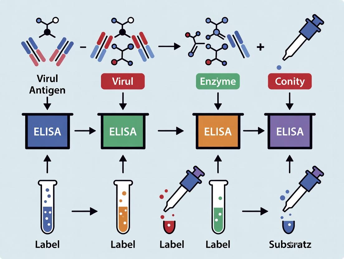
Fundamental Principles of Quantitative Antigen Capture ELISA
The quantification of viral antigens via sandwich ELISA is governed by several key principles:
Specificity through Immunosorbency: The assay relies on the high-affinity, specific binding of antibodies to target viral epitopes. A capture antibody, immobilized on a solid phase (typically a polystyrene microplate), selectively binds and retains the target antigen from a complex sample matrix.
Signal Amplification via Enzyme Conjugation: Detection is achieved through a second, enzyme-conjugated antibody that binds a different epitope on the captured antigen. This enzyme (e.g., Horseradish Peroxidase, Alkaline Phosphatase) catalyzes the conversion of a colorless substrate into a colored product, providing massive signal amplification from a single antigen molecule.
Quantification via Reference Standard Curve: The concentration of antigen in unknown samples is determined by interpolation from a standard curve. This curve is generated by assaying known, serially diluted concentrations of a purified viral antigen standard. The resulting optical density (OD) values establish the quantitative relationship between signal and antigen concentration.
Key Signaling Pathway & Workflow
The Scientist's Toolkit: Essential Research Reagent Solutions
| Reagent/Material | Function in Viral Antigen ELISA | Key Considerations |
|---|---|---|
| High-Binding Polystyrene Plate | Solid phase for passive adsorption of capture antibodies. | Optimal for proteins >10 kDa; ensures consistent binding capacity across wells. |
| Capture Antibody (Monoclonal) | Binds and immobilizes target viral antigen from sample. | Must be highly specific, affinity-purified, and bind a non-overlapping epitope from detection antibody. |
| Blocking Buffer (e.g., BSA, Casein) | Saturates non-specific protein-binding sites to reduce background noise. | Must be inert to assay components; choice affects sensitivity and specificity. |
| Purified Viral Antigen Standard | Provides known concentrations to generate the standard curve for quantification. | Must be identical to the target antigen; defines the assay's dynamic range and limit of detection (LOD). |
| Detection Antibody (Enzyme-Conjugated) | Binds captured antigen and provides enzymatic signal amplification. | Conjugation must not impair antibody affinity; enzyme choice (HRP/AP) dictates substrate options. |
| Chromogenic/TMB Substrate | Enzyme substrate that yields a measurable color change upon catalysis. | Stop solution required for HRP/TMB; kinetic vs. endpoint reading depends on substrate stability. |
| Microplate Spectrophotometer | Measures the optical density (absorbance) of the colored product in each well. | Must have appropriate filter/wavelength (e.g., 450 nm for TMB, 405 nm for pNPP). |
Detailed Protocol: Quantitative Sandwich ELISA for Viral Antigen
Coating
- Dilute the specific capture antibody in carbonate/bicarbonate coating buffer (pH 9.6) to a concentration of 2-10 µg/mL.
- Dispense 100 µL per well into a high-binding 96-well microplate.
- Seal plate and incubate overnight at 4°C (or 1-2 hours at 37°C).
Blocking
- Aspirate and wash plate 3x with 300 µL PBS containing 0.05% Tween-20 (PBST).
- Add 200 µL of blocking buffer (e.g., 3-5% BSA or 5% non-fat dry milk in PBS) to each well.
- Incubate for 1-2 hours at room temperature (RT) on a plate shaker.
Antigen Incubation
- Wash plate 3x with PBST.
- Prepare serial dilutions of the purified antigen standard in sample diluent (blocking buffer).
- Add 100 µL of standard, unknown samples, and blank (diluent only) to designated wells in duplicate/triplicate.
- Incubate for 2 hours at RT with shaking.
Detection Antibody Incubation
- Wash plate 3-5x with PBST thoroughly.
- Add 100 µL of the enzyme-conjugated detection antibody (optimally titrated concentration in blocking buffer) to each well.
- Incubate for 1-2 hours at RT with shaking.
Signal Development
- Wash plate 5x with PBST.
- Add 100 µL of substrate solution (e.g., TMB for HRP) to each well.
- Incubate in the dark for 10-30 minutes at RT. Monitor development.
- Stop the reaction by adding 50-100 µL of stop solution (e.g., 1M H₂SO₄ for TMB).
Data Acquisition & Analysis
- Read the Optical Density (OD) immediately at the appropriate wavelength (e.g., 450 nm for TMB).
- Subtract the average blank (background) OD from all standard and sample readings.
- Generate a standard curve by plotting the mean OD (y-axis) against the known standard concentration (x-axis) using a 4- or 5-parameter logistic (4PL/5PL) regression model.
- Interpolate the concentration of unknown samples from the standard curve.
Standard Curve & Performance Metrics
Table 1: Representative Standard Curve Data & Assay Performance
| Standard Concentration (pg/mL) | Mean OD (450 nm) | Standard Deviation | % CV |
|---|---|---|---|
| 0 (Blank) | 0.045 | 0.005 | 11.1 |
| 15.6 | 0.125 | 0.012 | 9.6 |
| 31.3 | 0.210 | 0.018 | 8.6 |
| 62.5 | 0.395 | 0.025 | 6.3 |
| 125 | 0.750 | 0.045 | 6.0 |
| 250 | 1.250 | 0.062 | 5.0 |
| 500 | 1.800 | 0.085 | 4.7 |
| 1000 | 2.100 | 0.095 | 4.5 |
Table 2: Calculated Assay Performance Metrics
| Metric | Calculation/Definition | Typical Target Value (Example) |
|---|---|---|
| Limit of Detection (LOD) | Mean blank OD + 3(SD blank) | ~5-10 pg/mL |
| Limit of Quantification (LOQ) | Mean blank OD + 10(SD blank) | ~15-20 pg/mL |
| Dynamic Range | Concentration between LOQ and upper asymptote | 15.6 - 1000 pg/mL |
| Assay Sensitivity | Slope of the linear portion of the standard curve | High (steep slope) |
| Inter-assay CV | Precision across multiple plates/runs | <15% (preferably <10%) |
| Intra-assay CV | Precision within a single plate | <10% (preferably <8%) |
| Coefficient of Determination (R²) | Goodness of fit for the standard curve | >0.99 |
Critical Protocol Considerations for Viral Targets
- Sample Matrix: Serum/plasma can cause non-specific interference. Use matched matrix for standard dilution or employ validated sample diluents.
- Hook Effect: At extremely high antigen concentrations, saturation can lead to falsely low signals. Samples should be run at multiple dilutions.
- Cross-Reactivity: Validate antibodies against related viral strains or common human proteins to ensure specificity.
- Temperature & Timing: Strict adherence to incubation times and temperatures is vital for reproducibility.
Application Notes
Within the context of a thesis on ELISA protocol for viral antigen detection, the selection and optimization of antibodies, plates, and enzymatic detection systems are critical for assay sensitivity, specificity, and reproducibility. This document provides current application notes and detailed protocols for researchers in virology and drug development.
1. Antibodies: The Foundation of Specificity The performance of a sandwich ELISA for viral antigen detection hinges on the capture and detection antibody pair. Monoclonal antibodies (mAbs) are preferred for their consistency and high specificity, reducing cross-reactivity with host proteins or other viral serotypes. Recent trends involve using recombinant antibodies for batch-to-batch consistency. The affinity constant (K_D) should ideally be <10 nM for high-sensitivity detection. For emerging viruses, neutralizing antibodies often serve as excellent detection reagents, linking detection to functional relevance.
2. Microplates: The Solid-Phase Substrate High-binding polystyrene plates (e.g., Nunc MaxiSorp) are standard. The binding capacity, typically 400-500 ng IgG/cm², directly impacts the standard curve's dynamic range. For antigens with hydrophobic epitopes or in complex matrices like serum, plates with modified polymer coatings can reduce non-specific binding (NSB). Recent studies show that plate geometry and well uniformity are crucial for automated high-throughput screening in drug discovery.
3. Enzymatic Detection Systems: Signal Amplification Horseradish peroxidase (HRP) and alkaline phosphatase (AP) remain the dominant enzymes. HRP, with its faster kinetics and higher specific activity, is favored for high-throughput assays. The choice of chromogenic (e.g., TMB, OPD) or chemiluminescent substrates (e.g., luminol-based) dictates sensitivity. Chemiluminescence can offer a 10- to 100-fold lower detection limit than chromogenic detection. Critical factors include enzyme-to-antibody ratio in conjugates and substrate stability.
Quantitative Comparison of Key ELISA Components
Table 1: Comparison of Common Enzymatic Detection Systems
| Component | Typical Enzyme | Common Substrates | Detection Limit (Typical) | Advantages | Disadvantages |
|---|---|---|---|---|---|
| Chromogenic | HRP | TMB, ABTS | 1-10 pg/well | Visible color change, simple instrumentation, cost-effective | Lower sensitivity than chemiluminescence |
| Chromogenic | AP | pNPP | 10-100 pg/well | Linear kinetics, stable signal | Slower than HRP, susceptible to phosphate inhibition |
| Chemiluminescent | HRP | Luminol + H₂O₂ enhancer | 0.1-1 pg/well | Very high sensitivity, wide dynamic range | Requires luminometer, signal can be transient |
| Chemiluminescent | AP | CDP-Star, CSPD | 0.1-1 pg/well | Stable, prolonged light emission | Slower kinetics than HRP, higher cost |
Table 2: Microplate Selection Guide for Viral Antigen ELISA
| Plate Type | Surface Chemistry | Binding Capacity (IgG) | Best For | Considerations for Viral Antigens |
|---|---|---|---|---|
| High-Binding | Polystyrene, hydrophobic | 400-500 ng/cm² | Most monoclonal/capture antibodies | Standard choice; optimal for hydrophobic proteins. |
| Medium-Binding | Polystyrene, slightly hydrophilic | 200-300 ng/cm² | Antigens prone to denaturation | Can help maintain antigen conformation. |
| Covalent/Linker | Activated (e.g., NHS) | Varies | Small peptides, fragmented antigens | Direct covalent linkage; orientation can be controlled. |
| Low-Binding | Polymer coating | Minimal | Samples with high NSB (e.g., serum) | Reduces background; may require high-affinity antibodies. |
Detailed Protocols
Protocol 1: Checkerboard Titration for Antibody Pair Optimization
Purpose: To determine the optimal concentrations of capture and detection antibodies for a sandwich ELISA targeting a viral nucleocapsid antigen. Reagents: See "The Scientist's Toolkit" below. Procedure:
- Coating: Prepare serial dilutions of the capture antibody (e.g., 10, 5, 2.5, 1.25 µg/mL) in carbonate-bicarbonate coating buffer (pH 9.6). Add 100 µL/well to a high-binding microplate. Incubate overnight at 4°C.
- Blocking: Aspirate and wash plate 3x with PBS + 0.05% Tween 20 (PBST). Add 300 µL/well of blocking buffer (1% BSA in PBS). Incubate for 2 hours at 37°C. Wash 3x with PBST.
- Antigen Incubation: Add 100 µL/well of a known positive control antigen (e.g., recombinant viral protein at 100 ng/mL in sample diluent) and negative control (diluent alone). Incubate 2 hours at 37°C. Wash 5x.
- Detection Antibody Titration: Prepare serial dilutions of the HRP-conjugated detection antibody (e.g., 1:2000, 1:4000, 1:8000, 1:16000) in blocking buffer. Add 100 µL/well in a cross-matrix pattern against the capture antibody concentrations. Incubate 1 hour at 37°C. Wash 5x.
- Signal Development: Add 100 µL/well of TMB substrate. Incubate in the dark for 10-15 minutes. Stop the reaction with 50 µL/well of 2M H₂SO₄.
- Analysis: Read absorbance at 450 nm. The optimal pair is the lowest concentration of each antibody that yields the highest signal-to-noise (positive/negative) ratio, typically >10.
Protocol 2: Chemiluminescent ELISA for High-Sensitivity Viral Titer Determination
Purpose: To quantify low-abundance viral surface antigen in cell culture supernatant with extended dynamic range. Reagents: See "The Scientist's Toolkit" below. Procedure:
- Coating & Blocking: Follow Protocol 1, steps 1-2.
- Sample & Standard Incubation: Prepare a standard curve of purified antigen in sample matrix (e.g., culture medium) across the expected range (e.g., 0.1 pg/mL to 10 ng/mL). Add 100 µL/well of standards and test samples. Incubate for 2 hours at room temperature with gentle shaking. Wash 5x.
- Detection Antibody Incubation: Add 100 µL/well of the optimal concentration of HRP-conjugated detection antibody. Incubate 1.5 hours at room temperature. Wash 7x thoroughly to minimize background.
- Chemiluminescent Development: Prepare luminol/peroxide/enhancer solution according to manufacturer instructions. Add 100 µL/well. Incubate for 2-5 minutes.
- Readout: Measure relative light units (RLU) immediately using a plate luminometer with an integration time of 100-500 ms/well.
- Data Analysis: Fit the standard curve using a 4- or 5-parameter logistic (4PL/5PL) model. Report sample concentrations from the linear range of the curve.
Diagrams
Title: Sandwich ELISA Workflow for Antigen Detection
Title: HRP-TMB Chromogenic Signal Generation
The Scientist's Toolkit: Essential Research Reagent Solutions
Table 3: Key Reagents for Viral Antigen ELISA Development
| Reagent/Material | Function & Critical Feature | Example/Notes |
|---|---|---|
| High-Affinity Capture Antibody | Binds target antigen with high specificity and immobilizes it to the plate. Monoclonal, virus-specific. | Recombinant mAb against SARS-CoV-2 nucleocapsid protein. |
| HRP-Conjugated Detection Antibody | Binds captured antigen at a distinct epitope; HRP enzyme catalyzes signal generation. Low non-specific binding conjugate. | Goat anti-virus spike protein IgG, HRP-linked. |
| High-Binding Microplates | Solid phase for antibody adsorption. Uniform well-to-well binding is critical. | Nunc MaxiSorp, polystyrene, flat-bottom. |
| Chromogenic Substrate (TMB) | HRP substrate yielding a soluble blue product upon oxidation, turns yellow when stopped. Sensitive, low background. | 3,3',5,5'-Tetramethylbenzidine, stabilized solution. |
| Chemiluminescent Substrate | HRP substrate yielding light emission upon oxidation. Offers highest sensitivity. | Luminol/enhancer/H2O2 solution. |
| Blocking Agent (BSA or Casein) | Coats uncovered plastic to prevent non-specific protein binding. Must not interfere with antibody-antigen binding. | Molecular biology grade Bovine Serum Albumin (BSA), protease-free. |
| Wash Buffer (PBST) | Removes unbound reagents; Tween-20 reduces non-specific binding. | Phosphate-Buffered Saline (PBS) with 0.05% Tween-20, pH 7.4. |
| Precision Pipettes & Tips | For accurate reagent transfer, especially for standard curve generation. | Calibrated single and multi-channel pipettes, low-retention tips. |
| Plate Reader | Measures absorbance (for chromogenic) or luminescence (for chemiluminescent) signal. | Multi-mode microplate reader with appropriate filters/luminometer. |
Within a thesis focused on developing and optimizing ELISA protocols for viral antigen detection, the selection of assay format is a foundational decision impacting sensitivity, specificity, and time-to-result. This application note details the core principles, comparative performance, and specific protocols for the four principal ELISA formats, enabling researchers to align their method with their virology research objectives.
Comparative Analysis of ELISA Formats
The following table summarizes the key quantitative and qualitative characteristics of each format, derived from current literature and reagent specifications.
Table 1: Comparison of Principal ELISA Formats for Viral Antigen Detection
| Feature | Direct ELISA | Indirect ELISA | Sandwich ELISA | Competitive ELISA |
|---|---|---|---|---|
| Key Principle | Antigen immobilized; detected directly with labeled primary antibody. | Antigen immobilized; detected with unlabeled primary, then labeled secondary antibody. | Antigen captured & detected between two matched antibodies. | Sample antigen competes with labeled reference antigen for limited antibody binding sites. |
| Typical Sensitivity | Low to Moderate (ng/mL range) | High (pg/mL - ng/mL) | Highest (pg/mL range) | Moderate (ng/mL range) |
| Specificity | Moderate (Depends on primary antibody only) | High (Amplification can increase background) | Very High (Requires two epitopes) | High (Competition format) |
| Steps & Time | Fewest steps; Fastest (~2-3 hrs) | Additional incubation; Moderate (~3-4 hrs) | Most steps; Longest (~4-5 hrs) | Moderate steps; Moderate (~3-4 hrs) |
| Signal Amplification | None | Yes (via secondary antibody) | Yes (via detection antibody system) | No (signal inversely proportional to analyte) |
| Primary Antibody Requirement | Must be conjugated/labeled | Can be unconjugated (more flexible) | Requires matched antibody pair | Specific for target antigen. |
| Best Suited For | High-abundance antigen screening, antibody conjugation validation. | General-purpose detection, especially with scarce primary antibodies. | Complex samples (serum, cell lysate), low-abundance antigens (e.g., viral coat proteins). | Detection of small antigens (haptens), or in samples with high antigenic similarity (viral variants). |
Detailed Experimental Protocols
Protocol 1: Sandwich ELISA for Viral Nucleocapsid Protein Detection
Objective: To quantify a specific viral nucleocapsid antigen in cell culture supernatant.
Materials: See "The Scientist's Toolkit" below. Procedure:
- Coating: Dilute capture antibody in 0.1 M carbonate-bicarbonate buffer (pH 9.6) to 2-10 µg/mL. Add 100 µL/well to a 96-well microplate. Seal and incubate overnight at 4°C.
- Blocking: Aspirate coating solution. Wash plate 3x with 300 µL/well PBS-T (0.05% Tween-20). Add 300 µL/well of blocking buffer (5% non-fat dry milk in PBS-T). Incubate for 1-2 hours at room temperature (RT). Wash 3x.
- Sample & Standard Incubation: Prepare serial dilutions of purified viral antigen standard in sample diluent (PBS-T with 1% BSA). Add 100 µL of standard or test sample per well in duplicate. Include blank wells (diluent only). Incubate for 2 hours at RT or 1 hour at 37°C. Wash 5x.
- Detection Antibody Incubation: Add 100 µL/well of biotinylated detection antibody (diluted in sample diluent per manufacturer's recommendation). Incubate for 1 hour at RT. Wash 5x.
- Enzyme Conjugate Incubation: Add 100 µL/well of Streptavidin-HRP conjugate (diluted in sample diluent). Incubate for 30-45 minutes at RT in the dark. Wash 7x thoroughly.
- Substrate Development: Add 100 µL/well of TMB substrate. Incubate for 5-15 minutes at RT in the dark until blue color develops.
- Stop & Read: Add 50 µL/well of 2M H₂SO₄ stop solution. Read absorbance immediately at 450 nm with a reference filter at 570/630 nm.
Protocol 2: Competitive ELISA for Detection of Cross-Reactive Viral Antigens
Objective: To measure serum antibodies against a specific viral strain in the presence of cross-reactive antibodies from related strains.
Procedure:
- Coating: Coat plate with purified viral antigen (1-5 µg/mL in coating buffer) overnight at 4°C.
- Blocking: Block as in Protocol 1.
- Competitive Reaction: Pre-mix a constant, limiting concentration of labeled (e.g., HRP-conjugated) specific monoclonal antibody with serial dilutions of the test serum sample (containing competing antibodies) for 1 hour at 37°C.
- Incubation: Transfer 100 µL of each mixture to the antigen-coated wells. Incubate for 1 hour at RT. The free antibody in the mixture binds to the immobilized antigen.
- Wash & Develop: Wash plate 5x to remove unbound antibody complexes. Add TMB substrate, incubate, and stop as in Protocol 1.
- Analysis: Higher sample antibody concentration leads to less labeled antibody bound, resulting in lower absorbance. Results are compared to a standard curve of a known inhibitor.
Visualization: ELISA Format Decision Pathway
Diagram Title: ELISA Format Selection Decision Tree
The Scientist's Toolkit: Essential Reagent Solutions
Table 2: Key Reagents for Viral Antigen ELISA Protocols
| Reagent/Material | Function & Rationale |
|---|---|
| High-Binding Polystyrene Microplate | Provides a hydrophobic surface for passive adsorption of proteins (antigens or antibodies). Critical for assay consistency. |
| Capture & Detection Antibody Pair | For sandwich ELISA, two antibodies binding non-overlapping epitopes ensure high specificity for the native viral antigen. |
| Recombinant Viral Antigen Standard | Purified antigen for generating a standard curve is essential for absolute quantification. Must match the native protein's conformation. |
| Biotin-Streptavidin System | Biotinylated detection antibody paired with Streptavidin-HRP enables significant signal amplification, boosting sensitivity. |
| HRP (Horseradish Peroxidase) Conjugate | Common enzyme label. Catalyzes colorimetric (e.g., TMB) or chemiluminescent substrate conversion for detection. |
| TMB (3,3',5,5'-Tetramethylbenzidine) Substrate | Chromogenic HRP substrate yielding a blue product measurable at 450 nm. Stable, sensitive, and safe. |
| Blocking Buffer (e.g., 5% BSA or Milk) | Saturates uncovered plastic surfaces to prevent non-specific binding of proteins, reducing background noise. |
| Wash Buffer (PBS with 0.05% Tween-20) | Removes unbound reagents. Tween-20 (a non-ionic detergent) reduces hydrophobic interactions and minimizes background. |
Within the broader thesis on developing and optimizing ELISA protocols for viral antigen detection, the success of the assay is fundamentally predicated on two interrelated factors: the intrinsic characteristics of the target antigen and the availability of its epitopes. The antigen's stability, conformation, and presentation modality directly dictate the choice of capture/detection antibodies and the conditions of the assay. This document provides detailed application notes and protocols to guide researchers in systematically evaluating these parameters to ensure robust, sensitive, and specific ELISA development.
Key Antigen Characteristics & Impact on ELISA Design
The following table summarizes critical antigen properties that must be characterized prior to assay development.
Table 1: Key Antigen Characteristics and Their Impact on ELISA Performance
| Characteristic | Description | Impact on ELISA Design | Typical Evaluation Method |
|---|---|---|---|
| Molecular Weight & Oligomeric State | Size and quaternary structure (monomer, dimer, trimer, etc.). | Determines pore size of solid phase, need for denaturation, and antibody accessibility. | SDS-PAGE, Native-PAGE, Size-Exclusion Chromatography. |
| Isoelectric Point (pI) | The pH at which the antigen has no net electrical charge. | Informs selection of coating buffer pH for optimal adsorption to plastic. | Isoelectric focusing, computational prediction. |
| Epitope Type | Linear (continuous amino acid sequence) or conformational (discontinuous, 3D structure). | Dictates whether native or denaturing conditions can be used; critical for antibody pair selection. | Western blot under reducing/non-reducing conditions, HDX-MS. |
| Glycosylation Status | Presence and extent of post-translational glycosylation. | Can mask epitopes; may require enzymatic deglycosylation for antibody access. | Lectin blot, PNGase F treatment, Mass Spectrometry. |
| Stability Profile | Sensitivity to pH, temperature, freeze-thaw cycles, and buffers. | Defines handling, storage, and assay incubation conditions to preserve native structure. | Differential Scanning Fluorimetry (DSF), Circular Dichroism (CD). |
| Source & Purity | Recombinant expression system (e.g., mammalian, insect, E. coli) and purification level. | Affects background noise, specificity, and the presence of confounding host cell proteins. | SDS-PAGE, Mass Spectrometry, Endotoxin assays. |
Protocols for Epitope Availability Assessment
A systematic evaluation of epitope availability is essential for selecting matched antibody pairs (for sandwich ELISA) or optimizing direct/competitive formats.
Protocol 3.1: Epitope Binning and Mapping via Bridging ELISA
Objective: To determine if two monoclonal antibodies (mAbs) bind to the same or different epitopes on the native antigen. Materials:
- Purified, native target antigen.
- Candidate monoclonal antibodies (capture and detection candidates).
- HRP-conjugated secondary antibody (species-specific).
- ELISA plates, coating buffer (e.g., Carbonate-Bicarbonate, pH 9.6), PBST, blocking buffer (e.g., 5% BSA in PBS), TMB substrate, stop solution.
Procedure:
- Coat ELISA plate with 100 µL/well of Antibody A (2-10 µg/mL in coating buffer). Incubate overnight at 4°C.
- Wash plate 3x with PBST. Block with 300 µL/well blocking buffer for 1-2 hours at RT.
- Wash 3x. Add a constant, saturating concentration of native antigen (in blocking buffer) to all wells. Incubate 1-2 hours at RT.
- Wash 3x. Add a titration of Antibody B (unlabeled) to wells. Include a well with no Antibody B as a maximum signal control. Incubate 1-2 hours at RT.
- Wash 3x. Add HRP-conjugated secondary antibody specific for the Fc region of Antibody B. Incubate 1 hour at RT.
- Wash 3x. Develop with TMB for 10-15 minutes. Stop with 1M H₂SO₄. Read absorbance at 450 nm.
- Interpretation: If Antibody B binds to a distinct, non-overlapping epitope from Antibody A, a strong signal will be generated (successful "bridge"). If Antibody B competes for the same or a sterically hindered epitope, signal will be low or absent.
Table 2: Example Bridging ELISA Results for Three mAbs
| Capture mAb | Detection mAb | Mean OD₄₅₀ (n=3) | % Signal vs. Control | Epitope Relationship Inference |
|---|---|---|---|---|
| mAb-1 | mAb-2 | 2.85 ± 0.12 | 95% | Different Epitope (Ideal Sandwich Pair) |
| mAb-1 | mAb-3 | 0.15 ± 0.05 | 5% | Same/Overlapping Epitope (Not suitable pair) |
| mAb-2 | mAb-3 | 2.70 ± 0.09 | 90% | Different Epitope (Ideal Sandwich Pair) |
Protocol 3.2: Evaluation of Epitope Accessibility Under Assay Conditions
Objective: To test if epitopes are accessible when antigen is immobilized on a plate or bound by a capture antibody. Materials: As in Protocol 3.1, plus chaotropic agents (e.g., urea, guanidine) or detergents if needed.
Procedure:
- Perform a standard sandwich ELISA setup using the intended capture antibody and antigen.
- In the detection step, compare signal generated by a panel of detection mAbs targeting different known regions of the antigen.
- Parallel Analysis: Run the same detection mAbs in a reverse format (coat with antigen directly) to compare epitope accessibility in solution-adsorbed vs. antibody-captured states.
- Variation: Pre-treat antigen with mild detergents (e.g., 0.1% Triton X-100) or reducing agents (e.g., 1-5 mM TCEP) before adding to the capture antibody. Compare signals to untreated antigen to assess the impact of partial denaturation on epitope availability.
- Interpretation: Identifies which detection mAbs are effective in the sandwich context and reveals if capture immobilization or mild denaturation enhances or reduces epitope exposure.
Visualization of Workflows and Relationships
Title: Antigen and Epitope Assessment Workflow for ELISA
Title: Factors Influencing Epitope Availability on an Antigen
The Scientist's Toolkit: Essential Research Reagent Solutions
Table 3: Key Reagents for Antigen and Epitope Characterization
| Reagent / Solution | Primary Function in Context | Key Consideration |
|---|---|---|
| High-Binding ELISA Plates (e.g., Polystyrene) | Passive adsorption of capture antibodies or antigens. | Lot-to-lot consistency is critical for assay reproducibility. |
| Cross-linking Buffers (e.g., DSS, BS³) | Stabilize protein-protein interactions; can fix antigen in a specific conformation. | Useful for studying transient interactions but may alter native structure. |
| Chaotropic Agents (Urea, Guanidine HCl) | Controlled denaturation of antigens to expose buried linear epitopes. | Concentration must be titrated to avoid complete, irreversible denaturation. |
| Glycosidase Enzymes (PNGase F, Endo H) | Remove N-linked glycans to assess impact of glycosylation on epitope masking. | Optimal activity requires specific buffer conditions (pH, temperature). |
| Reducing Agents (TCEP, DTT) | Break disulfide bonds to evaluate conformational vs. linear epitope dependence. | TCEP is more stable and does not require removal prior to labeling. |
| Epitope Mapping Peptide Libraries | Overlapping synthetic peptides spanning the antigen sequence. | Directly identifies linear epitopes; requires knowledge of full sequence. |
| Label-Free Biosensors (SPR, BLI) | Real-time analysis of antibody-antigen binding kinetics and epitope binning. | Provides quantitative data (KD, kon, koff) but requires specialized equipment. |
| Stabilization Cocktails | Preserve native antigen conformation during storage and assay steps. | Often proprietary; may contain polymers, salts, and non-specific proteins. |
Within the broader thesis focused on developing and optimizing ELISA protocols for viral antigen detection, adherence to biosafety guidelines is foundational. The accurate and safe detection of viral antigens from clinical or research samples is contingent upon the initial safe handling, inactivation, and processing of specimens. This document outlines the BSL classifications and provides specific application notes and protocols for handling viral samples prior to and during ELISA-based research.
Biosafety Levels are standardized protocols that define the containment principles, technologies, and practices required for working with biological agents. The level assigned is based on the agent's risk profile, including its transmissibility, pathogenicity, and available treatments.
Table 1: Summary of Biosafety Levels (BSLs) for Viral Agents
| BSL | Containment Level | Example Viral Agents | Primary Containment | Facility Requirements (Secondary Containment) |
|---|---|---|---|---|
| BSL-1 | Minimal Risk | Well-characterized agents not known to cause disease in healthy adults (e.g., Adeno-associated virus). | Standard microbiological practices. | Basic laboratory; no special containment. |
| BSL-2 | Moderate Risk | Agents associated with human disease of moderate hazard (e.g., Hepatitis B/C, HIV, Influenza, SARS-CoV-2*). | BSL-1 plus: PPE (lab coats, gloves, eye protection), biological safety cabinets (BSCs) for aerosol-generating procedures, biohazard signs, decontamination of waste. | Lab with self-closing doors, autoclave available, hands-free sink. |
| BSL-3 | High Risk | Indigenous or exotic agents with potential for aerosol transmission and serious/lethal disease (e.g., Mycobacterium tuberculosis, SARS-CoV, West Nile Virus, Rift Valley Fever virus). | BSL-2 plus: Respiratory protection, controlled lab access, all procedures performed in BSCs or other physical containment devices. | Physically separated corridor with double-door entry, directional airflow (inward), exhaust air not recirculated. |
| BSL-4 | Maximum Risk | Dangerous/exotic agents with high risk of life-threatening disease, aerosol transmission, and no available treatment/vaccine (e.g., Ebola, Marburg, Lassa fever viruses). | BSL-3 plus: Full-body, air-supplied positive pressure suit, mandatory shower-out, decontamination of all materials before exit. | Separate building or isolated zone, dedicated supply/exhaust, vacuum, and decontamination systems. |
Note: SARS-CoV-2 handling guidelines vary by country and research context (e.g., virus culture vs. inactivated clinical samples), often requiring BSL-2 with BSL-3 practices for propagation.
Application Notes for ELISA Research Workflow
For viral antigen detection ELISA, the sample journey from collection to plate must be managed within the appropriate BSL framework.
Sample Inactivation: A critical step for moving samples from higher containment (BSL-2/3) to lower containment (BSL-1/2) for downstream ELISA analysis. Common, validated methods include:
- Heat Inactivation: Incubation at 56°C for 30-60 minutes. Effectiveness varies by virus.
- Chemical Inactivation: Use of detergents (e.g., Triton X-100, NP-40) or chaotropic agents (e.g., Guanidinium thiocyanate) in lysis buffers. Must be validated to ensure antigen epitope integrity is preserved for antibody detection.
- Gamma Irradiation: Effective for complete pathogen inactivation while preserving protein structure.
Workflow Segmentation: The research workflow should be segmented by containment requirement:
- BSL-2/3 Area: Initial sample receipt, aliquoting, and primary inactivation.
- BSL-1/2 Area: Post-inactivation sample processing, plate coating, blocking, incubation with inactivated samples/antibodies, washing, and substrate development for ELISA.
Detailed Protocol: Inactivation of Enveloped Viral Samples for BSL-2 Downgrade Prior to ELISA
Objective: To safely inactivate enveloped viral samples (e.g., Influenza, SARS-CoV-2) using a detergent-based lysis buffer, enabling subsequent ELISA steps to be performed at BSL-1. Principle: Non-ionic detergents disrupt the viral lipid envelope and protein integrity, rendering the virus non-infectious while solubilizing viral antigens for detection.
Materials & Reagents (The Scientist's Toolkit): Table 2: Essential Research Reagent Solutions for Sample Inactivation and ELISA
| Item | Function in Protocol |
|---|---|
| Viral Transport Medium (VTM) | Preserves viral integrity during sample collection and transport. |
| Triton X-100 (1-2%) or NP-40 Lysis Buffer | Non-ionic detergent that disrupts viral membranes and inactivates enveloped viruses. |
| Protease Inhibitor Cocktail | Added to lysis buffer to prevent degradation of viral antigen proteins. |
| Phosphate-Buffered Saline (PBS) | Used for dilutions and as a buffer base. |
| Biosafety Cabinet (Class II) | Primary containment for handling uninactivated samples. |
| Personal Protective Equipment (PPE) | Lab coat, gloves, and safety goggles (face shield for splashes). |
| Nunc MaxiSorp ELISA Plates | High protein-binding plates for optimal coating of captured antibodies or viral antigens. |
| Blocking Buffer (e.g., 5% BSA/PBS) | Blocks non-specific binding sites on the ELISA plate. |
| Detection Antibodies (HRP-conjugated) | Enzyme-linked antibodies for specific antigen detection. |
| TMB Substrate Solution | Chromogenic substrate for HRP, produces measurable color change. |
| Stop Solution (e.g., 1M H₂SO₄) | Stops the HRP-TMB reaction at a defined endpoint. |
| Microplate Reader | Measures absorbance (450 nm for TMB) for data quantification. |
Methodology:
- Preparation: Perform all steps with uninactivated samples inside a certified Class II Biosafety Cabinet (BSC) in a BSL-2 laboratory. Wear appropriate PPE.
- Lysis/Inactivation: Combine 100 µL of viral sample (e.g., cell culture supernatant or VTM) with 100 µL of 2X Lysis Buffer (containing 2% Triton X-100 and 1X protease inhibitor in PBS). Mix thoroughly by vortexing.
- Incubation: Incubate the mixture at room temperature for 15-30 minutes. This duration is typically sufficient for complete inactivation of enveloped viruses.
- Validation: The inactivation protocol must be validated for each virus type. Validation involves attempting to culture the inactivated sample and confirming no cytopathic effect (CPE) occurs over 7-14 days.
- Post-Inactivation Handling: The lysed/inactivated sample can now be removed from the BSC. It may be clarified by centrifugation (10,000 x g, 5 min) if needed. The supernatant containing solubilized viral antigens can be used directly as analyte in a capture ELISA or aliquoted and stored at -80°C.
- Downstream ELISA: Proceed with standard ELISA protocol (coating, blocking, sample/antibody incubations, detection) at BSL-1 using the inactivated lysate.
Protocol: Indirect ELISA for Detecting Viral Antigen from Inactivated Samples
Objective: To detect and quantify a specific viral antigen within an inactivated sample lysate. Workflow Overview:
Title: Indirect ELISA Protocol Workflow for Viral Antigen Detection
Methodology:
- Coating: Dilute specific capture antibody in carbonate/bicarbonate coating buffer (pH 9.6). Add 100 µL/well to a MaxiSorp plate. Seal and incubate overnight at 4°C.
- Washing: Aspirate and wash plate 3x with 300 µL/well of PBS containing 0.05% Tween-20 (PBST).
- Blocking: Add 200 µL/well of blocking buffer (5% w/v BSA in PBST). Incubate 1-2 hours at room temperature (RT). Wash 3x with PBST.
- Sample Addition: Add 100 µL/well of inactivated viral sample lysate (and serial dilutions in sample diluent for a standard curve). Include appropriate negative controls. Incubate 1-2 hours at RT. Wash 3x.
- Primary Antibody: Add 100 µL/well of virus-specific, unlabeled primary detection antibody. Incubate 1 hour at RT. Wash 3x.
- Secondary Antibody: Add 100 µL/well of species-specific HRP-conjugated secondary antibody. Incubate 1 hour at RT in the dark. Wash 3x thoroughly.
- Detection: Add 100 µL/well of TMB substrate. Incubate in the dark at RT for 15-30 minutes until color develops.
- Stop & Read: Add 50 µL/well of 1M H₂SO₄ to stop the reaction. Immediately measure absorbance at 450 nm (with 570-620 nm reference) using a microplate reader.
Pathway: Biosafety Decision-Making for Sample Processing
The following logic diagram outlines the decision process for handling a sample in an ELISA research context.
Title: Biosafety Decision Logic for Viral Sample Processing
Step-by-Step Protocol: Optimized ELISA for Viral Antigen Quantification
Within the broader research thesis on ELISA for viral antigen detection, the pre-assay phase is critical for generating reliable, quantitative data. This Application Note details the foundational steps of sample preparation and standard curve design, which directly determine the accuracy, precision, and dynamic range of the assay. Failures in planning at this stage are a predominant source of error in diagnostic and drug development research.
Sample Preparation Protocols
Effective sample preparation ensures the target viral antigen is in an optimal state for detection while minimizing matrix interference.
Serum/Plasma Sample Handling
Detailed Protocol:
- Collection: Draw blood into collection tubes containing appropriate anticoagulants (e.g., K2EDTA for plasma). For serum, use clot-activated tubes.
- Separation: Allow serum tubes to clot for 30 minutes at room temperature (RT). Centrifuge both serum and plasma samples at 1,200-2,000 x g for 10 minutes at 4°C.
- Aliquoting: Immediately transfer the clear supernatant (serum/plasma) to fresh, pre-chilled polypropylene tubes. Avoid disturbing the buffy coat or pellet.
- Storage: Flash-freeze aliquots in liquid nitrogen or a dry-ice/ethanol bath. Store long-term at ≤ -80°C. Avoid repeated freeze-thaw cycles (max 2-3 cycles recommended).
Cell Culture Supernatant & Lysate Preparation
Detailed Protocol:
- Clarification: Centrifuge culture media at 300 x g for 5 minutes to remove cells. Transfer supernatant to a new tube.
- Concentration (Optional): For low-abundance antigens, use centrifugal concentrators (e.g., 10 kDa MWCO) per manufacturer's instructions to concentrate supernatant.
- Cell Lysis: For intracellular antigen detection, wash cell pellet twice with cold PBS. Resuspend in RIPA lysis buffer (with fresh protease inhibitors) at 1x10⁷ cells/mL. Incubate on ice for 30 minutes with vortexing every 10 minutes.
- Clarification: Centrifuge lysates at 16,000 x g for 15 minutes at 4°C. Collect supernatant (cleared lysate) for assay.
Key Considerations for Viral Antigens
- Inactivation: For pathogenic viruses, samples must be inactivated prior to handling (e.g., heat treatment at 56°C for 1 hour, or gamma irradiation) following BSL-2/3 guidelines.
- Stabilizers: Add protease and RNase inhibitors (for RNA virus antigens) immediately upon collection.
- Diluent: Use the sample matrix free of the target analyte (e.g., pooled negative control serum) or a validated commercial diluent to minimize matrix effects during assay dilution.
Standard Curve Design and Preparation
A well-characterized standard curve is non-negotiable for converting absorbance values into quantitative concentrations.
Selection and Reconstitution of the Standard
Detailed Protocol:
- Source: Use a purified, well-characterized viral antigen (e.g., recombinant spike protein). The standard should be immunologically identical to the target analyte.
- Reconstitution: Briefly centrifuge the vial. Reconstitute with the recommended buffer (often a protein-stabilizing PBS-based buffer with carrier protein like 1% BSA). Allow to equilibrate at RT for 10-15 minutes before gentle mixing.
- Stock Concentration: Calculate the stock concentration (C_stock) using the provided mass and volume, verifying with A280 absorbance if possible.
Serial Dilution Scheme
A minimum of 7 non-zero points across the expected dynamic range is essential. A log-based or semi-log dilution series is standard.
Detailed Protocol for 2-Fold Serial Dilution:
- Prepare a working dilution buffer matching the sample matrix (e.g., 1% BSA in PBS/0.05% Tween-20).
- Calculate the required top standard concentration (C_top), which should be at or above the assay's upper limit of quantification (ULOQ).
- Label 8 tubes (#1-8). Add an equal volume of dilution buffer to tubes #2-8 (e.g., 500 µL).
- Add 2x volume of C_top standard to tube #1 (e.g., 1000 µL).
- Perform a serial dilution: Transfer 500 µL from tube #1 to tube #2, mix thoroughly. Repeat from tube #2 to #3, and so on until tube #7. Discard 500 µL from tube #7 after mixing. Tube #8 is the zero standard (blank).
- Use freshly prepared dilutions immediately.
Figure 1: Workflow for 2-fold serial dilution of standard.
Quantitative Data and Acceptance Criteria
A robust standard curve is characterized by the following parameters.
Table 1: Standard Curve Performance Metrics and Acceptance Criteria
| Parameter | Ideal Value/Range | Typical Acceptance Criterion | Implication of Deviation |
|---|---|---|---|
| Linear Dynamic Range | 3-4 orders of magnitude | R² ≥ 0.99 for linear regression | Assay cannot quantify low/high samples accurately. |
| Lower Limit of Detection (LLOD) | As low as possible | Mean + 3SD of zero standard absorbance | Poor assay sensitivity. |
| Lower Limit of Quantification (LLOQ) | First point on curve | CV ≤ 20% at this concentration | Imprecision at low concentrations. |
| Upper Limit of Quantification (ULOQ) | Last point before plateau | CV ≤ 20% at this concentration | High-end hook effect or loss of precision. |
| Precision (Repeatability) | CV < 10% across mid-range | Intra-assay CV < 15% | Poor reproducibility. |
| Accuracy (% Recovery) | 80-120% | 70-130% at LLOQ/ULOQ; 80-120% else | Systematic bias in reported concentrations. |
| Calibrator Curve Fit | 4- or 5-Parameter Logistic (4PL/5PL) | R² ≥ 0.99 | Model mismatch leads to quantification errors. |
Integrated Pre-Assay Workflow
A systematic approach integrating sample and standard preparation is vital.
Figure 2: Integrated pre-assay planning workflow for ELISA.
The Scientist's Toolkit: Key Research Reagent Solutions
Table 2: Essential Materials for Sample & Standard Preparation
| Item | Function & Key Feature | Example/Consideration |
|---|---|---|
| Recombinant Viral Antigen Standard | Provides known analyte for calibration curve. Must be highly pure and characterized. | e.g., Recombinant SARS-CoV-2 Nucleocapsid protein, lyophilized, >95% purity. |
| Matrix-Matched Dilution Buffer | Dilutes standards and samples while mimicking sample composition to reduce matrix effects. | PBS with 1% BSA, 0.05% Tween-20, and 0.05% ProClin preservative. |
| Protease Inhibitor Cocktail | Preserves protein integrity in samples by inhibiting endogenous proteases. | Broad-spectrum, EDTA-free cocktail for serum/plasma and cell lysates. |
| Sterile, Low-Protein-Bind Tubes | Prevents loss of analyte via adsorption to tube walls during processing and storage. | Polypropylene, 0.5-2 mL, DNase/RNase-free. |
| Centrifugal Concentrators | Enables concentration of dilute samples (e.g., culture supernatant) to bring analyte within assay range. | 10 kDa molecular weight cut-off (MWCO), compatible with target antigen size. |
| CRP (C-Reactive Protein) or similar | Serves as an internal positive control for sample viability in inflammatory marker assays. | Purified human CRP for spiking into control samples. |
| Viral Inactivation Reagents | Ensures biosafety when handling infectious clinical samples. | Tri-reagent (for RNA/DNA/protein isolation) or beta-propiolactone. |
| Microplate Layout Template Software | Aids in randomizing sample and standard placement to minimize edge/position effects. | Tools like GraphPad Prism or dedicated ELISA analysis software. |
Within the broader thesis on ELISA protocol optimization for viral antigen detection research, the initial steps of plate coating and blocking are fundamental. These processes dictate the assay's ultimate sensitivity and specificity by maximizing the binding of the capture agent to the solid phase while minimizing non-specific interactions that cause background noise. This application note details current methodologies and data-driven strategies to achieve optimal performance in sandwich ELISA configurations for viral antigens.
Quantitative Data on Coating and Blocking Reagents
Table 1: Comparison of Common Coating Buffers for Viral Antigen Capture Antibody Immobilization
| Coating Buffer (pH) | Typical Coating Concentration (µg/mL) | Incubation Condition | Relative Binding Efficiency (%)* | Key Advantage | Key Disadvantage |
|---|---|---|---|---|---|
| Carbonate-Bicarbonate (pH 9.6) | 1-10 | Overnight, 4°C | 100 (Reference) | High passive adsorption efficiency for many antibodies. | Alkaline pH may denature some sensitive proteins. |
| PBS (pH 7.4) | 1-10 | Overnight, 4°C or 2h, 37°C | 75-90 | Physiological, gentle conditions. | Lower adsorption efficiency for some immunoglobulins. |
| Tris-HCl (pH 8.5) | 1-10 | Overnight, 4°C | 80-95 | Good buffering capacity at slightly alkaline pH. | Less commonly optimized than carbonate buffer. |
*Binding efficiency is normalized to the signal obtained with carbonate buffer under optimal conditions for a standard IgG.
Table 2: Efficacy of Common Blocking Agents in Reducing Background in Viral ELISA
| Blocking Agent | Typical Concentration & Incubation | % Background Reduction (vs. unblocked)* | Compatibility with Viral Targets/Biotin Systems | Potential Interference |
|---|---|---|---|---|
| BSA (Bovine Serum Albumin) | 1-5% in PBS, 1-2h, 37°C | 85-95% | High. Universal blocker. | May contain bovine IgG contaminants; can bind some lectins. |
| Casein | 1-3% in PBS, 1-2h, 37°C | 90-98% | Very High. Excellent for alkaline phosphatase (AP) systems. | Can form viscous solutions; variable purity. |
| Non-Fat Dry Milk | 1-5% in PBS, 1-2h, 37°C | 80-90% | Low cost. | Contains biotin and phosphoproteins; not for biotin-streptavidin systems. Can harbor proteases. |
| Fish Skin Gelatin | 0.5-1% in PBS, 1-2h, RT | 75-85% | Low cross-reactivity with mammalian samples. | Less effective for high-sensitivity applications. |
| Commercial Protein-Free Blockers | As per manufacturer | 90-99% | Excellent for biotin-streptavidin. Minimal cross-reactivity. | Can be expensive. |
*Representative data; actual reduction depends on sample matrix and detection system.
Detailed Experimental Protocols
Protocol 1: Optimal Plate Coating for Capture Antibody
Objective: To passively adsorb a virus-specific monoclonal antibody onto a 96-well polystyrene microplate.
Materials: See "The Scientist's Toolkit" below. Procedure:
- Antibody Dilution: Dilute the purified capture antibody in 0.05 M carbonate-bicarbonate coating buffer (pH 9.6) to a final concentration of 2 µg/mL. Note: A concentration range of 1-10 µg/mL should be empirically determined.
- Plate Coating: Dispense 100 µL of the antibody solution into each well of a high-binding polystyrene microplate. Seal the plate to prevent evaporation.
- Incubation: Incubate overnight (12-16 hours) at 4°C. Alternatively, incubate for 2 hours at 37°C, though 4°C overnight often yields more uniform coating.
- Washing: Aspirate the coating solution. Wash the plate three times with 300 µL of wash buffer (0.05% Tween 20 in PBS, PBS-T). Blot the plate firmly on clean paper towels after each wash to remove residual liquid.
Protocol 2: High-Efficiency Blocking for Low-Background Viral Detection
Objective: To saturate remaining protein-binding sites on the plate after coating.
Materials: See "The Scientist's Toolkit" below. Procedure:
- Blocking Solution Preparation: Prepare a 3% (w/v) solution of Bovine Serum Albumin (Fraction V, low IgG) in PBS-T. Alternatively, prepare a 1% (w/v) casein solution in PBS. Filter sterilize if necessary.
- Blocking: Immediately after the final wash from Protocol 1, dispense 200-300 µL of blocking solution into each well.
- Incubation: Incubate the plate for 1.5 to 2 hours at 37°C on a microplate shaker (gentle agitation).
- Preparation for Antigen Addition: Aspirate the blocking solution. Wash the plate twice with PBS-T. The plate is now ready for the addition of the sample/antigen. Note: Plates can be dried, sealed, and stored at 4°C for short-term use (up to 1 week) if kept desiccated.
Visualizations
The Scientist's Toolkit
Table 3: Essential Reagents and Materials for Coating and Blocking
| Item | Function & Rationale | Example/Note |
|---|---|---|
| High-Binding Polystyrene Microplates | Optimal surface chemistry for passive adsorption of proteins (IgG). | Costar 3369, Nunc MaxiSorp. |
| Carbonate-Bicarbonate Buffer (pH 9.6) | Alkaline pH increases hydrophobicity of protein, enhancing adsorption to plastic. | 0.05M or 0.1M. Prepare fresh or store at 4°C for ≤2 weeks. |
| PBS (Phosphate-Buffered Saline), pH 7.4 | Universal physiological buffer for dilution, washing, and some coating applications. | Contains NaCl, KCl, Na₂HPO₄, KH₂PO₄. |
| PBS-Tween (PBS-T) | Standard wash buffer. Tween 20 (non-ionic detergent) reduces non-specific binding. | Typical concentration: 0.05% (v/v) Tween 20. |
| Purified Capture Antibody | Virus-specific monoclonal or affinity-purified polyclonal antibody for antigen capture. | Must be protein A/G purified. Avoid antibody stabilizers (e.g., BSA, gelatin). |
| Bovine Serum Albumin (BSA), Fraction V | The most common blocking agent. Inert protein that occupies non-specific sites. | Use low-IgG, protease-free grade for critical assays. |
| Casein (Hammersten or Technical grade) | Highly effective blocking agent, especially for AP-based detection. | May require heating to dissolve. Commercial casein blockers are convenient. |
| Non-Fat Dry Milk | Cost-effective blocking agent for non-biotin systems. | Contains biotin and phosphoproteins; avoid in streptavidin systems. |
| Microplate Sealing Tape | Prevents evaporation and contamination during incubation steps. | Adhesive or heat-sealing films. |
| Microplate Washer or Manual Washer System | Ensures consistent and thorough washing, critical for low background. | Manual multichannel pipettes with reservoirs are acceptable. |
Within the broader context of developing a sensitive and specific ELISA protocol for viral antigen detection, the incubation steps involving primary and secondary antibodies are critical determinants of assay performance. Optimization of time, temperature, and concentration for these steps is paramount to maximize signal-to-noise ratio, ensure specificity, and achieve reliable quantitative results for research and drug development applications.
The Scientist's Toolkit: Key Research Reagent Solutions
| Reagent/Material | Function in ELISA |
|---|---|
| Coated Microplate (e.g., Polystyrene, 96-well) | Solid phase for antigen immobilization. |
| Viral Antigen (Purified or in sample matrix) | Target analyte for detection. |
| Blocking Buffer (e.g., BSA, Casein, Non-fat dry milk) | Saturates non-specific binding sites to reduce background. |
| Primary Antibody (Specific for target antigen) | Binds specifically to the captured antigen; defines specificity. |
| Secondary Antibody (Conjugated to HRP or AP) | Binds to Fc region of primary antibody; carries detection enzyme. |
| Wash Buffer (e.g., PBS or TBS with Tween-20) | Removes unbound antibodies and reagents, reducing background. |
| Colorimetric Substrate (e.g., TMB for HRP) | Enzyme substrate that produces a measurable color change. |
| Stop Solution (e.g., Sulfuric Acid) | Terminates the enzymatic reaction at a defined time. |
| Microplate Absorbance Reader | Quantifies the intensity of the colorimetric signal. |
Table 1: Typical Optimization Ranges for Antibody Incubation
| Parameter | Primary Antibody | Secondary Antibody | Notes |
|---|---|---|---|
| Concentration | 0.1 - 10 µg/mL | 0.01 - 1 µg/mL | Must be titrated against antigen load; high conc. can increase background. |
| Time | 1 - 2 hours (RT) or O/N (4°C) | 1 - 2 hours (RT) | Longer times (O/N) for primary can increase sensitivity but risk higher background. |
| Temperature | Room Temp (20-25°C) or 4°C | Room Temp (20-25°C) | 4°C for primary is preferred for labile antigens or O/N incubations. |
| Agitation | Gentle shaking (300-500 rpm) recommended | Gentle shaking (300-500 rpm) recommended | Improves kinetic binding, reduces incubation time, enhances uniformity. |
| Buffer | PBS/TBS with carrier protein (e.g., 0.1% BSA) | PBS/TBS with carrier protein (e.g., 0.1% BSA) | Carrier protein stabilizes antibodies and minimizes plate surface adsorption. |
Table 2: Impact of Incubation Conditions on Assay Metrics
| Condition Change | Typical Effect on Signal | Typical Effect on Background | Recommended Action for Optimization |
|---|---|---|---|
| Increased Primary [Ab] | Increases, then plateaus | Increases | Perform checkerboard titration vs. antigen to find optimal S/N ratio. |
| Increased Incubation Time | Increases, then plateaus | Slightly increases | Standardize time precisely; avoid over-incubation. |
| Increased Temperature | Increases kinetics | May increase | Use RT for consistency unless antigen is heat-labile. |
| Agitation vs. Static | Increases, speeds kinetics | Minimal effect | Implement gentle orbital shaking for all incubations. |
| Increased Secondary [Ab] | Increases, then plateaus sharply | Sharply increases | Titrate secondary Ab carefully; often optimal at manufacturer's suggestion. |
Detailed Experimental Protocols
Protocol 1: Checkerboard Titration for Primary and Secondary Antibody Optimization
Objective: To simultaneously determine the optimal pair of concentrations for primary and secondary antibodies that yield the highest signal-to-noise (S/N) ratio in a viral antigen detection ELISA.
Materials:
- Coated and blocked microplate with viral antigen and control wells.
- Primary antibody stock solution.
- Secondary antibody-enzyme conjugate stock solution.
- Assay buffer, wash buffer, substrate, and stop solution.
- Microplate reader.
Methodology:
- Plate Layout: Design a grid where columns represent serial dilutions of the primary antibody (e.g., 1:500 to 1:64,000) and rows represent serial dilutions of the secondary antibody (e.g., 1:1000 to 1:128,000). Include antigen-coated and blank (no antigen) wells for each condition.
- Primary Incubation: After blocking and washing, add the designated primary antibody dilutions to the plate. Incubate for 2 hours at room temperature with gentle agitation (500 rpm).
- Wash: Wash plate 5 times with wash buffer.
- Secondary Incubation: Add the designated secondary antibody dilutions to the appropriate wells. Incubate for 1 hour at room temperature with gentle agitation.
- Wash: Wash plate 5 times with wash buffer.
- Detection: Add substrate, incubate for a fixed time (e.g., 15 min), stop the reaction, and read absorbance.
- Analysis: For each well pair (antigen-coated vs. blank), calculate the S/N ratio (Absorbance sample / Absorbance blank). The combination yielding the highest S/N with adequate signal intensity is optimal.
Protocol 2: Time and Temperature Course for Primary Antibody Incubation
Objective: To assess the binding kinetics of the primary antibody at different temperatures to determine the most efficient incubation protocol.
Materials: As above, using predetermined optimal antibody concentrations.
Methodology:
- Plate Setup: Seed multiple identical antigen-coated and blank plates.
- Incubation: Add primary antibody to all plates. Incubate sets of plates at different temperatures (e.g., 4°C, Room Temperature, 37°C) for varying time points (e.g., 30 min, 1h, 2h, 4h, overnight).
- Processing: For each time point/temperature combination, process one plate through the standard protocol (wash, secondary Ab for 1h RT, detect).
- Analysis: Plot signal and background vs. time for each temperature. Identify the incubation condition that provides maximal specific signal (Signal - Background) within an acceptable assay timeframe.
Visualization of Workflows
Title: ELISA Antibody Incubation Optimization Workflow
Title: Troubleshooting Antibody Incubation Outcomes
Within the broader thesis on developing sensitive and reliable ELISA protocols for viral antigen detection, the selection and optimization of the substrate and detection system are critical. This Application Note details the principles, protocols, and comparative analysis of two dominant detection methodologies: spectrophotometric (colorimetric) and chemiluminescent. The choice between these methods directly impacts the assay's sensitivity, dynamic range, and suitability for high-throughput screening in diagnostic and drug development contexts.
Core Principles and Signaling Pathways
Spectrophotometric (Colorimetric) Detection
This method relies on the enzymatic conversion of a chromogenic substrate into a colored product. Horseradish Peroxidase (HRP) catalyzes the oxidation of substrates like TMB (3,3',5,5'-Tetramethylbenzidine) in the presence of hydrogen peroxide, producing a blue product that turns yellow upon acid stop. Alkaline Phosphatase (AP) dephosphorylates substrates like pNPP (p-Nitrophenyl Phosphate), yielding a yellow p-nitrophenol product. Signal intensity is proportional to the target antigen concentration and is measured as optical density (OD) at a specific wavelength.
Chemiluminescent Detection
Chemiluminescence involves the emission of light as a result of a chemical reaction. For HRP, substrates like luminol are oxidized in the presence of a peroxide buffer and a chemical enhancer (e.g., phenols), producing a sustained glow. For AP, substrates such as CDP-Star or CSPD are dioxetane phosphates that, upon dephosphorylation, decompose and emit light. The emitted photons are measured as Relative Light Units (RLUs) by a luminometer, offering a wider dynamic range and higher sensitivity than colorimetric methods.
Diagram 1: Key Enzyme-Substrate Pathways in ELISA Detection
Table 1: Comparative Analysis of Spectrophotometric vs. Chemiluminescent Detection Methods
| Parameter | Spectrophotometric (e.g., TMB/HRP) | Chemiluminescent (e.g., Luminol/HRP) |
|---|---|---|
| Detection Mechanism | Absorbance of colored product | Emission of photons |
| Readout | Optical Density (OD) | Relative Light Units (RLU) |
| Typical Sensitivity | Moderate (pg/mL range) | High (fg-pg/mL range) |
| Dynamic Range | Narrow (~2 logs) | Wide (>4-5 logs) |
| Signal Stability | Stable after stop solution | Transient (glow: minutes-hours; flash: seconds) |
| Instrumentation | Standard plate reader (450 nm) | Luminometer or capable plate reader |
| Throughput Speed | Fast (simultaneous reading) | Variable (sequential/fast injectors) |
| Cost per Test | Lower | Higher |
| Best For | Routine quantification, visual assessment | High-sensitivity applications, wide dynamic range needs |
Table 2: Common Substrate Systems for Viral Antigen ELISA
| Enzyme | Substrate Type | Example Product | Signal Measurement |
|---|---|---|---|
| Horseradish Peroxidase (HRP) | Chromogenic | TMB (Oxidized) | OD at 450 nm (acid stop) |
| Horseradish Peroxidase (HRP) | Chemiluminescent | Luminol + Peroxide + Enhancer | RLU (peak or integrated) |
| Alkaline Phosphatase (AP) | Chromogenic | pNPP | OD at 405 nm |
| Alkaline Phosphatase (AP) | Chemiluminescent | CDP-Star / CSPD | RLU (sustained glow) |
Detailed Experimental Protocols
Protocol 4.1: Spectrophotometric Detection using TMB for HRP
Application: Quantifying captured viral antigen in a sandwich ELISA format. Materials: See "The Scientist's Toolkit" below. Procedure:
- After completing the incubation with HRP-conjugated detection antibody and subsequent washing, prepare the TMB substrate solution. For commercial kits, equilibrate to room temperature. For lab-made solutions, mix component A (TMB in organic solvent) and component B (hydrogen peroxide in buffer) in equal volumes immediately before use.
- Add 100 µL of TMB substrate solution to each well of the microplate.
- Incubate the plate at room temperature, protected from light, for 10-20 minutes. Monitor blue color development visually or kinetically.
- To stop the reaction, add 50-100 µL of 1M H₂SO₄ or 2M H₃PO₄ stop solution to each well. The color will change from blue to yellow.
- Measure the absorbance (Optical Density) of each well within 30 minutes using a microplate reader fitted with a 450 nm filter. A reference wavelength of 620-650 nm may be used for background subtraction.
Protocol 4.2: Chemiluminescent Detection using Enhanced Luminol for HRP
Application: High-sensitivity detection of low-abundance viral antigens. Materials: See "The Scientist's Toolkit" below. Procedure:
- Following the final wash step after incubation with HRP-conjugated antibody, prepare the chemiluminescent substrate. Ensure the luminol/peroxide/enhancer working solution is prepared according to manufacturer instructions and is at room temperature.
- For optimal consistency, use a plate luminometer with injectors. If using manual addition, work quickly and consistently.
- Add 50-100 µL of substrate solution per well.
- If the substrate produces a "flash" signal (peak at seconds), read the plate immediately after a short, consistent incubation (e.g., 2 minutes). If it produces a "glow" signal (stable for minutes), incubate for the recommended time (e.g., 5 minutes) before reading.
- Read the plate in a luminometer, integrating the signal over a defined period (e.g., 100-500 ms/well). Record results in Relative Light Units (RLUs).
Diagram 2: ELISA Workflow with Detection Choice
The Scientist's Toolkit: Key Reagent Solutions
Table 3: Essential Materials for Substrate-Based Signal Detection
| Item | Function | Key Considerations |
|---|---|---|
| HRP-Conjugated Antibody | Binds specifically to target antigen; provides enzymatic activity for signal generation. | Titer for optimal signal-to-noise. Avoid sodium azide in storage buffers (inhibits HRP). |
| AP-Conjugated Antibody | Alternative enzyme conjugate for detection. | Requires different substrate and buffer (no EDTA/Tris which can inhibit AP). |
| TMB Substrate (Chromogenic for HRP) | Colorless substrate converted to blue chromogen by HRP/H₂O₂. | Commercial two-component (A+B) kits offer stability and consistency. Stop with acid. |
| pNPP Substrate (Chromogenic for AP) | Colorless substrate converted to yellow product by AP. | Supplied in diethanolamine or Tris buffer. Reaction stopped with NaOH. |
| Enhanced Chemiluminescent (ECL) Substrate (HRP) | Luminol/peroxide solution with enhancers (e.g., phenols) for sustained, amplified light output. | "Glow"-type substrates simplify high-throughput reading. Sensitive to light and temperature. |
| Dioxetane Substrate (AP) | Stable, phosphorylated dioxetane compound that emits light upon dephosphorylation by AP. | Very high sensitivity and long-lasting glow. Requires a compatible membrane or plate. |
| Microplate Washer | Removes unbound reagents between steps to reduce background. | Consistency in wash cycles and volumes is critical for assay precision. |
| Microplate Reader (Spectrophotometric) | Measures absorbance of colored products in each well. | Must have appropriate filter (e.g., 450 nm for acidified TMB). |
| Microplate Luminometer | Detects photon emission from chemiluminescent reactions. | Sensitivity, dynamic range, and injection capabilities are key selection criteria. |
| Stop Solution (Acid) | Stops HRP-TMB reaction, stabilizes color, and shifts absorbance maximum. | Typically 1-2M sulfuric or phosphoric acid. |
Introduction Within a thesis investigating ELISA protocol optimization for novel viral antigen detection, rigorous data analysis is the cornerstone of validating assay performance. This application note details the protocols and calculations for determining antigen concentration, assessing precision, and establishing key assay limits, forming the critical analytical framework for the broader research.
1. Quantitative Data Summary
Table 1: Representative Standard Curve Data for Recombinant Spike Protein (SARS-CoV-2)
| Standard Concentration (pg/mL) | Mean Absorbance (450 nm) | Standard Deviation (SD) | Coefficient of Variation (%CV) |
|---|---|---|---|
| 0 | 0.051 | 0.005 | 9.80 |
| 15.6 | 0.089 | 0.007 | 7.87 |
| 31.3 | 0.145 | 0.010 | 6.90 |
| 62.5 | 0.280 | 0.015 | 5.36 |
| 125 | 0.520 | 0.022 | 4.23 |
| 250 | 0.950 | 0.035 | 3.68 |
| 500 | 1.450 | 0.048 | 3.31 |
| 1000 | 1.900 | 0.055 | 2.89 |
Table 2: Intra- and Inter-Assay Precision Profile
| Sample (Spike Protein Conc.) | Intra-Assay Precision (n=10) | Inter-Assay Precision (n=3 assays) | ||
|---|---|---|---|---|
| Mean Conc. (pg/mL) | %CV | Mean Conc. (pg/mL) | %CV | |
| Low QC (85 pg/mL) | 82.4 | 6.2 | 84.1 | 8.5 |
| Mid QC (350 pg/mL) | 345.7 | 4.8 | 352.3 | 6.9 |
| High QC (750 pg/mL) | 738.9 | 3.5 | 761.2 | 5.1 |
Table 3: Calculated Assay Limits
| Parameter | Formula/Description | Calculated Value |
|---|---|---|
| Limit of Blank (LoB) | Meanblank + 1.645*(SDblank) | 0.059 OD |
| Limit of Detection (LoD) | LoB + 1.645*(SD_low concentration sample) | 0.072 OD (≈12 pg/mL) |
| Limit of Quantification (LoQ) | Concentration where %CV ≤ 20% (or accuracy 80-120%) | 25 pg/mL |
| Dynamic Range | From LoQ to upper asymptote of standard curve | 25 - 1000 pg/mL |
2. Experimental Protocols
2.1. Protocol: Standard Curve Generation and 4-PL Regression Objective: To generate a calibration model for interpolating unknown sample concentrations. Procedure:
- Prepare a dilution series of the purified viral antigen standard in sample diluent (e.g., 2-fold serial dilutions from 1000 pg/mL to blank).
- Analyze each standard in duplicate or triplicate alongside test samples using the validated ELISA protocol.
- Plot the mean absorbance (y-axis) against the known standard concentration (x-axis) using graphing software (e.g., GraphPad Prism, R).
- Fit the data to a 4-Parameter Logistic (4-PL) curve model:
y = d + (a - d) / (1 + (x/c)^b), wherea=minimum asymptote,d=maximum asymptote,c=inflection point (EC50),b=slope factor. - Validate the curve fit with an R² value >0.99.
- Interpolate unknown sample concentrations from the fitted curve.
2.2. Protocol: Determination of Precision (Intra- and Inter-Assay) Objective: To evaluate the repeatability and intermediate precision of the ELISA. Procedure for Intra-Assay Precision:
- Prepare three quality control (QC) samples (Low, Mid, High) spanning the assay range.
- Analyze each QC sample 10 times within a single assay run.
- Calculate the mean concentration, standard deviation (SD), and %CV for each QC level. Procedure for Inter-Assay Precision:
- Analyze the same three QC samples in triplicate across three independent assay runs performed on different days.
- Calculate the overall mean concentration, SD, and %CV for each QC level across all runs.
2.3. Protocol: Determination of LoD and LoQ Objective: To establish the lowest detectable and quantifiable levels of antigen. Procedure (Based on CLSI EP17-A2 Guidelines):
- LoB Measurement: Measure the zero standard (blank) at least 20 times. Calculate LoB = Meanblank + 1.645*SDblank.
- Low-Level Sample Analysis: Prepare a sample at a concentration expected to be near the LoD. Measure this sample at least 20 times independently.
- LoD Calculation: Calculate the SD of the low-level sample. LoD = LoB + 1.645*SD_low-level sample. Convert the resulting absorbance LoD to concentration via the standard curve.
- LoQ Determination: Analyze multiple samples with concentrations near the estimated LoD. The LoQ is the lowest concentration at which the total error (bias + 2*SD) meets predefined acceptability criteria (e.g., ≤20% CV and 80-120% recovery).
3. Diagrams
Title: ELISA Data Analysis Workflow for Assay Validation
Title: Sandwich ELISA Signal Generation Pathway
4. The Scientist's Toolkit: Research Reagent Solutions
Table 4: Essential Materials for ELISA Data Analysis
| Item | Function in Analysis Context |
|---|---|
| High-Purity Recombinant Antigen Standard | Provides the calibration curve for absolute quantification. Must be identical or immunologically similar to the target analyte. |
| Pre-coated ELISA Plates (96-well) | Solid phase for the immunoassay. Consistency in coating is critical for low inter-assay variability. |
| Precision Pipettes and Calibrated Tips | Ensure accurate and reproducible dispensing of standards and samples, directly impacting %CV. |
| Biotinylated Detection Antibody & HRP-Streptavidin | Key components of the signal amplification system. Lot-to-lot consistency is vital for stable assay sensitivity. |
| Stable Chromogenic Substrate (e.g., TMB) | Generates the measurable signal. Must have low background and consistent development kinetics for reliable OD readings. |
| Microplate Reader with 450nm Filter | Instrument for quantitative absorbance measurement. Regular calibration is required for data integrity. |
| Data Analysis Software (e.g., GraphPad Prism, SoftMax Pro, R) | Essential for performing 4-PL regression, calculating concentrations, and determining statistical parameters (LoD, LoQ, %CV). |
| Matrix-matched Sample Diluent / Assay Buffer | Minimizes matrix effects, ensuring accurate analyte recovery, especially in complex samples like serum. |
Solving Common ELISA Problems: A Troubleshooting Guide for Researchers
Within the context of optimizing an ELISA protocol for the detection of low-abundance viral antigens, managing background noise is a critical determinant of success. High background optical density (OD) readings compromise assay sensitivity, specificity, and the accurate determination of the limit of detection (LoD). This application note details the primary causes of elevated background in viral antigen ELISAs and provides strategic, actionable protocols for mitigation.
Causes and Quantitative Impact of High Background
High background noise in ELISA typically stems from non-specific binding (NSB) and assay interference. The following table summarizes common causes, their mechanisms, and their quantifiable impact on assay performance.
Table 1: Primary Causes of High Background Noise in Viral Antigen ELISA
| Cause Category | Specific Cause | Mechanism | Typical OD Increase vs. True Blank |
|---|---|---|---|
| Reagent-Based | Impure or Cross-Reactive Antibodies | Binds non-specifically to solid phase or sample components. | +0.15 to +0.35 |
| Enzyme Conjugate Polymerization | Forms aggregates with high enzymatic activity. | +0.10 to +0.25 | |
| Substrate Contamination/Oxidation | Spontaneous chromogen conversion. | +0.05 to +0.15 | |
| Assay Condition-Based | Inadequate Blocking | Leaves binding sites open on the plate. | +0.20 to +0.50 |
| Overly Stringent Wash Conditions | Disrupts antibody-antigen binding, increasing NSB. | Variable | |
| Insufficient Washing | Fails to remove unbound reagents. | +0.10 to +0.30 | |
| Sample/Matrix-Based | Heterophilic Antibodies (Human samples) | Bridge capture and detection antibodies. | +0.25 to >1.000 |
| Endogenous Enzymes (e.g., HRP in blood) | Directly catalyze substrate reaction. | +0.10 to +0.40 | |
| High Lipid or Protein Content | Increases viscous drag, impeding washing. | +0.08 to +0.20 |
Strategic Solutions: Detailed Protocols
Protocol 2.1: Systematic Troubleshooting for High Background
Objective: To identify the source of elevated background noise in a viral antigen ELISA. Materials: Coated ELISA plates, assay buffers, samples, detection system, plate reader. Procedure:
- Run an Extended Blank Series: Include the following wells in duplicate:
- True Blank: Coating Buffer only.
- Blocking Control: Coated, then blocked.
- Detection System Control: Coated, blocked, then all detection reagents (no sample).
- Sample Diluent Control: Coated, blocked, then sample diluent only.
- Conjugate-Only Control: Coated, blocked, then conjugate only.
- Substrate-Only Control: Substrate added to an untreated well.
- Incubate and develop according to the standard protocol.
- Analysis: Compare ODs. An elevated signal in the Detection System Control points to reagent issues. A high signal in the Sample Diluent Control indicates matrix interference. High Substrate-Only control suggests substrate instability.
Protocol 2.2: Optimization of Blocking and Washing to Minimize NSB
Objective: To establish optimal blocking conditions for a specific viral antigen-antibody pair. Materials: Coated plates, various blocking agents (BSA, Casein, Non-fat dry milk, commercial protein-free blockers), wash buffer (PBS/Tween-20). Procedure:
- Prepare 5 different blocking solutions: 1% BSA, 3% BSA, 1% Casein, 5% Non-fat dry milk, and a commercial protein-free blocker as per manufacturer's instruction.
- Block triplicate wells with each solution for 1 hour at 37°C or overnight at 4°C.
- Wash plates with a standardized wash buffer (0.05% Tween-20 in PBS) using an automated washer or consistent manual technique (3x washes, 300 µL/well).
- Proceed with the assay using a known negative sample and a low-positive control.
- Analysis: Calculate the Signal-to-Noise (S/N) ratio for the low-positive control against the negative for each blocker. Select the condition yielding the highest S/N and lowest negative control OD.
Protocol 2.3: Mitigation of Heterophilic Interference in Clinical Samples
Objective: To reduce false-positive signals caused by heterophilic antibodies in serum/plasma. Materials: Test samples, normal animal sera (e.g., mouse, goat), commercial heterophilic blocking tubes, sample diluent. Procedure:
- Pre-treatment: Split each sample into three aliquots.
- Aliquot A (Control): Dilute with standard diluent.
- Aliquot B (Animal Serum): Dilute with diluent containing 5-10% (v/v) normal serum from the same species as the detection antibody.
- Aliquot C (Commercial Blocker): Process according to commercial blocker tube instructions.
- Incubate all aliquots for 60 minutes at room temperature.
- Run the ELISA on all three sample preparations simultaneously.
- Analysis: A significant reduction (>30%) in OD for Aliquot B or C compared to Aliquot A confirms heterophilic interference. The effective blocker should be incorporated into the standard diluent.
Visualizing the Pathways to Background Noise
The Scientist's Toolkit: Key Reagents for Background Reduction
Table 2: Essential Reagents for Managing ELISA Background
| Reagent/Solution | Primary Function | Key Consideration for Viral Antigen ELISA |
|---|---|---|
| High-Purity, Virus-Specific Antibodies | Minimize cross-reactivity with host cell proteins or other non-target antigens. | Use monoclonal antibodies with confirmed specificity for the target viral epitope. |
| Protease-Free Blocking Agents (e.g., BSA, Casein) | Saturate uncovered binding sites on the microplate. | Casein often provides lower background than BSA for many viral systems; test empirically. |
| Commercial Heterophilic/Interference Blocking Reagents | Bind human anti-animal antibodies and other interfering factors in clinical samples. | Essential when testing human serum/plasma. More reliable than non-specific immunoglobulin addition. |
| Stable, Low-Background TMB Substrate | Provide sensitive chromogenic signal with low spontaneous conversion. | Use a ready-to-use, stabilized formulation containing H₂O₂ and TMB in an acidic buffer. |
| Wash Buffer with Optimal Detergent (e.g., 0.05% Tween-20) | Remove unbound reagents while maintaining specific interactions. | Concentration is critical; too high can denature antibodies, too low fails to reduce NSB. |
| Normal Sera from Detection Antibody Host Species | An alternative, cost-effective blocker for heterophilic interference. | Must be screened for absence of reactivity against the target virus or common human proteins. |
| Pre-Coated, Validated ELISA Plates | Provide consistent, high-binding capacity with low non-specific binding. | Saves time and reduces variability. Ensure the plate polymer is suitable for your target antigen size/charge. |
1. Introduction Within the broader thesis on ELISA protocol development for viral antigen detection, the optimization of antibody (Ab) concentrations and incubation parameters is critical. Suboptimal conditions are primary contributors to low signal or poor sensitivity, compromising assay robustness for research and diagnostic applications. This Application Note details systematic protocols for titer optimization and incubation step refinement to maximize the signal-to-noise ratio.
2. Key Optimization Parameters and Quantitative Data Summary Table 1: Summary of Key Optimization Variables and Their Impact on Assay Performance
| Parameter | Typical Test Range | Optimal Outcome | Impact on Sensitivity |
|---|---|---|---|
| Capture Ab Concentration | 0.5 - 10 µg/mL | Lowest conc. yielding max signal | Defines antigen-binding capacity |
| Detection Ab Concentration | 0.1 - 5 µg/mL | Lowest conc. yielding max signal | Directly influences final signal strength |
| Sample/Antigen Incubation Time | 60 - 180 min | Time yielding signal plateau | Ensures complete antigen binding |
| Detection Ab Incubation Time | 30 - 120 min | Time yielding signal plateau | Ensures sufficient Ab-antigen complex formation |
| Enzyme-Conjugate Incubation Time* | 15 - 60 min | Time yielding signal plateau | Determines enzyme loading for substrate conversion |
| *If using a tertiary (enzyme-labeled) reagent. For direct conjugates, this step is combined with Detection Ab incubation. |
3. Detailed Experimental Protocols
Protocol 3.1: Checkerboard Titration for Antibody Pair Optimization Objective: To empirically determine the optimal combination of capture and detection antibody concentrations. Materials: See "Research Reagent Solutions" table. Procedure:
- Coating: Prepare serial dilutions of the capture antibody (e.g., 10, 5, 2.5, 1.25 µg/mL) in coating buffer. Dispense 100 µL/well across the columns of a 96-well microplate. Include wells with coating buffer only as blanks. Seal and incubate overnight at 4°C.
- Blocking: Aspirate and wash plate 3x with Wash Buffer. Add 300 µL/well of Blocking Buffer. Incubate for 1-2 hours at room temperature (RT). Wash 3x.
- Antigen Addition: Add a fixed, moderate concentration of the target viral antigen (e.g., 100 µL/well of a mid-range standard dilution) in Assay Diluent to all wells. Incubate for 2 hours at RT. Wash 3-5x.
- Detection Ab Titration: Prepare serial dilutions of the detection antibody (e.g., 2, 1, 0.5, 0.25 µg/mL) in Assay Diluent. Add 100 µL/well, distributing different concentrations across the plate rows to create a matrix with all capture/detection Ab combinations. Incubate for 1-2 hours at RT. Wash 3-5x.
- Conjugate & Substrate: Add enzyme-conjugated secondary Ab (if needed) at manufacturer's recommended dilution. Incubate 1 hour at RT. Wash 3-5x. Add substrate (e.g., TMB) for development. Stop reaction after optimal development time.
- Analysis: Read absorbance. The optimal pair is the lowest combination of concentrations yielding the highest signal for the target antigen with a low background (blank) signal.
Protocol 3.2: Kinetic Incubation Time Course Experiment Objective: To determine the minimum incubation times required for signal saturation at each assay step. Materials: As in Protocol 3.1, using antibody concentrations identified as optimal. Procedure:
- Setup: Coat and block plate as per optimized protocol. Distribute antigen-positive and antigen-negative controls across the plate.
- Variable Incubation: For the step being optimized (e.g., antigen incubation), add reagents simultaneously to all rows. Place the plate on the shaker.
- Time-Point Sampling: At defined time points (e.g., 30, 60, 90, 120, 180 min), immediately aspirate and wash the wells of one entire row. Proceed immediately to the next step (blocking or adding next reagent) for that row only.
- Completion: Complete the remaining assay steps (detection Ab, conjugate, substrate) with fixed, previously optimized times for all rows.
- Analysis: Plot signal vs. incubation time. Select the minimum time at which the signal reaches a plateau for efficient workflow.
4. Visualizing the Optimization Workflow and Critical Pathways
Title: ELISA Sensitivity Optimization Decision Workflow
Title: Key ELISA Signal Generation Pathways
5. The Scientist's Toolkit: Research Reagent Solutions Table 2: Essential Materials for ELISA Optimization
| Reagent/Material | Function in Optimization | Key Considerations |
|---|---|---|
| High-Binding 96-Well Plates | Solid phase for protein (capture Ab) immobilization. | Polystyrene, clear, flat-bottom for consistent coating and optical reading. |
| Purified Capture Antibody | Binds target viral antigen with high specificity. | Monoclonal recommended for consistency; concentration is primary variable. |
| Purified Detection Antibody | Binds a distinct epitope on captured antigen. | Conjugated directly (e.g., HRP) or used with a secondary conjugate; concentration is key variable. |
| Recombinant Viral Antigen Standard | Provides known positive control for signal optimization. | Critical for determining limit of detection (LOD) and establishing standard curve. |
| Blocking Buffer (e.g., BSA, Casein) | Covers unsaturated binding sites to reduce nonspecific signal. | Must be protein-based and compatible with the antibody-antigen system. |
| Wash Buffer (PBS/Tween-20) | Removes unbound reagents, reducing background. | Typical Tween-20 concentration is 0.05-0.1%; critical for stringency. |
| Enzyme Substrate (TMB, pNPP) | Converted by enzyme to colored product for detection. | TMB/HRP is common; choice dictates stop solution (acid) and readout wavelength. |
| Microplate Spectrophotometer | Quantifies colorimetric signal (Absorbance). | Must be capable of reading at appropriate wavelength (e.g., 450 nm for TMB). |
Within the broader thesis on optimizing ELISA protocols for viral antigen detection, achieving high precision is critical for robust, reproducible research and downstream drug development. High inter- and intra-assay variability compromises data reliability, leading to false conclusions. This document outlines the primary sources of variability in quantitative ELISA and provides detailed application notes and protocols for their mitigation, focusing on both technical execution and reagent quality.
The following table summarizes common sources of variability and their typical impact on the Coefficient of Variation (CV%), based on current literature and manufacturer data.
Table 1: Primary Sources of ELISA Variability and Their Impact
| Source Category | Specific Source | Typical Impact on CV% | Potential Magnitude of Error |
|---|---|---|---|
| Reagent Quality | Lot-to-lot antibody variation | 15-25% increase | High (Can shift standard curve) |
| Reagent Quality | Substrate instability/contamination | 10-30% increase | Medium-High (Affects kinetics) |
| Technical | Inconsistent washing (manual) | 20-40% increase | Very High (Primary source of error) |
| Technical | Pipetting error (serial dilution) | 5-15% increase per step | Medium (Propagates through assay) |
| Technical | Incubation time/temp fluctuation | 10-20% increase | Medium |
| Plate & Read | Edge effects (evaporation) | 10-25% increase (edge vs. center) | High (Spatial bias) |
| Signal | Substrate development time | 5-10% variation per minute | Medium (Kinetic dependence) |
Detailed Experimental Protocols for Mitigation
Protocol 3.1: Standardized Plate Washing Procedure to Minimize Technical Variability
Objective: To eliminate inconsistencies in manual washing, the major contributor to poor precision. Materials: Coated ELISA plate, wash buffer (PBS + 0.05% Tween-20), automated microplate washer or manual washer reservoir, absorbent towels. Procedure:
- Aspiration: Using an automated washer or a manual multi-channel pipettor with a reservoir, aspirate liquid from all wells simultaneously. If manual, tilt the plate at a 45° angle. Place the tip at the side of the well, just above the liquid meniscus to avoid scratching the coating.
- Dispensing: Fill each well completely with wash buffer (typically 300 µL). Do not allow the dispensing tips to touch the well contents or walls to avoid cross-contamination.
- Soaking: Allow the plate to soak for 30 seconds on the benchtop to dissociate non-specifically bound material.
- Aspiration: Repeat step 1. Thoroughly tap the inverted plate on a stack of clean absorbent towels to remove residual droplets.
- Repetition: Repeat steps 2-4 for the total number of washes specified in the assay (typically 3-5 times).
- Final Removal: After the last wash and tapping, perform a final brief centrifugation of the plate (1 minute at 1000 x g) to collect all residual liquid at the bottom of the well for consistent removal.
Protocol 3.2: Serial Dilution for Calibration Curve with Precision
Objective: To generate an accurate standard curve with minimal propagation of pipetting error. Materials: Standard stock solution, assay diluent, low-retention microcentrifuge tubes, calibrated pipettes with fresh tips (preferably positive displacement for viscous samples). Procedure:
- Prepare a 1.5 mL working volume of each dilution in separate, labeled tubes. Do not perform serial dilutions in the plate or in a single tube.
- Calculate the required concentration for the highest standard. Prepare this High Standard by diluting the stock in assay diluent in a microcentrifuge tube. Mix thoroughly via gentle vortexing for 5 seconds, then briefly centrifuge.
- Prepare the next standard by taking the calculated volume from the High Standard tube and adding it to a fresh tube containing the calculated volume of diluent. This creates the second point in the curve.
- Repeat step 3 sequentially to create all required standard points. This "parallel dilution" method minimizes error propagation compared to a classic serial dilution.
- Once all standard points are prepared in their individual tubes, transfer the required volume (e.g., 100 µL) in replicate (e.g., n=3) from each tube to the assay plate.
Visualization of Workflows and Relationships
Diagram Title: Systematic Strategy to Minimize ELISA Variability
Diagram Title: Critical QC Steps in ELISA Protocol Flow
The Scientist's Toolkit: Key Research Reagent Solutions
Table 2: Essential Reagents and Tools for High-Precision ELISA
| Item | Function & Rationale | Recommendation for Precision |
|---|---|---|
| Monoclonal Capture Antibody (Master Batch) | Binds target antigen with high specificity. Lot-to-lot variation is a major variability source. | Purchase a large, single lot sufficient for entire thesis project. Aliquot and store at -80°C. |
| Stable Chemiluminescent Substrate | Generates light signal proportional to enzyme activity. Signal stability impacts read-time variability. | Use a commercially available, QC-tested luminol-based substrate with a long glow signal (>30 min). |
| Automated Microplate Washer | Removes unbound material consistently. Manual washing is the largest technical error source. | Utilize a calibrated washer with adjustable aspiration height, speed, and soak time. Validate wash efficiency. |
| Low-Protein Binding Plates | Minimizes non-specific adsorption of proteins (antigens, antibodies) to well surfaces. | Use plates certified for ELISA, with high well-to-well consistency (CV% < 10% by manufacturer). |
| Assay Diluent with Protein & Stabilizers | Matrix for standards and samples; prevents non-specific binding and stabilizes proteins. | Use a commercial diluent or optimize a homemade buffer (e.g., PBS with 1% BSA, 0.05% Proclin-300). |
| Precision Pipettes & Tips | Accurate liquid handling for serial dilutions and reagent addition. | Use calibrated, regularly serviced pipettes. Match tip type (low-retention for viscous samples). |
| Plate Sealer/Adhesive Film | Prevents evaporation during incubations, eliminating "edge effect" variability. | Use a high-quality, optically clear adhesive seal. Apply firmly without bubbles. |
| Reference Control Sample (Positive/Negative) | Monitors inter-assay precision and validates the entire assay run. | Include a validated, aliquoted control sample in triplicate on every plate. Track using a Levey-Jennings chart. |
Hook Effect and Prozone Phenomenon in Sandwich ELISA
Application Notes
Within a thesis focused on developing robust ELISA protocols for novel viral antigen detection, understanding the hook effect is critical for assay validation and ensuring accurate clinical and research data. The hook effect, or prozone phenomenon, is a high-dose anomaly in sandwich immunoassays where an excess of target analyte saturates both capture and detection antibodies, preventing the formation of the "sandwich" complex. This leads to a falsely low or negative signal, potentially resulting in missed diagnoses or inaccurate quantitative results. Recent studies underscore its relevance in high-concentration scenarios like systemic infections or certain biomarker monitoring.
Table 1: Characteristic Signal Response in Hook Effect-Prone Sandwich ELISA
| Analyte Concentration (pg/mL) | Expected OD (450 nm) | Observed OD (450 nm) with Hook Effect | Signal Deviation (%) |
|---|---|---|---|
| 10^2 | 0.15 | 0.14 | -6.7 |
| 10^3 | 0.48 | 0.47 | -2.1 |
| 10^4 | 1.20 | 1.22 | +1.7 |
| 10^5 | 2.50 | 2.55 | +2.0 |
| 10^6 | 3.00 | 1.80 | -40.0 |
| 10^7 | 3.20 | 0.95 | -70.3 |
Table 2: Key Factors Contributing to the Hook Effect
| Factor | High Risk Condition | Mitigation Strategy |
|---|---|---|
| Antibody Affinity | Low affinity (<10^8 M^-1) | Use high-affinity monoclonal pairs. |
| Antibody Concentration | Low coating/detection antibody concentration | Optimize antibody titration. |
| Incubation Time | Short antigen incubation | Extend incubation; kinetic measurements. |
| Sample Matrix | Undiluted patient serum | Implement mandatory serial dilution protocol. |
Protocols
Protocol 1: Identifying the Hook Effect in Viral Antigen ELISA
Objective: To detect and characterize the prozone phenomenon for a target viral antigen.
- Coating: Coat a 96-well plate with 100 µL/well of capture antibody (2 µg/mL in carbonate-bicarbonate buffer, pH 9.6). Incubate overnight at 4°C.
- Blocking: Wash 3x with PBS-0.05% Tween 20 (PBST). Add 200 µL/well of blocking buffer (1% BSA in PBS). Incubate 2 hours at 37°C. Wash 3x.
- Antigen Incubation: Prepare the viral antigen standard in a 12-point 10-fold serial dilution, spanning from 10^8 pg/mL down to 1 pg/mL, using sample diluent. Include a blank (diluent only). Add 100 µL/well of each dilution in duplicate. Incubate 2 hours at 37°C. Wash 5x.
- Detection Antibody: Add 100 µL/well of biotinylated detection antibody (optimized concentration, e.g., 1 µg/mL in diluent). Inculate 1.5 hours at 37°C. Wash 5x.
- Streptavidin-Enzyme Conjugate: Add 100 µL/well of Streptavidin-HRP (1:5000 dilution). Incubate 30 minutes at RT, protected from light. Wash 7x.
- Signal Development: Add 100 µL/well of TMB substrate. Incubate 15 minutes at RT.
- Stop and Read: Add 50 µL/well of 2M H2SO4. Measure absorbance immediately at 450 nm with 620 nm reference.
- Analysis: Plot log(concentration) vs. OD450. A non-sigmoidal curve or a decrease in signal at the highest concentrations indicates the hook effect.
Protocol 2: Mitigating the Hook Effect via Sample Serial Dilution
Objective: To obtain an accurate quantitative result from a sample suspected of causing a high-dose hook effect.
- Follow Protocol 1 steps 1-2 for plate preparation.
- Prepare a 1:10, 1:100, 1:1000, and 1:10,000 dilution of the test sample in the provided assay diluent.
- Alongside standard curve points, add 100 µL/well of each sample dilution in duplicate.
- Complete Protocol 1 from step 4 onward.
- Analysis: Calculate the apparent concentration from the linear range of the standard curve for each sample dilution. The valid result is from the dilution yielding the highest calculated concentration that is consistent across dilutions (after multiplying by the dilution factor).
Visualizations
Title: Mechanism of the Hook Effect in ELISA
Title: Workflow for Hook Effect Detection & Mitigation
The Scientist's Toolkit
Table 3: Essential Research Reagent Solutions for Hook Effect Analysis
| Item | Function & Relevance to Hook Effect Studies |
|---|---|
| High-Affinity Matched Antibody Pair (Capture/Detection) | Forms the core immunoassay. High affinity reduces risk, but optimal concentration is key to defining the assay's hook point. |
| Recombinant Viral Antigen Standard | Provides known high-concentration material to experimentally induce and characterize the hook effect for assay validation. |
| Streptavidin-HRP (Horseradish Peroxidase) Conjugate | Common detection amplifer. Must be titrated to avoid signal saturation independent of the hook effect. |
| TMB (3,3',5,5'-Tetramethylbenzidine) Substrate | Chromogenic HRP substrate. Linear range must be known to interpret high-dose signal decreases accurately. |
| Blocking Buffer (e.g., 1-5% BSA or Casein) | Reduces nonspecific binding. Inadequate blocking can mimic high background, obscuring the hook effect. |
| Wash Buffer (PBS with 0.05-0.1% Tween 20) | Critical for removing unbound antigen and detection antibody. Insufficient washing in high-dose wells can artifactually elevate signal. |
| Microplate Reader (450 nm filter) | Quantifies endpoint signal. Essential for generating the full dose-response curve to visualize the characteristic hook curve. |
| Sample Dilution Buffer (Protein-based, e.g., 0.1% BSA in PBS) | Matrix for creating serial dilutions of samples and standards. Must maintain antibody and antigen stability. |
Within the broader thesis on developing a robust ELISA protocol for detecting novel viral antigens, optimizing the concentrations of capture and detection antibodies is a critical, non-intuitive step. The checkerboard titration is the definitive experiment for this purpose, simultaneously determining the optimal pairing of reagent concentrations to maximize assay sensitivity and dynamic range while minimizing cost and background.
Protocol: Checkerboard Titration for Indirect and Sandwich ELISA
Objective: To empirically determine the optimal concentrations of antigen (for indirect ELISA) or capture antibody and detection antibody (for sandwich ELISA) for viral antigen detection.
Principle: A two-dimensional serial dilution of one reagent (e.g., capture antibody) is cross-titrated against a serial dilution of a second reagent (e.g., detection antibody) in a plate format. The resulting signal matrix identifies the combination that yields the highest signal-to-noise ratio (SNR).
Materials & Pre-Experiment Preparation
- Microplate: 96-well, high-binding polystyrene plate.
- Coating Buffer: 0.05 M carbonate-bicarbonate buffer, pH 9.6.
- Wash Buffer: PBS or Tris-buffered saline (TBS) with 0.05% Tween 20 (PBST/TBST).
- Blocking Buffer: 3-5% Bovine Serum Albumin (BSA) or non-fat dry milk in wash buffer.
- Antigen: Purified viral antigen of interest.
- Antibodies: Capture antibody (if sandwich ELISA), primary detection antibody (if indirect ELISA), and enzyme-conjugated secondary antibody.
- Detection Substrate: TMB (3,3’,5,5’-Tetramethylbenzidine) or other appropriate chromogenic/chemiluminescent substrate.
- Stop Solution: 1M or 2M sulfuric acid (for TMB).
- Plate Reader: Capable of measuring absorbance at 450 nm (for TMB).
The Scientist's Toolkit: Essential Research Reagent Solutions
| Reagent/Solution | Function in Checkerboard Titration |
|---|---|
| Carbonate-Bicarbonate Buffer (pH 9.6) | High-pH coating buffer promotes passive adsorption of proteins (antibodies or antigens) to the polystyrene plate. |
| PBST/TBST (0.05% Tween 20) | Wash buffer; Tween 20 is a non-ionic detergent that reduces non-specific binding by disrupting hydrophobic interactions. |
| Blocking Agent (BSA/Casein) | Occupies non-specific protein-binding sites on the plate after coating, minimizing background signal. |
| TMB Substrate | Chromogenic peroxidase substrate. Enzymatic conversion produces a soluble blue product measurable at 450nm. |
| High-Affinity Antibody Pair | Matched monoclonal or polyclonal antibodies targeting non-overlapping epitopes on the viral antigen. Critical for sandwich assay specificity. |
Detailed Methodology
Part A: Checkerboard Titration for Sandwich ELISA (Viral Antigen Detection)
Plate Coating:
- Prepare a 2-fold serial dilution of the capture antibody in coating buffer across a range (e.g., 10 µg/mL to 0.08 µg/mL). Use eight concentrations.
- Add 100 µL of each dilution to the wells of a 96-well plate by column (e.g., Column 1: highest conc., Column 8: lowest conc.). Perform in duplicate or triplicate.
- Seal and incubate overnight at 4°C.
Washing and Blocking:
- Aspirate coating solution. Wash plate 3x with 300 µL wash buffer per well.
- Add 300 µL of blocking buffer to each well. Incubate for 1-2 hours at room temperature (RT).
- Wash plate 3x as before.
Antigen Addition:
- Add a fixed, predetermined concentration of the viral antigen (e.g., a mid-range concentration from a prior experiment) in 100 µL of blocking buffer to all wells. Incubate 2 hours at RT.
- Wash plate 3x.
Detection Antibody Titration:
- Prepare a 2-fold serial dilution of the detection antibody (conjugated or unconjugated) in blocking buffer across a similar range (e.g., 5 µg/mL to 0.04 µg/mL). Use eight concentrations.
- Add 100 µL of each dilution to the plate by row (e.g., Row A: highest conc., Row H: lowest conc.).
- Incubate for 1-2 hours at RT. Wash 3x.
- If using an unconjugated detection antibody: Add a fixed, optimal concentration of enzyme-conjugated secondary antibody. Incubate 1 hour. Wash 3x.
Signal Detection & Analysis:
- Add 100 µL of TMB substrate. Incubate in the dark for 5-15 minutes.
- Stop the reaction with 100 µL of stop solution.
- Read absorbance at 450 nm immediately.
Part B: Checkerboard Titration for Indirect ELISA (Antibody Characterization)
- Antigen Coating: Dilute viral antigen as per Part A, Step 1, coating by column.
- Blocking: As per Part A, Step 2.
- Primary Antibody Titration: Dilute test antiserum or primary antibody as per Part A, Step 4, adding by row.
- Secondary Antibody: Add a fixed, optimal concentration of enzyme-conjugated secondary antibody.
- Detection: As per Part A, Step 5.
Data Presentation and Analysis
The optimal combination is not merely the highest signal but the lowest concentration of both antibodies that yields a signal near the plateau of the maximum OD, providing a high SNR and cost-efficiency.
Table 1: Representative Checkerboard Titration Data (Absorbance at 450nm) Detection Antibody Concentration (µg/mL) vs. Capture Antibody Concentration (µg/mL)
| Det Ab / Cap Ab | 2.0 | 1.0 | 0.5 | 0.25 | 0.125 | 0.0625 | 0.031 | Blank |
|---|---|---|---|---|---|---|---|---|
| 1.0 | 3.200 | 2.980 | 2.501 | 1.880 | 1.210 | 0.650 | 0.301 | 0.055 |
| 0.5 | 2.950 | 2.850 | 2.450 | 1.920 | 1.300 | 0.720 | 0.350 | 0.052 |
| 0.25 | 2.501 | 2.400 | 2.200 | 1.801 | 1.250 | 0.680 | 0.320 | 0.050 |
| 0.125 | 1.900 | 1.850 | 1.750 | 1.501 | 1.100 | 0.601 | 0.280 | 0.049 |
| 0.0625 | 1.300 | 1.250 | 1.200 | 1.050 | 0.800 | 0.450 | 0.210 | 0.048 |
| 0.031 | 0.750 | 0.720 | 0.700 | 0.620 | 0.480 | 0.290 | 0.150 | 0.047 |
| 0.0156 | 0.400 | 0.390 | 0.380 | 0.350 | 0.280 | 0.180 | 0.100 | 0.046 |
| 0.0 (Blank) | 0.055 | 0.053 | 0.051 | 0.050 | 0.049 | 0.048 | 0.047 | 0.045 |
Interpretation: In this example, the combination of 0.5 µg/mL Capture Antibody and 0.5 µg/mL Detection Antibody (OD~2.45) may be selected over 2.0/1.0 µg/mL (OD~3.20), as it uses 75% less antibody for ~76% of the maximal signal, representing a more efficient operational point.
Visualization: Experimental Workflow
Diagram Title: Checkerboard Titration ELISA Workflow and Plate Layout
Ensuring Reliability: ELISA Validation and Comparison to Alternative Methods
Thesis Context: This document details the critical validation parameters for a sandwich ELISA protocol developed for the detection of a novel viral antigen (e.g., SARS-CoV-2 nucleocapsid protein) in clinical serum samples, as part of a broader thesis on immunodiagnostic assay development.
Specificity
Definition: The ability of the assay to measure solely the analyte of interest in the presence of other potentially cross-reactive components.
Application Note: For viral antigen detection, specificity is paramount to avoid false positives from host proteins, related viral strains, or matrix interferents.
Experimental Protocol: Cross-Reactivity Assessment
- Preparation: Coat microplate with capture antibody (1 µg/mL in carbonate buffer, 100 µL/well, overnight at 4°C).
- Blocking: Block with 5% BSA in PBS-T (200 µL/well, 2 hours, 37°C).
- Sample Incubation: Add the following to separate wells (in triplicate, 100 µL/well, 2 hours, 37°C):
- Target Analyte: Recombinant viral antigen at 2x the expected positive cutoff.
- Potential Interferents: Structurally similar antigens from related viruses, common human serum proteins (e.g., albumin, immunoglobulins), and sample collection additives (e.g., heparin, EDTA). Use at physiologically high concentrations (e.g., 100 µg/mL).
- Negative Control: Assay buffer only.
- Detection: Proceed with standard ELISA steps (detection antibody, enzyme conjugate, substrate).
- Analysis: Calculate signal relative to the negative control. A signal increase of <5% for interferents compared to the target analyte signal indicates acceptable specificity.
Key Research Reagent Solutions
| Reagent/Solution | Function in Specificity Testing |
|---|---|
| Cross-Reactive Antigens (e.g., from related coronaviruses) | Assess antibody pair uniqueness and potential for false positives. |
| High-Purity Human Serum Albumin & Immunoglobulins | Test for non-specific binding in the sample matrix. |
| Heterophilic Blocking Reagent | Minimize interference from human anti-mouse antibodies (if using murine antibodies). |
| Defined Negative Serum Pool | Establish a true biological baseline for specificity calculations. |
Sensitivity
Definition: The lowest concentration of analyte that can be consistently distinguished from zero (the blank). Often defined by the Limit of Blank (LoB).
Application Note: Determines the assay's ability to detect early or low-level viral infections.
Experimental Protocol: Determination of Limit of Blank (LoB)
- Sample Analysis: Run at least 20 independent replicates of a blank sample (matrix without the target analyte, e.g., confirmed negative serum).
- Calculation: LoB = Mean(blank) + 1.645 * SD(blank). Assumes a 5% error rate (95% confidence) where results above this are not due to blank variability.
Limit of Detection (LOD)
Definition: The lowest concentration of analyte that can be reliably detected, but not necessarily quantified, with stated probability (typically 95%).
Application Note: Defines the clinical detection threshold for the viral antigen.
Experimental Protocol: Determination of LOD
- Preparation: Prepare a sample spiked with analyte at a concentration near the expected LoB/LOD (e.g., 5-10 replicates).
- Analysis: Measure each replicate in the assay.
- Calculation: LOD = LoB + 1.645 * SD(Low Concentration Sample). Alternatively, LOD = Mean(blank) + 3 * SD(blank) is a common approximation.
Limit of Quantification (LOQ)
Definition: The lowest concentration of analyte that can be quantified with acceptable precision (typically ≤20% CV) and accuracy (typically 80-120% recovery).
Application Note: Establishes the threshold for reliable quantitative measurement of viral load, crucial for monitoring disease progression.
Experimental Protocol: Determination of LOQ
- Preparation: Prepare at least 5 samples spiked with analyte at a low concentration expected to be near the LOQ. Include calibration standards.
- Analysis: Run each sample in a minimum of 5 replicates across multiple assay runs (different days, analysts).
- Calculation: LOQ is the lowest concentration where both criteria are met:
- Precision: Coefficient of Variation (CV) ≤ 20%.
- Accuracy: Mean measured concentration is within 80-120% of the nominal (spiked) concentration.
Linearity
Definition: The ability of the assay to obtain results that are directly proportional to the concentration of the analyte within a given range.
Application Note: Defines the working dynamic range for quantifying viral antigen concentrations.
Experimental Protocol: Linearity and Range Assessment
- Preparation: Create a dilution series of the analyte in the sample matrix (e.g., serum) to span the entire expected range (e.g., from LOQ to maximum expected concentration). Use 5-7 concentration levels.
- Analysis: Assay each concentration in triplicate.
- Analysis: Plot measured concentration (or signal) vs. expected concentration. Perform linear regression analysis (y = mx + c). Evaluate the coefficient of determination (R²). Acceptance criterion is typically R² ≥ 0.990.
Table 1: Summary of Quantitative Validation Parameters for Viral Antigen ELISA
| Parameter | Symbol | Experimental Value (Example) | Acceptance Criterion |
|---|---|---|---|
| Limit of Blank | LoB | 0.08 ng/mL | Calculated statistically. |
| Limit of Detection | LOD | 0.12 ng/mL | > LoB; Detected in 19/20 replicates. |
| Limit of Quantification | LOQ | 0.25 ng/mL | CV ≤ 20%; Accuracy 85-115%. |
| Linearity Range | - | 0.25 - 50 ng/mL | R² ≥ 0.990. |
| Specificity | - | <1% cross-reactivity | Signal <5% vs. target for all interferents tested. |
Specificity: Antigen-Antibody Binding Specificity in Sandwich ELISA
Hierarchical Determination of Assay Sensitivity Metrics
Experimental Workflow for Determining LoB, LOD, and LOQ
Assessing Cross-Reactivity with Related Viral Strains or Host Proteins
Application Notes
In the context of developing and validating ELISA protocols for viral antigen detection, assessing cross-reactivity is a critical step to ensure assay specificity. Cross-reactivity can occur with antigens from related viral strains (e.g., different SARS-CoV-2 variants, influenza subtypes) or with proteins present in the host sample matrix, leading to false-positive results and compromising diagnostic or research conclusions.
Recent studies emphasize the need for systematic cross-reactivity panels. For instance, a 2023 evaluation of a SARS-CoV-2 nucleocapsid (N) protein ELISA demonstrated significant cross-reactivity with sera from patients infected with endemic human coronaviruses (HCoV-OC43, HCoV-229E) at rates of 15-22%, underscoring the necessity of using conserved region-depleted antigens or competitive assay formats. Similarly, assays for dengue virus NS1 antigen must be validated against other flaviviruses like Zika, where cross-reactivity rates can exceed 30% in polyclonal antibody-based assays.
Quantitative data from recent cross-reactivity assessments are summarized below:
Table 1: Representative Cross-Reactivity Data in Viral Antigen ELISAs
| Target Antigen | Potential Cross-Reactant | Assay Type | % Cross-Reactivity | Key Mitigation Strategy |
|---|---|---|---|---|
| SARS-CoV-2 N Protein | HCoV-OC43 N Protein | Indirect IgG ELISA | 22.4% | Use of variant-specific monoclonal antibody pairs |
| Dengue Virus NS1 | Zika Virus NS1 | Sandwich ELISA | 31.7% | Epitope mapping and chimeric antibody engineering |
| Influenza A H1N1 HA | Influenza A H3N2 HA | Competitive ELISA | 8.5% | Absorption with heterologous HA protein |
| HIV-1 gp41 | Human Autoantibodies (e.g., anti-nuclear) | Rapid Diagnostic Test | 12.1% | Sample pre-treatment with blocking reagents |
Experimental Protocols
Protocol 1: Cross-Reactivity Panel Testing for Sandwich ELISA Objective: To determine the specificity of a monoclonal antibody (mAb) pair used in a viral antigen capture ELISA. Materials: Coating mAb (1C2), detection mAb-biotin (5F11), target purified antigen (Virus A), related viral strain antigens (Virus B, C, D), recombinant host proteins (e.g., ACE2, serum albumin), streptavidin-HRP, TMB substrate, wash buffer, plate reader. Procedure:
- Coat high-binding 96-well plate with 100 µL/well of capture mAb 1C2 (2 µg/mL in PBS). Incubate overnight at 4°C.
- Wash plate 3x with PBS + 0.05% Tween-20 (PBST). Block with 200 µL/well of 3% BSA in PBS for 2 hours at 37°C.
- Wash 3x. Add 100 µL/well of serial dilutions (0-100 ng/mL) of the following antigens in separate wells: Target Virus A, Related Viruses B, C, D, and host proteins. Incubate 1 hour at 37°C.
- Wash 5x. Add 100 µL/well of detection mAb 5F11-biotin (1 µg/mL in dilution buffer). Incubate 1 hour at 37°C.
- Wash 5x. Add 100 µL/well of streptavidin-HRP (1:5000 dilution). Incubate 30 mins at RT, protected from light.
- Wash 7x. Add 100 µL TMB substrate. Incubate 15 mins. Stop with 50 µL 2M H₂SO₄.
- Read absorbance at 450 nm. Calculate cross-reactivity % as (OD of cross-reactant at EC₅₀ of target / OD of target at its EC₅₀) * 100.
Protocol 2: Competitive Inhibition Assay for Epitope Specificity Objective: To confirm whether cross-reactive signals share an identical epitope. Materials: As in Protocol 1, plus purified, unlabeled competitor antibodies or soluble recombinant proteins. Procedure:
- Perform steps 1-3 from Protocol 1, using a single concentration of target antigen (~EC₈₀) and the highest-concentration cross-reactant.
- Pre-incubate the detection antibody with a 10-fold molar excess of potential competitor (unrelated mAb, homologous antigen from other strain) for 30 mins before adding to the plate in step 4.
- Complete steps 4-7. A >50% reduction in signal for both target and cross-reactant indicates shared epitope recognition.
Mandatory Visualizations
Title: Sources and Consequences of ELISA Cross-Reactivity
Title: Workflow for Cross-Reactivity Panel Testing
The Scientist's Toolkit
Table 2: Essential Research Reagent Solutions for Cross-Reactivity Assessment
| Item | Function in Cross-Reactivity Assessment |
|---|---|
| Recombinant Viral Antigens (Multiple Strains) | Serve as the primary panel of potential cross-reactants for specificity testing. |
| Monoclonal Antibody Pairs (Variant-Specific) | Provide high specificity; preferred over polyclonals to minimize non-specific binding. |
| Host Protein Lysates (e.g., Lung, Serum) | Identify non-specific binding to proteins in the sample matrix. |
| Competitive Inhibitors (Soluble Antigens/Antibodies) | Determine if signal is epitope-specific through competitive assays. |
| Heterologous Blocking Agents (e.g., Animal Sera) | Reduce background and non-specific interactions in complex samples. |
| High-Stringency Wash Buffer (e.g., with High Salt/Detergent) | Dissociate low-affinity, cross-reactive binding during plate washing steps. |
| Cross-Adsorbed Secondary Antibodies | Secondary antibodies pre-adsorbed against host proteins to minimize background. |
| Reference Standards (WHO International Standards) | Calibrate assays and allow comparison of cross-reactivity data across labs. |
Application Notes
The selection between Enzyme-Linked Immunosorbent Assay (ELISA) and Lateral Flow Assays (LFAs) is a pivotal decision in viral antigen detection research. This choice fundamentally hinges on the trade-off between analytical sensitivity and quantification versus rapid, point-of-care applicability. Within a thesis focused on developing a high-sensitivity ELISA protocol for novel viral antigen detection, LFAs represent a complementary, rapid screening tool. ELISA remains the gold standard for generating quantitative, high-fidelity data for kinetics, epitope mapping, and vaccine immunogenicity studies. In contrast, LFAs are indispensable for field deployment, rapid patient triage, and scenarios demanding results in minutes without sophisticated instrumentation.
Quantitative Comparison of Key Performance Metrics
Table 1: Comparative Analysis of ELISA and LFA for Viral Antigen Detection
| Parameter | Quantitative Sandwich ELISA | Rapid Lateral Flow Assay (LFA) |
|---|---|---|
| Assay Time | 3 - 6 hours (including incubation and development) | 10 - 30 minutes (from sample application to readout) |
| Limit of Detection (LoD) | 1 - 100 pg/mL (high sensitivity) | 1 - 10 ng/mL (moderate sensitivity) |
| Quantitative Output | Continuous data (precise concentration via standard curve) | Semi-quantitative/Qualitative (visual or reader-based intensity) |
| Throughput | High (96 or 384-well plates, automated processing) | Low (single test or small batch processing) |
| Instrumentation Required | Plate washer, plate reader (spectrophotometer/fluorometer) | Minimal (optional reflectance reader for quantitation) |
| Sample Volume | 50 - 100 µL | 50 - 100 µL (plus running buffer) |
| Assay Complexity | Multi-step, requires technical skill | Simple, user-friendly, minimal training |
| Primary Application Context | Laboratory research, biomarker validation, drug development | Point-of-care testing, rapid screening, field diagnostics |
| Data Robustness | High (internal controls, replicates) | Moderate (control line validation) |
| Cost per Test | Moderate | Low |
Experimental Protocols
Protocol 1: Quantitative Sandwich ELISA for Viral Antigen Detection
Context: Core protocol for thesis research on characterizing antigen-antibody binding kinetics and serum antibody titers.
Objective: To quantitatively detect and measure the concentration of a target viral antigen in a complex sample (e.g., cell culture supernatant, patient serum).
Key Research Reagent Solutions & Materials:
- Capture Antibody: High-affinity monoclonal antibody specific to target viral antigen. Function: Immobilized on plate to capture antigen from sample.
- Detection Antibody: Biotin-conjugated monoclonal antibody to a different epitope on the antigen. Function: Binds captured antigen for signal generation.
- Streptavidin-HRP (Horseradish Peroxidase): Enzyme conjugate. Function: Binds to biotin on detection antibody, catalyzes colorimetric reaction.
- Blocking Buffer: 5% BSA or non-fat dry milk in PBS-T. Function: Prevents non-specific binding to plate wells.
- Wash Buffer: PBS with 0.05% Tween 20 (PBS-T). Function: Removes unbound reagents.
- TMB (3,3',5,5'-Tetramethylbenzidine) Substrate: Chromogenic HRP substrate. Function: Yields blue color upon enzymatic cleavage.
- Stop Solution: 1M or 2M Sulfuric Acid (H₂SO₄). Function: Halts enzymatic reaction, turns solution yellow for measurement.
- Microplate Reader: Spectrophotometer capable of measuring absorbance at 450 nm. Function: Quantifies color intensity proportional to antigen amount.
Methodology:
- Coating: Dilute capture antibody in carbonate-bicarbonate coating buffer (pH 9.6). Add 100 µL per well to a 96-well microplate. Seal and incubate overnight at 4°C.
- Washing: Aspirate solution and wash plate 3 times with 300 µL wash buffer per well using a plate washer or manual pipetting.
- Blocking: Add 200 µL of blocking buffer to each well. Incubate for 1-2 hours at room temperature (RT). Wash 3 times.
- Antigen Incubation: Prepare serial dilutions of antigen standard and add test samples (100 µL/well) in duplicate. Include blank wells (diluent only). Incubate for 2 hours at RT or 1 hour at 37°C. Wash 3-5 times.
- Detection Antibody Incubation: Add 100 µL of diluted biotinylated detection antibody to each well. Incubate for 1-2 hours at RT. Wash 3-5 times.
- Enzyme Conjugate Incubation: Add 100 µL of diluted Streptavidin-HRP to each well. Incubate for 30-60 minutes at RT in the dark. Wash 3-5 times thoroughly.
- Signal Development: Add 100 µL of TMB substrate solution to each well. Incubate in the dark at RT for 5-30 minutes until color develops in standard wells.
- Reaction Stop: Add 100 µL of stop solution to each well. The blue color will turn yellow immediately.
- Measurement & Analysis: Measure the absorbance at 450 nm (reference 570-650 nm) within 30 minutes. Generate a standard curve (log antigen concentration vs. absorbance) using software (e.g., 4- or 5-parameter logistic curve) and interpolate unknown sample concentrations.
Diagram Title: Step-by-Step Quantitative ELISA Protocol Workflow
Protocol 2: Rapid Lateral Flow Assay (LFA) for Viral Antigen Screening
Context: Supplementary protocol for rapid screening of column fractions or preliminary patient sample assessment.
Objective: To perform a rapid, qualitative or semi-quantitative detection of a target viral antigen in a sample.
Key Research Reagent Solutions & Materials:
- Nitrocellulose Membrane: Porous matrix with capillary flow properties. Function: Solid support for immobilized test and control lines.
- Conjugate Pad: Contains dried detection antibody conjugated to colored or fluorescent nanoparticles (e.g., gold, latex). Function: Releases conjugate upon sample application.
- Sample Pad: Cellulose pad for initial sample application. Function: Filters particulates and regulates sample flow.
- Absorbent Pad: High-capacity wicking pad at the end of the strip. Function: Drives capillary flow by creating a volume sink.
- Test Line (T): Immobilized capture antibody specific to the target antigen. Function: Captures antigen-conjugate complex to generate signal.
- Control Line (C): Immobilized antibody specific to the conjugate antibody (e.g., anti-species IgG). Function: Captures excess conjugate to validate strip function.
- Running Buffer: PBS-based buffer with surfactants and blockers. Function: Optimizes flow and reduces non-specific binding; often used to dilute viscous samples.
Methodology:
- Sample Preparation: If required, dilute the sample (e.g., nasal swab in extraction buffer, serum) with the provided running buffer. High-viscosity samples should be clarified by brief centrifugation.
- Test Assembly: Place the LFA cassette on a flat, dry surface. For dip-style strips, place the strip in a suitable tube.
- Sample Application: Precisely pipette the recommended volume (e.g., 75-100 µL) of prepared sample onto the sample pad or into the sample well of the cassette.
- Buffer Application (if required): Some assays require immediate addition of 1-2 drops of running buffer to the buffer well to initiate flow.
- Lateral Flow: Allow the test to develop undisturbed. Capillary action will move the sample across the conjugate pad, mobilizing the detection conjugate, and then across the nitrocellulose membrane.
- Result Interpretation: Read the result at the exact time specified by the manufacturer (typically 10-20 minutes). Do not read after the maximum time (e.g., 30 minutes) due to potential background.
- Positive: Both Control (C) line and Test (T) line are visible.
- Negative: Only the Control (C) line is visible.
- Invalid: Control (C) line fails to appear. The test must be repeated with a new device.
- Semi-Quantitative Analysis (Optional): Use a portable reflectance reader to measure the intensity of the test line. Intensity correlates with antigen concentration and can be compared to a calibration curve.
Diagram Title: Lateral Flow Assay Components and Detection Mechanism
Within the broader thesis on ELISA protocol development for viral antigen detection, understanding the complementary role of Immunofluorescence (IFA) and Immunohistochemistry (IHC) is critical. The fundamental distinction governing assay selection is the nature of the target antigen: soluble versus cellular/structural.
ELISA (Enzyme-Linked Immunosorbent Assay) is the benchmark for quantifying soluble antigens (e.g., viral coat proteins in lysates or patient serum) or antibodies in a solution phase. It provides high-throughput, quantitative data but loses spatial and morphological context.
IFA (Immunofluorescence) & IHC (Immunohistochemistry) are imaging-based techniques used to detect antigens in their native cellular or tissue context. They provide critical qualitative and semi-quantitative data on antigen localization, distribution, and cellular infection status, but are generally lower throughput and less easily quantifiable.
Comparative Analysis: Key Parameters
The choice between these techniques depends on the research question. The following table summarizes the core differences.
Table 1: Core Comparison of ELISA, IFA, and IHC
| Parameter | ELISA | IFA / IHC |
|---|---|---|
| Primary Antigen Type | Soluble, extracted antigens | Cellular, fixed antigens in situ |
| Primary Output | Quantitative (optical density/concentration) | Qualitative/Semi-quantitative (visual localization, fluorescence/color intensity) |
| Throughput | High (96/384-well plates) | Low to Medium (microscope slides) |
| Spatial Context | None | Excellent (subcellular, cellular, and tissue-level resolution) |
| Key Application in Virology | Viral load quantification, serology, vaccine potency testing | Confirmation of viral infection in cell culture, tissue tropism studies, pathogenesis research |
| Typical Detection Method | Enzymatic colorimetric/chemiluminescent reaction in plate reader | Fluorescence microscopy (IFA) or Brightfield microscopy (IHC) |
| Data Complexity | Simple numerical data | Complex image data requiring analysis software |
Table 2: Quantitative Performance Metrics
| Metric | Indirect ELISA (for Antibody Titer) | Direct IFA (for Antigen Detection) |
|---|---|---|
| Typical Sensitivity | ~0.1-1 ng/mL target analyte | ~10-100 antigen copies per cell |
| Assay Time | 3-5 hours | 4-6 hours (excluding imaging) |
| Sample Throughput | 96 samples in ~4 hours | 10-20 samples in ~6 hours |
| Coefficient of Variation (Inter-assay) | 8-12% | 15-25% (due to manual processing) |
Detailed Protocols
Protocol: Indirect ELISA for Detection of Anti-Viral Antibodies
Application: Measuring humoral immune response in serum from infected or vaccinated hosts.
Materials: 96-well microplate coated with purified viral antigen, blocking buffer (5% BSA in PBST), test serum samples (serial dilutions in PBST), primary antibody detection conjugate (e.g., HRP-anti-species IgG), TMB substrate solution, stop solution (1M H₂SO₄), wash buffer (PBST), plate reader.
Procedure:
- Coating: Viral antigen (100 µL/well at 1-5 µg/mL in carbonate buffer) is incubated overnight at 4°C. Wash 3x with PBST.
- Blocking: Add 200 µL/well blocking buffer. Incubate 1-2 hours at room temperature (RT). Wash 3x.
- Primary Incubation: Add 100 µL/well of serially diluted test serum. Incubate 2 hours at RT or 1 hour at 37°C. Wash 3-5x.
- Secondary Incubation: Add 100 µL/well of HRP-conjugated anti-species IgG. Incubate 1 hour at RT in the dark. Wash 3-5x.
- Detection: Add 100 µL/well TMB substrate. Incubate 10-15 minutes in the dark.
- Stop & Read: Add 50 µL/well stop solution. Measure absorbance immediately at 450 nm with a reference at 620 nm.
Protocol: Direct Immunofluorescence Assay (IFA) for Viral Antigen Detection in Infected Cells
Application: Confirming active viral infection and observing cytopathic effect in cell culture.
Materials: Virus-infected cell monolayer on glass coverslip or chamber slide, ice-cold methanol or 4% PFA fixative, permeabilization buffer (0.1% Triton X-100 in PBS), blocking buffer (5% normal serum/BSA in PBS), fluorophore-conjugated primary antibody specific to viral antigen, mounting medium with DAPI, fluorescence microscope.
Procedure:
- Fixation: Aspirate media from cells. Wash gently with PBS. Fix with ice-cold methanol for 10 min at -20°C OR 4% PFA for 15 min at RT. Wash 3x with PBS.
- Permeabilization (if using PFA): Incubate with 0.1% Triton X-100 for 10 min at RT. Wash 3x with PBS.
- Blocking: Incubate with blocking buffer for 1 hour at RT in a humidified chamber.
- Primary Antibody Incubation: Apply fluorophore-conjugated anti-viral antibody diluted in blocking buffer. Incubate for 1-2 hours at RT or overnight at 4°C in a dark, humidified chamber.
- Washing: Wash 3x for 5 minutes each with PBS in the dark.
- Mounting: Mount coverslip onto slide using mounting medium containing DAPI to stain nuclei.
- Imaging: Visualize using a fluorescence microscope with appropriate filter sets. Capture images for analysis.
The Scientist's Toolkit: Essential Reagent Solutions
Table 3: Key Research Reagents for ELISA and IFA/IHC
| Reagent/Solution | Primary Function | Typical Example in Protocol |
|---|---|---|
| Blocking Buffer | Prevents non-specific binding of antibodies to plate or tissue. | 5% BSA or non-fat dry milk in PBST (ELISA); 5% normal serum in PBS (IFA). |
| PBST (Wash Buffer) | Removes unbound reagents; Tween-20 reduces surface tension and non-specific binding. | 0.05% or 0.1% Tween-20 in Phosphate Buffered Saline (PBS). |
| TMB Substrate | Chromogenic substrate for HRP enzyme, produces soluble blue product turning yellow when stopped. | 3,3',5,5'-Tetramethylbenzidine (TMB) in stable peroxide solution. |
| Fluorophore Conjugate | Provides detectable signal for microscopy; must match microscope filter sets. | FITC (green, 488 nm ex), TRITC (red, 552 nm ex), Alexa Fluor dyes. |
| Mounting Medium with DAPI | Preserves sample, reduces photobleaching, and provides nuclear counterstain. | Aqueous or permanent mounting media containing DAPI (blue fluorescence, binds DNA). |
| Permeabilization Agent | Creates pores in cell membranes to allow antibodies access to intracellular antigens. | 0.1-0.5% Triton X-100, Tween-20, or saponin. |
Experimental Workflow and Pathway Visualization
Title: Assay Selection Workflow for Viral Antigen Analysis
Title: Direct ELISA Signal Generation Pathway
Title: Direct IFA Protocol Steps for Viral Detection
Within viral antigen detection research, selecting the appropriate assay is foundational. Enzyme-Linked Immunosorbent Assay (ELISA) and Polymerase Chain Reaction (PCR)-based methods target fundamentally different biomolecules, defining their applications, strengths, and limitations.
Core Distinction:
- ELISA detects proteins (antigens or antibodies) using specific antibody-antigen interactions, coupled with an enzyme-mediated colorimetric, chemiluminescent, or fluorescent readout.
- PCR detects nucleic acids (DNA or RNA) by enzymatically amplifying a specific target sequence to detectable levels, often quantified in real-time (qPCR/RT-qPCR).
The choice between these methods hinges on the research question: Is the goal to confirm active viral protein presence (ELISA) or to identify the genetic material of the virus, potentially before significant protein expression (PCR)?
Quantitative Comparison Table
Table 1: Head-to-Head Comparison of ELISA and PCR-Based Methods
| Feature | ELISA (e.g., Sandwich ELISA for Antigen) | PCR-Based (e.g., RT-qPCR for Viral RNA) |
|---|---|---|
| Target Molecule | Protein (Antigen) | Nucleic Acid (DNA or RNA) |
| Detection Principle | Antibody-Antigen Binding + Enzymatic Signal Generation | In vitro Enzymatic Amplification of Target Sequence |
| Typical Sensitivity | Picogram (pg) to nanogram (ng) per mL (e.g., 1-100 pg/mL for high-sensitivity kits) | Copy number per reaction (e.g., 5-100 copies/µL) |
| Throughput | High (96 or 384-well plate formats) | Moderate to High (96 or 384-well plate formats) |
| Speed (Hands-on + Assay Time) | Moderate (4-6 hours, often less hands-on) | Fast to Moderate (1.5 - 3 hours, often more hands-on pre-PCR) |
| Quantification | Relative (vs. standard curve) or Semi-Quantitative | Absolute (with standard curve) or Relative |
| Key Advantage | Detects functional proteins/post-translational modifications; simpler workflow. | Extremely high sensitivity and specificity; can detect latent or early infection. |
| Key Limitation | Dependent on antibody quality/availability; cannot detect viral genome directly. | Cannot distinguish between viable and non-viable virus; prone to contamination. |
| Primary Application in Viral Research | Viral antigenemia, vaccine immunogenicity (antibody titer), protein expression analysis. | Viral load quantification, early diagnosis, genotyping, subclinical infection detection. |
Detailed Application Protocols
Protocol 1: Sandwich ELISA for Direct Viral Antigen Detection This protocol is central to a thesis investigating the kinetics of a viral nucleocapsid antigen in cell culture supernatants.
A. Research Reagent Solutions (The Scientist's Toolkit)
| Reagent/Material | Function in Protocol |
|---|---|
| Capture Antibody (Monoclonal, specific to target antigen) | Immobilized on plate to bind antigen from sample. |
| Blocking Buffer (e.g., 5% BSA in PBS) | Covers unsaturated binding sites to reduce non-specific background. |
| Test Samples & Standard (Purified antigen for standard curve) | Unknown samples quantified against a known concentration series. |
| Detection Antibody (Biotin-conjugated monoclonal) | Binds a different epitope on the captured antigen; provides specificity. |
| Streptavidin-HRP (Horseradish Peroxidase) Conjugate | Binds biotin; enzyme catalyzes colorimetric reaction. |
| Chromogenic Substrate (e.g., TMB) | Colorless solution oxidized by HRP to a blue product, stopped to yellow. |
| Stop Solution (1M H₂SO₄ or HCl) | Halts enzymatic reaction, stabilizes signal for reading. |
| Microplate Reader | Measures absorbance (e.g., 450 nm for TMB). |
B. Step-by-Step Methodology:
- Coating: Dilute capture antibody in carbonate/bicarbonate coating buffer (pH 9.6). Add 100 µL/well to a 96-well microplate. Seal and incubate overnight at 4°C.
- Washing: Aspirate and wash plate 3x with 300 µL PBS containing 0.05% Tween-20 (PBST) using a plate washer or manual squirt bottle.
- Blocking: Add 200 µL/well of blocking buffer. Incubate for 1-2 hours at room temperature (RT). Wash 3x with PBST.
- Sample & Standard Incubation: Add 100 µL/well of diluted samples and serially diluted standard in duplicate. Include blank wells (diluent only). Incubate 2 hours at RT. Wash 5x.
- Detection Antibody Incubation: Add 100 µL/well of diluted biotinylated detection antibody. Incubate 1-2 hours at RT. Wash 5x.
- Enzyme Conjugate Incubation: Add 100 µL/well of diluted Streptavidin-HRP. Incubate 30 minutes at RT in the dark. Wash 7x.
- Signal Development: Add 100 µL/well of TMB substrate. Incubate in the dark for 10-20 minutes at RT until standard curve shows clear gradient.
- Stop & Read: Add 50 µL/well of stop solution. Gently tap plate to mix. Read absorbance at 450 nm within 30 minutes.
Protocol 2: One-Step RT-qPCR for Viral RNA Quantification This complementary protocol quantifies viral genomic copies, correlating with antigen data from ELISA.
A. Research Reagent Solutions (The Scientist's Toolkit)
| Reagent/Material | Function in Protocol |
|---|---|
| Viral RNA Extraction Kit (Silica-membrane based) | Isolates and purifies high-quality RNA from samples (e.g., supernatant, lysate). |
| One-Step RT-qPCR Master Mix | Contains reverse transcriptase, DNA polymerase, dNTPs, buffer, and Mg²⁺ in an optimized mix. |
| Sequence-Specific Primers & Probe (TaqMan) | Primers amplify target cDNA; fluorescently labeled probe enables specific, real-time detection. |
| Nuclease-Free Water | Solvent for preparing reactions, free of RNases and DNases. |
| Quantitative PCR Standard (e.g., RNA transcript of known concentration) | Generates standard curve for absolute quantification of copy number in unknowns. |
| Optical 96-Well Reaction Plate & Seal | Compatible with real-time PCR cycler. |
| Real-Time PCR Cycler | Performs thermal cycling and monitors fluorescence in real-time. |
B. Step-by-Step Methodology:
- RNA Extraction: Extract total RNA from 100-200 µL of sample/standard using the kit protocol. Elute in 30-60 µL nuclease-free water.
- Reaction Plate Setup (on ice): In a pre-labeled optical plate, add the following per well in triplicate:
- Nuclease-Free Water: Variable volume to reach 20 µL total.
- 2X One-Step RT-qPCR Master Mix: 10 µL.
- Forward Primer (10 µM): 0.8 µL.
- Reverse Primer (10 µM): 0.8 µL.
- TaqMan Probe (10 µM): 0.2 µL.
- Template RNA (sample, standard, or no-template control): 5 µL.
- Seal & Centrifuge: Apply optical adhesive seal. Centrifuge plate briefly to collect contents at bottom.
- Run RT-qPCR Program: Place plate in cycler. Run a program such as:
- Reverse Transcription: 50°C for 10-15 minutes.
- Initial Denaturation/Enzyme Activation: 95°C for 2 minutes.
- 45 Cycles of:
- Denaturation: 95°C for 15 seconds.
- Annealing/Extension: 60°C for 1 minute (acquire fluorescence).
- Data Analysis: Set threshold line in exponential phase of amplification plots. The cycler software generates a standard curve (Ct vs. log10 concentration) and calculates copy numbers for unknown samples.
Experimental Workflow and Pathway Diagrams
ELISA Experimental Workflow
PCR Amplification Principle
Assay Selection Decision Tree
Conclusion
ELISA remains a cornerstone technique for precise viral antigen detection, offering quantifiable, high-throughput data critical for research and drug development. Mastery requires a solid grasp of foundational principles, meticulous execution of an optimized protocol, adept troubleshooting skills, and rigorous validation against established benchmarks. While emerging technologies offer complementary advantages, the versatility, robustness, and quantitative nature of ELISA ensure its enduring role. Future directions involve integration with multiplex platforms, development of ultra-sensitive digital ELISA formats, and application in point-of-care diagnostics, continually expanding its impact on virology, vaccinology, and therapeutic monitoring.