Overcoming Enzyme Denaturation: AI-Driven Stabilization Strategies for Harsh Conditions in Biomedicine
This article provides a comprehensive overview of innovative strategies to combat enzyme denaturation, a critical challenge that compromises therapeutic efficacy and industrial application.
Overcoming Enzyme Denaturation: AI-Driven Stabilization Strategies for Harsh Conditions in Biomedicine
Abstract
This article provides a comprehensive overview of innovative strategies to combat enzyme denaturation, a critical challenge that compromises therapeutic efficacy and industrial application. Tailored for researchers and drug development professionals, it explores the fundamental mechanisms of denaturation caused by temperature, pH, and chemical stressors. The scope extends to cutting-edge methodological advances, including machine learning-guided enzyme engineering, the use of stabilizing additives, and data-driven protein design. It further covers practical troubleshooting for optimizing enzyme stability in formulation and manufacturing, alongside rigorous validation frameworks required for regulatory compliance and clinical translation. By synthesizing foundational knowledge with the latest technological breakthroughs, this resource aims to equip scientists with the tools to develop more robust and effective enzyme-based solutions.
The Enzyme Stability Crisis: Understanding Denaturation Mechanisms in Hostile Environments
FAQs: Understanding Denaturation
What is denaturation, and why is it a critical concern in biopharmaceutical research? Denaturation is the process where proteins or DNA lose their functional, three-dimensional structure due to external stressors. This loss of structure leads directly to a loss of function. In drug development, this is critical because it can deactivate therapeutic enzymes, cause irreversible aggregation of protein-based drugs, and compromise the validity of high-throughput screening assays, leading to failed experiments and costly losses.
Is denaturation always irreversible? No, denaturation can be either reversible or irreversible. The reversibility depends on the protein and the severity of the denaturing conditions. Irreversible denaturation often occurs when the conditions cause the protein to aggregate, as unfolded regions that are normally buried become exposed and stick together. However, some proteins can refold into their native structure when the denaturing stress is removed [1].
What are the most common causes of denaturation in a laboratory setting? The primary causes of denaturation can be categorized as follows:
- Temperature: High temperatures provide enough kinetic energy to break the weak hydrogen bonds and ionic interactions that stabilize protein structure. While cold temperatures slow enzyme function, they do not typically cause denaturation [1].
- pH Extremes: Highly acidic or basic conditions can alter the charge of amino acid side chains, disrupting the salt bridges and hydrogen bonding networks essential for structure [1].
- Organic Solvents: Water-miscible solvents can strip the essential hydration shell from a protein's surface and bind to the partially dehydrated protein, leading to a conformational change and loss of activity [2].
- Chemical Denaturants: Chaotropic agents like urea and guanidine hydrochloride (GdmCl) disrupt the hydrogen bonding and hydrophobic interactions within a protein, leading to unfolding [3].
- High Salt Concentration: For non-halophilic proteins, very high salt concentrations can compete for essential water molecules, leading to dehydration, aggregation, and precipitation [4].
How can we experimentally confirm that our protein sample has denatured? Several biophysical techniques can confirm denaturation:
- Circular Dichroism (CD) Spectroscopy: A loss of signal in the far-UV region indicates a disruption of the protein's secondary structure (e.g., alpha-helices, beta-sheets) [4].
- Differential Scanning Calorimetry (DSC): Measures the heat capacity of a protein as it is heated. Denaturation is observed as an endothermic peak, and the midpoint of this transition (Tm) indicates the protein's thermal stability [5].
- Fluorescence Spectroscopy: Tryptophan residues buried in the native protein become exposed to the solvent upon unfolding, causing a shift in their intrinsic fluorescence emission spectrum [3].
- Mass Photometry: Can be used under denaturing conditions (dMP) to monitor the dissociation of protein complexes into monomers, confirming the loss of quaternary structure [6].
Troubleshooting Guide: Preventing and Managing Denaturation
This guide addresses common experimental issues related to denaturation.
| Problem | Potential Cause | Solution |
|---|---|---|
| Loss of enzyme activity after purification | Exposure to low salt buffers, leading to unfolding. | Dialyze into a stabilizing buffer. For halophilic enzymes, maintain high salt (e.g., 1-4 M NaCl or KCl) as per the specific enzyme's requirement [4]. |
| Protein aggregation during storage | Repeated freeze-thaw cycles, or storage in a dilute, salt-free buffer. | Add stabilizing agents like glycerol or sucrose. Store in aliquots to avoid freeze-thaw cycles. Increase protein concentration or add a carrier protein like BSA [2]. |
| Inconsistent enzyme kinetics data | Partial, temperature-dependent denaturation during the assay. | Use a temperature control unit. Characterize your enzyme's activity and stability at the assay temperature, as kinetics are highly temperature-dependent [5]. |
| Unexpected bands on SDS-PAGE after cross-linking | Formation of non-specific, denatured aggregates due to harsh cross-linking conditions. | Optimize cross-linker concentration, reaction time, and temperature. Use denaturing Mass Photometry (dMP) for rapid optimization, as it provides accurate mass identification and quantification of all species [6]. |
Advanced Stabilization Strategies
For experiments in highly denaturing conditions, such as those involving organic solvents, consider these advanced methodologies:
- Enzyme Immobilization: Covalently attaching enzymes to a solid support provides high conformational rigidity, allowing them to retain activity in the presence of significant amounts of organic co-solvents [2].
- Forming Polyplexes: Forming complexes between enzymes and polyelectrolytes (e.g., polycations or polyanions) through multiple electrostatic interactions can protect the enzyme from inactivation. These complexes remain stable in low-dielectric media like organic solvents [2].
- Surface Modification: Chemically modifying the enzyme's surface with polar or charged groups (e.g., using pyromellitic anhydride) increases its affinity for water, retarding dehydration caused by organic solvents [2].
- Learning from Halophiles: Halophilic (salt-loving) enzymes have evolved acidic, hydrophilic surfaces that maintain a hydration shell in high-salt environments. This principle can be mimicked in protein engineering to create more stable enzymes [4].
Experimental Protocols & Data
Protocol: Assessing Thermal Stability via Multi-Temperature Crystallography
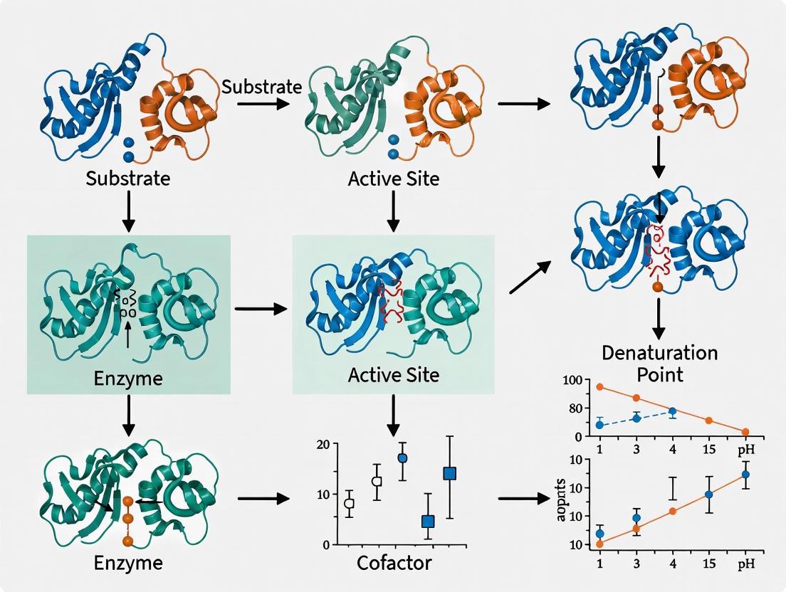
This protocol uses serial crystallography to capture enzyme structure and kinetics at physiological temperatures [5].
- Protein Crystallization: Generate a large batch of microcrystals of your target enzyme (e.g., β-lactamase CTX-M-14 or xylose isomerase) to ensure consistency.
- Environmental Control: Load crystals onto a fixed-target 'HARE' chip and place it within an environmental control box. This box uses closed-loop control circuits to maintain precise temperature and relative humidity.
- Data Collection: Raster-scan the chip through an X-ray beam to collect diffraction data. Collect sequential datasets at a range of temperatures (e.g., from 10°C to 70°C).
- Reaction Initiation (for time-resolved studies): Use the LAMA (liquid-application method) to mix soluble ligands (substrates) with the crystals directly on the chip, initiating the enzymatic reaction.
- Data Analysis: Determine the enzyme's structure at each temperature. Analyze the Atomic Displacement Parameters (ADPs) to quantify structural dynamics. Correlate structural changes with measured turnover kinetics (kcat) at each temperature.
The workflow for this protocol is illustrated below.
Quantitative Data on Denaturation
The following table summarizes experimental data on how environmental factors induce denaturation and the corresponding protective effects of stabilization strategies.
Table 1: Denaturation Triggers and Stabilization Efficacy
| Denaturation Stressor | Observed Effect on Protein | Measured Protective Strategy | Result of Stabilization |
|---|---|---|---|
| Organic Solvents (e.g., Methanol) | Threshold-like drop in activity at ~40 vol.% [2]. | Chymotrypsin immobilized to polyacrylamide gel [2]. | Retained activity at 60 vol.% methanol (20% higher than native) [2]. |
| Urea (Chemical Denaturant) | Unfolding midpoint (C_M) of BsCspB at 2.7 M [3]. | Adding 120 g/L PEG1 (crowding agent) [3]. | C_M shifted to 3.3 M urea; ΔΔG increased by ~1.4 kJ/mol [3]. |
| Temperature (on CTX-M-14 β-lactamase) | Modulation of turnover kinetics and structural dynamics [5]. | N/A (Inherent property measured) | Direct correlation between temperature, atomic displacement dynamics, and catalytic rate [5]. |
| Low Salt (on Halophilic Malate Dehydrogenase) | Complete loss of activity and secondary structure at 0 M NaCl [4]. | Maintaining 4.0 M NaCl [4]. | Full enzymatic activity and structured ellipticity in CD spectra [4]. |
The Scientist's Toolkit: Key Research Reagents
This table details essential materials used in research focused on understanding and preventing denaturation.
Table 2: Essential Reagents for Denaturation Research
| Reagent | Function in Experiment | Key Characteristic |
|---|---|---|
| Chaotropes (Urea, Guanidine HCl) | Chemical denaturants used to induce unfolding and measure protein stability [3]. | Disrupts hydrogen bonding networks within the protein. |
| Crowding Agents (PEG, Dextran) | Mimic the crowded intracellular environment; can stabilize proteins via the excluded volume effect [3]. | Increases thermodynamic stability and can shift unfolding midpoints. |
| Halophilic Enzymes (e.g., Halophilic MDH) | Model systems for studying structural adaptations to extreme conditions [4]. | Rich in acidic surface residues, requires high salt for stability. |
| Chemical Cross-linkers (e.g., DSS/BS3) | Covalently link proximal amino acids to stabilize protein complexes and provide structural insights [7]. | Homo-bifunctional, amine-reactive, with defined spacer arm lengths. |
| Polyelectrolytes (e.g., Polybrene) | Form multi-point, non-covalent complexes with enzymes to increase rigidity in organic solvents [2]. | Provides reversible stabilization through electrostatic interactions. |
The relationships between different stabilization strategies and their core principles are mapped in the following diagram.
Troubleshooting Guide: Investigating Protein Denaturation
This guide helps researchers diagnose and understand the fundamental stressors that disrupt protein structure during experiments.
| Stressor Type | Primary Mechanism of Action | Observed Experimental Consequences | Key Structural Levels Affected |
|---|---|---|---|
| Temperature (High) | Disrupts weak, non-covalent interactions (hydrogen bonds, hydrophobic interactions); increases molecular agitation leading to unfolding [8] [9]. | Loss of activity; increased turbidity or aggregation; observable precipitation [8] [10]. | Secondary, Tertiary, Quaternary [8]. |
| Temperature (Low/Cold) | Disrupts the hydrophobic effect and hydration shells, potentially destabilizing the protein core below the freezing point [8]. | Loss of activity, often reversible upon warming; can occur below solution freezing point [8]. | Tertiary, Quaternary [8]. |
| pH (Extremes) | Alters the charge states of amino acid side chains, disrupting ionic bonds and hydrogen bonding networks [9] [11]. | Loss of solubility; precipitation; sharp decline in enzymatic activity outside optimal range [9] [11]. | Secondary, Tertiary, Quaternary [9]. |
| Chemical Denaturants (Urea, GdnHCl) | Destabilize the native structure by forming more favorable interactions with the peptide backbone than the protein-internal hydrogen bonds, thereby solubilizing unfolded state [12]. | Unfolding and loss of function; increased susceptibility to proteolysis; used to measure protein stability (Tm) [8] [12]. | Secondary, Tertiary [8] [12]. |
| Organic Solvents | Perturbs the dielectric constant of the medium and disrupts hydrophobic interactions critical for the protein core [8] [9]. | Loss of activity; possible conformational change or precipitation [9]. | Tertiary, Quaternary [9]. |
| Detergents (e.g., SDS) | Binds extensively to the protein chain, overwhelming native interactions and imparting a strong negative charge, which unfolds the structure [8]. | Extensive unfolding; loss of native function; used in denaturing gel electrophoresis [8]. | Secondary, Tertiary, Quaternary [8]. |
| Heavy Metals (e.g., Hg, Pb) | Form complexes with functional side chains (e.g., thiols of cysteine), oxidize residues, or displace essential metal cofactors [9]. | Irreversible inactivation; non-specific aggregation or precipitation [9]. | Tertiary (can disrupt primary if disulfides cleaved) [9]. |
| Interfacial Stresses (Ice-Water, Air-Water) | Disruption of the protein's hydrophobic core due to direct interactions with the interface [8]. | Surface-induced denaturation and aggregation [8]. | Tertiary [8]. |
Frequently Asked Questions (FAQs)
What are the most immediate signs that my protein has denatured during an experiment?
The most common indicators are a precipitate or increased turbidity in your solution and a complete or near-complete loss of biological activity. In assays, you may observe a sudden drop in the reaction rate. Techniques like differential scanning fluorimetry (DSF) or circular dichroism (CD) can directly confirm a loss of native structure by showing a decrease in the melting temperature (Tm) or a change in the secondary structure spectrum [8] [10].
Is protein denaturation always irreversible?
No, denaturation can be reversible. Reversible denaturation occurs when the polypeptide chain is stabilized in its unfolded state by the denaturing agent but can spontaneously refold into its native, active conformation once the stressor is removed, provided the primary structure is intact. Irreversible denaturation often happens when unfolded chains interact and form stabilized aggregates, or when reactive groups like thiols become exposed and form incorrect disulfide bonds upon unfolding [8] [9]. For example, the protein ribonuclease A can refold after denaturation by urea and reduction, while a boiled egg white protein cannot [8] [9].
How can I determine the thermodynamic stability (ΔG) of my protein, and what are the challenges?
Protein stability (ΔG0D) is thermodynamically defined as the Gibbs free energy change for the transition from the native (N) to the denatured (D) state under physiological conditions. In practice, this quantity is typically measured by inducing denaturation with chemical denaturants like guanidinium chloride (GdnHCl) or urea and monitoring the unfolding transition using spectroscopic methods. A significant challenge is that the stability value under physiological conditions (ΔG0D) must be extrapolated from data obtained in denaturant solutions, and different extrapolation methods can yield different values, eroding confidence in the absolute number [12]. Furthermore, these in vitro measurements in dilute buffers ignore the "crowding effect" of the in vivo environment [12].
My therapeutic protein is prone to aggregation. What formulation strategies can enhance its stability?
Yes, formulation buffers are a primary tool for increasing stability. Key strategies include:
- pH Optimization: Finding the right pH is critical for stability [10].
- Additives/Excipients:
- Salts: Can help balance ionic strength [10].
- Sugars (e.g., sucrose, trehalose): Act as stabilizers and help with protein hydration [10].
- Amino Acids (e.g., glycine, arginine): Can help balance charges and suppress aggregation [10].
- Surfactants (e.g., Polysorbate 80): Protect the protein from aggregation at interfaces by acting as emulsifiers [8] [10].
- It is essential to verify that any additive does not negatively impact the protein's structure or function [10].
What is enzyme immobilization, and how can it protect against denaturation?
Enzyme immobilization involves binding or entrapping enzyme molecules to a solid support or within a carrier material. This process can significantly enhance stability against temperature, pH, solvents, and impurities. It works by limiting the mobility of the enzyme molecule, thereby reducing its tendency to unfold, and can provide a protective microenvironment. Common techniques include adsorption, covalent binding, and encapsulation [13]. For instance, covalent immobilization can create multiple attachment points between the enzyme and the support, dramatically rigidifying the structure and preventing unfolding [13].
Experimental Protocols & Methodologies
Protocol 1: Determining Melting Temperature (Tm) via Differential Scanning Fluorimetry (DSF)
Purpose: To quantify the thermal stability of a protein and the potential stabilizing effects of ligands or buffer conditions.
Principle: A fluorescent dye (e.g., SYPRO Orange) binds to hydrophobic patches of the protein that become exposed during unfolding. The fluorescence intensity increases as the protein denatures, allowing the melting temperature (Tm) to be determined [10].
Procedure:
- Sample Preparation: Prepare a protein solution (e.g., 0.1 - 1 mg/mL) in the buffer of interest. Include a 1:1000 dilution of SYPRO Orange dye. If testing, include your ligand or additive in the experimental sample.
- Plate Setup: Load the samples into a real-time PCR plate or a suitable thermostable plate.
- Run the Assay: Using a real-time PCR machine or other thermal gradient instrument, heat the plate from 25°C to 95°C with a gradual ramp rate (e.g., 1°C per minute). Monitor the fluorescence signal continuously.
- Data Analysis: Plot the fluorescence intensity as a function of temperature. The Tm is defined as the midpoint of the unfolding transition, where 50% of the protein is unfolded [10]. A higher Tm indicates a more stable protein.
Protocol 2: Assessing Structural Stability Using Microfluidic Modulation Spectroscopy (MMS)
Purpose: To detect subtle, stress-induced changes in protein secondary structure (alpha-helix, beta-sheet) that may indicate instability.
Principle: MMS measures the infrared absorption spectrum of a protein, which is sensitive to its secondary structure. It compares the sample spectrum to a control and highlights differences, making it highly sensitive to conformational changes [10].
Procedure:
- Stress Application: Subject your protein sample to the stress of interest (e.g., incubate at 50°C overnight, agitate, or expose to different pH buffers).
- Control Sample: Keep an aliquot of the protein under non-stressful conditions as a control.
- Spectra Acquisition: Analyze both the stressed and control samples using an MMS instrument.
- Data Interpretation: The instrument generates a delta plot by subtracting the second derivative spectrum of the control from the stressed sample. A loss of alpha-helix or beta-sheet signals, or the formation of intermolecular beta-sheet (associated with aggregation), indicates stability loss. The technique can also demonstrate the stabilizing effect of ligands or excipients by showing reduced structural change after stress [10].
Research Reagent Solutions
| Reagent/Category | Primary Function in Stability Research | Example Use-Case |
|---|---|---|
| Chemical Denaturants | To progressively unfold the protein in a controlled manner to study stability and folding pathways [12]. | Determining the free energy of unfolding (ΔG) by urea or GdnHCl titration [12]. |
| Stabilizing Excipients | To protect the native protein structure from various stressors in formulation buffers [10]. | Adding polysorbate 80 to prevent surface-induced aggregation of a monoclonal antibody [10]. |
| Surfactants (e.g., Tween 80) | To stabilize proteins at interfaces (e.g., air-water, ice-water) by competing for the surface, preventing protein unfolding [8]. | Added to protein formulations to prevent denaturation at air-liquid interfaces during shaking [8]. |
| Immobilization Carriers | To provide a solid support for covalent or non-covalent enzyme attachment, rigidifying structure and enabling reuse [13]. | Covalently binding an enzyme to chitosan beads to enhance its thermal and pH stability for industrial catalysis [13]. |
| Cross-linkers (e.g., Glutaraldehyde) | To create covalent bonds within or between protein molecules, increasing rigidity and stability [13]. | Used in enzyme immobilization protocols to form stable, cross-linked enzyme aggregates (CLEAs) [13]. |
Protein Denaturation and Stabilization Pathways
Troubleshooting Guide: Active Site Stability
Problem 1: How does mutation of a single amino acid near the catalytic triad lead to activity loss?
Issue: A glycine residue adjacent to the catalytic histidine was mutated to an asparagine (G282N) to promote hydrogen bond formation. The mutant enzyme showed a significant drop in activity, especially at sub-optimal temperatures [14].
Root Cause: The mutation restricts the necessary flexibility of the catalytic "His-loop." The wild-type enzyme uses a glycine at this position to allow for dynamic loop movement essential for catalysis. Introducing a bulkier side chain (asparagine) forms a new hydrogen bond that locks the loop in a less flexible conformation, impeding the conformational changes needed for efficient substrate turnover [14].
Solutions:
- Revert to Glycine: Switch back to the wild-type glycine residue to restore loop flexibility.
- Engineer Compensatory Flexibility: If the mutation is essential for another property, consider introducing flexibility at another nearby, non-critical loop region to compensate for the rigidity introduced by the mutation.
- Optimize Reaction Conditions: Perform the reaction at the enzyme's optimal temperature, as the deleterious effect of rigidity is often more pronounced at lower temperatures [14].
Problem 2: Why does my enzyme lose activity after incubation at elevated (but sub-optimal) temperatures?
Issue: An EstE1 G282N mutant gradually lost residual activity when pre-incubated at 50–60°C, retaining only 25% activity at 70°C, while the wild-type remained stable [14].
Root Cause: The mutation compromises active-site stability. The new hydrogen bond that restricts flexibility also makes the active site more vulnerable to thermal denaturation. The local rigidity may prevent the enzyme from absorbing thermal energy through harmless conformational dynamics, leading to irreversible unfolding or distortion of the catalytic geometry [14].
Solutions:
- Screen Stabilizing Excipients: Add stabilizers like sugars (sucrose, trehalose) or certain amino acids (e.g., arginine) to the formulation. These can create a protective hydration shell and prevent aggregation during thermal stress [15].
- Consider Immobilization: Covalently immobilize the enzyme on a solid support. This can increase the enzyme's rigidity and resistance to denaturation by restricting unfolding [13].
Problem 3: What strategies can I use to improve the stability of an enzyme with a flexible, unstable active site?
Issue: A mesophilic enzyme (rPPE) has a globally flexible structure but a locally rigid His-loop stabilized by hydrogen bonding, limiting its activity [14].
Root Cause: The inherent flexibility of the enzyme's scaffold, combined with rigid loops in the active site, creates a sub-optimal balance between stability and activity.
Solutions:
- Enzyme Immobilization: This is a primary technique for stabilizing free enzymes.
- Covalent Binding: Create stable complexes by forming covalent bonds between enzyme functional groups (e.g., from lysine or cysteine) and a carrier matrix. This method prevents enzyme leakage and often improves thermal stability [13].
- Adsorption: Bind the enzyme to a support material (e.g., chitosan, porous silica) via weak forces. This is a simpler, reversible method but can lead to enzyme leakage under shifting pH or ionic strength [13].
- Protein Engineering: Use directed evolution or rational design to introduce stabilizing mutations. Machine learning tools can now predict mutations that enhance stability, solubility, and function without compromising activity [16].
- Chemical Modification: Conjugate the enzyme with chemically modified polysaccharides to enhance its physicochemical characteristics [13].
Frequently Asked Questions (FAQs)
Q1: Can enzyme denaturation be reversed? In some cases of mild denaturation, if the changes in temperature or pH are not too severe or prolonged, the enzyme can refold into its functional shape—a process called renaturation. However, many instances of denaturation, especially those that cause irreversible tangling of the polypeptide chain, are permanent [17].
Q2: Besides temperature, what other factors can denature an enzyme and distort the active site? Deviations from the optimal pH can alter the charge of amino acid residues, disrupting ionic and hydrogen bonds that maintain the enzyme's three-dimensional structure, including the active site. Exposure to organic solvents, certain salts, and mechanical stress (e.g., from pumping or agitation) can also cause denaturation [13] [17] [15].
Q3: How can I experimentally monitor changes in active-site flexibility? Intrinsic fluorescence spectroscopy can be used. Tryptophan or tyrosine residues in the active-site wall contribute to the overall fluorescence signal. A temperature-dependent fluorescence decrease can indicate conformational changes. Acrylamide quenching can further probe flexibility; a more rigid mutant will exhibit reduced quenching compared to the flexible wild-type [14].
Q4: We discovered a novel enzyme with great activity but poor stability. Is formulation a viable solution? Yes. A well-designed formulation creates a microenvironment that protects the enzyme's native structure. This involves finding the optimal combination of pH, ionic strength, and stabilizing excipients (e.g., sugars, surfactants, antioxidants) to protect the molecule through storage and delivery. A data-driven approach using high-throughput screening and machine learning can efficiently identify the right stabilizers for a specific enzyme [15].
Experimental Data & Protocols
Key Quantitative Data on His-Loop Mutations
Table 1: Impact of His-loop mutations on esterase activity and stability. WT = Wild-Type. Data adapted from [14].
| Enzyme | Mutation | Residual Activity vs. WT | Key Stability Observation |
|---|---|---|---|
| EstE1 (Hyperthermophilic) | G282N | 71% (at 70°C), 54% (at 30°C) | Lost ~75% activity after pre-incubation at 70°C |
| EstE1 (Hyperthermophilic) | G282Q | 65% (at 70°C), 32% (at 30°C) | Greater flexibility than WT, but lower stability |
| rPPE (Mesophilic) | D287G | 153% (at 30°C) | Increased flexibility enhanced activity |
| rPPE (Mesophilic) | D287E | 116% (at 30°C) | Stronger hydrogen bond improved stability and affinity |
Protocol 1: Assessing Active-Site Stability via Residual Activity
Purpose: To determine an enzyme's ability to maintain its catalytic function after exposure to stressful conditions.
Methodology:
- Pre-incubation: Aliquot the enzyme solution into small tubes. Incubate each tube at a specific elevated temperature (e.g., from 40°C to 70°C) for a fixed period (e.g., 10-60 minutes).
- Cooling: Immediately place the tubes on ice to stop the thermal denaturation process.
- Activity Assay: Measure the remaining enzymatic activity of each pre-incubated sample under the enzyme's standard optimal assay conditions (e.g., at its optimal temperature and pH, using a specific substrate like p-nitrophenyl esters).
- Calculation: Express the activity of each heat-treated sample as a percentage of the activity of an untreated control sample kept on ice [14].
Protocol 2: Probing Flexibility with Intrinsic Tryptophan Fluorescence
Purpose: To monitor conformational changes and flexibility in the enzyme's structure, particularly near the active site.
Methodology:
- Sample Preparation: Prepare a purified enzyme solution in an appropriate buffer.
- Temperature Ramp: Place the sample in a spectrofluorometer equipped with a temperature controller. Gradually increase the temperature from a low (e.g., 4°C) to a high value (e.g., 90°C).
- Fluorescence Measurement: At regular temperature intervals, excite the sample at 295 nm (to selectively excite tryptophan residues) and record the emission spectrum, typically from 300 to 400 nm.
- Data Analysis: Plot the maximum fluorescence intensity versus temperature. A gradual decrease suggests a flexible structure undergoing continuous unfolding, while a sharp drop indicates a cooperative, more rigid unfolding transition [14].
Visualizing the Concepts
Diagram 1: Active Site Flexibility Trade-Off
Diagram 2: Experimental Workflow for Stability Analysis
The Scientist's Toolkit: Research Reagent Solutions
Table 2: Essential materials and methods for studying and mitigating active site geometry loss.
| Research Reagent / Method | Function | Key Consideration |
|---|---|---|
| Site-Directed Mutagenesis Kits | Introduces targeted point mutations to test the role of specific residues in active-site geometry and flexibility. | Critical for establishing causality between a specific amino acid and observed functional changes. |
| Intrinsic Tryptophan Fluorescence | Probes conformational changes and flexibility in the enzyme's structure, often near the active site. | A non-destructive method that can monitor real-time unfolding. |
| Covalent Immobilization Carriers | Provides a solid support for stable enzyme attachment via covalent bonds, restricting unfolding. | Prevents enzyme leakage but requires careful selection to avoid blocking the active site [13]. |
| Stabilizing Excipients | Protects the enzyme's native structure in solution against various stressors. | Sucrose/Trehalose: Form protective hydration shells.Polysorbates: Shield against interfacial stress.Antioxidants: Prevent chemical degradation [15]. |
| AI/Machine Learning Platforms | Analyzes sequence-structure-function data to predict stabilizing mutations and optimal formulation conditions. | Dramatically accelerates the engineering and formulation process compared to traditional trial-and-error [18] [15]. |
Troubleshooting Guides & FAQs
This technical support center provides solutions for common challenges in enzyme stability research, directly supporting thesis work on overcoming denaturation in harsh conditions.
Frequently Asked Questions
Q1: Our enzyme rapidly loses activity in liquid formulation at room temperature. What are our most effective strategies to improve shelf-life?
Liquid enzyme formulations are prone to degradation, but several strategies can significantly enhance stability.
- Add Stabilizing Excipients: Incorporate polyhydric alcohols like glycerol (typically 25-50%) or sugars like sucrose and trehalose. These act as cryoprotectants and stabilizers by forming a protective hydration shell around the enzyme, preventing unfolding and aggregation [19] [20] [21]. Antioxidants (e.g., DTT) and chelating agents (e.g., EDTA) can also be added to prevent chemical instability like oxidation [19] [15].
- Optimize Buffer Conditions: The buffer's pH and ionic strength are critical. Most enzymes are stable only within a specific pH range, and the buffer type itself can sometimes inhibit activity [19]. Extensive screening is required to find the optimal buffer.
- Consider a Solid Formulation: Converting a liquid enzyme to a solid granulate through processes like wet granulation can dramatically improve long-term stability. One study showed that a granulated enzyme was stable for up to 24 months at 30°C, compared to a liquid form which was stable for only 1 day at room temperature after reconstitution [22]. Lyophilization (freeze-drying) is another common method, often facilitated by glycerol-free formulations for better water removal [20].
Q2: How can we stabilize an enzyme for repeated use in an industrial bioreactor?
For reusable industrial biocatalysts, immobilization is the preferred strategy as it enhances stability and allows easy separation from the reaction mixture.
- Covalent Bonding: This technique creates stable complexes by forming covalent bonds between the enzyme's functional groups (e.g., amino groups from lysine) and a solid carrier matrix (e.g., porous silica, agarose). It is highly effective due to strong enzyme/support interaction, which prevents enzyme leakage and often improves thermal stability [23] [13].
- Entrapment: The enzyme is physically trapped within an inert matrix, such as calcium alginate beads or a silk fibroin film. Entrapment in silk films has been shown to allow enzymes to retain significant activity even when stored at 37°C for ten months [23] [24].
- Cross-Linking: Enzyme molecules are cross-linked to each other using linkers like glutaraldehyde to form a stable aggregate. This method, known as Cross-Linked Enzyme Aggregates (CLEAs), "locks" the enzyme structure and has shown large improvements in stability for industrial biocatalysts [24].
Q3: We need to ship diagnostic enzymes to remote areas without cold chain access. How can we achieve ambient-temperature stability?
Achieving ambient-temperature stability is a key goal in diagnostic and therapeutic development.
- Develop Glycerol-Free Lyophilized Reagents: Glycerol, while good for cold storage, hinders lyophilization. Formulating glycerol-free enzymes with optimized buffers containing specific stabilizers allows for successful lyophilization, creating stable powders that can be shipped and stored at ambient temperature without losing activity [20].
- Use Solid Stabilization Matrices: Embedding enzymes in biocompatible, solid-state protein matrices like silk fibroin can stabilize them without refrigeration. These systems are ingestible, biocompatible, and offer remarkable long-term stability, making them ideal for distribution in challenging environments [24].
Q4: Can we predict and engineer an enzyme to be inherently more stable under high temperature or pressure?
Yes, advanced computational and simulation methods are now used to guide enzyme engineering for enhanced stability.
- Molecular Dynamics (MD) Simulations: MD simulations under varying temperature and pressure levels can reveal an enzyme's structural adaptability. By analyzing metrics like root mean square deviation (RMSD) and radius of gyration (Rg), researchers can understand how the enzyme's structure fluctuates and denatures under stress [25].
- Conformational Biasing and ProteinMPNN: This computational pipeline uses stable enzyme conformations derived from MD simulations (e.g., from states at 273 K/1 bar and 333 K/4000 bar) to identify mutation sites. The ProteinMPNN neural network model then designs mutant sequences with a bias toward more stable conformations, effectively engineering enzymes for harsh conditions [25].
Key Experimental Protocols
Protocol 1: Enzyme Immobilization via Covalent Binding to a Solid Support
This methodology details the covalent immobilization of an enzyme, which improves reusability and resistance to temperature and pH changes [13].
- Principle: Create stable covalent bonds between functional groups on the enzyme (e.g., amino groups from Lysine) and an activated carrier matrix.
- Materials:
- Purified enzyme
- Carrier matrix (e.g., porous silica, agarose, chitin/chitosan)
- Coupling agent (e.g., Glutaraldehyde or Carbodiimide)
- Appropriate buffer solutions
- Procedure:
- Activate the Carrier: Incubate the carrier matrix with a coupling agent like glutaraldehyde to create an electrophilic group on its surface.
- Couple the Enzyme: Mix the activated carrier with the enzyme solution in a suitable buffer. The nucleophilic groups on the enzyme (e.g., amino groups) will covalently bind to the activated support.
- Incubate: Allow the reaction to proceed for several hours to ensure complete binding and formation of a self-assembled monolayer (SAM).
- Wash: Thoroughly wash the immobilized enzyme preparation to remove any unbound enzyme or reagents.
- Troubleshooting Tip: A loss of activity after immobilization may indicate that the covalent binding involved amino acid residues critical for the enzyme's catalytic function. Testing different coupling chemistries or carrier materials may be necessary [13].
Protocol 2: Assessing Stability via Molecular Dynamics (MD) Simulations
This protocol describes a computational method to study enzyme stability under harsh conditions like high temperature and pressure, providing a theoretical foundation for experimental engineering [25].
- Principle: Use MD simulations to model the physical movements of atoms and molecules over time under defined environmental stresses to analyze structural changes.
- Materials:
- Enzyme 3D structure (e.g., from AlphaFold3 or crystal structure)
- MD simulation software (e.g., GROMACS)
- High-performance computing cluster
- Procedure:
- System Setup: Place the enzyme structure in a simulation box with explicit water molecules (e.g., TIP4P model). Add ions to neutralize the system's charge.
- Energy Minimization: Perform energy minimization using a method like steepest descent to relieve any steric clashes.
- Set Conditions: Define the simulation parameters, including a range of temperatures (e.g., 273 K to 333 K) and pressures (e.g., 1 bar to 4000 bar).
- Run Simulation: Perform multiple independent MD simulation replicates for each temperature-pressure condition (e.g., 60 ns each).
- Trajectory Analysis: Calculate key structural metrics from the simulation data:
- Root Mean Square Deviation (RMSD): Measures overall structural stability.
- Root Mean Square Fluctuation (RMSF): Identifies flexible regions.
- Radius of Gyration (Rg): Assesses protein compactness.
- Solvent Accessible Surface Area (SASA): Monitors unfolding.
The workflow for this computational protocol is summarized in the following diagram:
Quantitative Data on Enzyme Stabilization Strategies
Table 1: Comparison of Enzyme Formulation Strategies for Shelf-Life Extension
| Strategy | Key Components | Storage Condition | Stability Outcome | Key Advantage |
|---|---|---|---|---|
| Liquid Formulation (with stabilizers) [19] [21] | Glycerol (25-50%), Sucrose/Trehalose, Antioxidants | 0°C to 4°C, sometimes -20°C with glycerol | Varies; gradual activity loss over weeks/months | Convenient for ready-to-use solutions |
| Solid Granulate [22] | Enzyme, Excipients (e.g., Citrate), processed via wet granulation | 30°C | Stable for 24 months; 3 days after reconstitution | Dramatically improved ambient-temperature shelf-life |
| Silk Fibroin Entrapment [24] | Silk protein matrix, Enzymes (e.g., Glucose Oxidase) | 37°C | Retained >90% activity after 10 months | Biocompatible and stable under physiological conditions |
| Lyophilized (Glycerol-Free) [20] | Optimized salts, PCR enhancers, stabilizers | Ambient temperature | Maintains stability and performance post-lyophilization | Eliminates cold chain for shipping and storage |
Table 2: Structural Metrics from Molecular Dynamics Simulations for Stability Analysis [25]
| Simulation Condition | Impact on RMSD (Overall Stability) | Impact on Rg (Compactness) | Impact on SASA (Solvent Exposure) | Molecular Interpretation |
|---|---|---|---|---|
| Increasing Temperature | Increase | Increase | Increase | Elevated molecular entropy leads to unfolding and loss of structure. |
| Increasing Pressure | Decrease (at moderate levels) | Decrease | Decrease | Collapse of internal cavities and organized molecular order, leading to tighter packing. |
| Coupled High Temp. & Pressure | Varies, can be stabilizing | Varies, can be compacting | Varies | Pressure can counteract some denaturing effects of heat, revealing structural adaptability. |
The Scientist's Toolkit: Key Research Reagent Solutions
Table 3: Essential Materials for Enzyme Stabilization Research
| Reagent / Material | Function in Research & Development |
|---|---|
| Glycerol [19] [21] | A cryoprotectant and stabilizer; prevents ice crystal formation and protein aggregation in cold storage. |
| Silk Fibroin [24] | A biocompatible protein matrix for enzyme entrapment; provides a stabilizing microenvironment for long-term activity. |
| Glutaraldehyde [13] | A cross-linking agent; used for covalent enzyme immobilization on supports and creating Cross-Linked Enzyme Aggregates (CLEAs). |
| Chitosan / Alginate [23] [13] | Natural polymer supports for enzyme immobilization via adsorption or covalent binding; cost-effective and biodegradable. |
| Trehalose / Sucrose [15] | Stabilizing excipients that form a protective hydration shell around enzymes, used in both liquid and lyophilized formulations. |
| Polysorbate Surfactants [15] | Protect enzymes from interfacial and mechanical stress (e.g., from agitation) by occupying air-liquid interfaces. |
Engineering Resilience: AI, Additives, and Advanced Strategies for Enzyme Stabilization
Core Concepts: Machine Learning for Enzyme Stability
How can Machine Learning help overcome enzyme denaturation in harsh industrial conditions? Machine Learning (ML) guides enzyme engineering by rapidly predicting sequence modifications that enhance stability, moving beyond traditional trial-and-error methods. ML models learn from vast datasets of sequence-function relationships to map fitness landscapes and identify mutations that improve structural robustness against stressors like extreme pH, temperature, and solvents [26] [27]. This data-driven approach is particularly effective for designing stabilized enzymes for bioreactors and drug development, where maintaining activity in demanding environments is critical [15] [28].
What are the primary ML strategies for designing stable enzymes? ML integration in enzyme engineering has evolved through multiple stages, each contributing to stability prediction:
- Classical Machine Learning: Uses algorithms like ridge regression with handcrafted features to predict stability from sequence data [26].
- Deep Neural Networks (DNNs): Model complex, non-linear relationships in protein sequences for more accurate stability predictions [27].
- Protein Language Models (pLMs): Leverage evolutionary information from protein sequences to make zero-shot stability predictions without explicit experimental data [26] [27].
- Multimodal Models: Integrate diverse data types, such as sequence, structure, and functional assays, for a holistic and generalizable stability assessment [27].
Troubleshooting Guide: ML-Guided Enzyme Engineering Workflow
| Problem Area | Specific Problem | Possible Cause | Proposed Solution |
|---|---|---|---|
| Data Generation & Quality | Limited sequence-function data for model training. | Low-throughput screening methods; high cost of experimental characterization. | Adopt high-throughput cell-free gene expression (CFE) systems to rapidly generate large fitness datasets [26]. |
| Model Performance & Prediction | ML model fails to predict variants with improved stability. | Insufficient or low-quality training data; model cannot generalize to new sequence space. | Augment models with evolutionary zero-shot fitness predictors; use ensemble methods combining multiple algorithms [26] [27]. |
| Experimental Validation | Predicted stable enzymes perform poorly in real-world bioreactors. | Model trained on idealized lab conditions; neglects process stresses like shear forces or interfaces. | Include stability data from industrial-relevant conditions (e.g., presence of solvents, high concentration) in training sets [15] [28]. |
| Stability in Application | Enzyme loses activity upon immobilization or in final formulation. | Stabilizing mutations may block key functional sites or alter surface charges crucial for immobilization. | Employ multimodal ML models that simultaneously optimize for stability, activity, and surface properties for downstream application [27] [29]. |
Detailed Experimental Protocols
Protocol 1: ML-Guided Engineering of a Thermostable Amide Synthetase
This protocol is adapted from a study that engineered amide synthetases using an ML-guided, cell-free platform [26].
1. Objective: Improve enzyme activity and stability under industrial-relevant conditions for pharmaceutical synthesis.
2. Methods:
- Data Generation (Build):
- Library Construction: Use cell-free DNA assembly and PCR-based site-saturation mutagenesis to generate a library of 1,216 single-point mutants targeting 64 residues enclosing the active site and substrate tunnels [26].
- High-Throughput Screening (Test): Express mutant enzymes using cell-free gene expression (CFE). Perform functional assays with target substrates under conditions of high substrate concentration and low enzyme loading to simulate industrial stress. Collect conversion data as a fitness score [26].
- Machine Learning (Learn):
- Model Training: Use the sequence-function data (1,217 variants tested in 10,953 reactions) to train supervised ridge regression models.
- Model Augmentation: Augment the model with an evolutionary zero-shot fitness predictor to improve extrapolation.
- Variant Prediction: Use the trained ML model to predict higher-order mutants with increased activity and stability [26].
- Validation:
- Synthesize and test the top ML-predicted variants. Reported results showed variants with 1.6- to 42-fold improved activity relative to the parent enzyme for synthesizing nine pharmaceutical compounds [26].
Protocol 2: Enzyme Stabilization via Surface Immobilization
Immobilization is a key method to combat denaturation, and ML can help select optimal enzyme-support pairs [28] [29].
1. Objective: Enhance enzyme stability and reusability by covalent immobilization on a solid support.
2. Methods:
- Support Activation:
- Activate the carrier surface (e.g., porous silica, chitosan) with a linker molecule like glutaraldehyde. This creates an electrophilic group on the carrier [29].
- Enzyme Coupling:
- Incubate the enzyme with the activated carrier. Covalent bonds form between the carrier's electrophilic groups and nucleophilic amino acid residues on the enzyme (e.g., lysine's amino group). This creates a stable, multi-point attachment [29].
- Analysis:
The Scientist's Toolkit: Research Reagent Solutions
| Item | Function in ML-Guided Enzyme Engineering |
|---|---|
| Cell-free Gene Expression (CFE) System | Enables rapid, high-throughput synthesis and testing of thousands of enzyme variants without cloning, accelerating data generation for ML models [26]. |
| Ridge Regression ML Model | A supervised learning algorithm used to predict enzyme fitness from sequence data, helping to navigate the protein fitness landscape and identify stabilizing mutations [26]. |
| Augmented ML Model (e.g., with zero-shot predictor) | Enhances predictive power by combining experimentally derived sequence-function data with evolutionary information from related protein sequences [26]. |
| Covalent Immobilization Support (e.g., Chitosan, Porous Silica) | Provides a solid matrix for enzyme attachment, restricting molecular movement and enhancing stability against denaturation from heat, pH, and solvents [29]. |
| Linker Molecules (e.g., Glutaraldehyde) | Acts as a cross-linking agent to form stable, covalent bonds between the enzyme and the support material during immobilization [29]. |
Comparison of Enzyme Stabilization Techniques
When designing your enzyme stabilization strategy, the choice of method depends on your application requirements. The table below compares common techniques.
| Stabilization Method | Key Mechanism | Relative Cost | Stability Improvement | Reusability | Best For Applications Requiring: |
|---|---|---|---|---|---|
| ML-Guided Protein Engineering | Altering amino acid sequence to improve intrinsic stability [26] [27]. | High (R&D) | High (tailored) | N/A (inherent) | Permanent, intrinsic stability without supports. |
| Covalent Immobilization | Forming strong covalent bonds between enzyme and solid support [29]. | Medium | High | Excellent | Continuous flow reactors, multiple reuses without enzyme leakage [28]. |
| Adsorption Immobilization | Binding via weak forces (ionic, hydrophobic) [29]. | Low | Low to Medium | Poor | Low-cost, one-off use where some enzyme loss is acceptable. |
| Chemical Modification | Modifying surface residues with soluble polymers (e.g., polysaccharides) [29]. | Medium | Medium | N/A | Improved solubility and stability in liquid formulations. |
Workflow Visualization
ML-Guided Engineering Workflow
Enzyme Stabilization via Immobilization
Enzyme thermostability—the ability to retain structure and function at high temperatures—is a critical property for industrial and research applications, from pharmaceutical synthesis to biofuel production. Overcoming enzyme denaturation in harsh conditions is a central challenge in biocatalysis. Traditional methods like directed evolution are often costly, time-consuming, and labor-intensive [30] [31]. The emergence of a data-driven research paradigm, powered by machine learning (ML) and large-scale biological databases, is revolutionizing this field. This approach leverages vast, curated datasets to build predictive models that guide rational enzyme design, significantly accelerating the engineering of robust biocatalysts [30]. This technical support center is designed to empower researchers in navigating this new landscape, providing practical guidance on utilizing key resources like BRENDA and ThermoMutDB to overcome the persistent problem of enzyme denaturation.
Frequently Asked Questions (FAQs)
Q1: What are the primary data resources for enzyme thermostability engineering, and how do I choose between them? Your choice of database depends on the specific data type required for your project. The table below summarizes the core characteristics of major resources.
Table 1: Key Databases for Enzyme Thermostability Research
| Database Name | Primary Data Type | Scale | Key Features & Advantages | Primary Use Case |
|---|---|---|---|---|
| BRENDA [30] [31] | Enzyme function & properties (e.g., optimal temperature) | >32 million sequences; ~41,000 optimal temp. labels [30] | Manually curated from literature; wide enzyme coverage; actively maintained. | Finding wild-type enzyme properties and temperature activity profiles. |
| ThermoMutDB [30] [32] | Mutant thermodynamic data (e.g., ΔTm, ΔΔG) | ~14,669 mutations across 588 proteins [30] | Manually curated; flexible search/API; focuses on mutation effects. | Analyzing the stabilizing/destabilizing effects of specific point mutations. |
| AlphaFold DB [33] | Predicted 3D protein structures | Over 200 million entries [33] | Open access; high accuracy; broad coverage of UniProt. | Generating structural models for rational design when experimental structures are unavailable. |
| ProThermDB [30] | Mutant thermal stability data | >32,000 proteins; ~120,000 data points [30] | Extensive, high-quality experimental data; continuously updated. | Accessing a large volume of experimentally derived thermal stability parameters. |
Q2: My dataset is small and imbalanced, with few examples of thermostable enzymes. How can I train an effective machine learning model? Data scarcity and imbalance, where enzymes with mid-range temperatures are over-represented, are common challenges [31]. To address this:
- Use Specialized Datasets: Leverage recently developed, pre-curated datasets designed for thermal stability modeling, which implement clustering strategies to minimize sequence similarity between training and test sets, thus improving generalizability [31].
- Apply Weighted Loss Functions: During model training, use a weighted Root Mean Square Error (RMSE) loss function. This technique assigns higher importance to underrepresented samples (e.g., hyper-thermostable enzymes), forcing the model to learn these patterns more effectively [31].
- Leverage Transfer Learning: Start with a model pre-trained on a large, general protein sequence database (like UniProt) and fine-tune it on your smaller, specific thermostability dataset. This allows the model to learn fundamental protein principles before specializing.
Q3: How can I predict thermostability for an enzyme when the optimal growth temperature (OGT) of its source organism is unknown? Earlier models relied heavily on OGT, limiting their applicability [31]. Newer, state-of-the-art models like the Segment Transformer are OGT-independent. They predict temperature stability directly from the amino acid sequence by learning segment-level features that contribute unequally to thermal behavior, achieving high accuracy without organism-specific metadata [31].
Troubleshooting Guides
Guide: Interpreting and Validating Computational Predictions
Problem: Predictions from a machine learning model for a newly designed enzyme variant do not align with initial experimental results. Solution: Follow this systematic workflow to diagnose and resolve the discrepancy.
Steps:
- Check Input Data Quality:
- Sequence Verification: Ensure the input amino acid sequence for prediction is accurate and free of errors. A single misplaced residue can significantly alter the predicted structure and stability.
- Database Context: Cross-reference your enzyme's sequence and properties (e.g., from BRENDA) to ensure it falls within the model's trained scope. Performance may degrade for highly novel enzymes distant from the training data.
Assess Model Limitations:
- Understand the Training Data: Recognize that models trained on databases like BRENDA may be biased towards mesophilic enzymes, as data for extreme thermophiles is underrepresented [31]. A predicted stability of 70°C might have a higher error margin than one at 50°C.
- Use Uncertainty Estimates: If the model provides confidence intervals (e.g., the Segment Transformer outputs fluctuation ranges [31]), treat the prediction as a guide rather than an absolute value. A wide range suggests lower reliability.
Validate Experimental Conditions:
- Protocol Consistency: Ensure your experimental protocol for measuring stability (e.g., melting temperature (T_m)) matches the assumptions underlying the data in resources like ThermoMutDB (e.g., buffer pH, ionic strength). Inconsistent conditions are a major source of apparent disagreement [30].
- Replicate Experiments: Perform multiple experimental replicates to confirm the initial result and rule out technical errors.
Iterate and Re-design:
- Use the initial experimental data as a new data point to refine your understanding. Even a "failed" prediction provides valuable information about the model's performance for your specific enzyme family, guiding future design cycles.
Guide: Designing a Thermostability Engineering Workflow
Problem: My team wants to initiate a thermostability engineering project but needs a standardized, effective workflow. Solution: Implement a synergistic data-driven pipeline that integrates computational prediction with experimental validation.
Steps:
- Define Goal and Select Parent Enzyme: Clearly define the target temperature stability and select a parent enzyme with the desired catalytic activity.
- Data Mining and Analysis:
- Mine BRENDA for homologous enzymes to understand natural sequence and stability variation.
- Query ThermoMutDB to identify point mutations (e.g., ΔTm, ΔΔG values) known to stabilize similar protein folds.
- Obtain a 3D Structure from the AlphaFold Database to visualize and guide rational design.
- Run ML Models like the Segment Transformer to get initial stability predictions and identify potential stability "hotspots" [31].
- Generate Variant Library: Synthesize a library of enzyme variants based on the computational predictions, prioritizing mutations with high predicted stability scores and avoiding the enzyme's active site.
- High-Throughput Experimental Testing: Express and purify the variants, then test them using high-throughput assays for activity and thermal stability (e.g., melting temperature (T_m) assays, residual activity after heat shock).
- Model Refinement and Next Cycle: Feed the experimental results back into the machine learning model as new training data. This improves the model's accuracy for your specific enzyme, creating a virtuous cycle of design, test, and learn.
The Scientist's Toolkit: Research Reagent Solutions
Table 2: Essential Resources for Data-Driven Thermostability Engineering
| Tool / Resource | Type | Function in Experimentation |
|---|---|---|
| BRENDA Database [30] | Data Resource | Provides reference data on wild-type enzyme temperature optima and stability for benchmarking and homolog analysis. |
| ThermoMutDB [32] | Data Resource | Offers a curated list of characterized point mutations and their thermodynamic impact (ΔTm, ΔΔG) to inform rational design. |
| AlphaFold DB [33] | Structural Resource | Supplies accurate 3D protein structure predictions for visualizing mutations, analyzing folds, and conducting in silico analysis. |
| Segment Transformer Model [31] | Software Model | A deep learning tool that predicts enzyme temperature stability directly from sequence, enabling rapid in silico screening of designs. |
| MMseqs2 [31] | Software Tool | Used for sensitive sequence clustering and searching; crucial for creating non-redundant training and test sets to avoid data bias. |
| Thermal Shift Assay (e.g., T_m) | Experimental Assay | A standard high-throughput method to measure protein melting temperature, providing a key experimental validation metric (ΔTm). |
| Residual Activity Assay | Experimental Assay | Measures the percentage of enzymatic activity remaining after exposure to a high temperature, indicating operational stability. |
Frequently Asked Questions (FAQs)
1. What is the primary function of DTT in enzyme stabilization? Dithiothreitol (DTT) is a potent reducing agent that protects enzymes by reducing disulfide bonds and preventing the formation of incorrect intermolecular disulfide bridges that can lead to aggregation and inactivation. Its main role is to maintain cysteine residues in their reduced (-SH) state, which is crucial for the catalytic activity of many enzymes. Once oxidized, DTT forms a stable six-membered ring, which drives the reduction reaction forward and prevents the reformation of disulfide bonds [34] [35].
2. How does cysteine act as a stabilizing agent? Cysteine can function as a stabilizing additive through its thiol group, which serves as an antioxidant. It helps control reactive oxygen species (ROS) that can cause oxidative stress and damage to enzymes. By scavenging ROS, cysteine prevents the oxidation of sensitive amino acid residues in the enzyme's active site, thereby preserving its native structure and function [36]. Furthermore, its thiol group can participate in redox reactions, helping to maintain a reducing environment in the solution [37].
3. What is the protective mechanism of phosphocholine? Phosphocholine, a phospholipid, contributes to enzyme stabilization by mimicking the native membrane environment for membrane-associated enzymes. Many enzymes, such as cytochromes P450, are embedded in lipid membranes. In vitro, phosphocholine helps preserve the structural integrity and catalytic activity of such enzymes by maintaining the hydrophobic and phospholipid interactions necessary for their correct folding and oligomerization [36].
4. When should I choose DTT over other reducing agents like TCEP? DTT is highly effective at pH values above 7, but its reducing power diminishes in acidic conditions. In contrast, Tris(2-carboxyethyl)phosphine (TCEP) is more stable and effective over a broader pH range, including acidic conditions. However, TCEP is a bulkier molecule and may reduce disulfide bonds in folded proteins more slowly than DTT. Choose DTT for standard, pH-neutral conditions and TCEP when working under acidic conditions or when a more stable, odorless alternative is needed [34] [35].
5. Why is my enzyme still losing activity despite adding DTT? A common reason is the oxidation of DTT in solution. DTT is susceptible to air oxidation, and its half-life can be as short as 1.4 hours at pH 8.5 and 20°C. To ensure efficacy, always prepare fresh DTT solutions and store the powder in a desiccated environment at -20°C. The inclusion of EDTA in the solution can chelate divalent metal ions and considerably extend DTT's half-life [35]. Furthermore, note that DTT cannot reduce buried (solvent-inaccessible) disulfide bonds; for these, denaturing conditions may be required prior to reduction [35].
6. Can these additives be used together? Yes, these additives are often used in combination in complex stabilization cocktails. Research focused on stabilizing cytochrome P450 enzymes screened various classes of additives—including sugars, salts, amino acids, phospholipids, and antioxidants—simultaneously. The key is to test different combinations and concentrations, as synergistic effects are possible, but incompatibilities must also be ruled out [36].
Troubleshooting Guide
| Problem | Possible Cause | Solution |
|---|---|---|
| Rapid loss of enzyme activity | Additives have oxidized or degraded.Incorrect storage conditions. | Prepare fresh DTT and cysteine solutions daily [35]. Store stock solutions in aliquots at -20°C under inert gas [34]. |
| Enzyme precipitation or aggregation | Lack of reducing agent leading to disulfide bond formation.Incorrect ionic strength. | Increase DTT concentration (e.g., 1-5 mM) [34]. Desalt the enzyme preparation to remove contaminants [38]. |
| Poor enzyme kinetics in assays | Stabilizing additives interfering with the reaction.Mass transfer limitations. | Include control experiments without the substrate to identify inhibition [36]. Dilute the enzyme-additive mix to minimize interference. |
| Inconsistent results between batches | Variable oxidation state of additives.Enzyme sensitivity to minor buffer changes. | Standardize buffer preparation and use high-purity reagents. Confirm the pH of all solutions, as DTT's efficacy is pH-dependent [35]. |
Stabilizing Additives at a Glance
The table below summarizes the key properties and applications of DTT, cysteine, and phosphocholine for easy comparison.
| Additive | Core Function | Typical Working Concentration | Key Considerations |
|---|---|---|---|
| DTT | Reduces and prevents disulfide bond formation [34] [35]. | 1 - 10 mM | Unstable in solution; prepare fresh. Ineffective at low pH [35]. |
| Cysteine | Antioxidant; scavenges ROS [36]. | Concentration data not available in search results | Can form mixed disulfides; its effectiveness is concentration and context-dependent [37]. |
| Phospholipids (e.g., Phosphocholine) | Mimics native membrane environment; stabilizes structure [36]. | Concentration data not available in search results | Critical for membrane-bound enzymes (e.g., CYPs); may form micelles at high concentrations [36]. |
The Scientist's Toolkit: Research Reagent Solutions
| Reagent | Function in Enzyme Stabilization |
|---|---|
| Dithiothreitol (DTT) | Potent reducing agent to keep cysteines reduced and prevent aggregation [34]. |
| L-Cysteine | Acts as an antioxidant to mitigate oxidative damage from reactive oxygen species (ROS) [36]. |
| Phospholipids | Provides a lipid bilayer-like environment to maintain the structure of membrane-associated enzymes [36]. |
| Trehalose | Stabilizes protein structure through preferential exclusion and water replacement mechanisms [36]. |
| Bovine Serum Albumin (BSA) | Prevents surface adsorption and stabilizes enzymes by hydrophobic interactions [36]. |
| EDTA | Chelates metal ions to prevent metal-catalyzed oxidation and extends the half-life of DTT [35]. |
| Glycerol | Acts as a kosmotropic agent to reduce molecular mobility and stabilize the hydrated structure of enzymes [39]. |
Experimental Workflow for Testing Stabilizing Additives
The following diagram illustrates a logical workflow for systematically testing and optimizing stabilizing additives for an enzyme.
Enzyme Stabilization Workflow
Mechanism of DTT in Reducing Disulfide Bonds
This diagram details the step-by-step chemical mechanism by which DTT reduces a protein's disulfide bond.
DTT Reduction Mechanism
Cell-Free Platforms and High-Throughput Screening for Rapid Biocatalyst Development
Troubleshooting Guides
Common Issues in Cell-Free Biocatalyst Development
Problem: Low or No Protein Yield in Cell-Free Expression
- Potential Cause 1: Suboptimal DNA Template. The DNA template may be impure, have low concentration, or contain sequence errors.
- Solution: Ensure the DNA template is pure and not contaminated with salts, ethanol, or RNases. Verify the sequence, including the presence of an ATG initiation codon and correct reading frame. Avoid gel purification methods that can inhibit the reaction [40].
- Potential Cause 2: Inefficient Reaction Conditions. The reaction may be operating at a non-optimal temperature or with insufficient feeding.
- Solution: For larger proteins, reduce the incubation temperature to 25–30°C to aid proper folding. Use a thermomixer or incubator with shaking. Instead of a single feed, perform multiple feeding steps with smaller volumes of feed buffer (e.g., every 30-45 minutes) to sustain the reaction [40].
- Potential Cause 3: Protein Misfolding or Lack of Cofactors.
- Solution: Add mild detergents (e.g., up to 0.05% Triton-X-100) or molecular chaperones to the reaction to improve folding. If the enzyme requires co-factors for activity or stability, add them directly to the protein synthesis reaction [40].
Problem: High Variability and False Results in High-Throughput Screening (HTS)
- Potential Cause 1: Manual Process Variability. Inter- and intra-user variability during liquid handling can lead to inconsistent reagent volumes and concentrations.
- Solution: Implement automated liquid handling systems to standardize workflows. Technologies with in-built verification, such as droplet detection, can confirm dispensed volumes and enhance reproducibility [41].
- Potential Cause 2: Sample Interference in Cell-Free Systems. The lack of a physical cell membrane means system components are more exposed to interference from complex sample matrices.
- Solution: Incorporate design strategies that reduce interference. This can include using mathematical simulations to optimize assay kinetics or engineering two-filter systems to improve specificity [42].
- Potential Cause 3: Data Handling Challenges. The vast volume of multiparametric data generated by HTS can be difficult to manage and analyze consistently.
- Solution: Utilize automated data management and analytics platforms to streamline analysis, enable rapid insights, and ensure hit compounds are accurately identified [41].
Problem: Enzyme Instability and Denaturation in Harsh Conditions
- Potential Cause 1: Dehydration in Organic Solvents. In water-co-solvent mixtures, organic solvents displace essential water molecules from the enzyme's hydration shell, leading to denaturation.
- Solution: Enhance the enzyme's hydration shell by covalent modification with polar or charged groups (e.g., using pyromellitic anhydride). This increases the enzyme's affinity for water, retarding dehydration-driven denaturation [2].
- Solution: Use immobilization techniques. Multi-point covalent attachment to a support or formation of complexes with polyelectrolytes can increase the conformational rigidity of the enzyme, making it more stable in the presence of organic solvents [2].
- Potential Cause 2: Thermal Degradation. At high temperatures, enzymes can undergo irreversible inactivation through processes like deamidation of asparagine and glutamine or succinimide formation at aspartate and glutamate residues.
- Solution: While this is inherent to the enzyme's structure, one stabilization strategy is "nanoarmoring." Wrapping the enzyme with a synthetic polymer (e.g., polyacrylic acid) reduces the conformational entropy of the denatured state, effectively raising the free energy required for unfolding and stabilizing the native state [2].
Frequently Asked Questions (FAQs)
Q1: What are the key advantages of using cell-free platforms over traditional cell-based methods for biocatalyst screening? Cell-free systems offer several distinct advantages: they provide high biosafety as no live cells are involved, enable fast material transport without membrane barriers for quicker reactions, and permit direct control over the reaction environment. This allows for high sensitivity and the ability to screen conditions that would be toxic to cells [42].
Q2: How can I improve the portability and shelf-life of my cell-free biosensor? Freeze-drying technology is a key strategy. You can lyophilize the cell-free transcription-translation system onto substrates like paper, creating stable, ready-to-use tests that can be activated simply by adding water. These freeze-dried systems have been shown to remain stable at room temperature for months [42].
Q3: Our AI-driven enzyme discovery is successful in silico, but the enzymes perform poorly in the lab. What is the most likely cause? This is a common bottleneck. AI predicts structure and function, but it may not fully account for critical real-world factors like enzyme production yield, solubility, cofactor requirements, or stability under actual process conditions (e.g., in the presence of solvents or at elevated temperatures). Bridging this gap requires integrated wet-lab testing with rapid feedback loops to characterize and optimize the computationally designed enzymes [43].
Q4: What are some practical methods to stabilize enzymes in organic solvents? Beyond the methods in the troubleshooting guide, you can:
- Use protein engineering: Introduce mutations that increase rigidity or metal-chelating histidine residues to create stabilizing internal coordination bonds [2].
- Form complexes with oligoamines: Compounds like spermine can electrostatically bind to enzymes, providing additional stabilization under harsh conditions [2].
- Choose solvents wisely: Hydrophilic solvents like glycerol and ethylene glycol have a much lower denaturing capacity than hydrophobic solvents like tetrahydrofuran (THF) or butanol [2].
Q5: How can automation specifically address challenges in HTS for biocatalyst development? Automation directly tackles the core issues of reproducibility and data quality. It minimizes human error and variability in liquid handling, which is crucial for reliable results. Furthermore, automation enables miniaturization, drastically reducing reagent consumption and costs by up to 90%, while increasing throughput by allowing large libraries to be screened at multiple concentrations [41].
Performance Data and Stabilization Strategies
The tables below summarize quantitative data and methodological details to aid in experimental planning and problem-solving.
Table 1: Performance Metrics of Selected Cell-Free Biosensors This table provides examples of detection capabilities for various targets using cell-free systems, illustrating their sensitivity and versatility [42].
| Target Substance | Limit of Detection / Range | Response Time | Output Signal |
|---|---|---|---|
| Benzoic Acid | 10 µM | ~1 hour | sfGFP |
| 12 Amino Acids | 0.1–1 µM | 1 hour | sfGFP |
| Theophylline | 1 mM | <90 minutes | lacZ |
| Zika Virus RNA | 2 aM | 2.5 hours | lacZ |
| Mercury | 6 µg/L | ~1 hour | sfGFP |
| Tetracycline | 10–10,000 ng/mL | <90 minutes | Luciferase |
| Arsenic | 0.5 µM | 2 hours | XylE |
Table 2: Comparison of Enzyme Stabilization Techniques Against Denaturation A summary of common strategies to combat enzyme denaturation, particularly in non-ideal environments [2].
| Stabilization Method | Mechanism of Action | Example Application / Result |
|---|---|---|
| Covalent Immobilization | Multi-point attachment to a support increases conformational rigidity. | Chymotrypsin on polyacrylamide remained active at 60% methanol, 20% higher than native enzyme. |
| Polyelectrolyte Complexation | Multiple electrostatic interactions protect the enzyme from denaturation. | Chymotrypsin-polyanion complex enabled peptide synthesis in 60% DMF. |
| Surface Modification | Introduces groups that strongly bind water, retarding dehydration. | Pyromellitic anhydride-modified chymotrypsin retained activity in broader ethanol range. |
| Nanoarmoring | Synthetic polymer wrapping reduces conformational entropy of denatured state. | Polyacrylic acid wrapping stabilizes the native enzyme state via "entropy control". |
| Metal Chelation | Introduced residues create internal coordination bonds for rigidity. | Enzymes with engineered metal-chelating histidines showed higher stability in organic solvents. |
Experimental Protocols
Protocol 1: Stabilizing an Enzyme via Polyelectrolyte Complexation for Use in Organic Solvents
This protocol describes a method to form a non-covalent complex between an enzyme and a polyelectrolyte to enhance its stability in water-organic solvent mixtures [2].
- Solution Preparation: Prepare a low-salt aqueous buffer (e.g., 1-5 mM) at a pH where your enzyme is charged. For a negatively charged enzyme, use a pH above its isoelectric point (pI).
- Complex Formation: Add the polycation (e.g., polybrene) to the enzyme solution under gentle stirring. The amount of polyelectrolyte required must be determined empirically, but is typically based on charge ratios.
- Incubation: Allow the mixture to incubate for 30-60 minutes at 4°C to form the complex.
- Application: The enzyme-polyelectrolyte complex can be used directly in the water-organic solvent mixture. The complex is stable in low-dielectric media but can be dissociated by adding high concentrations of salt if needed.
Protocol 2: Miniaturized HTS using Automated Liquid Handling
This protocol outlines a general workflow for a high-throughput screening assay in a microplate format, emphasizing automation to minimize variability [41].
- Assay Design: Define the experimental parameters, including compound concentrations, controls, and replication scheme.
- Plate Reformatting: Use an automated liquid handler to transfer compounds from a library stock plate into the assay microplate. Non-contact dispensers are preferred to avoid cross-contamination.
- Reagent Dispensing: Dispense the cell-free reaction mix or buffer containing the target and detection reagents into all wells of the assay plate. Automated systems with drop-detection technology can verify dispensed volumes.
- Initiation: If required, use the dispenser to add a substrate to initiate the enzymatic reaction.
- Incubation and Reading: Incubate the plate at the designated temperature and time, then read the output signal (e.g., fluorescence, luminescence) using a plate reader.
- Data Analysis: Automatically stream the raw data to an analysis platform for normalization, hit identification, and quality control checks.
Workflow and Pathway Visualizations
HTS Automation Workflow
Enzyme Denaturation and Stabilization
The Scientist's Toolkit: Key Research Reagent Solutions
Table 3: Essential Reagents for Cell-Free and HTS Biocatalyst Development
| Item | Function / Application |
|---|---|
| Pure, Linear DNA Template | Directly used in cell-free protein synthesis reactions to program the production of the target biocatalyst. Must be free of contaminants like salts and RNases [40]. |
| Cell-Free Transcription-Translation Kit | A pre-mixed system containing all essential cellular components (ribosomes, tRNAs, enzymes, nucleotides) for protein synthesis in vitro. Examples include Expressway or similar systems [40]. |
| MembraneMax Reagent | A proprietary scaffold used in cell-free systems to facilitate the proper folding and integration of membrane proteins, which are often difficult to express functionally [40]. |
| Polyelectrolytes (e.g., Polybrene) | Used to form reversible complexes with enzymes, providing stabilization against denaturation in organic solvents and other harsh conditions through multi-point electrostatic binding [2]. |
| Non-Contact Liquid Handler | Automated instrument for precise, high-throughput dispensing of nanoliter to microliter volumes of compounds, reagents, and cell-free mixes into microplates, minimizing variability and cross-contamination [41]. |
| Molecular Chaperones | Proteins that can be added to cell-free reactions to assist in the proper folding of the synthesized target enzyme, thereby improving the yield of active biocatalyst [40]. |
| All-trans Retinal | A required cofactor for the functional folding and activity of certain enzymes, such as bacteriorhodopsin. Must be stored in the dark due to photosensitivity [40]. |
Troubleshooting Guides
Troubleshooting Guide: Enzyme Instability in Organic Solvents
| Problem | Possible Cause | Recommended Solution |
|---|---|---|
| Low or no activity in co-solvent reaction | Enzyme unfolding due to co-solvent concentration [44] | Determine the ( c{U{50}}^T ) parameter to identify the solvent concentration threshold where activity drops precipitously [44]. |
| Incorrect stability metric used for enzyme selection [44] | Select enzymes based on ( c{U{50}}^T ) instead of melting temperature (( Tm )), as ( Tm ) does not correlate well with activity in solvents [44]. | |
| Unexpected enzyme ranking | Ranking enzymes by ( T_m ) gives different results than ranking by solvent tolerance [44] | For solvent-based applications, prioritize screening results from ( c{U{50}}^T ) analysis over thermal stability data [44]. |
| Rapid inactivation during storage | Physical denaturation and aggregation [15] | Optimize buffer pH and add stabilizers like sucrose or trehalose [15]. For liquid formulations, include surfactants (e.g., polysorbates) to protect against interfacial stress [15]. |
| Chemical degradation (e.g., oxidation) [15] | Include antioxidants and chelating agents in the formulation. Use inert gas overlays in storage vials [15]. |
Troubleshooting Guide: Enzyme Instability at Non-Optimal pH
| Problem | Possible Cause | Recommended Solution |
|---|---|---|
| Loss of activity at alkaline pH | Deprotonation of critical amino acid side chains, disrupting electrostatic interactions [45] [46] | Use rational design to mutate surface charges. For Protein A, mutating acidic residues (Glu, Asp) to Ala improved alkaline tolerance [45]. |
| Irreversible inactivation after exposure to extreme pH | Irreversible denaturation or chemical modification [47] | Perform directed evolution, screening variants for stability and activity under the desired pH conditions [47]. |
| Disruption of electrostatic interactions | Altering the protonation state of amino acids (e.g., Lys, Asp) [46] | Carefully buffer the enzyme's environment to maintain its optimal pH [46]. |
Frequently Asked Questions (FAQs)
Q1: Why is thermostability not a reliable predictor of an enzyme's performance in organic solvents?
While thermostability and solvent tolerance are sometimes correlated, they are distinct properties [44] [47]. Research on ene reductases has shown that an enzyme's melting temperature (( Tm )) does not correlate with its specific activity in the presence of co-solvents like DMSO or alcohols [44]. An enzyme with a high ( Tm ) can lose activity rapidly at low solvent concentrations, whereas a less thermostable enzyme might retain significant activity. Therefore, stability should be assessed directly under the conditions in which the enzyme will operate [44].
Q2: What is a more effective parameter than melting temperature for screening solvent-tolerant enzymes?
The concentration of a co-solvent that causes 50% unfolding of the protein at a specific temperature, denoted as ( c{U{50}}^T ), is a more predictive metric [44]. This parameter directly indicates the solvent concentration where the enzyme's activity drops most sharply. Ranking enzymes based on their ( c{U{50}}^T ) values for a specific solvent can yield a completely different, and more application-relevant, order than ranking based on melting temperature [44].
Q3: What are the key strategies for engineering an enzyme to withstand alkaline conditions?
Rational design is a powerful strategy. This involves:
- Surface Charge Engineering: Replacing deprotonatable acidic residues (like glutamic acid or aspartic acid) on the protein surface with neutral amino acids (like alanine) can significantly improve alkaline stability, as demonstrated in the engineering of Protein A [45].
- Rigidifying the Structure: Introducing rigid linkers between protein domains or using consensus design can reduce flexibility and enhance resistance to alkaline unfolding [45].
- Directed Evolution: Creating a diverse library of enzyme variants and screening them under high-pH conditions can select for mutations that confer stability, even without a complete structural understanding [47].
Q4: My enzyme is unstable in a liquid formulation. What formulation strategies can help?
Developing a stable liquid formulation involves protecting the enzyme from various stressors [15]:
- Against Aggregation: Use stabilizers like sugars (sucrose, trehalose) or amino acids (e.g., arginine) to form a protective hydration shell and prevent molecules from sticking together.
- Against Interfacial Stress: Add surfactants (e.g., polysorbates) which preferentially occupy interfaces (like air-liquid or container walls), shielding the enzyme from denaturation.
- Against Chemical Degradation: Include antioxidants and chelating agents to protect oxidation-prone residues like methionine and cysteine.
Stability Parameters for Enzyme Selection
| Parameter | Definition | Application | Key Insight |
|---|---|---|---|
| Melting Temperature (( T_m )) | Temperature at which 50% of the protein is unfolded [44]. | Measures thermal stability. | Does not correlate with enzyme activity in the presence of organic co-solvents [44]. |
| ( c{U{50}}^T ) | Concentration of a co-solvent causing 50% protein unfolding at a specific temperature T [44]. | Measures solvent tolerance. | Directly indicates the solvent concentration where enzyme activity drops fastest; provides a more reliable ranking for solvent-based applications [44]. |
Effects of Common Co-solvents on Enzyme Stability
| Co-solvent | Relative Impact on Melting Temperature (( T_m )) [44] | Notes on Activity [44] |
|---|---|---|
| DMSO | Smallest decrease | Effect on activity varies by enzyme; can sometimes boost activity. |
| Methanol | Moderate decrease | Effect on activity is enzyme-dependent. |
| Ethanol | Moderate decrease | Effect on activity is enzyme-dependent. |
| 2-Propanol | Significant decrease | Effect on activity is enzyme-dependent. |
| n-Propanol | Largest decrease | Effect on activity is enzyme-dependent. |
Experimental Protocols
Protocol 1: Determining ( c{U{50}}^T ) for Solvent Tolerance
Purpose: To identify the concentration of an organic co-solvent at which an enzyme is 50% unfolded at a fixed temperature, providing a predictive metric for solvent stability [44].
Materials:
- Purified enzyme sample
- Appropriate assay buffer
- Water-miscible organic co-solvent
- Fluorescent dye or instrument for monitoring unfolding
- Thermostatted spectrophotometer or fluorometer
Procedure:
- Prepare a series of enzyme solutions in buffer with increasing concentrations of the target organic co-solvent.
- Incubate all samples at your desired reaction temperature.
- Monitor the protein unfolding state. For fluorescent proteins like ene reductases, you can track the intrinsic fluorescence of the cofactor. Alternatively, use a fluorescent dye that binds to unfolded proteins.
- Plot the fluorescence signal (or another unfolding signal) against the co-solvent concentration.
- Fit a sigmoidal curve to the data. The co-solvent concentration at the inflection point, where 50% of the protein is unfolded, is the ( c{U{50}}^T ) value.
Protocol 2: Rational Design for Improved Alkaline Stability
Purpose: To engineer a protein for enhanced stability under alkaline conditions through targeted mutagenesis of surface charges [45].
Materials:
- Plasmid containing the gene of interest
- Site-directed mutagenesis kit
- Expression host
- SDS-PAGE equipment
- Equipment for activity and stability assays
Procedure:
- Sequence and Structural Analysis: Use bioinformatics tools to analyze the protein's surface and identify solvent-exposed acidic residues.
- Mutagenesis Design: Design primers to mutate selected acidic residues to neutral residues like alanine.
- Generate Variants: Perform site-directed mutagenesis to create the desired variants.
- Express and Purify: Express the wild-type and mutant proteins and purify them.
- Stability Testing: Incubate the purified proteins in alkaline buffers and measure residual activity over time compared to the wild-type. Confirm stability using techniques like circular dichroism.
- Characterization: For successful mutants, determine kinetic parameters and perform further structural analysis if possible.
Research Reagent Solutions
| Reagent / Material | Function in Research |
|---|---|
| Sucrose / Trehalose | Stabilizers that form a protective hydration shell around enzymes, preventing denaturation and aggregation in liquid formulations [15]. |
| Polysorbate Surfactants | Occupy air-liquid and solid-liquid interfaces to shield enzymes from mechanical and interfacial stresses during manufacturing, shipping, and administration [15]. |
| Site-Directed Mutagenesis Kit | Enables rational protein design by allowing precise introduction of point mutations to replace specific amino acids (e.g., for surface charge engineering) [45]. |
| High-Throughput Screening Platform | Allows for the rapid testing of thousands of enzyme variants for stability and activity under harsh conditions, enabling directed evolution campaigns [15]. |
Diagrams
Enzyme Engineering Workflow
Solvent vs. Thermal Stability
From Lab to Vial: Troubleshooting Stability in Drug Development and Manufacturing
For researchers and drug development professionals, the journey of an enzyme therapeutic from a promising catalyst to a stable, efficacious drug is fraught with unique Chemistry, Manufacturing, and Controls (CMC) challenges. Unlike small molecules, enzymes are complex proteins whose delicate three-dimensional structures are essential for activity and are highly susceptible to degradation. This technical support center is framed within our broader thesis on overcoming enzyme denaturation in harsh conditions. It provides targeted troubleshooting guides and FAQs to help you navigate the specific, often unanticipated, obstacles in formulating and manufacturing enzyme-based therapeutics.
FAQs & Troubleshooting Guides
Myth: "A standard liquid formulation is sufficient for long-term enzyme storage."
Issue: My enzyme therapeutic is losing activity rapidly during storage, leading to a short shelf-life and failed stability protocols.
Background: Enzymes are inherently prone to physical and chemical degradation. Physical instability involves unfolding, aggregation, and adsorption to surfaces, while chemical instability includes processes like oxidation of methionine residues or deamidation of asparagine [15]. A standard buffer developed for a monoclonal antibody will not necessarily protect the complex and specific active site of an enzyme.
Troubleshooting Steps:
- Diagnose the Root Cause: First, identify the primary degradation pathway.
- Perform Size-Exclusion Chromatography (SEC): A growing peak in the high molecular weight region indicates aggregation.
- Use Differential Scanning Calorimetry (DSC): Determine the melting temperature (Tm) to understand the enzyme's innate thermal stability.
- Run Ion-Exchange Chromatography (IEX): New peaks can indicate chemical modifications like deamidation that alter the protein's charge.
- Implement Corrective Formulation Strategies:
- For Aggregation: Introduce stabilizers that act as molecular chaperones. Sucrose and trehalose form stabilizing hydration shells, while certain amino acids like arginine can suppress aggregation [15].
- For Surface-Induced Denaturation: Incorporate surfactants like polysorbates. They preferentially occupy interfaces (e.g., air-liquid, container walls), shielding the enzyme from mechanical stress [15].
- For Chemical Degradation: Add antioxidants (e.g., methionine) to scavenge free radicals and use chelating agents (e.g., EDTA) to remove trace metal ions that catalyze oxidation [15].
- Consider a Paradigm Shift: If a liquid formulation proves insufficient, develop a lyophilized (freeze-dried) product. While it adds complexity, lyophilization removes water and can dramatically enhance long-term stability [15].
Research Reagent Solutions:
- Sucrose/Trehalose: Bulking agents and stabilizers that form a protective matrix during lyophilization.
- L-Histidine: A common buffer providing optimal pH control.
- Polysorbate 80: A non-ionic surfactant to mitigate interfacial stress.
- Methionine: An antioxidant to prevent oxidation.
Myth: "Enzyme immobilization is a straightforward process with universal carriers."
Issue: The immobilized enzyme shows low activity retention, leaks from the carrier, or becomes inaccessible to its substrate.
Background: Immobilization enhances enzyme stability, facilitates reusability, and simplifies separation from reaction mixtures [13]. However, its success is highly dependent on the enzyme, the carrier material, and the chosen method. An inappropriate strategy can block the active site, cause conformational changes, or introduce mass transfer limitations.
Troubleshooting Steps:
- Analyze Immobilization Efficiency: Measure the activity and protein concentration in the solution before and after immobilization to calculate the immobilization yield and efficiency.
- Select the Carrier and Method Based on Application:
- Adsorption: Uses weak forces (van der Waals, ionic). It's simple and reversible but can lead to enzyme leakage due to desorption [13].
- Covalent Binding: Creates stable bonds between enzyme and carrier. It prevents leakage but may involve complex chemistry and reduce activity if the active site is involved [13].
- Encapsulation/Entrapment: Physically confines the enzyme within a polymer matrix (e.g., alginate) or inorganic framework (e.g., CaCO₃) [48] [49] [13].
- Optimize the Process: Fine-tune parameters like pH, ionic strength, enzyme-to-carrier ratio, and reaction time. Multi-point covalent attachment can significantly rigidify the enzyme structure, enhancing stability [13].
The following workflow outlines a systematic approach to enzyme immobilization, helping you select the right strategy and materials based on your specific enzyme and application goals.
Research Reagent Solutions:
- Calcium Carbonate (CaCO₃): An abundant, eco-friendly inorganic carrier with high porosity for enzyme loading [48].
- Sodium Alginate: A natural polysaccharide for encapsulation via ionic gelation with calcium ions [49].
- Glutaraldehyde: A common crosslinker for covalent immobilization [13].
- Chitosan: A biocompatible polymer with functional groups for covalent attachment [13].
- Guar Gum: A viscoelastic polysaccharide that can be co-immobilized to enhance stability during drying [48].
Myth: "PEGylation completely solves immunogenicity and stability problems."
Issue: Despite PEGylating our therapeutic enzyme, we are observing reduced efficacy in later treatment cycles, potentially due to anti-drug antibodies.
Background: PEGylation—the attachment of polyethylene glycol (PEG) chains—is a well-established strategy to increase hydrodynamic size, shield immunogenic epitopes, and prolong circulation half-life [50]. However, the immune system can produce anti-PEG antibodies, which accelerate blood clearance and reduce therapeutic efficacy upon repeated administration [50].
Troubleshooting Steps:
- Monitor Immune Response: Implement sensitive assays (e.g., ELISA or microarray-based immunoassays) to detect the development of anti-drug antibodies (ADAs), including anti-PEG antibodies, in patient sera [50] [51].
- Explore Alternative Stealth Technologies:
- Polymer Conjugation: Investigate alternatives to PEG, such as polysarcosine, hydroxyethyl starch (HES), or other biocompatible polymers.
- Nanoparticle Encapsulation: Encapsulate the enzyme within lipid or polymeric nanoparticles. This creates a physical barrier against the immune system and can also improve stability [50].
- Consider Enzyme Miniaturization: Explore engineering a smaller, yet fully functional, version of the enzyme. Smaller enzymes can be less immunogenic and may also exhibit improved folding efficiency, stability, and expressibility [52].
Research Reagent Solutions:
- mPEG-NHS Ester: A common activated PEG derivative for amine-directed conjugation.
- Lipid Nanoparticles (LNPs): A delivery system for encapsulating and protecting enzymes.
- Polysarcosine-NHS: An alternative, low-immunogenicity polymer for protein conjugation.
Quantitative Data on Enzyme Stabilization Strategies
The following table summarizes experimental data from recent studies, providing a quantitative comparison of the effectiveness of different stabilization strategies.
Table 1: Efficacy of Different Enzyme Stabilization Strategies
| Stabilization Strategy | Enzyme Model | Key Performance Metric | Result | Citation |
|---|---|---|---|---|
| CaCO₃ Immobilization with Guar Gum | Serine Protease | Residual activity after vacuum drying | ~70% (vs. significant loss for free enzyme) | [48] |
| Alginate Encapsulation (Spray Drying) | Phytase | Residual activity after heat treatment | 79% (vs. 20% for free enzyme) | [49] |
| Enzyme Miniaturization | General Class | Typical length for efficient soluble expression | Proteins < 200 residues fold efficiently; solubility drops for proteins > 400 residues | [52] |
| PEGylation | Uricase | Primary Limitation | Development of anti-PEG antibodies, reducing long-term efficacy | [50] |
Essential Research Reagent Toolkit
Table 2: Key Reagents for Enzyme Stabilization and Formulation
| Reagent / Material | Function / Application | Key Consideration |
|---|---|---|
| Trehalose / Sucrose | Stabilizer / Cryoprotectant | Forms a glassy matrix to protect against denaturation during drying and storage. |
| Polysorbate 80 / 20 | Surfactant | Protects enzymes from interfacial stresses at air-liquid and solid-liquid interfaces. |
| Calcium Carbonate (CaCO₃) | Inorganic Immobilization Carrier | Mesoporous structure offers high enzyme loading and improved thermal stability. |
| Sodium Alginate | Polymer for Encapsulation | Forms gentle hydrogel beads via ionic crosslinking with Ca²⁺, protecting enzymes in harsh GI conditions [53] [49]. |
| L-Methionine | Antioxidant | Scavenges reactive oxygen species to prevent oxidation of susceptible residues. |
| Glutaraldehyde | Crosslinker | Used for covalent immobilization and carrier-free cross-linked enzyme aggregates (CLEAs). |
Troubleshooting Guide and FAQs
This technical support center provides targeted solutions for common challenges in enzyme formulation, specifically addressing shear stress, excipient interactions, and lyophilization processes. These guides are designed to help researchers maintain enzyme stability and activity under harsh conditions.
Shear Stress: Troubleshooting Guide
Q: What is shear stress and how does it cause enzyme denaturation in bioprocessing? A: Shear stress occurs when parallel forces act on a material's surface, causing layers to slide against each other [54]. In bioprocessing, this can disrupt an enzyme's three-dimensional structure by breaking the delicate hydrogen bonds, ionic bonds, and disulfide bridges that maintain its functional shape [17]. The resulting loss of structural integrity particularly affects the active site, preventing proper substrate binding and catalytic activity [17].
Q: What are the visual indicators of shear-induced denaturation in my enzyme solution? A: Look for these signs:
- Unexpected precipitation or aggregation in previously clear solutions
- Foaming or bubble formation during mixing or transfer
- Reduced catalytic activity without other explanatory factors
- Increased viscosity suggesting protein unfolding and aggregation
Experimental Protocol: Quantifying Shear Stress Impact
Objective: To determine the shear tolerance of your enzyme and establish safe processing parameters.
Materials:
- Enzyme solution at standard working concentration
- Controlled-stress rheometer or high-speed mixer
- Activity assay reagents specific to your enzyme
- SDS-PAGE equipment for structural analysis
Method:
- Prepare identical samples of your enzyme solution
- Subject each to increasing shear rates (e.g., 100-10,000 s⁻¹) for fixed durations
- Immediately assay remaining enzymatic activity
- Analyze structural changes via SDS-PAGE and circular dichroism if available
- Plot activity retention versus applied shear stress to determine tolerance threshold
Table 1: Shear Stress Calculation Methods for Different Scenarios
| Scenario | Formula | Parameters | Application Note |
|---|---|---|---|
| Simple Shear | τ = F/A | τ=shear stress (Pa), F=parallel force (N), A=area (m²) [54] | Applicable to surface interactions |
| Fluid Flow | τ = μ(du/dy) | μ=dynamic viscosity, du/dy=velocity gradient [54] | For pumping, mixing, or filtration processes |
| Beam Analysis | τ = VQ/It | V=internal shear force, Q=statical moment of area, I=moment of inertia, t=material thickness [54] | Relevant to equipment design |
Mitigation Strategies:
- Reduce flow rates during transfer and processing
- Minize sudden pressure changes or constrictions in flow path
- Use appropriate impellers that maintain mixing with lower shear
- Consider protective additives like sugars or polyols that stabilize enzyme structure
- Control temperature to avoid combining shear stress with thermal stress
Excipient Interactions: Troubleshooting Guide
Q: How can excipients intended to stabilize my enzyme actually cause degradation? A: Excipients can initiate, propagate, or participate in chemical or physical interactions with enzymes, potentially compromising stability and performance [55]. Even excipients considered "inert" may contain impurities that catalyze degradation reactions, or they may directly interact with functional groups on the enzyme surface [56].
Q: What are the hidden incompatibilities I should check for in my enzyme formulation? A: The most challenging incompatibilities are not immediately visible. Watch for:
- Charge interactions between ionizable excipients and charged residues on enzyme surface [55]
- Hydrogen-donating interactions with excipients like polyvinylpyrrolidone (PVP) [55]
- Moisture-mediated effects where excipient-bound water becomes available for hydrolytic reactions [55]
- pH-modifying residues in excipients that alter local microenvironment [55]
Experimental Protocol: Excipient Compatibility Screening
Objective: Systematically evaluate potential excipient interactions during formulation development.
Materials:
- Purified enzyme
- Candidate excipients (buffers, stabilizers, bulking agents)
- Forced degradation equipment (elevated temperature, humidity)
- Analytical methods (HPLC, activity assays, spectroscopy)
Method:
- Prepare enzyme samples with each excipient at proposed use concentration
- Include control without excipients
- Store under accelerated conditions (e.g., 40°C/75% RH) and sample at predetermined intervals
- Monitor for:
- Activity retention
- Formation of degradation products
- Physical changes (precipitation, color)
- Structural changes (via SDS-PAGE, spectroscopy)
- Rank excipients by compatibility
Table 2: Common Excipient Interactions and Mitigation Strategies
| Interaction Type | Mechanism | Signs | Prevention Approach |
|---|---|---|---|
| Charge-Based | Ionic attraction/repulsion between ionizable groups [55] | Precipitation, reduced solubility | Match pI of enzyme with formulation pH; use non-ionic excipients |
| Hydrogen Bonding | H-donor/acceptors between enzyme and excipient [55] | Conformational changes, activity loss | Select low H-bonding potential excipients; control moisture |
| Impurity-Mediated | Reactive residues in excipients (e.g., peroxides) [56] | Oxidation products, color changes | Source high-purity grades; use antioxidants |
| Moisture Redistribution | Bound water becomes available for hydrolysis [55] | Hydrolytic degradation products | Use non-hygroscopic excipients; control RH during processing |
Mitigation Strategies:
- Select low-reactivity excipients like partially pregelatinized starch known for minimal interaction [56]
- Control moisture content through proper packaging and storage conditions
- Consider competitive binding where excipients like cyclodextrins can be used strategically to shield enzymes [56]
- Employ impurity profiling of excipient lots to identify potential catalysts for degradation
Lyophilization: Troubleshooting Guide
Q: Why does my enzyme show inconsistent activity after lyophilization across different vial positions? A: This "edge effect" occurs when vials at the shelf periphery experience different thermal environments than central vials [57]. Outer vials contact colder chamber walls and doors, leading to different freezing rates and potential variability in residual moisture content [57].
Q: What causes stoppers to 'pop out' or slide into vials during lyophilization? A: Stoppers may pop out if the product isn't completely frozen and contains highly concentrated, non-solidified inclusions that evaporate explosively when vacuum is applied [57]. Stoppers sliding into vials typically occurs during freezing, especially at very low shelf temperatures, due to dimensional changes in stopper material [57].
Experimental Protocol: Lyophilization Process Optimization
Objective: Develop a robust freeze-drying cycle that preserves enzyme activity and ensures batch uniformity.
Materials:
- Enzyme formulation solution
- Laboratory-scale lyophilizer with controllable shelf temperature
- Temperature probes (product, shelf, condenser)
- Residual moisture analyzer
- Activity assay reagents
Method:
- Freezing Stage Optimization:
- Cool shelves to -40°C to -50°C
- Consider controlled nucleation to standardize ice crystal formation
- Ensure complete solidification before initiating vacuum
Primary Drying Optimization:
- Apply vacuum (typically 0.1-0.2 mbar)
- Gradually increase shelf temperature while monitoring product temperature
- Maintain product temperature below collapse temperature
- End when all ice has sublimated (determined by pressure rise test)
Secondary Drying Optimization:
- Gradually increase shelf temperature to 20-25°C
- Hold for 4-6 hours to reduce residual moisture
- Monitor moisture content to target specification
Table 3: Lyophilization Troubleshooting for Common Issues
| Problem | Possible Causes | Diagnostic Steps | Corrective Actions |
|---|---|---|---|
| Prolonged Evacuation Time | Excess ice on shelves from condensation during loading [57] | Check condenser temperature vs pressure reached [57] | Keep chamber pressure with dry gas during loading; slowly raise shelf temperature [57] |
| Slow Pressure Increase | Release of dissolved gases from frozen ice; misplaced vacuum pump suction [57] | Check for constant pressure rise despite decreasing condenser temperature [57] | Increase vacuum pump capacity; verify proper condenser connection placement [57] |
| Product Collapse | Exceeding collapse temperature during primary drying | Monitor product temperature vs known collapse point | Lower shelf temperature during primary drying; adjust formulation to increase collapse temperature |
| High Residual Moisture | Inadequate secondary drying; improper stopper placement | Measure moisture across batch positions; check stoppering force | Extend secondary drying; ensure complete stoppering under vacuum |
Mitigation Strategies:
- Use controlled nucleation to standardize the freezing step and improve batch uniformity [58]
- Implement process analytical technology (PAT) for real-time monitoring of critical parameters [58]
- Characterize thermal properties of your formulation to determine critical temperatures
- Employ automated loading systems to minimize chamber exposure and ice condensation [58]
Research Reagent Solutions
Table 4: Essential Materials for Enzyme Stabilization Research
| Reagent/Material | Function/Purpose | Example Applications | Key Considerations |
|---|---|---|---|
| Chitosan | Natural polymer support for enzyme immobilization [13] | Covalent or ionic enzyme attachment [13] | Biocompatible, biodegradable, multiple functional groups [13] |
| Partially Pregelatinized Starch | Low-interacting excipient with moisture sequestration ability [56] | Stabilization of moisture-sensitive APIs (e.g., aspirin) [56] | Does not make moisture available for hydrolytic reactions [56] |
| Silica Nanoparticles | Mesoporous support for adsorption-based immobilization [13] | Energy applications, biocatalysis [13] | High surface area, tunable pore size |
| Cyclodextrins | Form complex with APIs to increase solubility and bioavailability [56] | Solubility enhancement of poorly soluble enzymes/compounds [56] | Can be used strategically to shield enzymes from denaturing conditions [56] |
| Alginate | Ionic gel polymer for entrapment and encapsulation [59] | Enzyme immobilization for dairy processing, dye removal [59] | Mild gelation conditions, high enzyme loading capacity [59] |
| Cryoprotectants (Sucrose, Mannitol) | Stabilize protein structure during freezing and drying [58] | Lyophilization of biologics, protein formulations [58] | Prevent denaturation/aggregation during freeze-thaw cycles [58] |
Frequently Asked Questions
Q: Can enzyme denaturation from shear stress be reversed? A: In some cases of mild denaturation where the polypeptide chain remains intact, enzymes can refold into their functional shape when optimal conditions are restored (renaturation) [17]. However, severe or prolonged exposure to high shear typically causes irreversible denaturation as the polypeptide chain becomes permanently tangled or fragmented [17].
Q: How do I know if my enzyme-excipient interaction is beneficial or detrimental? A: Beneficial interactions typically show improved stability under accelerated conditions, maintained or enhanced activity, and consistent performance over time [56]. Detrimental interactions manifest as decreasing potency, new degradation products, physical changes (precipitation, color), or increasing variability between batches [55]. Always test excipient compatibility under both recommended storage and stress conditions.
Q: What is the most frequently overlooked parameter in lyophilization cycle development? A: The controlled nucleation step is often underestimated. Uncontrolled ice crystal formation leads to heterogeneous cake structure and variable drying rates [58]. Implementing controlled nucleation techniques standardizes the freezing phase, resulting in more uniform product quality and often shorter cycle times [58].
Q: How can I quickly screen multiple excipients for compatibility with my sensitive enzyme? A: Use a micro-scale forced degradation approach: prepare small samples (100-200 µL) of enzyme with each excipient, subject to controlled stress conditions (elevated temperature, agitation, or light), and monitor activity loss and physical changes over 1-2 weeks. This accelerated approach can identify incompatibilities before committing to full formulation development.
Frequently Asked Questions (FAQs)
FAQ 1: What are the primary clinical consequences of immunogenicity for enzyme therapeutics? The immune response to enzyme therapeutics can lead to a spectrum of clinical consequences, ranging from mild to severe. These include:
- Loss of Efficacy: The development of Neutralizing Anti-Drug Antibodies (NAbs) can directly inhibit the enzyme's pharmacological activity, reducing or completely negating its therapeutic benefit. This is often associated with worse clinical prognosis and accelerated disease progression [60] [61].
- Altered Pharmacokinetics: ADA can lead to either accelerated clearance of the drug, reducing systemic exposure, or in some cases, sustain drug levels by decreasing clearance rates [62].
- Infusion-Related Reactions (IRRs): The presence of ADA increases the risk of reactions during or after intravenous infusion, which are typically anaphylactoid (non-IgE mediated) rather than true anaphylaxis [61].
- Safety Risks: In rare cases, immunogenicity can lead to severe adverse events, including deficiency syndromes if the ADA cross-reacts with and neutralizes an essential endogenous protein [62].
FAQ 2: What factors influence the immunogenicity of a therapeutic enzyme? Immunogenicity is influenced by a combination of product-, patient-, and treatment-related factors [61] [62]:
- Product Factors: The enzyme's amino acid sequence (degree of "foreignness" compared to human proteins), post-translational modifications (e.g., glycosylation patterns), the cell line used for production (e.g., human, CHO, or plant-based), and the presence of aggregates or impurities in the final formulation.
- Patient Factors: The patient's immune status, genetic background (e.g., MHC haplotype), and whether they have any residual native enzyme (Cross-Reactive Immunological Material or CRIM status). CRIM-negative patients, who lack the native protein, are at a significantly higher risk of developing a robust immune response [61].
- Treatment Factors: The route of administration (intravenous vs. subcutaneous), dosing frequency, and treatment duration.
FAQ 3: What strategies can be used to mitigate the immunogenicity of enzyme therapeutics? Several strategies are employed to de-immunize therapeutic enzymes:
- Protein Engineering and Humanization: Modifying the amino acid sequence to remove or alter T-cell epitopes, a process known as deimmunization [63]. Computational algorithms (e.g., DP2) can guide this process by predicting and optimizing mutations that reduce immunogenicity while maintaining stability and activity [63] [64].
- PEGylation: Covalently attaching polyethylene glycol (PEG) chains to the enzyme. This can mask immunogenic epitopes, increase the enzyme's hydrodynamic size, and prolong its circulating half-life, potentially reducing immunogenicity. For example, pegunigalsidase alfa, a pegylated enzyme for Fabry disease, shows a lower immunogenicity profile [61].
- Enzyme Stabilization: Enhancing structural stability through methods like immobilization on solid supports or chemical modification with polysaccharides can make the enzyme less susceptible to denaturation and proteolytic processing, which can in turn reduce immunogen exposure [13].
- Immune Tolerance Induction: In clinical practice, protocols involving immunosuppression may be used to induce tolerance in high-risk patients.
FAQ 4: How is immunogenicity monitored and assessed in a clinical setting? International recommendations highlight the importance of systematic monitoring [61]:
- Anti-Drug Antibody (ADA) Assays: Bridging immunoassays (e.g., ELISA) are used to detect the presence and titer of binding antibodies in patient serum.
- Neutralizing Antibody (NAb) Assays: Cell-based or enzyme activity-based bioassays are used to confirm if the detected ADA can neutralize the drug's pharmacological function.
- Biomarker Monitoring: In diseases like Fabry disease, monitoring the accumulation of substrate (e.g., lyso-Gb3) is crucial to correlate ADA/NAb presence with loss of treatment efficacy [60] [61].
- Pharmacokinetic Profiling: Measuring drug concentration in plasma over time to identify accelerated clearance associated with a "clearing ADA response" [62].
Troubleshooting Guides
Guide 1: Troubleshooting Loss of Enzyme Therapeutic Efficacy
A sudden or gradual loss of therapeutic effect can be a key indicator of immunogenicity.
| Step | Investigation | Methodology & Expected Outcome |
|---|---|---|
| 1. Clinical Symptom Check | Correlate clinical symptoms with laboratory data. Review patient records for disease progression markers. | Compare biomarker levels (e.g., plasma lyso-Gb3 for Fabry disease) before and during treatment. An increase suggests loss of efficacy [61]. |
| 2. ADA/NAb Assay | Test patient serum for the presence of Anti-Drug Antibodies (ADA) and Neutralizing Antibodies (NAb). | Protocol: Use a validated bridging ELISA. 1. Coat plates with the therapeutic enzyme. 2. Incubate with diluted patient serum. 3. Detect bound human IgG using an enzyme-conjugated anti-human IgG antibody. A positive signal indicates ADA presence. Confirm neutralization with a cell-based or activity-based bioassay [62]. |
| 3. Pharmacokinetic Analysis | Determine if drug clearance is accelerated. | Protocol: Collect serial blood samples after drug infusion. Measure drug concentration using a specific ELISA or activity assay. Compare the concentration-time curve (AUC, half-life) to established profiles from patients without ADA. A reduced AUC and shorter half-life suggest a "clearing ADA response" [62]. |
Guide 2: Troubleshooting Immunogenicity in Preclinical Enzyme Design
This guide helps address high immunogenicity risks during the enzyme design and optimization phase.
| Step | Investigation | Methodology & Expected Outcome |
|---|---|---|
| 1. Epitope Mapping | Identify immunogenic T-cell epitopes within the enzyme's sequence. | Protocol (Computational): Use algorithms like TepiTool or integrated suites like the DP2 algorithm. Input the enzyme's amino acid sequence to predict peptides that bind to common MHC II alleles. Peptides with high prediction scores are candidate epitopes for deletion [63]. |
| 2. Computational Deimmunization | Re-engineer the enzyme to remove epitopes while maintaining function. | Protocol: Use the DP2 algorithm or similar tools. The algorithm integrates epitope prediction with sequence conservation data to recommend optimal, function-preserving mutations. Output is a set of variant sequences with predicted reduced immunogenicity and high stability [63]. |
| 3. In Vitro Immunogenicity Assessment | Validate the reduced immunogenicity of designed variants. | Protocol: Use an in vitro MHC association assay. 1. Incubate synthetic peptides (wild-type vs. deimmunized) with purified human MHC II molecules. 2. Measure binding affinity. A significant reduction in binding for deimmunized peptides confirms successful epitope deletion [63]. |
| 4. Functional & Stability Validation | Ensure deimmunized variants retain catalytic activity and stability. | Protocol: Express and purify the variant proteins. Perform standard enzyme activity assays (e.g., spectrophotometric monitoring of substrate conversion) and stability assays (e.g., thermal shift assays). The variant should exhibit wild-type-like activity and improved stability [64]. |
Table 1: Immunogenicity Prevalence in Fabry Disease Enzyme Replacement Therapies (ERT) [61]
| Therapeutic Enzyme | Production System | Approximate Prevalence of Neutralizing Antibodies (NAbs) in Male Patients | Key Characteristics |
|---|---|---|---|
| Agalsidase Alfa | Human fibrosarcoma cells (HT-1080) | ~24% | Lower approved dose (0.2 mg/kg) |
| Agalsidase Beta | Chinese Hamster Ovary (CHO) cells | ~40% (Most patients develop ADAs) | Higher approved dose (1 mg/kg); Higher immunogenicity risk |
| Pegunigalsidase Alfa | Plant (tobacco) cells, PEGylated | Induced in 16% of patients (at 1 mg/kg Q2W) | Pegylation masks epitopes and extends half-life; lower immunogenicity in clinical trials |
Table 2: Experimental Success Rates of Computationally Generated Enzymes [64]
| Generative Model | Enzyme Family | Experimental Success Rate (Active Enzymes) | Key Finding |
|---|---|---|---|
| Ancestral Sequence Reconstruction (ASR) | Malate Dehydrogenase (MDH) | 10 of 18 sequences (55.6%) | High success rate; often has stabilizing effect |
| Ancestral Sequence Reconstruction (ASR) | Copper Superoxide Dismutase (CuSOD) | 9 of 18 sequences (50.0%) | Robust performance even with sequence truncation |
| Generative Adversarial Network (ProteinGAN) | Malate Dehydrogenase (MDH) | 0 of 18 sequences (0%) | Initial naive generation resulted in mostly inactive sequences |
| Protein Language Model (ESM-MSA) | Copper Superoxide Dismutase (CuSOD) | 0 of 18 sequences (0%) | Success improved with computational filters (COMPSS) |
Experimental Protocols
Protocol 1: In Vitro Assessment of Neutralizing Antibodies (NAb) Using an Enzyme Activity Inhibition Assay
Principle: Patient serum containing potential NAbs is incubated with the therapeutic enzyme. The residual enzyme activity is measured and compared to a control without serum. A reduction in activity indicates the presence of NAbs.
Materials:
- Purified therapeutic enzyme.
- Patient and control (ADA-negative) serum samples.
- Enzyme-specific substrate.
- Reaction buffer (optimal pH and ionic strength for the enzyme).
- Spectrophotometer or fluorometer (depending on the assay).
Procedure:
- Serum Pre-treatment: Heat-inactivate all serum samples at 56°C for 30 minutes to destroy complement activity.
- Incubation: Mix a fixed concentration of the enzyme with serial dilutions of patient serum. Include a control with enzyme and buffer only (maximum activity control) and enzyme with control serum (negative control).
- Reaction: Incubate the mixture for 1-2 hours at 37°C to allow antibody-enzyme binding.
- Activity Measurement: Add the substrate to the mixture and immediately monitor the reaction (e.g., change in absorbance per minute) for a fixed period.
- Calculation: Calculate the percentage of enzyme activity inhibition for each patient serum dilution compared to the maximum activity control. A sample is considered positive for NAb if the inhibition exceeds a pre-defined threshold (e.g., >20-30%) and is dose-dependent [61] [62].
Protocol 2: Enzyme Stabilization via Covalent Immobilization
Principle: Covalently attaching enzymes to a solid support enhances stability by restricting conformational mobility and protecting against denaturation and proteolysis, which can also reduce immunogenicity by shielding epitopes.
Materials:
- Enzyme of interest.
- Support matrix (e.g., chitosan beads, porous silica, agarose).
- Cross-linker (e.g., glutaraldehyde or carbodiimide).
- Coupling buffer (e.g., 0.1 M phosphate buffer, pH 7.0).
- Washing solutions (buffer, high-salt buffer).
Procedure:
- Support Activation: Activate the support matrix. For example, incubate chitosan beads with glutaraldehyde (e.g., 2.5% v/v) in coupling buffer for 2 hours at room temperature with gentle mixing.
- Washing: Thoroughly wash the activated support with coupling buffer to remove excess glutaraldehyde.
- Enzyme Coupling: Incubate the purified enzyme solution with the activated support for 4-16 hours at 4°C with gentle agitation.
- Quenching and Washing: Block any remaining active sites by incubating with a quenching agent (e.g., 1 M ethanolamine, pH 8.0, or 0.1 M glycine) for 1-2 hours. Wash the immobilized enzyme extensively with coupling buffer followed by a high-salt buffer (e.g., 1 M NaCl) to remove any physically adsorbed enzyme.
- Storage: Store the immobilized enzyme in an appropriate storage buffer at 4°C [13].
Visualizations
Immunogenicity Risk Assessment Workflow
Computational Deimmunization Logic
The Scientist's Toolkit: Key Research Reagents
Table 3: Essential Reagents for Immunogenicity and Stability Research
| Item | Function/Application | Example in Context |
|---|---|---|
| Human MHC II Molecules | In vitro binding assays to directly measure the affinity of enzyme-derived peptides for immune recognition molecules [63]. | Purified HLA-DR molecules for epitope mapping studies. |
| Support Matrices for Immobilization | Provide a solid surface for covalent enzyme attachment to enhance stability and reusability. | Chitosan beads, porous silica, agarose, epoxy-activated supports [13]. |
| Cross-linking Reagents | Create stable covalent bonds between the enzyme and the support matrix during immobilization. | Glutaraldehyde, carbodiimide (EDC) [13]. |
| Enzyme-Specific Substrates | Measure catalytic activity before and after modification to ensure function is retained. | Chromogenic or fluorogenic substrates for spectrophotometric/fluorometric activity assays [64]. |
| Computational Tools | Predict T-cell epitopes and optimize enzyme sequences for reduced immunogenicity. | DP2 algorithm, ESM-MSA, TepiTool [63] [64]. |
| ADA/NAb Assay Kits | Detect and characterize anti-drug antibodies in serum samples for immunogenicity monitoring. | Validated bridging ELISA kits and cell-based neutralization assay components [62]. |
Managing Raw Material Variability to Ensure Lot-to-Lot Consistency
Troubleshooting Guides
Guide 1: Troubleshooting Inconsistent Enzyme Activity Assays
Problem: Measured enzyme activity varies significantly between different lots of the same raw material, leading to inconsistent experimental results.
| Potential Cause | Corrective Action | Preventive Measure |
|---|---|---|
| Presence of proteases in biological raw materials degrading the enzyme [65]. | Add protease inhibitors to the assay buffer. Re-test activity with a new sample from the retained lot [65]. | Source raw materials from suppliers who perform and provide data from protease activity assays [65]. |
| Denatured albumin in serum-based raw materials causing unpredictable analyte binding [65]. | Test the suspect lot in a different, validated assay platform to check for platform-specific discrepancies [65]. | Require suppliers to provide CoAs with data on albumin conformation and avoid materials exposed to heat/pH stress [65]. |
| Inconsistent inactivation processes (e.g., heat treatment) by the supplier, which can variably affect protein integrity [65]. | Use a biological activity assay specific to your application, not just chemical purity tests, to qualify new lots [66]. | Work with suppliers to understand their inactivation and purification methods and set joint specifications [65]. |
Guide 2: Addressing Poor Enzyme Stability in Formulations
Problem: The enzyme formulation loses activity rapidly during storage or under process conditions, which may be linked to raw material variability.
| Potential Cause | Corrective Action | Preventive Measure |
|---|---|---|
| Variability in excipient purity or physical properties (e.g., sugar, buffer, or surfactant quality) between lots [15]. | Perform accelerated stability studies (e.g., at elevated temperatures) to quickly identify subpar excipient lots [15]. | Establish strict Chemical and Physical Specifications for all formulation excipients, including vendor CoA requirements [66]. |
| Trace metal ion contamination in water or buffers, promoting oxidation [15]. | Add chelating agents (e.g., EDTA) or antioxidants to the formulation to scavenge free radicals [15]. | Use high-purity water (e.g., Milli-Q) and specify HPLC-grade or higher buffers. Test for metal ions if oxidation is a known risk [66]. |
| Inherent enzyme instability exacerbated by minor changes in the raw material microenvironment [13]. | Consider enzyme immobilization on a solid support to enhance stability and reusability [13]. | During development, use High-Throughput Screening to test formulation robustness against expected raw material variability [15]. |
Frequently Asked Questions (FAQs)
Q1: What are the most critical tests to run on a new lot of a biological raw material before using it in my enzyme assay? The most critical tests depend on your specific application, but should always include:
- Purity Analysis: Using techniques like HPLC or GC to verify chemical composition [66].
- Functional/Biological Assay: A standardized assay that measures the material's performance in a system as close to your end-use as possible [66].
- Consistency Check: Compare the results of the above tests against the data from your previous, qualified lot and the supplier's Certificate of Analysis (CoA) [66] [65].
Q2: How can I minimize the risk of enzyme denaturation when my process requires harsh conditions? Two primary strategies are employed to enhance enzyme robustness:
- Enzyme Immobilization: Binding enzymes to a solid carrier (e.g., chitosan, porous silica) can dramatically improve their stability against heat, pH, and organic solvents [13]. This also allows for enzyme reuse, reducing costs [13].
- Chemical Modification: Covalently attaching stabilizing molecules, such as polysaccharides, to the enzyme's surface can alter its physicochemical properties and increase resistance to denaturation [13] [67].
Q3: Our supplier provides a Certificate of Analysis (CoA). Is that sufficient to guarantee a new lot will perform identically? A CoA is a necessary starting point but is often not sufficient on its own. A CoA verifies the material meets the supplier's general specifications, but it may not assess parameters critical for your unique application [65]. You should always perform your own application-specific qualification using a small sample of the new lot before putting it into full production [66].
Q4: What should I do immediately if I suspect a raw material lot is causing inconsistent results?
- Quarantine the suspect lot.
- Retest the material using your standard qualification protocols.
- Compare the results against your retained samples from the previous, well-performing lot [66] [65].
- Contact your supplier with your data. A reliable supplier will investigate their retained samples and work with you to resolve the issue [65].
Experimental Protocols for Verification
Protocol 1: Qualification of a New Raw Material Lot
This protocol outlines the steps to validate a new lot of a critical raw material (e.g., a buffer component or cofactor) against a qualified reference lot.
1. Objective: To ensure a new lot of a raw material performs equivalently to the current qualified lot and will not negatively impact enzyme stability or activity.
2. Materials:
- New lot of raw material.
- Currently qualified lot of raw material (reference).
- All other standardized reagents and equipment for the relevant enzyme activity assay.
3. Methodology:
- Step 1: Receiving and Documentation. Upon arrival, verify the new lot's CoA against your predefined specifications [66].
- Step 2: Solution Preparation. Prepare a working solution of the raw material from both the new and reference lots using the same standardized procedure.
- Step 3: Experimental Testing. Test both solutions in your enzyme activity or stability assay. The assay should be run in at least triplicate for statistical power.
- Step 4: Data Analysis. Calculate the mean activity and standard deviation for both the new and reference lots. Perform a t-test to determine if any observed difference is statistically significant (typically, p < 0.05 is considered significant).
4. Acceptance Criteria: The new lot is considered qualified if the measured enzyme activity (or stability metric) is not statistically significantly different from the reference lot.
Protocol 2: Immobilization of Enzyme on Chitosan Beads to Enhance Stability
This protocol provides a methodology for immobilizing enzymes via covalent bonding to improve stability against denaturing conditions, thereby reducing sensitivity to raw material variability [13].
1. Objective: To stabilize a free enzyme by covalent immobilization onto chitosan beads, enhancing its reusability and resistance to temperature and pH shifts [13].
2. Materials:
- Purified enzyme solution.
- Chitosan beads (or other suitable carrier).
- Glutaraldehyde solution (e.g., 2.5% v/v).
- Coupling buffer (e.g., 0.1 M phosphate buffer, pH 7.0).
- Washing buffer (same as coupling buffer, possibly with added salt).
- Vacuum filtration setup.
3. Methodology:
- Step 1: Support Activation. Suspend chitosan beads in glutaraldehyde solution. Incubate for 1-2 hours with gentle agitation. Wash thoroughly with coupling buffer to remove excess glutaraldehyde [13].
- Step 2: Enzyme Coupling. Incubate the activated beads with the enzyme solution in coupling buffer for several hours (or overnight) at 4°C with gentle agitation [13].
- Step 3: Washing. Recover the immobilized enzyme beads via filtration or mild centrifugation. Wash extensively with coupling buffer followed by a higher ionic strength buffer to remove any non-covalently bound enzyme [13].
- Step 4: Storage. Store the final immobilized enzyme preparation in an appropriate storage buffer at 4°C [13].
4. Expected Outcome: The immobilized enzyme should retain a significant portion of its initial activity and demonstrate improved stability when exposed to high temperature or denaturants compared to the free enzyme. It should also be reusable for multiple cycles [13].
Visualizations
Enzyme Immobilization Workflow
Lot Qualification Decision Process
The Scientist's Toolkit: Research Reagent Solutions
| Item | Function & Rationale |
|---|---|
| Certificate of Analysis (CoA) | A document from the supplier providing test results for key parameters (purity, concentration) for a specific lot; the first checkpoint for consistency [66] [65]. |
| Retained Samples | A small portion of material stored from each qualified lot; essential for troubleshooting and direct comparison when a new lot is suspected of causing issues [66] [65]. |
| Chitosan/Other Carriers | Natural polymers used as supports for enzyme immobilization; they are cost-effective, biocompatible, and offer functional groups for stable covalent enzyme attachment [13]. |
| Cross-linkers (e.g., Glutaraldehyde) | Bifunctional reagents used to create stable covalent bonds between an enzyme and a solid support, preventing enzyme leakage and enhancing stability [13]. |
| Protease Inhibitors | Added to buffers and formulations to inhibit protease activity that may be present in biological raw materials and that can degrade the enzyme of interest [65]. |
| Chelating Agents (e.g., EDTA) | Added to formulations to bind trace metal ions that can catalyze oxidation reactions, thereby protecting susceptible amino acids in the enzyme [15]. |
| Stabilizing Excipients (e.g., Sucrose, Trehalose) | Sugars and polyols that act as lyoprotectants and stabilizers in liquid and lyophilized formulations, forming a protective shell around enzymes [15]. |
| Surfactants (e.g., Polysorbates) | Added to formulations to reduce interfacial and shear stress by occupying air-liquid interfaces, preventing surface-induced denaturation of enzymes [15]. |
Frequently Asked Questions (FAQs)
FAQ 1: Why is enzyme kinetics analysis so critical for defining CQAs in biotherapeutic development? Enzyme kinetics provide a direct, quantitative measure of a biotherapeutic's biological function and stability. For most therapeutic proteins, binding to a target is essential for pharmacological activity, making binding activity a key surrogate for biological activity [68]. Kinetics parameters (e.g., KD, ka, kd) are highly sensitive to subtle changes in the enzyme's three-dimensional structure caused by post-translational modifications or process-induced stresses. A change in a kinetic parameter in response to a stressor can directly identify a potential CQA, as it signals a meaningful impact on the product's function [68] [69].
FAQ 2: We see activity loss in our enzyme formulation under stress. How can we determine if it's due to a drop in binding affinity or a loss of active molecules? An SPR-based relative binding activity method, which incorporates both binding affinity (KD) and binding response (Rmax), is designed to answer this exact question [68].
- Relative KD tells you if the strength of binding between your enzyme and its substrate has changed (a reduction).
- Relative Rmax indicates if the proportion of active molecules in your sample has changed (a loss). By analyzing both, you can distinguish between a situation where all molecules bind more weakly versus one where a fraction of molecules is completely inactive, enabling more precise root-cause analysis [68].
FAQ 3: Can minor chemical modifications in my enzyme's structure really have a significant functional impact? Yes. Even low-abundance modifications in critical regions can substantially impair function. For instance, a case study identified Asn33 deamidation in the light chain complementarity-determining region (CDR) as a potential CQA. Despite deamidation abundances ranging only from 4.2% to 27.5%, the stressed samples showed a measurable binding affinity change (KD from 1.76 nM to 2.16 nM) [68]. This highlights the need for sensitive analytical techniques to detect such critical modifications.
FAQ 4: What are the most common degradation pathways that lead to enzyme denaturation and loss of function? The major degradative mechanisms for enzymes under stress are:
- Deamidation of asparagine (Asn) and glutamine (Gln) residues.
- Isomerization and succinimide formation at aspartate (Asp) and glutamate (Glu) residues, which can lead to peptide bond hydrolysis [70]. These reactions are highly dependent on the conformational freedom of the susceptible residues, meaning that a loss of structural integrity (unfolding) can accelerate degradation [70].
Troubleshooting Guide: Investigating Enzyme Denaturation
| Problem Area | Possible Cause | Investigation Approach & Methodologies |
|---|---|---|
| Reduced Binding Affinity | - Asp isomerization or Asn deamidation in CDR loops.- Disruption of critical hydrogen bonds or salt bridges in the active site.- Subtle unfolding that alters active site geometry. | - SPR Kinetics Analysis: Monitor for an increased dissociation rate (kd).- Peptide Mapping with Mass Spectrometry: Identify and quantify specific modifications [68]. |
| Loss of Binding Capacity | - Aggregation leading to a loss of active monomers.- Fragmentation of the polypeptide chain.- Irreversible, complete denaturation of a subpopulation. | - SPR Analysis: Observe a decrease in the maximum binding response (Rmax).- Size Exclusion Chromatography (SEC): Quantify soluble aggregates and fragments [68].- CE-SDS: Check for protein fragmentation under reducing and non-reducing conditions [68]. |
| Activity Loss in Organic Solvents | - Dehydration of the enzyme's essential hydration shell.- Organic solvent binding to the partially dehydrated protein, inducing a conformational transition to a denatured state [2]. | - Covalent Immobilization: Use multi-point attachment to a support to increase conformational rigidity [2].- Polyelectrolyte Complexation: Form complexes with polycations/anions to protect against inactivation [2]. |
| Inactivation at High Temperature | - Breakage of non-covalent bonds (hydrogen bonds, ionic bonds, hydrophobic interactions) stabilizing the 3D structure [70] [17] [71].- Exposure of hydrophobic residues, leading to aggregation. | - Differential Scanning Calorimetry (DSC): Determine the melting temperature (Tm) and measure conformational stability.- Use of Thermostable Enzymes: Source enzymes from thermophilic archaea that are naturally stable at high temperatures [72]. |
Key Kinetic Parameters and Their Significance in CQA Assessment
Table 1: Summary of key enzyme kinetic and binding parameters used in CQA assessment.
| Parameter | Description | What it Reveals About Product Quality |
|---|---|---|
| KD (Equilibrium Dissociation Constant) | Measure of binding affinity (strength). A lower KD indicates tighter binding. | A change suggests altered functional strength, often due to modifications near the binding interface that affect interaction energy [68]. |
| kd (Dissociation Rate Constant) | The rate at which the enzyme-substrate complex dissociates. | An increase suggests a less stable complex, often a sensitive indicator of structural changes impacting the binding site [68]. |
| ka (Association Rate Constant) | The rate at which the enzyme-substrate complex forms. | A decrease can indicate steric hindrance or electrostatic changes that impede productive binding [68]. |
| Rmax (Maximum Binding Response) | Proportional to the number of active molecules captured on the sensor surface. | A decrease indicates a loss of binding capacity, pointing to issues like aggregation, fragmentation, or irreversible denaturation [68]. |
| Relative Binding Activity | A composite metric combining relative KD and relative Rmax. | Provides an overall picture of binding potency, crucial for assessing the total impact of a quality attribute on function [68]. |
Table 2: Common enzyme-related Critical Quality Attributes and their functional impact.
| Potential CQA | Degradation Pathway | Typical Impact on Enzyme Kinetics & Function |
|---|---|---|
| Asp Isomerization | Conversion to isoaspartic acid in flexible or CDR regions [68]. | Significant reduction in binding affinity (increased KD), primarily through an increased dissociation rate (kd) [68]. |
| Asn Deamidation | Conversion of asparagine to aspartic/isoaspartic acid, especially in CDR loops [68] [70]. | Can lead to a reduction in binding affinity or, in some cases, a complete loss of binding if the modification is severe [68]. |
| Fragmentation | Peptide backbone hydrolysis [68] [70]. | Loss of binding capacity (decreased Rmax) due to the generation of non-functional protein fragments [68]. |
| Methionine Oxidation | Oxidation of methionine residues to methionine sulfoxide. | Can lead to a reduction in binding affinity or conformational stability, depending on the location of the residue [68]. |
| Altered Glycosylation | Changes in glycan profile or site occupancy [69]. | Can impact stability, solubility, and binding affinity for receptors (e.g., FcγR binding for antibodies) [69]. |
| Disulfide Bond Scrambling | Incorrect formation or breakage of disulfide bonds [69]. | Can lead to misfolding, aggregation, and a consequent loss of binding capacity and affinity [69]. |
Experimental Protocol: SPR-Based Relative Binding Activity Assessment
This protocol outlines a method to precisely determine the relative binding activity of enzyme or antibody samples, enabling the identification of CQAs through forced degradation studies [68].
1. Objective: To characterize the binding activity alterations of therapeutic proteins under stress conditions by simultaneously measuring changes in binding affinity (KD) and binding capacity (Rmax).
2. Materials and Reagents:
- Biosensor Instrument: Surface Plasmon Resonance (SPR) instrument.
- Sensor Chip: CM5 or similar carboxymethylated dextran chip.
- Running Buffer: HBS-EP (10 mM HEPES, 150 mM NaCl, 3 mM EDTA, 0.05% v/v Surfactant P20, pH 7.4) or a buffer suitable for your specific protein.
- Capture Reagent: A high-affinity, specific antibody or ligand to immobilize your protein of interest.
- Standard Reference: A well-characterized, non-degraded reference standard of your protein.
- Test Samples: Stressed samples (e.g., heat-stressed, pH-stressed, light-exposed).
- Regeneration Solution: Glycine-HCl (pH 1.5-3.0) or another solution that removes bound analyte without damaging the capture surface.
3. Methodology: Step 1: Surface Preparation Immobilize the capture reagent onto the sensor chip surface using standard amine-coupling chemistry to achieve a low-density capture surface.
Step 2: Experimental Run
- Dilute the reference standard and test samples into running buffer.
- For each cycle: a. Capture Phase: Inject the reference or test sample for a fixed time to achieve a consistent capture level. b. Association Phase: Inject the antigen/substrate at a single concentration or a series of concentrations for kinetic analysis. c. Dissociation Phase: Replace the antigen/substrate solution with running buffer to monitor dissociation. d. Regeneration Phase: Inject the regeneration solution to remove the captured protein, preparing the surface for the next cycle.
Step 3: Data Analysis
- Kinetic Analysis: Fit the sensorgram data to a 1:1 Langmuir binding model to determine the association rate (ka), dissociation rate (kd), and equilibrium dissociation constant (KD) for each sample.
- Normalize Rmax: Normalize the maximum binding response (Rmax) of each sample by its respective capture level to obtain the Normalized Rmax.
- Calculate Relative Measures:
- Relative Rmax = (Normalized Rmaxtest) / (Normalized Rmaxreference)
- Relative KD = KDreference / KDtest
- Relative Binding Activity = Relative Rmax × Relative KD
4. Data Interpretation:
- A Relative Rmax < 1 indicates a loss of active protein (e.g., due to aggregation or fragmentation).
- A Relative KD < 1 indicates a reduction in binding affinity (e.g., due to a modification in the binding site).
- The Relative Binding Activity provides an overall potency value relative to the reference standard.
The workflow below illustrates the logical relationship and data analysis pathway for this method.
The Scientist's Toolkit: Essential Research Reagents & Materials
Table 3: Key reagents and materials for characterizing enzyme CQAs.
| Item | Function in CQA Assessment |
|---|---|
| Surface Plasmon Resonance (SPR) Biosensor | The core instrument for label-free, real-time analysis of binding kinetics (ka, kd, KD) and binding response (Rmax) [68]. |
| CM5 Sensor Chip | A carboxymethylated dextran sensor chip used for immobilizing capture ligands via amine coupling chemistry. |
| Size Exclusion Chromatography (SEC) Columns | For separating and quantifying high molecular weight aggregates and low molecular weight fragments in protein samples [68]. |
| Mass Spectrometer | For peptide mapping to identify and quantify specific post-translational modifications (e.g., deamidation, oxidation) [68]. |
| Forced Degradation Buffers | A range of buffers at different pH levels and with excipients to intentionally stress the protein and reveal instability-prone sites [68]. |
| Capture Antibody/Ligand | A high-affinity reagent specific for the protein of interest, used to immobilize it on the SPR sensor chip in the correct orientation. |
| Polyelectrolytes (e.g., Polybrene) | Used to form reversible complexes with enzymes, increasing conformational rigidity and stability in harsh conditions like organic solvents [2]. |
| Cross-linking Reagents (e.g., glutaraldehyde) | Used for enzyme immobilization, creating multi-point attachments that increase rigidity and resistance to denaturation [2]. |
Proving Stability: Validation Frameworks and Comparative Analysis of Stabilization Techniques
Multi-Dimensional Comparability Studies for Process Changes
Technical Support Center: Troubleshooting Enzyme Stability
Troubleshooting Guide: Common Enzyme Instability Issues
This guide assists in diagnosing and resolving common enzyme stability and activity problems during experimental processes.
| Problem | Possible Cause | Recommended Solution |
|---|---|---|
| Incomplete Digestion/Reaction [73] | Inhibition by salt contaminants from purification kits.Cleavage blocked by DNA methylation.Using the incorrect reaction buffer. | Desalt DNA prior to digestion; ensure DNA solution is ≤25% of total reaction volume [73].Check enzyme's methylation sensitivity; grow plasmid in dam-/dcm- E. coli strains if needed [73].Always use the manufacturer's recommended buffer [73]. |
| Low Enzyme Activity/Stability [17] [67] | Enzyme denaturation due to non-optimal pH or temperature.Loss of activity upon storage after activation. | Determine and maintain optimal pH/temperature for your enzyme; even slight deviations can cause denaturation [17] [67].Activate enzymes immediately before use; do not store activated enzymes for extended periods [74]. |
| Enzyme Leakage from Support [13] [59] | Weak physical bonds (adsorption/ionic) in immobilization support. | Switch to a covalent immobilization method using a cross-linker like glutaraldehyde for a stable, leak-proof complex [13]. |
| Extra Bands on Gel (Star Activity) [73] | Non-specific cleavage due to high glycerol concentration, excess enzyme, or prolonged incubation. | Ensure glycerol concentration is <5% v/v; use minimum units of enzyme required; reduce incubation time [73]. |
Frequently Asked Questions (FAQs)
Q1: What are the primary strategies to prevent enzyme denaturation in harsh industrial conditions? The two most common and cost-effective strategies are enzyme immobilization and chemical modification [13]. Immobilization involves binding the enzyme to a solid support, which enhances its rigidity, stability, and reusability [13]. Chemical modification, often using polysaccharides, alters the enzyme's surface properties to improve its resistance to factors like heat and extreme pH [67].
Q2: How do I choose between adsorption and covalent binding for immobilization? The choice depends on your priority between simplicity and stability. Adsorption is simpler, reversible, and better for activity retention but carries a high risk of enzyme leakage [13]. Covalent binding is more complex and can sometimes lower initial activity, but it prevents enzyme leakage and often provides superior thermal and operational stability [13] [59].
Q3: Our immobilized enzyme loses activity quickly. The enzyme is firmly attached to the carrier. What could be wrong? This is a classic sign of a poorly designed immobilization protocol. If the immobilization process involves uncontrolled, multi-point interactions between the enzyme and the support, it can distort the enzyme's active site or critical conformation, leading to accelerated deactivation even though the enzyme remains bound [59]. Review and optimize your immobilization chemistry.
Q4: We are setting up a new enzyme process. Should we use free or immobilized enzymes? For most industrial processes, immobilized enzymes are preferred. They offer the critical advantages of reusability over multiple batches, easy separation from the reaction mixture, and typically enhanced stability under process conditions, which significantly reduces the overall operational cost [13] [59].
Q5: Can a denatured enzyme recover its activity? In some cases of mild denaturation, yes. If the damaging condition (e.g., a slight temperature increase) is removed quickly and the polypeptide chain is not permanently damaged, the enzyme can sometimes refold into its active conformation in a process called renaturation [17]. However, severe or prolonged exposure to denaturing conditions usually causes irreversible damage [17].
Quantitative Data on Enzyme Stabilization Techniques
The following table summarizes key metrics for assessing the effectiveness of enzyme stabilization methods, which are crucial for comparability studies.
Table 1: Key Metrics for Enzyme Stabilization Studies
| Metric | Description | Significance in Comparability Studies |
|---|---|---|
| Half-life (t₁/₂) | Time required for the enzyme to lose 50% of its initial activity under specific conditions [67]. | A direct measure of operational longevity. An increased t₁/₂ indicates successful stabilization. |
| Immobilization Yield | Percentage of enzyme activity successfully bound to the support compared to the initial activity used. | Determines the efficiency of the immobilization process itself. A low yield suggests issues with the protocol or support compatibility [59]. |
| Activity Retention | Percentage of catalytic activity retained by the immobilized enzyme relative to the free enzyme [13]. | Indicates whether the immobilization process has negatively impacted the enzyme's inherent catalytic power. |
| Reusability (Cycle Number) | The number of batch reactions an immobilized enzyme can be used for while retaining a defined level of activity (e.g., >80%) [59]. | A critical economic parameter for industrial applications. Demonstrates robustness and cost-effectiveness. |
Experimental Protocols for Stability Assessment
Protocol 1: Determining Enzyme Half-Life at Elevated Temperature
This protocol is used to quantify thermal stability, a key parameter in process development.
- Preparation: Prepare identical samples of the enzyme (free or immobilized) in the appropriate reaction buffer.
- Incubation: Incubate the samples at the desired elevated temperature (e.g., 60°C).
- Sampling: At predetermined time intervals (e.g., 0, 15, 30, 60, 120 minutes), remove a sample and immediately place it on ice.
- Activity Assay: Measure the residual enzyme activity for each sample under standard, optimal assay conditions.
- Data Analysis: Plot the natural logarithm of residual activity (%) versus time. The half-life (t₁/₂) can be calculated from the slope (k) of the linear regression using the formula: t₁/₂ = ln(2) / k [67].
Protocol 2: Covalent Immobilization of Enzymes using Glutaraldehyde
A common method for creating stable, reusable biocatalysts [13].
- Support Activation:
- Wash the chosen porous support (e.g., chitosan beads, porous silica) with a suitable buffer.
- Incubate the support with a 2-5% (v/v) glutaraldehyde solution in buffer for 1-2 hours at room temperature with gentle agitation. Glutaraldehyde acts as a linker, forming an electrophilic layer on the support.
- Washing: Thoroughly wash the activated support with buffer to remove any unbound glutaraldehyde.
- Enzyme Coupling:
- Add the enzyme solution to the activated support. The pH of the coupling buffer should be optimized to favor the reaction between glutaraldehyde and nucleophilic amino groups (e.g., lysine) on the enzyme surface.
- Incubate for several hours (2-12 hours) at 4-25°C with gentle mixing.
- Quenching and Washing: After coupling, block any remaining active groups by incubating with a quenching agent (e.g., 1M Tris-HCl, pH 8.0). Wash the final immobilized enzyme preparation extensively to remove any non-covalently bound enzyme.
- Storage: Store the immobilized enzyme in a suitable storage buffer at 4°C.
Visualization of Immobilization Techniques
The following diagram illustrates the logical decision process for selecting an appropriate enzyme immobilization strategy, which is central to enhancing stability.
The Scientist's Toolkit: Research Reagent Solutions
Table 2: Essential Reagents for Enzyme Stabilization and Immobilization
| Item | Function / Application |
|---|---|
| Glutaraldehyde | A homobifunctional crosslinker used for activating supports and covalently immobilizing enzymes via amino groups [13]. |
| Chitosan | A natural, biodegradable, and low-cost polymer used as a support for immobilization due to its biocompatibility and multiple functional groups [13]. |
| BSA (Bovine Serum Albumin) | Often used as an inert carrier protein to stabilize enzymes during storage by preventing surface adsorption and degradation [74]. |
| Mesoporous Silica Nanoparticles (MSNs) | Inorganic carriers with high surface area and tunable pore size, ideal for adsorption-based immobilization in biocatalytic applications [13]. |
| Polyacrylamide / Alginate Gels | Polymers used for entrapping or encapsulating enzymes, creating a protective physical barrier against the external environment [59]. |
| His-Tag & Affinity Resins | For site-specific immobilization. A genetically engineered His-tag on the enzyme allows for controlled orientation binding to metal-ion (e.g., Ni-NTA) functionalized supports [59]. |
Frequently Asked Questions
What are orthogonal and complementary methods?
- Orthogonal methods are measurements that use different physical or chemical principles to measure the same property or Critical Quality Attribute (CQA) of the same sample. The goal is to minimize the method-specific biases and interferences inherent in any single technique, providing independent confirmation and a more accurate description of a single, important property [75] [76].
- Complementary methods are techniques that provide information about different sample attributes or analyze the same attribute but over a different dynamic range. They corroborate each other to support a common decision about the overall quality of the product [75] [76].
Why is a single bioassay insufficient for confirming the potency of a biologic? A single bioassay, such as a content assay based on SEC-HPLC, can quantify the amount of a protein but does not provide information about its biological effect or higher-order structure [77]. For example, a chromatogram may show a protein as mostly monomeric, but this does not confirm that the protein is correctly folded and biologically active. A combination of methods is required to fully define the identity, purity, potency, and quality of a complex biologic [78] [77].
My therapeutic protein is prone to aggregation. Which orthogonal methods should I consider? For a comprehensive picture of protein aggregation, you need methods that cover different particle size ranges.
- For subvisible particles (2-100 μm): Flow Imaging Microscopy (FIM) and Light Obscuration (LO) are orthogonal methods. FIM provides digital images for size, count, and morphology, while LO (required for pharmacopeia compliance like USP <788>) provides a count and size distribution based on light blockage [75].
- For nanoparticles (<1 μm): Techniques like Dynamic Light Scattering (DLS) and Size Exclusion Chromatography (SEC) are complementary to the above methods, as they analyze the same attribute (size) but in a different dynamic range [75].
What are the key recommendations for developing a potency assay for a monoclonal antibody? Regulatory guidances recommend that potency assays for monoclonal antibodies should be based on the mechanism of action [77]. The following table summarizes recommendations for different assay types:
| Assay Type | Regulatory Recommendation |
|---|---|
| Binding Assays | Develop as inhibition assays (e.g., inhibition SPR/ELISA) instead of direct binding assays [77]. |
| Viral Neutralization Assays | Confirm the mechanism of action using wild-type virus, pseudotyped virus, or virus-like particles [77]. |
| Fc-effector Function Assays | Assess complement activity and antibody-dependent cellular cytotoxicity if Fc-mediated effector functions are part of the mechanism of action [77]. |
Troubleshooting Guides
Guide 1: Designing an Orthogonal Method Strategy
Problem: My current analytical methods are not providing a complete picture of my enzyme's stability, leading to unexpected potency loss after storage.
Solution: Implement a phase-appropriate orthogonal strategy.
- Step 1: Define Critical Quality Attributes (CQAs). Identify the essential characteristics for your enzyme's efficacy and safety (e.g., biological activity, aggregation state, primary structure) [75] [77].
- Step 2: Map CQAs to Analytical Techniques. Select methods based on different physical principles for each CQA. The diagram below illustrates a strategy for an enzyme therapeutic.
- Step 3: Validate for Intended Use. Qualify and validate methods according to the clinical phase of development (Phase I, II, or III) [77].
Guide 2: Troubleshooting Method Transfer Failures
Problem: My validated LC method for peptide purity shows peak broadening and shifting retention times when transferred to another lab's system.
Solution: Investigate common sources of system dispersion and variation.
- Step 1: Check System Dispersion.
- Step 2: Verify Gradient Dwell Volume.
- The dwell volume (gradient delay volume) is the volume between the point where the mobile phases mix and the head of the column. A change in this volume between systems can cause significant shifts in retention times, especially with fast gradients [79].
- Step 3: Confirm Mobile Phase pH and Column History.
- Ensure mobile phases are prepared identically, as small pH variations can alter separation.
- Check that the same orthogonal column type (same chemistry and lot) is being used [79].
The Scientist's Toolkit: Research Reagent Solutions
The following table details key reagents and materials used in developing and running orthogonal methods for enzyme characterization.
| Item | Function / Explanation |
|---|---|
| Orthogonal Chromatography Columns | Columns with different surface chemistries (e.g., C18, phenyl, ion-exchange). Screening these helps ensure separations are based on different principles, improving method robustness [79]. |
| Stable Cell Line / Master Cell Bank | A qualified cell bank is crucial for generating reproducible and relevant results in cell-based potency assays, ensuring the biological response is consistent [77]. |
| Relevant Positive & Negative Controls | Controls are essential for potency assays to ensure there are no matrix effects and that the system is functioning as intended [77]. |
| Wild-type Virus / Pseudotyped Particles | For antiviral mAbs, these are used in viral neutralization assays to confirm the mechanism of action in a biologically relevant context [77]. |
| Cross-linking Reagents (e.g., Glutaraldehyde) | Used in enzyme immobilization studies (e.g., CLEAs) to stabilize enzyme structure and study the impact of immobilization on activity and stability [80]. |
For researchers combating enzyme denaturation in harsh industrial conditions, computational protein design offers a powerful path to engineered stability. The current landscape is dominated by tools that fall into two main categories: structure prediction networks (like AlphaFold2 and RoseTTAFold) that predict how a sequence will fold, and inverse folding models (like ProteinMPNN) that find sequences which will fold into a desired structure. A newer, integrated approach uses diffusion models (like ProteinGenerator) to generate sequence and structure pairs simultaneously [81].
Understanding the strengths and weaknesses of these tools is critical for designing enzymes that remain stable and functional under extreme temperatures, non-aqueous solvents, or atypical pH levels. This guide provides a practical benchmarking and troubleshooting resource to help you select and effectively implement the right tool for your stabilization projects.
Core Tool Functions & Performance Metrics
The table below summarizes the primary function and key performance characteristics of each major tool, crucial for planning your experimental workflow.
| Tool Name | Primary Function | Key Performance Metric | Experimental Success Indicator |
|---|---|---|---|
| Rosetta (Relax) [82] | Physics-based structure refinement & design | Energy minimization (Ref2015 score); GDT-TS improvement | High stability, experimental structure agreement |
| AlphaFold2 (AF2) [83] | Structure prediction from sequence | pLDDT (>90 high conf.); RMSD to experimental structures | Accurate ab initio prediction of protein folds |
| ProteinMPNN [83] [84] | Inverse folding (sequence design for a backbone) | Sequence recovery; AF2/ESMFold self-consistency (RMSD, pLDDT) | High solubility, high stability, designed structure accuracy |
| ProteinGenerator (PG) [81] | Joint sequence & structure generation | AF2/ESMFold self-consistency; ESM pseudo-perplexity | High thermostability (up to 95°C), correct folding by CD |
| RFdiffusion [81] | Structure generation via diffusion | Motif scaffolding accuracy (motif RMSD < 1 Å) | Successful scaffolding of functional motifs |
| DynamicMPNN [84] | Multi-state inverse folding | AFIG self-consistency (RMSD, pLDDT) | Sequence compatibility with multiple conformational states |
Troubleshooting FAQs and Solutions
Tool Selection and Strategy
Q: Which tool chain should I use to design a novel, thermostable enzyme from scratch? A: For de novo design of a stable enzyme, a robust pipeline combines several tools. A highly successful protocol is the AF2seq-MPNN pipeline [83]:
- Use AF2seq (an inverse of AlphaFold2) or a structure diffusion model like RFdiffusion to generate a novel protein backbone that scaffolds your active site.
- Use ProteinMPNN to design a stable amino acid sequence for that backbone. Using backbones from AF2seq, followed by ProteinMPNN, can generate high sequence diversity for a desired fold [83].
- Filter designs using AlphaFold2 or ESMFold self-consistency checks (pLDDT > 80, TM-score > 0.8) [81] [83].
- For further side-chain optimization and energy minimization, use Rosetta Relax [82].
Q: How can I design a protein that is stable in non-aqueous solvents or has a specific isoelectric point? A: Use a method that allows for sequence-based guidance. ProteinGenerator (PG), which diffuses in sequence space, can be guided by custom sequence potentials to control for properties like hydrophobicity or charge composition [81]. This is a key advantage over pure structure-first approaches for tailoring biophysical properties.
Computational Performance and Output
Q: My ProteinMPNN-designed sequences show low AF2 confidence (pLDDT) when folded. What is wrong? A: Low pLDDT indicates the designed sequence may not reliably fold into the target structure. This is a common failure mode.
- Solution 1: Filter more aggressively. Select only designs with a high pLDDT (>85) and a low RMSD to the design backbone (<2 Å) [81].
- Solution 2: If using a simple ProteinMPNN run on a single backbone, try a more diverse backbone generation strategy first (e.g., with AF2seq or diffusion models) to create more designable scaffolds [83].
- Solution 3: For multi-state proteins, use DynamicMPNN, which is explicitly trained for this task and outperforms ProteinMPNN on AFIG metrics [84].
Q: I need to run predictions but have limited computational resources. Are there efficient models? A: Yes. Consider LightRoseTTA, a lightweight version of RoseTTAFold for structure prediction. It requires only 1.4M parameters and can be trained on a single GPU in about a week, offering competitive performance with its larger counterpart [85].
Experimental Validation
Q: My computationally stable designs are aggregating or misfolding in the lab. How can I improve experimental success? A: Low experimental success rates, especially for complex tasks like multi-state design, are a known challenge [84].
- Solution 1: Prioritize designs with native-like ESM pseudo-perplexity, a metric highly indicative of experimental success [81].
- Solution 2: For de novo designs, incorporate stabilizing elements explicitly. ProteinGenerator has been used to successfully design proteins enriched with disulfide bonds (by guiding for high cysteine content) and proteins with high beta-sheet content (by guiding for valine) [81].
- Solution 3: Use Rosetta Relax or the described Relax-DE memetic algorithm to refine your final designs. This step optimizes side-chain packing and relieves atomic clashes, producing more physically realistic models [82].
Experimental Protocol: Designing a Thermostable Enzyme
This protocol outlines the integrated use of AI tools to design and validate a novel thermostable enzyme, a critical need for industrial biocatalysis [86].
Objective: To computationally design a novel enzyme sequence that folds into a target functional topology and exhibits high thermal stability.
Materials & Computational Tools:
- Hardware: GPU-accelerated computing cluster.
- Software: AlphaFold2, ProteinMPNN, Rosetta Relax suite, circular dichroism (CD) spectroscopy software, size-exclusion chromatography (SEC) system.
Procedure:
Step 1: Backbone Generation and Motif Scaffolding
- Define the active site motif or structural fold you wish to scaffold.
- Use a structure diffusion model like RFdiffusion or the AF2seq inversion method to generate a plurality of novel protein backbones that successfully scaffold your functional motif. RFdiffusion is particularly proficient at motif scaffolding for larger proteins [81].
- Success Criteria: Generated backbone should have a motif RMSD < 1 Å to the target and a full-structure RMSD < 2 Å when the designed sequence is fed back into AlphaFold2 [81].
Step 2: Sequence Design via Inverse Folding
- Input the highest-quality generated backbone into ProteinMPNN to design sequences that are predicted to fold into it.
- For initial screening, generate a large number (e.g., 100-1000) of sequence candidates.
- Success Criteria: ProteinMPNN should produce diverse sequences with low sequence recovery from native scaffolds, indicating novelty [83].
Step 3: In Silico Validation and Filtering
- Process all ProteinMPNN-designed sequences through AlphaFold2 or ESMFold to obtain predicted structures.
- Filter sequences based on:
- Success Criteria: A subset of designs (e.g., 10-20%) passing these filters.
Step 4: All-Atom Refinement
- Take the top-ranking designs and submit them to all-atom refinement using Rosetta Relax or the Relax-DE memetic algorithm [82].
- This step minimizes the full-atom energy function (Ref2015), resolves side-chain collisions, and improves the overall stereochemical quality of the model.
- Success Criteria: A significant drop in the Ref2015 energy score and the absence of atomic clashes in the refined model.
Step 5: Experimental Characterization
- Clone, express, and purify the top refined designs.
- Assess solubility and monomericity via Size-Exclusion Chromatography (SEC).
- Confirm secondary structure and thermostability via Circular Dichroism (CD) spectroscopy, performing thermal denaturation melts to determine melting temperature (Tm). Successful designs from similar pipelines are stable up to 95°C [81].
- Success Criteria: A soluble, monomeric protein with the designed secondary structure and a high Tm.
The Scientist's Toolkit: Research Reagent Solutions
| Reagent / Resource | Function in Workflow | Application in Enzyme Stabilization |
|---|---|---|
| ProteinMPNN [87] | Inverse folding for sequence design | Generates stable, foldable sequences for de novo backbones or for recapitulating a stable fold. |
| AlphaFold2 [83] | Structure prediction & self-consistency check | Validates that a designed sequence folds into the intended structure; used for in silico filtering. |
| Rosetta Relax [82] | All-atom energy minimization | Refines designed models to relieve atomic clashes and improve side-chain packing, increasing realism. |
| ESMFold [81] | Rapid structure prediction | Alternative to AF2 for high-throughput self-consistency checks and sequence quality (pseudo-perplexity) evaluation. |
| CAPE-Beam [88] | ProteinMPNN decoding strategy | Re-designs proteins to minimize cytotoxic T-lymphocyte immunogenicity, crucial for therapeutic enzymes. |
| DynamicMPNN [84] | Multi-state inverse folding | Designs sequences that are compatible with multiple conformational states, important for allosteric enzymes. |
Cytochrome P450 (CYP) enzymes are heme-containing monooxygenases crucial for the metabolism of most pharmaceuticals, with enzymes like CYP1A2, CYP2C9, CYP2C19, CYP2D6, and CYP3A4 responsible for approximately 90% of Phase I drug metabolism [89]. However, a significant challenge in both drug development and fundamental pharmacogenetic research is the inherent instability of these enzymes in in vitro settings [36] [90]. Traditional in vitro models, such as liver microsomes and S9 fractions, often fail to account for individual variability, while more advanced personalized models like 3D organoids may not fully replicate physiological processes, especially for extrahepatic CYP families [36] [90].
The rapid degradation of CYP enzymes ex vivo, particularly in extrahepatic tissues with low enzymatic content, often prevents the accurate assessment of their native activity and functionality [36]. This instability poses a major obstacle for reliable drug testing, pharmacogenetic studies, and the accurate prediction of drug-drug interactions. This case study explores a systematic approach to stabilizing native CYP enzymatic activity in vitro through buffer engineering, with a specific focus on CYP2D6 due to its clinical significance in brain metabolism and neuroprotection [36] [90].
Troubleshooting Guide: Common Issues and Solutions
| Problem Area | Specific Issue | Possible Cause | Recommended Solution |
|---|---|---|---|
| General Enzyme Stability | Rapid loss of catalytic activity during assays | Enzyme denaturation or degradation [36] | Incorporate 45 µM cysteine, 4 mM DTT, and 300 µM phosphocholine (PC) in the assay buffer [90]. |
| Experimental Results | High variability in reaction rates between replicates | Oxidative damage from reactive oxygen species (ROS) formed during the uncoupled reaction cycle [36] | Include antioxidants like dithiothreitol (DTT) in the stabilization buffer to control ROS [36]. |
| Brain & Extrahepatic Tissues | Inability to detect CYP2D6 activity in brain tissue samples | Fast enzymatic degradation and low native enzymatic content [36] [90] | Apply the optimized stabilization buffer (Cysteine, DTT, PC) to preserve native activity in primary human brain tissue [90]. |
| Enzyme Storage | Loss of function after freezing/thawing or storage | Loss of structural integrity and long-term instability [36] | Use stabilizing agents like sugars, phospholipids, and amino acids that preserve structural integrity [36]. |
Frequently Asked Questions (FAQs)
Q1: Why is stabilizing Cytochrome P450 enzymes like CYP2D6 so important for drug development?
CYP enzymes are responsible for metabolizing over 90% of known pharmaceuticals [89]. Their instability in in vitro systems leads to unreliable data, which can mispredict a drug's metabolic fate, leading to costly late-stage clinical failures. Stabilizing them allows for more accurate assessment of drug metabolism, drug-drug interactions, and individual pharmacogenetic variations, which is crucial for patient safety and effective therapy [36] [90] [91].
Q2: What are the primary causes of CYP enzyme denaturation and loss of activity in vitro?
The main causes are:
- Uncoupling: During the catalytic cycle, unstable intermediates can decompose, generating Reactive Oxygen Species (ROS) that cause oxidative damage to the enzyme itself [36].
- Physical Denaturation: The delicate structural integrity of these membrane-associated enzymes can be disrupted outside their native cellular environment [36].
- Proteolytic Degradation: Enzymes can be broken down by other proteases present in tissue preparations, especially in complex samples like brain tissue [36].
Q3: Beyond the optimal stabilizers, what other classes of compounds can be tested for enzyme stabilization?
Based on a systematic review, the following substance classes are known to have stabilizing properties and can be explored [36]:
- Sugars (e.g., Trehalose): Act as compatible osmolytes that preserve protein structure [36].
- Amino Acids (e.g., Glutamate, Proline): Can protect against aggregation and stabilize the protein fold [36].
- Phospholipids: Help maintain the membrane-like environment necessary for many CYP enzymes [36].
- Metal Ions: Certain metal ions can contribute to structural stability [36].
- Other Proteins (e.g., Bovine Serum Albumin): Can prevent surface adsorption and stabilize the enzyme [36].
Q4: How was the effectiveness of the stabilizing buffer formulation confirmed?
The applicability of the stabilizers (cysteine, DTT, and phosphocholine) was confirmed in primary human brain tissue, where they successfully enabled the determination of native CYP2D6 activity, which is otherwise difficult to detect due to rapid degradation [90]. This demonstrated the buffer's utility for enhancing CYP functionality in diverse and challenging tissue types.
Detailed Experimental Protocol: Stabilization Buffer Formulation and Testing
This protocol outlines the methodology for creating an optimized buffer to stabilize CYP enzyme activity, based on the systematic study by Yamoune et al. [36] [90].
Materials and Equipment
| Research Reagent | Function / Explanation |
|---|---|
| Recombinant CYP Supersomes | Commercially available recombinant CYP enzymes (e.g., CYP2D6) used for initial standardized testing [36]. |
| Human Liver Microsomes (HLMs) | A common in vitro system derived from human liver tissue, used to validate findings in a more physiologically relevant matrix [36] [92]. |
| Dithiothreitol (DTT) | A reducing agent that acts as an antioxidant, controlling Reactive Oxygen Species (ROS) and preventing oxidative damage to the enzyme [36] [90]. |
| L-Cysteine | An amino acid that serves as an antioxidant and can help stabilize the enzyme's structure [36] [90]. |
| Phosphocholine (PC) | A phospholipid that helps maintain the structural integrity of the enzyme's native membrane-like environment [36] [90]. |
| Standard CYP Substrate | A probe substrate specific to the CYP enzyme being studied (e.g., bufuralol for CYP2D6) to measure enzymatic activity [93]. |
| NADPH Regenerating System | Provides the necessary reducing equivalents (NADPH) to drive the CYP catalytic cycle [93]. |
| Stop Solution & Analytical Instrumentation | Solution to terminate reactions (e.g., acetonitrile with internal standard) and an LC-MS/MS system for quantifying metabolite formation [93]. |
Step-by-Step Procedure
- Buffer Preparation: Prepare a standard incubation buffer (e.g., phosphate or Tris buffer at physiological pH). To this base buffer, add the stabilizing agents to achieve the final optimized concentrations: 45 µM L-Cysteine, 4 mM DTT, and 300 µM Phosphocholine [90].
- Enzyme Incubation with Stabilizers: Pre-incubate the CYP enzyme source (recombinant supersomes or human liver microsomes) in the stabilization buffer for a short period (e.g., 10-15 minutes) at the assay temperature (typically 37°C).
- Activity Assay: Initiate the enzymatic reaction by adding the substrate and the NADPH regenerating system.
- Reaction Termination: At predetermined time points, aliquot the reaction mixture and transfer it to a tube containing stop solution (e.g., ice-cold acetonitrile) to precipitate proteins and halt the reaction.
- Sample Analysis: Centrifuge the stopped samples to remove precipitated proteins. Analyze the supernatant using a suitable analytical method (e.g., LC-MS/MS) to quantify the formation of the specific metabolite.
- Data Comparison: Compare the measured enzyme activity (e.g., metabolite formation rate) against a control assay performed in the base buffer without the stabilizing additives.
The systematic screening of various substance classes identified key stabilizers for CYP enzymes. The table below summarizes the most effective compounds and their proposed mechanisms of action.
Table 1: Key Stabilizing Additives for CYP Enzymes and Their Mechanisms.
| Stabilizing Additive | Optimal Concentration | Proposed Mechanism of Action | Key Findings |
|---|---|---|---|
| Dithiothreitol (DTT) | 4 mM | Reduces oxidative stress by scavenging Reactive Oxygen Species (ROS) generated during the uncoupled catalytic cycle [36] [90]. | Protects the heme iron and the protein structure from oxidative damage, preserving catalytic function. |
| L-Cysteine | 45 µM | Acts as an antioxidant; may also contribute to metal-binding or structural stabilization [36] [90]. | Effectively stabilized activity, particularly in primary human brain tissue samples. |
| Phosphocholine | 300 µM | Mimics the native membrane environment, helping to maintain the structural integrity of the enzyme [36] [90]. | Preserves the functional conformation of the enzyme, preventing denaturation. |
Visualizing the Experimental Workflow and Stabilization Strategy
The following diagram illustrates the logical workflow for developing and testing the stabilized buffer system, from identifying the problem of instability to confirming the solution in complex tissues.
Diagram 1: Workflow for developing a stabilization buffer for CYP enzymes.
Regulatory Expectations for Enzyme Replacement Therapies (ERTs) and Complex Biologics
For researchers and drug development professionals, navigating the regulatory landscape for Enzyme Replacement Therapies (ERTs) and complex biologics presents unique challenges. These sophisticated therapeutic products, produced from living systems, require rigorous manufacturing controls and comprehensive characterization to ensure patient safety and efficacy. This technical support center provides essential guidance on regulatory frameworks, troubleshooting common development hurdles, and implementing robust experimental protocols to successfully advance these critical therapies from bench to bedside.
FAQs: Core Regulatory and Scientific Principles
What are the fundamental regulatory definitions for complex biologics?
Regulatory bodies worldwide have established specific definitions for biologics and biosimilars. The US Food and Drug Administration (FDA) defines a biosimilar as "a biological product that is highly similar to a US-licensed reference product notwithstanding minor differences in clinically inactive components, and for which there are no clinically meaningful differences between the biological product and the reference product in terms of safety, purity, and potency of the product" [94]. The European Medicines Agency (EMA) and World Health Organization (WHO) maintain similar but distinct definitions, emphasizing the requirement for extensive comparability to an already authorized reference product [94].
What are the key challenges in ERT and biologic development?
Developing ERTs and complex biologics involves overcoming several scientific and technical challenges:
- Immunogenicity: The immune system may recognize therapeutic enzymes as foreign, producing anti-drug antibodies (ADAs) that neutralize the enzyme or accelerate its clearance. This is a particular concern for uricase-based therapies and other recombinant enzymes [95] [50].
- Product Stability and Denaturation: Enzymes are sensitive to temperature, pH, and proteolytic degradation, which can compromise therapeutic efficacy. Uricase, for example, demonstrates optimal activity at specific temperatures (approximately 40°C) and pH ranges (8.5-10 depending on the source), with deviations leading to rapid activity loss [50].
- Manufacturing Complexity: Biologics manufacturing requires robust process and facility design, rigorous regulatory compliance, and meticulous handling to maintain product integrity [96].
- Characterization Difficulties: The complex structure of biologics, including higher-order protein structure and post-translational modifications, makes comprehensive analytical characterization challenging yet essential [94].
How is the regulatory landscape for biosimilars evolving?
The FDA has recently issued draft guidance that could dramatically streamline biosimilar approval by reducing the need for comparative efficacy studies (CES) [97]. This modernized approach emphasizes that current analytical technologies can often detect minor differences more precisely than clinical trials. Under the new framework, if a comparative analytical assessment (CAA) demonstrates high similarity to the reference product and is supported by human pharmacokinetic (PK) similarity and immunogenicity assessment, a CES may not be needed [97]. This shift could reduce biosimilar development costs (traditionally $100-300 million) and timelines (typically 6-9 years) [97].
Troubleshooting Guides
Challenge: Enzyme Instability and Denaturation
Problem: Therapeutic enzyme loses activity during production, storage, or administration.
Potential Causes and Solutions:
Cause: Exposure to suboptimal temperature or pH during processing.
- Solution: Implement strict temperature controls and monitor pH throughout manufacturing. For uricase, maintain temperatures below 50°C and within optimal pH range (8.5-10 depending on source) [50].
- Experimental Verification: Conduct stability studies under various temperature and pH conditions to establish optimal parameters.
Cause: Proteolytic degradation during production or administration.
Cause: Structural denaturation during freezing/thawing cycles.
- Solution: Implement controlled freezing rates and incorporate cryoprotectants (e.g., sucrose, trehalose) in the formulation [98].
- Experimental Verification: Compare enzyme activity recovery after multiple freeze-thaw cycles with and without cryoprotectants.
Challenge: Immunogenicity of Therapeutic Enzymes
Problem: Patients develop anti-drug antibodies that reduce therapeutic efficacy.
Potential Causes and Solutions:
Cause: Immune recognition of foreign protein epitopes.
Cause: Protein aggregates acting as immunogenic triggers.
Cause: Suboptimal dosing frequency leading to immune sensitization.
- Solution: Optimize dosing regimens to maintain therapeutic levels while minimizing immune recognition through half-life extension technologies.
- Experimental Verification: Conduct PK/PD studies with various dosing intervals to identify regimens that minimize immunogenicity while maintaining efficacy.
Data Presentation: ERT Market and Therapeutic Landscape
Table 1: Global Enzyme Replacement Therapy Market Forecast (2025-2035)
| Metric | Value |
|---|---|
| Market Size (2025) | USD 11.41 Billion |
| Projected Market Size (2035) | USD 21.62 Billion |
| CAGR (2025-2035) | 6.6% |
| Leading Therapeutic Condition (2025) | Mucopolysaccharidosis (42.3%) |
| Dominant Route of Administration (2025) | Injectable (87.4%) |
| Primary Distribution Channel (2025) | Hospital Pharmacies (48.6%) |
Source: Future Market Insights [99]
Table 2: Select Enzyme Replacement Therapies and Indications
| Disease/Condition | Deficient Enzyme | Therapeutic Enzyme (Example Brands) |
|---|---|---|
| Gaucher Disease | Glucocerebrosidase | Imiglucerase, Velaglucerase Alfa [95] |
| Fabry Disease | α-galactosidase A | Agalsidase Alfa/Beta [95] |
| Pompe Disease | Acid α-glucosidase | Alglucosidase Alfa [95] |
| Mucopolysaccharidosis I (Hurler) | α-L-iduronidase | Laronidase [95] |
| Mucopolysaccharidosis II (Hunter) | Iduronate-2-sulfatase | Idursulfase [95] |
| ENPP1 Deficiency | Ectonucleotide pyrophosphatase/phosphodiesterase 1 | INZ-701 (investigational) [100] |
| MPS IIIB (Sanfilippo B) | N-Acetyl-Alpha-Glucosaminidase | Tralesinidase Alfa (investigational) [101] |
Experimental Protocols
Protocol 1: Assessing Enzyme Stability Under Physiological Conditions
Purpose: To evaluate the stability of therapeutic enzyme formulations under simulated physiological conditions to predict in vivo performance.
Materials Needed:
- Purified therapeutic enzyme
- Physiological buffer (e.g., PBS, pH 7.4)
- Incubation system with temperature control
- Protease solutions (e.g., trypsin, pepsin)
- Activity assay reagents specific to the enzyme
- HPLC or spectrophotometric equipment
Procedure:
- Prepare enzyme solutions at therapeutic concentration in physiological buffer.
- Aliquot samples into controlled temperature environments (4°C, 25°C, 37°C, 40°C).
- At predetermined time points (0, 1, 2, 4, 8, 24, 48 hours), remove aliquots and measure:
- Residual enzyme activity using specific functional assay
- Structural integrity via SDS-PAGE and size-exclusion HPLC
- Aggregation status by dynamic light scattering
- For proteolytic stability assessment, add relevant proteases to separate aliquots and measure activity loss over time.
- Calculate half-life of enzyme activity under each condition and identify optimal stabilizers.
Troubleshooting Tips:
- If activity loss is rapid, consider adding stabilizers (BSA, glycerol) or implementing PEGylation [50].
- If aggregation occurs, optimize buffer composition or incorporate surfactants.
Protocol 2: Monitoring Immune Response to Enzyme Therapies
Purpose: To detect and quantify anti-drug antibodies (ADAs) during enzyme therapy development.
Materials Needed:
- Serum samples from treated subjects
- Purified therapeutic enzyme
- Reference standards (positive and negative controls)
- ELISA plates and reagents or microarray platform
- Detection antibodies (anti-species IgG, IgM)
- Plate reader or microarray scanner
Procedure:
- Coat ELISA plates with purified therapeutic enzyme (1-5 µg/mL).
- Block nonspecific binding sites with appropriate blocking buffer.
- Add serum samples (diluted series) and incubate to allow antibody binding.
- Wash plates and add enzyme-linked secondary antibody.
- Develop with substrate and measure absorbance.
- For higher throughput analysis, utilize microarray platforms with immobilized enzyme [95].
- Establish cutoff values for positivity based on negative controls.
- Monitor ADA levels throughout treatment course.
Troubleshooting Tips:
- High background signal may require optimization of blocking buffer and wash stringency.
- For drugs with PEG components, include assays specifically detecting anti-PEG antibodies [50].
Visualization: Experimental and Regulatory Workflows
Enzyme Stability Assessment
Biosimilar Development Pathway
The Scientist's Toolkit: Research Reagent Solutions
Table 3: Essential Reagents for ERT Development and Characterization
| Reagent/Category | Function in ERT Development |
|---|---|
| Recombinant Enzymes | Serve as the active therapeutic ingredient; require high purity and specific activity [95]. |
| PEGylation Reagents | Modify enzymes to extend half-life and reduce immunogenicity; includes various PEG sizes and activation chemistries [50]. |
| Chromatography Systems | Purify enzymes from cell culture; includes affinity, ion-exchange, and size-exclusion chromatography [98]. |
| Enzyme Activity Assays | Quantify functional capacity of therapeutic enzymes; must be specific, sensitive, and reproducible [95]. |
| Cell-Based Uptake Assays | Evaluate cellular internalization of enzyme therapies; crucial for lysosomal targeting [101]. |
| Immunogenicity Tools | Detect and characterize anti-drug antibodies; includes ELISA, BLI, and microarray platforms [95] [50]. |
| Stabilizers & Cryoprotectants | Maintain enzyme stability during storage and shipping; includes sugars, polyols, and amino acids [98]. |
| Nanoparticle Delivery Systems | Protect therapeutic enzymes and enable targeted delivery; includes lipid-based and polymeric nanoparticles [50]. |
Conclusion
Overcoming enzyme denaturation is no longer a matter of simple temperature control but a sophisticated engineering challenge at the intersection of biophysics, computational biology, and regulatory science. The synthesis of insights from foundational mechanisms to AI-driven design reveals a clear path forward: the integration of high-quality data, intelligent machine learning models, and robust experimental validation is paramount for success. The advancements in predictive algorithms and high-throughput screening are dramatically accelerating the creation of stable biocatalysts. For biomedical and clinical research, these progressions promise not only more effective enzyme replacement therapies and targeted drugs but also more reliable and scalable manufacturing processes. Future efforts must focus on closing the remaining gaps in data annotation, improving the accuracy of generative models for de novo enzyme design, and developing standardized regulatory frameworks that accommodate the unique complexities of enzyme-based therapeutics. Embracing this data-driven, multi-faceted approach is essential for unlocking the full potential of enzymes in medicine and ushering in a new era of sustainable, precise biopharmaceuticals.
References
- 1. Changes in Enzyme Activity – MHCC Biology 112 [https://openoregon.pressbooks.pub/mhccbiology112/chapter/changes-in-enzyme-activity/]
- 2. Enzyme Denaturation - an overview [https://www.sciencedirect.com/topics/immunology-and-microbiology/enzyme-denaturation]
- 3. All atom insights into the impact of crowded environments ... [https://pmc.ncbi.nlm.nih.gov/articles/PMC7666220/]
- 4. Protective role of salt in catalysis and maintaining structure of halophilic... [https://pmc.ncbi.nlm.nih.gov/articles/PMC3988381/]
- 5. Probing the modulation of enzyme kinetics by multi- ... [https://www.nature.com/articles/s41467-025-61631-2]
- 6. Denaturing mass photometry for rapid optimization of ... [https://www.nature.com/articles/s41467-024-47732-4]
- 7. Chemical cross-linking with mass spectrometry: A tool for ... [https://pmc.ncbi.nlm.nih.gov/articles/PMC6382564/]
- 8. Denaturation - an overview [https://www.sciencedirect.com/topics/biochemistry-genetics-and-molecular-biology/denaturation]
- 9. Denaturation (biochemistry) [https://en.wikipedia.org/wiki/Denaturation_(biochemistry)]
- 10. Track Protein Stability and Structural Changes [https://www.redshiftbio.com/measurements/stability]
- 11. Factors affecting enzyme activity - Student Academic Success [https://www.monash.edu/student-academic-success/biology/regulation-of-biochemical-pathways/factors-affecting-enzyme-activity]
- 12. Protein stability [determination] problems - PMC [https://pmc.ncbi.nlm.nih.gov/articles/PMC9388781/]
- 13. Review of research progress in immobilization and chemical ... [https://microbialcellfactories.biomedcentral.com/articles/10.1186/s12934-025-02791-0]
- 14. Catalytic His-loop flexibility drives high activity in ... [https://pmc.ncbi.nlm.nih.gov/articles/PMC12502808/]
- 15. Why Is Enzyme Formulation So Difficult? Unpack Stability [https://www.leukocare.com/blog/why-is-enzyme-formulation-so-difficult]
- 16. Current status and emerging frontiers in enzyme engineering [https://www.sciencedirect.com/science/article/pii/S2405844024087048]
- 17. Denatured Enzyme: AP® Biology Review [https://www.albert.io/blog/denatured-enzyme-ap-biology-review/]
- 18. How enzymatic technology is reinventing materials ... [https://www.weforum.org/stories/2025/07/enzymatic-technology-materials-production-manufacturing/]
- 19. Enzyme Stability and Preservation [https://www.creative-enzymes.com/blog/enzyme-stability-and-preservation/]
- 20. Solving Enzyme Stability by Tackling Glycerol-Free ... [https://www.meridianbioscience.com/lifescience-blog/solving-enzyme-stability-by-tackling-glycerol-free-formulation-challenges/]
- 21. Enzymes Storage Best Practices to Prevent Pre [https://k2sci.com/blogs/news/enzymes-storage-best-practices-to-prevent-premature-degradation?srsltid=AfmBOorR5UdbSvDIfXFxRNBi_AqoN2YaZ5OZ1Z5TkVdaRqIe5q1FFO0j]
- 22. Stability Improvement of a Liquid Enzyme Product - PMC [https://pmc.ncbi.nlm.nih.gov/articles/PMC2799593/]
- 23. Top 4 Strategies To Improve The Stability Of Enzyme [https://infinitabiotech.com/blog/stability-of-enzyme/]
- 24. Stabilization of Enzymes in Silk Films - PMC [https://pmc.ncbi.nlm.nih.gov/articles/PMC2705330/]
- 25. Function and Engineering of a Food Enzyme Under Coupled ... [https://pmc.ncbi.nlm.nih.gov/articles/PMC12294462/]
- 26. Accelerated enzyme engineering by machine-learning ... [https://www.nature.com/articles/s41467-024-55399-0]
- 27. From Machine Learning to Multimodal Models: The AI ... [https://www.sciencedirect.com/science/article/pii/S2693125725000457]
- 28. Enzyme immobilization advances: a key to unlocking ... [https://pubs.rsc.org/ja-jp/content/articlehtml/2025/gc/d5gc03388h]
- 29. Review of research progress in immobilization and ... [https://pmc.ncbi.nlm.nih.gov/articles/PMC12273361/]
- 30. Data-driven strategies for enzyme thermostability design - PMC [https://pmc.ncbi.nlm.nih.gov/articles/PMC10160227/]
- 31. Modeling enzyme temperature stability from sequence ... [https://arxiv.org/html/2507.19755v1]
- 32. ThermoMutDB | Protein Mutation Thermodynamics Database [https://biosig.lab.uq.edu.au/thermomutdb/]
- 33. AlphaFold Protein Structure Database [https://alphafold.ebi.ac.uk/]
- 34. Dithiothreitol (DTT) Common Applications [https://agscientific.com/blog/dithiothreitol-dtt-applications.html]
- 35. Dithiothreitol - Wikipedia [https://en.wikipedia.org/wiki/Dithiothreitol]
- 36. Evaluation of stabilizing additives to protect activities of cytochrome P450 enzymes for in vitro drug testing and pharmacogenetic studies: Focus on CYP2D6 - ScienceDirect [https://www.sciencedirect.com/science/article/abs/pii/S0304416525000157]
- 37. Role of Cysteines in the Stability and DNA-Binding Activity of ... [https://pmc.ncbi.nlm.nih.gov/articles/PMC3792123/]
- 38. Restriction Enzyme Troubleshooting Guide [https://www.neb.com/en-us/tools-and-resources/troubleshooting-guides/restriction-enzyme-troubleshooting-guide?srsltid=AfmBOoqMMYuPjzoJ63z5SbjYw2OCKREo-Q_yNQunNBTG7S_KvT_-8ygK]
- 39. Preserving enzyme conformation and catalytic efficiency in ... [https://pmc.ncbi.nlm.nih.gov/articles/PMC12394905/]
- 40. Cell-Free Expression Support—Troubleshooting [https://www.thermofisher.com/us/en/home/technical-resources/technical-reference-library/protein-expression-support-center/cell-free-expression-support/cell-free-expression-support-troubleshooting.html]
- 41. How Automation Overcomes Variability & Limitations in ... [https://dispendix.com/blog/how-automation-overcomes-variability-limitations-in-hts-troubleshooting]
- 42. Advances, Challenges and Future Trends of Cell-Free ... [https://pmc.ncbi.nlm.nih.gov/articles/PMC9138237/]
- 43. From AI to Application: Turning Enzyme Discovery into… [https://www.biocatalysts.com/media-resources/ai-enzyme-discovery]
- 44. Solvent concentration at 50% protein unfolding may reform ... [https://www.nature.com/articles/s41467-024-49774-0]
- 45. Engineering an alkaline-stable protein A through rational ... [https://www.sciencedirect.com/science/article/abs/pii/S0006291X25007053]
- 46. Working with Enzymes: Stability, Purification and Activity [https://bitesizebio.com/6249/working-with-enzymes-stability-purification-and-activity/]
- 47. Review Directed evolution of enzyme stability [https://www.sciencedirect.com/science/article/abs/pii/S1389034405000079]
- 48. Serine protease–viscoelastic polysaccharide interaction in ... [https://www.sciencedirect.com/science/article/abs/pii/S0144861725012056]
- 49. Improved enzyme thermal stability, loading and ... [https://www.sciencedirect.com/science/article/abs/pii/S0268005X22009055]
- 50. overcoming therapeutic challenges in enzyme replacement ... [https://link.springer.com/article/10.1007/s40005-025-00733-x]
- 51. Enzyme Therapy: Current Challenges and Future ... [https://www.mdpi.com/1422-0067/22/17/9181]
- 52. Enzyme Miniaturization: Revolutionizing Future Biocatalysts [https://pmc.ncbi.nlm.nih.gov/articles/PMC12226312/]
- 53. Barriers and Strategies for Oral Peptide and Protein ... [https://pmc.ncbi.nlm.nih.gov/articles/PMC12030352/]
- 54. What Is Shear Stress and Why Is It Important? [https://prototek.com/article/what-is-shear-stress-and-why-is-it-important/]
- 55. BASIC PRINCIPLES OF DRUG- EXCIPIENTS ... [https://pmc.ncbi.nlm.nih.gov/articles/PMC7250125/]
- 56. How Can Excipients Influence Drug Interactions? [https://www.colorcon.com/education-insights/how-can-excipients-influence-drug-interactions]
- 57. Overcoming Common Issues during Lyophilization [https://www.austarunion.com/index.php?c=news&a=center&id=153]
- 58. What's hot in freeze drying? Your lyophilization questions ... [https://www.patheon.com/us/en/insights-resources/blog/your-lyophilization-questions-answered.html]
- 59. A Comprehensive Guide to Enzyme Immobilization: All You ... [https://pmc.ncbi.nlm.nih.gov/articles/PMC11857967/]
- 60. Current status of the immunogenicity of enzyme ... [https://pubmed.ncbi.nlm.nih.gov/40420145/]
- 61. Current status of the immunogenicity of enzyme replacement ... [https://ojrd.biomedcentral.com/articles/10.1186/s13023-025-03705-4]
- 62. Assessment and Reporting of the Clinical Immunogenicity of ... [https://pmc.ncbi.nlm.nih.gov/articles/PMC4070270/]
- 63. Design and analysis of immune-evading enzymes for ADEPT ... [https://pmc.ncbi.nlm.nih.gov/articles/PMC3449401/]
- 64. Computational scoring and experimental evaluation of ... [https://www.nature.com/articles/s41587-024-02214-2]
- 65. Achieving Lot-to-Lot Consistency in IVD Manufacturing [https://logicalbiological.com/quality-control-calibrator-achieving-lot-to-lot-consistency-in-ivd-manufacturing/]
- 66. How to Verify Lot-to-Lot Consistency of Biochemical Raw ... [https://synapse.patsnap.com/article/how-to-verify-lot-to-lot-consistency-of-biochemical-raw-materials]
- 67. Enzyme Stability - an overview [https://www.sciencedirect.com/topics/agricultural-and-biological-sciences/enzyme-stability]
- 68. Early determination of potential critical quality attributes ... [https://pmc.ncbi.nlm.nih.gov/articles/PMC11225922/]
- 69. Analytical Characterization of Biotherapeutic Products, Part I [https://www.chromatographyonline.com/view/analytical-characterization-biotherapeutic-products-part-i-quality-attributes]
- 70. The denaturation and degradation of stable enzymes at high ... [https://pmc.ncbi.nlm.nih.gov/articles/PMC1217448/]
- 71. How Temperature Affects Enzyme Activity: Denaturation vs. ... [https://synapse.patsnap.com/article/how-temperature-affects-enzyme-activity-denaturation-vs-optimization]
- 72. Biotechnological applications of archaeal enzymes from ... [https://biolres.biomedcentral.com/articles/10.1186/s40659-018-0186-3]
- 73. Restriction Enzyme Troubleshooting Guide [https://www.neb.com/en-us/tools-and-resources/troubleshooting-guides/restriction-enzyme-troubleshooting-guide?srsltid=AfmBOoqXKtl8mbYcW9jqHVlTW7-8_u_2EcLmK4f2YRIcZfRe4tLUxgoE]
- 74. Help & FAQs: Proteins and Enzymes [https://www.rndsystems.com/resources/faqs/proteins-and-enzymes]
- 75. What is a Particle Analysis "Orthogonal Method"? [https://www.fluidimaging.com/blog/orthogonal-method-particle-analysis]
- 76. Orthogonal Measurements | NIST [https://www.nist.gov/programs-projects/orthogonal-measurements]
- 77. Do Your Biologics Assays Tell the Whole Story? [https://premier-research.com/perspectives/do-your-biologics-assays-tell-the-whole-story/]
- 78. Instability Challenges and Stabilization Strategies of ... [https://pmc.ncbi.nlm.nih.gov/articles/PMC9699111/]
- 79. Five Tricks for Troubleshooting Your LC Methods [https://www.chromatographyonline.com/view/five-tricks-for-troubleshooting-your-lc-methods]
- 80. Putting precision and elegance in enzyme immobilisation with ... [https://pubs.rsc.org/en/content/articlehtml/2022/cs/d1cs01004b]
- 81. Multistate and functional protein design using ... [https://www.nature.com/articles/s41587-024-02395-w]
- 82. Protein structure refinement with a memetic algorithm [https://link.springer.com/article/10.1007/s11047-025-10027-8]
- 83. Computational design of soluble and functional membrane ... [https://www.nature.com/articles/s41586-024-07601-y]
- 84. Multi-state Protein Design with DynamicMPNN [https://arxiv.org/html/2507.21938v1]
- 85. LightRoseTTA: High‐Efficient and Accurate Protein Structure ... [https://pmc.ncbi.nlm.nih.gov/articles/PMC12097069/]
- 86. Protein Engineering for Industrial Biocatalysis: Principles ... [https://www.mdpi.com/2073-4344/15/2/147]
- 87. A critical address to advancements and challenges in ... [https://www.sciencedirect.com/science/article/abs/pii/S1476927125000908]
- 88. A novel decoding strategy for ProteinMPNN to design with ... [https://pmc.ncbi.nlm.nih.gov/articles/PMC12396444/]
- 89. Curated CYP450 Interaction Dataset: Covering the Majority ... [https://www.nature.com/articles/s41597-025-05753-8]
- 90. Evaluation of stabilizing additives to protect activities ... [https://pubmed.ncbi.nlm.nih.gov/39875004/]
- 91. How In Vitro DMPK Studies Can Prevent Late-Stage Drug ... [https://blog.crownbio.com/how-in-vitro-dmpk-studies-can-prevent-late-stage-drug-development-failures]
- 92. A Review on New Frontiers in Drug-Drug Interaction ... [https://www.mdpi.com/1999-4923/17/6/747]
- 93. Optimized Experimental Design for the Estimation of ... [https://www.sciencedirect.com/science/article/abs/pii/S0090955624061786]
- 94. Biosimilars: Key regulatory considerations and similarity ... [https://pmc.ncbi.nlm.nih.gov/articles/PMC5698755/]
- 95. Enzyme Therapy: Current Challenges and Future ... [https://pmc.ncbi.nlm.nih.gov/articles/PMC8431097/]
- 96. Managing unexpected events in the manufacturing of ... [https://pubmed.ncbi.nlm.nih.gov/23529766/]
- 97. FDA Guidance to Remove One of the Largest Barriers ... [https://www.ajmc.com/view/fda-guidance-to-remove-one-of-the-largest-barriers-to-biosimilar-development]
- 98. Biologic Manufacturing: Process, Steps, and Challenges [https://www.susupport.com/blogs/knowledge/biologics-manufacturing-process-steps-challenges]
- 99. Enzyme Replacement Therapy Market Trends 2025-2035 [https://www.futuremarketinsights.com/reports/enzyme-replacement-therapy-market]
- 100. BioMarin Strengthens Enzyme Therapy Business with ... [https://www.biomarin.com/news/press-releases/biomarin-strengthens-enzyme-therapy-business-with-acquisition-of-inozyme-pharma/]
- 101. News Release Details [https://investors.sprucebio.com/news-releases/news-release-details/spruce-biosciences-receives-us-fda-breakthrough-therapy]