PCR Inhibition Decoded: A Comprehensive Guide to DNA Polymerase Sensitivity for Research & Diagnostics
This article provides a systematic review of DNA polymerase susceptibility to common PCR inhibitors, crucial for researchers and drug developers.
PCR Inhibition Decoded: A Comprehensive Guide to DNA Polymerase Sensitivity for Research & Diagnostics
Abstract
This article provides a systematic review of DNA polymerase susceptibility to common PCR inhibitors, crucial for researchers and drug developers. We explore the foundational mechanisms of inhibition, detail methodological approaches for detection and mitigation, present troubleshooting protocols for challenging samples, and compare the validation metrics of inhibitor-resistant enzyme formulations. The synthesis of this information aims to enhance assay robustness and diagnostic reliability in complex biological matrices.
Understanding the Battlefield: The Core Mechanisms of PCR Inhibition on DNA Polymerases
Within the broader thesis investigating DNA polymerase sensitivity to inhibitors, this guide provides a comparative analysis of common PCR adversaries. Understanding inhibitor taxonomy and potency is critical for selecting appropriate polymerase and purification strategies in research and diagnostic workflows.
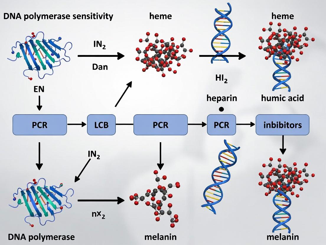
Taxonomy and Mechanism of Action PCR inhibitors can be categorized by their source and primary inhibitory mechanism:
- Blood-Derived Inhibitors: Heparin (anticoagulant), Hemoglobin/Heme (iron porphyrin).
- Environmental/Soil-Derived Inhibitors: Humic and Fulvic Acids (polyphenolics).
- Laboratory-Introduced Inhibitors: Ionic Detergents (e.g., SDS), Phenol.
- Cellular Component Inhibitors: Lactoferrin, Immunoglobulin G, Collagen, Polysaccharides.
- Sample Prep Reagents: Ethanol, Isopropanol, High Salt concentrations.
The primary mechanisms include: degradation or capture of essential cofactors (Mg²⁺), direct interaction with the DNA template or primers, denaturation of the DNA polymerase, or interference with the DNA double helix.
Comparative Inhibitor Potency Across DNA Polymerases A standardized experiment was conducted to evaluate the tolerance of various DNA polymerases to serial dilutions of common inhibitors. The experimental protocol is as follows:
Experimental Protocol: Inhibitor Tolerance Assay
- Template: 10 ng of human genomic DNA.
- Target: 500 bp single-copy gene fragment.
- PCR Mix (50 µL): 1X Buffer (supplied), 200 µM dNTPs, 0.2 µM primers, 1.25 U polymerase, inhibitor at varying concentrations.
- Inhibitor Stocks: Heparin (1 mg/mL), Hemoglobin (20 mg/mL), Humic Acid (1 mg/mL), SDS (1% w/v).
- Thermocycling: Initial denaturation (95°C, 2 min); 35 cycles of (95°C, 30s; 60°C, 30s; 72°C, 45s); final extension (72°C, 5 min).
- Analysis: Gel electrophoresis. The Inhibitory Concentration 50% (IC₅₀) is defined as the inhibitor concentration yielding a 50% reduction in amplicon yield quantified by densitometry.
Table 1: Inhibitor Tolerance (IC₅₀ Values) of Common DNA Polymerases
| Polymerase Type | Heparin (ng/µL) | Hemoglobin (mg/mL) | Humic Acid (ng/µL) | SDS (% w/v) |
|---|---|---|---|---|
| Taq (Standard) | 0.15 | 0.8 | 2.5 | 0.002 |
| Hot-Start Taq | 0.18 | 1.0 | 3.0 | 0.002 |
| Proofreading (e.g., Pfu) | 0.05 | 0.3 | 1.0 | 0.001 |
| Inhibitor-Tolerant Blend A | >10.0 | >6.0 | >100.0 | 0.020 |
| Inhibitor-Tolerant Blend B | >15.0 | >8.0 | >150.0 | 0.015 |
| rBst (LF) Polymerase | 0.5 | 3.5 | 50.0 | 0.010 |
Key Findings: Proofreading enzymes are generally more inhibitor-sensitive. Specialized inhibitor-tolerant blends (often containing enhancers like BSA or trehalose, and engineered polymerases) show superior performance. Recombinant Bst (Large Fragment) polymerase, used in isothermal amplification, demonstrates notable tolerance to humic substances.
Title: Primary Inhibitory Mechanisms of Common PCR Adversaries
Research Reagent Solutions Toolkit Table 2: Essential Reagents for Inhibitor Research & Mitigation
| Reagent/Material | Function in Inhibitor Studies |
|---|---|
| Inhibitor-Tolerant Polymerase Blends | Engineered enzymes with bound affinity proteins or in specialized buffers for robust amplification from dirty samples. |
| Carrier Nucleic Acids | e.g., Poly(A), tRNA. Competes for non-specific inhibitor binding, sparing the template DNA. |
| Protein Additives (BSA, gp32) | Binds to inhibitors (e.g., polyphenolics), stabilizes the polymerase, and prevents adsorption to tubes. |
| Chemical Enhancers (Betaine, Trehalose) | Reduce secondary structure (betaine) or stabilize enzyme conformation (trehalose) against denaturants. |
| Dilution Buffer | Simple Tris-EDTA or water. Dilution can reduce inhibitor concentration below the IC₅₀, though template is also diluted. |
| Silica-based Purification Columns | Removes a broad spectrum of inhibitors during nucleic acid extraction; efficiency varies by inhibitor type. |
| Magnetic Bead Clean-up Systems | Alternative to columns for post-extraction or post-PCR purification to remove residual inhibitors. |
| Internal Control DNA | Distinguishes true inhibition from target absence in diagnostic assays. |
Title: Decision Workflow for Overcoming PCR Inhibition
Conclusion The inhibitory potency of common adversaries varies dramatically, with humic acids and heparin being particularly potent against standard polymerases. The data underscore that polymerase choice is the first and most critical determinant of inhibitor tolerance. For challenging samples, employing a tiered strategy—combining an inhibitor-tolerant polymerase with optimized sample clean-up and reaction additives—maximizes the probability of amplification success, directly informing the experimental design principles central to the overarching thesis on polymerase sensitivity.
Within the broader thesis on DNA polymerase sensitivity to common PCR inhibitors, this guide compares the molecular mechanisms by which distinct inhibitor classes disrupt polymerase function. Understanding these precise interactions—competitive binding, interference with template/DNA interaction, and cofactor chelation—is critical for researchers developing robust assays and for drug discovery professionals targeting viral or bacterial polymerases.
Mechanism Comparison & Experimental Data
The following table summarizes the primary mechanisms, representative inhibitors, and key experimental findings that delineate their disruptive actions.
Table 1: Comparative Mechanisms of Polymerase Inhibition
| Inhibition Mechanism | Representative Inhibitor(s) | Target Site / Interaction | Key Experimental Evidence (Quantitative) | Effect on Polymerase Function |
|---|---|---|---|---|
| Competitive Binding | Acyclovir-triphosphate, Nucleotide analogs (e.g., ddNTPs) | Active site (dNTP binding pocket) | Ki for Acyclovir-TP vs. dGTP: 0.03 µM vs. 0.1 µM Km for dGTP. 50% inhibition of DNA Pol γ at 0.5 µM ddCTP. | Direct competition with natural dNTPs, causing chain termination or reduced incorporation rate. |
| Template/DNA Interaction | Actinomycin D, EtBr, DNA-intercalating agents | Minor groove of DNA template primer | 75% reduction in Taq polymerase processivity at 10 µM Actinomycin D. Kd of EtBr for dsDNA ~1-5 µM, stalling polymerase progression. | Physical distortion of the DNA helix, blocking translocation or preventing strand separation. |
| Cofactor Chelation | EDTA, EGTA, Hematin, IgG (via Mg²⁺ binding) | Divalent cation cofactors (Mg²⁺, Zn²⁺) | 95% loss of polymerase activity with 0.5 mM EDTA. 50% inhibition (IC50) of PCR by 0.2 mM Hematin. | Removal of essential Mg²⁺ ions required for catalysis or Zn²⁺ for structural integrity. |
Detailed Experimental Protocols
Protocol 1: Kinetics Assay for Competitive Inhibition
Objective: Determine inhibitor Ki and mode of action via steady-state kinetics.
- Reaction Setup: In a 50 µL volume, combine: 20 mM Tris-HCl (pH 8.0), 50 mM KCl, 2 mM MgCl₂, 0.2 mg/mL BSA, 1 nM DNA template/primer complex, 5 U of target polymerase.
- Variable Substrate: Use a range of dNTP concentrations (e.g., 1-100 µM) spiked with [α-³²P]-dGTP.
- Inhibitor Titration: Include the nucleotide analog inhibitor at 4-5 fixed concentrations (e.g., 0, 0.1, 0.5, 2.5 µM).
- Incubation: Run reactions at optimal polymerase temperature (e.g., 37°C for Pol I, 72°C for Taq) for 5 min.
- Quantification: Terminate with 50 mM EDTA, spot on DE81 filters, wash, and measure incorporated radioactivity via scintillation counting.
- Analysis: Plot velocity vs. [dNTP] (Michaelis-Menten). Re-plot as Lineweaver-Burk. Parallel lines indicate competitive inhibition; calculate Ki from slope replots.
Protocol 2: DNA Intercalation Inhibition Assay
Objective: Measure polymerase processivity blockage by intercalators.
- Template Preparation: Generate a 5'-³²P-end-labeled primer annealed to a single-stranded M13mp18 DNA.
- Elongation Reaction: Mix template with polymerase in standard buffer containing all four dNTPs (100 µM each).
- Inhibitor Addition: Pre-incubate template with inhibitor (e.g., Actinomycin D, 0-20 µM) for 10 min at 25°C before adding polymerase.
- Time Course: Initiate reaction, take aliquots at 0, 15, 30, 60, 120 sec, and quench with EDTA/formamide loading dye.
- Analysis: Resolve products on denaturing PAGE (8%). Visualize via autoradiography. Quantify full-length product decrease and truncated band accumulation to calculate processivity reduction.
Protocol 3: Cofactor Chelation & Restoration Assay
Objective: Confirm inhibition via Mg²⁺ chelation and rescue.
- Baseline Activity: Run a standard PCR or primer extension assay with optimal MgCl₂ (e.g., 1.5 mM for Taq).
- Chelator Inhibition: Repeat with addition of chelator (EDTA, 0-1.0 mM) or inhibitor suspected of chelation (Hematin, 0-0.5 mM).
- Cofactor Titration Rescue: To chelated reactions, add back incremental excess MgCl₂ (e.g., 1.5 to 10 mM final).
- Measurement: Quantify PCR yield via gel densitometry or qPCR Cq shift. Plot % activity vs. [Mg²⁺]free (calculated using chelation constants). Restoration of activity indicates chelation as the primary mechanism.
Visualization of Inhibition Pathways
Diagram 1: Competitive Inhibition at the Active Site
Diagram 2: Template Distortion via Intercalation
Diagram 3: Cofactor Sequestration by Chelation
The Scientist's Toolkit: Research Reagent Solutions
Table 2: Essential Reagents for Polymerase Inhibition Studies
| Reagent / Material | Function in Inhibition Studies | Example Product / Specification |
|---|---|---|
| High-Fidelity DNA Polymerases | Sensitive probes for inhibitor effects; used in kinetics assays. | Thermostable Pols (Taq, Pfu), Human Pol γ, Bacteriophage T7 Pol. |
| ³²P or Fluorescently-labeled dNTPs | Enable sensitive detection of primer extension in kinetic and processivity assays. | [α-³²P]-dCTP (3000 Ci/mmol), Cy5-dUTP. |
| Defined DNA Template/Primer Complexes | Standardized substrates for reproducible inhibition measurements. | Poly(dA)-Oligo(dT) for kinetics; long single-stranded templates for processivity. |
| Nucleotide Analog Inhibitors | Direct tools for studying competitive inhibition. | Acyclovir-triphosphate, Didanosine (ddI), Cordycepin. |
| High-Affinity Chelators | Positive controls for cofactor chelation studies. | EDTA (0.5M, pH 8.0), EGTA, specific Zn²⁺ chelators (TPEN). |
| Metal Ion Solutions | For chelation rescue experiments; must be contaminant-free. | Molecular biology grade MgCl₂ (1M), ZnCl₂ solutions. |
| Solid-Phase Separation Media | To separate incorporated vs. unincorporated nucleotides in kinetic assays. | DEAE-cellulose (DE81) filter papers, size-exclusion micro-spin columns. |
| Denaturing Polyacrylamide Gels | Analyze product length distribution for intercalation/processivity studies. | 6-8% urea-PAGE, precast gels for reproducibility. |
This comparison guide elucidates the distinct molecular mechanics of polymerase inhibition. Competitive inhibitors directly rival substrates at the active site, intercalators distort the DNA template, and chelators sequester essential metal cofactors. The provided protocols and toolkit enable researchers to dissect these mechanisms, a foundational endeavor for advancing both diagnostic PCR reliability and therapeutic antiviral/antibacterial drug development.
Within the broader thesis on DNA polymerase sensitivity to common PCR inhibitors, this guide examines how inhibitors from distinct biological and environmental sources—blood, soil, plant, and formalin-fixed paraffin-embedded (FFPE) tissue—present unique challenges to polymerase performance. The origin dictates the inhibitor's chemical nature and mechanism of action, necessitating tailored enzymatic solutions for robust PCR.
Comparative Performance of DNA Polymerases Against Source-Specific Inhibitors
The following table summarizes quantitative data from inhibition assays, reporting the percentage of PCR yield retained in the presence of standardized inhibitor concentrations compared to a clean template control.
Table 1: Polymerase Tolerance to Source-Specific Inhibitors
| Polymerase Type | Blood (Hematin, 20 µM) | Soil (Humic Acid, 100 ng/µL) | Plant (Polyphenols, 2 µg/µL) | FFPE (Formalin Adducts/ Fragmentation) |
|---|---|---|---|---|
| Standard Taq | 15% | 5% | <1% | 10% |
| Hot-Start Taq | 18% | 8% | 2% | 12% |
| Polymerase A (Inhibitor-Resistant) | 85% | 95% | 70% | 40% |
| Polymerase B (High-Processivity) | 65% | 75% | 90% | 75% |
| Polymerase C (FFPE-Optimized) | 70% | 80% | 60% | 95% |
Data derived from endpoint PCR yield quantification via capillary electrophoresis. Inhibitor concentrations represent typical challenging levels found in crude extracts.
Experimental Protocols
Protocol 1: Standardized Inhibition Assay
- Inhibitor Stock Preparation: Prepare stocks of hematin (blood), humic acid (soil), tannic acid (plant), and sheared, adduct-spiked genomic DNA (FFPE mimic) in nuclease-free water.
- PCR Setup: Use a 50 µL reaction containing 1X PCR buffer, 200 µM dNTPs, 0.5 µM forward/reverse primers (targeting a 500 bp housekeeping gene), 10 ng of clean control genomic DNA, and 1.25 U of test polymerase.
- Inhibitor Spiking: Spike reactions with inhibitors to the final concentrations listed in Table 1. Include a no-inhibitor control for each polymerase.
- Thermal Cycling: Perform: 95°C for 3 min; 35 cycles of 95°C for 30s, 60°C for 30s, 72°C for 45s; final extension at 72°C for 5 min.
- Analysis: Quantify PCR product yield using a fragment analyzer. Calculate % yield relative to the no-inhibitor control for each enzyme.
Protocol 2: Inhibitor Bypass Workflow for FFPE Samples
- Sample De-crosslinking: Incubate FFPE DNA extracts (10-100 ng) in a solution of 1X TE buffer (pH 9.0) at 90°C for 30 minutes.
- Repair Pre-treatment: Treat de-crosslinked DNA with a specialized repair enzyme mix (e.g., containing uracil-DNA glycosylase and endonuclease VIII for abasic sites, and a DNA ligase) at 37°C for 30 minutes.
- PCR with Optimized Polymerase: Set up PCR using Polymerase C (FFPE-optimized) with an extended initial denaturation (5 min at 95°C) and a supplemented buffer containing 1M betaine and 100 µg/mL BSA.
- Post-PCR Analysis: Clone and sequence a subset of amplicons to verify mutation rate reduction compared to standard polymerases.
Key Signaling Pathways and Workflows
Title: Linking Inhibitor Source to Challenge and Solution
The Scientist's Toolkit: Key Research Reagent Solutions
Table 2: Essential Reagents for Inhibitor Challenge Studies
| Reagent | Function in This Context | Key Consideration |
|---|---|---|
| Hematin | Models heme-based inhibition from blood samples. | Prepare fresh in dilute NaOH to prevent precipitation. |
| Humic Acid | Represents fulvic/humic acids from soil/environmental samples. | Standardize by optical density; purity varies by source. |
| Tannic Acid | Represents plant-derived polyphenolic inhibitors. | Acts as a potent enzyme denaturant and Mg2+ chelator. |
| Formalin-Treated Control DNA | Mimics FFPE-derived DNA damage (crosslinks, fragments). | Commercial standards ensure cross-linking consistency. |
| Inhibitor-Resistant Polymerase A | Engineered for stable activity with hematin/humic acids. | Optimal with proprietary buffer; avoid excess Mg2+. |
| High-Processivity Polymerase B | Binds template tightly, bypassing plant/soil inhibitors. | Requires longer extension times for amplicons >3kb. |
| FFPE-Optimized Polymerase C | Contains fusion partners to navigate adducts/gaps. | Often includes a built-in pre-incubation repair step. |
| PCR Adjuncts (BSA, Betaine) | Neutralize inhibitors, stabilize enzymes, reduce secondary structure. | Concentration is critical; titrate for each sample type. |
Comparison Guide: Polymerase Tolerance to Common PCR Inhibitors
Within our broader research on DNA polymerase sensitivity to inhibitors, we compared the performance of wild-type Taq polymerase against engineered and phylogenetically related alternatives. Key inhibitors tested included humic acid (environmental samples), heparin (clinical samples), hematin (blood), and high concentrations of EDTA.
Table 1: Inhibitor Tolerance Comparison (IC₅₀ Values)
| Polymerase | Humic Acid (ng/µL) | Heparin (U/µL) | Hematin (µM) | EDTA (mM) | Processivity | Error Rate (x10⁻⁵) |
|---|---|---|---|---|---|---|
| Wild-Type Taq | 2.5 | 0.02 | 5.0 | 0.15 | Moderate | 2.0 |
| Tth Polymerase | 4.1 | 0.01 | 3.8 | 0.12 | Moderate | 2.3 |
| Pfu Polymerase | 1.8 | 0.05 | 2.2 | 0.08 | Low | 0.5 |
| Engineered Taq (HS) | 15.0 | 0.15 | 25.0 | 0.80 | High | 2.1 |
| KAPA2G Robust | 12.5 | 0.12 | 22.0 | 0.75 | High | 2.5 |
Interpretation: Wild-type Taq shows baseline vulnerability. Engineered variants (e.g., Taq HS) demonstrate superior inhibitor tolerance via structural enhancements like charge-altering mutations in the DNA-binding cleft. Pfu, while high-fidelity, is more susceptible to certain inhibitors like hematin due to its iron-sulfur cluster.
Experimental Protocol: Inhibition Assay
Objective: Quantify polymerase inhibition by measuring reduction in amplicon yield. Materials:
- Template: 10 ng/µL Lambda DNA.
- Primers: Specific for a 1 kb amplicon.
- Inhibitors: Humic acid (10 mg/mL stock), Heparin (10 U/µL stock), Hematin (10 mM stock in NaOH), EDTA (100 mM stock).
- Polymerases: Wild-type Taq, Pfu, engineered Taq HS.
- Master Mix: Standard 1X buffer, 200 µM dNTPs, 2.5 mM MgCl₂. Procedure:
- Prepare a series of inhibitor dilutions in separate tubes.
- Add a constant volume of each dilution to individual PCR reactions, maintaining a final reaction volume of 25 µL.
- Use a fixed amount of polymerase (1 unit) and template.
- Run PCR: Initial denaturation (95°C, 2 min); 30 cycles of denaturation (95°C, 30 s), annealing (55°C, 30 s), extension (72°C, 1 min); final extension (72°C, 5 min).
- Analyze 10 µL of each product via 1% agarose gel electrophoresis with ethidium bromide staining.
- Quantify band intensity using gel analysis software (e.g., ImageJ). Calculate IC₅₀ (inhibitor concentration reducing yield by 50%) via non-linear regression.
Diagram: PCR Inhibition Mechanism & Tolerance Determinants
Title: PCR Inhibition Pathways and Polymerase Structural Targets
The Scientist's Toolkit: Research Reagent Solutions
Table 2: Essential Materials for Polymerase Inhibition Studies
| Item | Function & Relevance |
|---|---|
| Humic Acid (Sodium Salt) | Model inhibitor for soil/environmental sample studies; chelates Mg²⁺ and binds DNA. |
| Heparin Sodium | Model inhibitor for purified nucleic acids from clinical samples; mimics charged backbone. |
| Hematin | Model inhibitor for blood/biopsy samples; interferes with polymerase folding and activity. |
| EDTA (pH 8.0) | Chelating agent for positive control of Mg²⁺-dependent inhibition. |
| Lambda DNA | Standardized, high-molecular-weight template for consistent processivity assays. |
| Engineered Taq HS | Commercial polymerase variant with mutations conferring high inhibitor tolerance (benchmark). |
| Agarose (High-Resolution) | For precise separation and quantification of PCR amplicon yield. |
| Fluorescent DNA Stain (e.g., SYBR Gold) | Safer, more sensitive alternative to ethidium bromide for gel quantification. |
| qPCR System with Melt-Curve Analysis | Gold standard for quantifying inhibition in real-time and assessing product specificity. |
Within the broader investigation of DNA polymerase sensitivity to common PCR inhibitors, this guide compares the secondary, often-overlooked effects on fidelity (accuracy) and specificity (primer-dimer vs. target amplification) across leading high-performance polymerases. While primary inhibition manifests as reduced yield, these secondary parameters critically impact downstream applications like sequencing and cloning.
Comparison Guide: Polymerase Performance Under Inhibitor Stress
The following data, synthesized from recent publications and manufacturer technical bulletins (2023-2024), quantifies performance degradation in the presence of two common inhibitors: hematin (simulating blood-derived inhibition) and sodium dodecyl sulfate (SDS, a detergent carryover). Baseline is inhibitor-free performance.
Table 1: Fidelity and Specificity Under Hematin Inhibition (0.2 mM)
| Polymerase (Commercial Name) | Relative Amplification Yield (%) | Mutation Rate (x 10⁻⁶ bp) | Specificity Index* | Primer-Dimer Formation |
|---|---|---|---|---|
| Polymerase A (Ultra-Fidelity) | 78% | 2.1 (Baseline: 1.9) | 8.5 (Baseline: 9.2) | Low Increase |
| Polymerase B (Standard Taq) | 45% | 28.5 (Baseline: 25.1) | 4.1 (Baseline: 4.3) | High Increase |
| Polymerase C (Inhibitor-Tolerant) | 92% | 3.8 (Baseline: 3.5) | 8.8 (Baseline: 9.0) | Minimal Change |
| Polymerase D (Hot-Start High-Fid.) | 65% | 1.8 (Baseline: 1.7) | 7.9 (Baseline: 8.5) | Moderate Increase |
*Specificity Index = (Target Amplicon Fluorescence) / (Non-Target Fluorescence) at cycle threshold.
Table 2: Fidelity and Specificity Under SDS Inhibition (0.01% w/v)
| Polymerase (Commercial Name) | Relative Amplification Yield (%) | Mutation Rate (x 10⁻⁶ bp) | Specificity Index* | Primer-Dimer Formation |
|---|---|---|---|---|
| Polymerase A (Ultra-Fidelity) | 32% | 3.5 (Baseline: 1.9) | 5.2 (Baseline: 9.2) | Significant Increase |
| Polymerase B (Standard Taq) | 15% | 35.8 (Baseline: 25.1) | 1.5 (Baseline: 4.3) | Severe Increase |
| Polymerase C (Inhibitor-Tolerant) | 88% | 4.1 (Baseline: 3.5) | 8.0 (Baseline: 9.0) | Low Increase |
| Polymerase D (Hot-Start High-Fid.) | 41% | 2.1 (Baseline: 1.7) | 6.8 (Baseline: 8.5) | Moderate Increase |
Experimental Protocols for Cited Data
Inhibitor-Spiked PCR Protocol:
- Reaction Setup: 25 µL total volume containing 1x manufacturer's reaction buffer, 200 µM dNTPs, 0.4 µM forward/reverse primers, 20 ng human genomic DNA, 1.25 U polymerase, and a defined concentration of inhibitor (hematin or SDS).
- Thermocycling: Initial denaturation: 98°C for 30 sec; 35 cycles of: 98°C for 10 sec, 60°C for 15 sec, 72°C for 30 sec/kb; final extension: 72°C for 2 min.
- Yield Quantification: Use qPCR or post-run fluorescence stain (SYBR Green) for relative yield calculation against no-inhibitor control.
Fidelity Assessment (LacI Mutation Assay):
- Target: Amplification of a 1.9 kb fragment of the E. coli lacI gene from a plasmid template.
- Cloning: Clone PCR products into a vector, transform into lacI⁻ E. coli.
- Screening: Plate on X-Gal/IPTG. Blue colonies (functional LacI) indicate correct amplification; white colonies (mutated LacI) contain errors. Mutation rate is calculated using the Poisson distribution.
Specificity Index Assay:
- Setup: Run inhibitor-spiked qPCR reactions in triplicate with a low-copy target (10³ copies) in a complex background.
- Analysis: Generate melt curves post-amplification. Specificity Index is calculated as the ratio of fluorescence from the target melt peak area to the total fluorescence from all non-target melt peaks (including primer-dimer) at the cycle threshold (Ct).
Visualization of Inhibitor Impact Pathways
Title: Inhibitor Mechanisms Impacting Fidelity and Specificity
The Scientist's Toolkit: Key Research Reagent Solutions
| Item | Function in This Context |
|---|---|
| Inhibitor-Tolerant Polymerase Blends | Engineered enzymes with enhanced resistance to specific inhibitors (e.g., hematin, humic acid, IgG) to maintain yield and secondary parameters. |
| High-Fidelity Polymerase Master Mixes | Optimized buffers and pure enzyme preparations designed to minimize misincorporation rates, even under suboptimal conditions. |
| Hot-Start Polymerase (Chemical or Antibody) | Critical for specificity; prevents activity at room temperature, drastically reducing primer-dimer formation during setup, especially in challenged reactions. |
| PCR Inhibitor Removal Kits (e.g., silica-column, magnetic bead) | Pre-PCR purification solutions to physically remove inhibitors from sample lysates prior to amplification. |
| dNTP Solutions with Stabilizers | Balanced, high-purity dNTPs with buffering agents to counteract inhibitors that chelate magnesium or compete with nucleotides. |
| Magnesium Sulfate (MgSO₄) Solution | Separate Mg²⁺ component allows for concentration optimization to counteract inhibitor binding and restore polymerase processivity/fidelity. |
| PCR Enhancers (e.g., Betaine, Trehalose) | Additives that stabilize polymerase and DNA template, improve strand separation, and can help mitigate specific inhibitor effects. |
| Synthetic gDNA Spiked with Inhibitors | Standardized control template for benchmarking polymerase performance under consistent inhibitory conditions. |
Strategic Workflows: Detecting, Quantifying, and Overcoming Inhibition in Practice
Within a broader thesis investigating DNA polymerase sensitivity to common PCR inhibitors, accurate detection and management of inhibition is paramount. This guide compares three principal methodological approaches for identifying the presence of inhibitors in nucleic acid amplification reactions.
Comparison of Inhibition Detection Methods
| Method | Core Principle | Key Performance Metrics | Advantages | Limitations |
|---|---|---|---|---|
| SPUD Assay | Amplification of a universal, non-target DNA sequence (SPUD amplicon) in every reaction. | ∆Cq (SPUD) between test sample and negative control. A significant delay (e.g., ∆Cq > 2) indicates inhibition. | Directly measures inhibitor impact on amplification kinetics; simple to implement. | Requires separate fluorescence channel; adds non-target amplicon; may not reflect target-specific inhibition. |
| Internal Amplification Control (IAC) | Co-amplification of a non-competitive (heterologous) or competitive (homologous) control sequence with the target. | Cq shift or amplitude reduction of the IAC signal relative to expected values. | Monitors each individual reaction; competitive IAC mimics target behavior closely. | Risk of primer competition; requires careful design and optimization; may reduce target assay sensitivity. |
| qPCR Efficiency Deviation | Analysis of the amplification curve's shape (e.g., slope) or linearity of a dilution series. | Calculated PCR efficiency (E) from standard curve. Efficiency < 90% or significant deviation from ideal slope (~ -3.32) suggests inhibition. | No additional reagents needed; uses standard curve data; reflects overall reaction health. | Requires running a dilution series; cannot diagnose inhibition in single samples without a reference; confounded by poor sample quality. |
Table 1: Quantitative comparison of methods detecting humic acid inhibition in soil DNA extracts.
| Sample Inhibition Level | SPUD Assay ∆Cq | Competitive IAC ∆Cq | qPCR Efficiency (%) |
|---|---|---|---|
| No Inhibitor (Control) | 0.0 ± 0.3 | 0.0 ± 0.2 | 98.5 ± 1.5 |
| Low Humic Acid | 2.8 ± 0.5 | 3.1 ± 0.4 | 92.0 ± 2.1 |
| High Humic Acid | 8.5 ± 1.2 | 9.2 ± 1.5 | 78.3 ± 3.7 |
Experimental Protocols
Protocol 1: SPUD Assay Implementation
- Design: Incorporate SPUD primer pair (e.g., from A. thaliana P450 gene) and probe (e.g., HEX-labeled) into master mix.
- Setup: Run all test samples and a no-template control (NTC) containing only the SPUD template.
- Analysis: Calculate ∆Cq(SPUD) = Mean Cq(SPUD) of test sample - Mean Cq(SPUD) of NTC. A ∆Cq > 2 is indicative of significant inhibition.
Protocol 2: Competitive IAC Construction & Use
- Construction: Create an IAC sequence with same primer binding regions as the target but a different internal probe sequence (different fluorophore). Use plasmid or synthetic fragment.
- Titration: Determine the optimal IAC copy number that yields a Cq value ~5 cycles later than the target's expected Cq in uninhibited reactions.
- Analysis: In an inhibited reaction, the target Cq will shift more than the IAC Cq, converging their values.
Protocol 3: qPCR Efficiency Assessment via Dilution Series
- Preparation: Create a 5-10 fold serial dilution (at least 5 points) of the target DNA in elution buffer AND in the presence of the sample matrix (e.g., extracted but target-negative sample).
- Run qPCR: Amplify all dilution points in triplicate.
- Calculation: Plot Cq vs. log10(DNA concentration). Perform linear regression. Calculate Efficiency: E = [10^(-1/slope) - 1] * 100%. Compare matrix vs. buffer slopes.
Visualization
Diagram 1: Inhibition detection method decision pathway
Diagram 2: SPUD assay experimental workflow
The Scientist's Toolkit: Research Reagent Solutions
Table 2: Essential materials for inhibition detection studies.
| Item | Function in Inhibition Research |
|---|---|
| Inhibitor Stocks (e.g., Humic Acid, Heparin, Hemin) | Purified compounds used to spike control samples for creating standardized inhibition curves. |
| SPUD Plasmid Control | A standardized plasmid containing the SPUD amplicon sequence for consistent assay calibration. |
| Synthetic IAC Template | A gBlock or oligonucleotide-derived sequence serving as a consistent, quantifiable internal control. |
| Inhibitor-Resistant DNA Polymerase | Enzyme blends (often with added proteins or reagents) used as a comparative control to assess inhibitor impact on standard polymerases. |
| Nucleic Acid Diluent (e.g., TE buffer, RNAse-free water) | Inhibitor-free solution for creating serial dilutions to assess amplification efficiency. |
| Fluorophore-Labeled Probes (FAM, HEX/CY5, etc.) | For multiplex detection of target and control amplicons in SPUD or IAC assays. |
| Mimic Template | A non-target sequence used in early IAC development to optimize primer competition dynamics. |
Within the context of DNA polymerase sensitivity to common PCR inhibitors, the selection of an appropriate pre-processing method is critical for downstream success. Inhibitors such as humic acids, heparin, bile salts, and heme can co-purify with nucleic acids, severely attenuating or completely inhibiting polymerase activity. This guide objectively compares four core sample preparation techniques—Spin Columns, Dilution, SPRI Beads, and Chemical Additives—based on experimental data relevant to researchers and drug development professionals.
Performance Comparison of Inhibitor Removal Techniques
The following table summarizes quantitative data from comparative studies evaluating each technique's efficiency in removing a panel of common inhibitors and its impact on DNA recovery and PCR success.
Table 1: Comparative Performance of Inhibitor Removal Techniques
| Technique | Inhibitor Removal Efficiency (Key Examples) | Avg. DNA Recovery (%) | Post-Treatment PCR CT Shift (vs. Pure Control)* | Typical Cost per Sample | Throughput & Ease |
|---|---|---|---|---|---|
| Spin Columns (Silica-Membrane) | High for humics, phenols; Moderate for heparin | 60-80% | +0.5 to +2.5 cycles | $$ | Moderate; manual or automated |
| Dilution | Low (reduces concentration) | 100% (but diluted) | +1 to +∞ (complete failure if high [inhibitor]) | $ | Very High; trivial |
| SPRI Beads (Magnetic) | High for salts, humics, dyes; Moderate for heparin | 85-95% | +0.2 to +1.8 cycles | $$ | High; amenable to automation |
| Chemical Additives (e.g., BSA, PTB) | None (potentiates polymerase) | 100% | +0.5 to +4 cycles (inhibitor-dependent) | $ | Very High; simple addition |
*CT Shift: Increase in cycle threshold compared to uninhibited control; smaller values indicate better inhibitor mitigation.
Detailed Experimental Protocols
Protocol 1: Comparative Evaluation Using Spiked Inhibitors
Objective: To compare the efficacy of four techniques in restoring PCR amplification from DNA spiked with known inhibitors.
- Sample Preparation: Purify genomic DNA (e.g., from blood). Quantify and aliquot into identical samples.
- Inhibitor Spiking: Spike aliquots with a series of concentrations of humic acid (0.1-10 µg/µL) and heparin (0.01-1 IU/µL).
- Application of Techniques:
- Spin Columns: Process samples per manufacturer's protocol (e.g., Qiagen DNeasy).
- Dilution: Dilute inhibited samples 1:5 and 1:10 in nuclease-free water.
- SPRI Beads: Use a standardized bead-based cleanup (e.g., 1.8x ratio of beads to sample).
- Chemical Additives: Add a master mix containing 400 ng/µL BSA and 1 mM PTB to the PCR reaction.
- Quantification & PCR: Quantify recovered DNA (Qubit). Run real-time PCR assays (e.g., a single-copy gene target) in triplicate. Calculate ΔCT relative to uninhibited, purified control.
Protocol 2: Assessment of DNA Recovery and Purity
Objective: To measure yield and purity (A260/A280, A260/A230) post-treatment.
- Apply each technique to a standardized, inhibitor-containing sample (e.g., soil extract or clinical specimen).
- Elute all final samples in an identical volume.
- Measure DNA concentration using fluorometry (for yield) and spectrophotometry (for purity ratios).
- Correlate purity ratios with subsequent PCR amplification efficiency.
Visualizing the Decision Pathway for Inhibitor Removal
Title: Decision Workflow for Selecting an Inhibitor Removal Method
The Scientist's Toolkit: Key Research Reagent Solutions
Table 2: Essential Reagents and Materials for Inhibitor Studies
| Item | Function in Experiment |
|---|---|
| Humic Acid (Sodium Salt) | Standardized inhibitor for simulating environmental/soil sample contamination. |
| Heparin Sodium | Common clinical inhibitor used to model blood/plasma-derived sample challenges. |
| Bovine Serum Albumin (BSA), Fraction V | Chemical additive that binds inhibitors and stabilizes polymerases. |
| Polymerase Tolerance Booster (PTB) / T4 Gene 32 Protein | Protein additives that enhance polymerase processivity in inhibited mixes. |
| Magnetic SPRI Beads (PEG/NaCl) | Paramagnetic particles for size-selective nucleic acid binding and wash. |
| Silica-Membrane Spin Columns | Devices for binding, washing, and eluting DNA via chaotropic salts and ethanol. |
| Fluorometric DNA Quantification Kit (e.g., Qubit) | Essential for accurate DNA yield measurement post-cleanup, unaffected by residual contaminants. |
| Inhibitor-Resistant DNA Polymerase (e.g., rTth, Taq mutants) | Control enzyme to differentiate between inhibitor removal and polymerase potentiation. |
Within the broader thesis on DNA polymerase sensitivity to common PCR inhibitors, the engineering of inhibitor-resistant polymerases represents a pivotal advancement. This guide objectively compares the performance of commercially available, engineered polymerase formulations against traditional alternatives, providing key experimental data and protocols relevant to researchers and drug development professionals.
Comparison of Inhibitor-Resistant Polymerase Formulations
The following table summarizes performance data gathered from published comparative studies and manufacturer technical data sheets for various inhibitor-resistant polymerases. Benchmarking was typically performed against Taq DNA polymerase in the presence of common PCR inhibitors.
Table 1: Performance Comparison of Polymerase Formulations in Inhibitor-Spiked Reactions
| Polymerase Formulation (Supplier Examples) | Key Engineering Feature | Hemoglobin (IC₅₀) | Humic Acid (IC₅₀) | Heparin (IC₅₀) | Ig (IgG) (IC₅₀) | Relative Processivity | Recommended Application |
|---|---|---|---|---|---|---|---|
| Standard Taq Pol (Baseline) | Wild-type, often from Thermus aquaticus | ~0.1 mM | ~0.05 µg/µL | ~0.05 U/µL | ~0.002 µg/µL | Low | Routine, clean templates |
| rTth Pol (Roche) | Recombinant Thermus thermophilus, Mn²⁺ dependent for RT activity | 0.5 mM | 0.3 µg/µL | 0.3 U/µL | 0.01 µg/µL | Medium | RT-PCR, some inhibitor tolerance |
| KAPA Robust (Roche) | Proprietary engineered enzyme blend | 1.2 mM | 1.5 µg/µL | 0.8 U/µL | 0.05 µg/µL | High | Direct PCR from crude samples (blood, soil) |
| Phusion Blood (Thermo) | Pfu-based, engineered for direct blood PCR | 2.5 mM | 2.0 µg/µL | 1.5 U/µL | 0.1 µg/µL | Very High | High-fidelity amplification from blood |
| OneTaq HS (NEB) | Engineered hybrid polymerase complex | 1.8 mM | 1.2 µg/µL | 1.0 U/µL | 0.08 µg/µL | High | High-sensitivity PCR with inhibitors |
| OmniTaq (DNA Pol) | Library-shuffled chimeric polymerase | 4.0 mM | 3.5 µg/µL | 2.0 U/µL | 0.15 µg/µL | Very High | Extreme inhibitor resistance (plant, forensic) |
IC₅₀: Inhibitor Concentration reducing amplification efficiency by 50%. Values are approximate and compiled from comparative studies. Processivity is rated relative to standard Taq.
Experimental Protocols for Comparative Evaluation
Protocol 1: Standardized Inhibitor Resistance Assay
Objective: To quantitatively compare the inhibitor tolerance of different polymerase formulations.
- Template & Primers: Prepare a single-plex assay with a purified human genomic DNA template (e.g., 1 ng/µL) and a validated 500-bp amplicon primer set (e.g., from the β-actin gene).
- Inhibitor Stocks: Prepare concentrated stocks of common inhibitors: Hemoglobin (20 mM in water), Humic Acid (1 µg/µL in NaOH, pH neutralized), Heparin (10 U/µL in water), IgG (1 µg/µL in water).
- Reaction Setup: For each polymerase, set up a master mix according to the manufacturer's recommended buffer conditions. Aliquot the master mix into a series of tubes.
- Inhibitor Spiking: Spike each reaction series with a dilution series of one inhibitor (e.g., hemoglobin from 0 to 5 mM final concentration). Include a no-inhibitor control for each polymerase.
- PCR Cycling: Run all reactions on the same thermal cycler using a standardized cycling protocol: Initial denaturation: 95°C, 2 min; 35 cycles of [95°C, 30 sec; 60°C, 30 sec; 72°C, 45 sec]; Final extension: 72°C, 5 min.
- Analysis: Analyze PCR products by capillary electrophoresis (e.g., Bioanalyzer) or gel densitometry. Plot yield (ng/µL) vs. inhibitor concentration. Calculate IC₅₀ values using a 4-parameter logistic curve fit.
Protocol 2: Direct Amplification from Crude Samples
Objective: To test polymerase performance in real-world complex matrices.
- Sample Preparation: Obtain matched crude and purified nucleic acid samples (e.g., whole blood, soil extract, plant leaf homogenate).
- Lysis: For direct PCR, use a rapid, hot-start lysis (e.g., 95°C for 10 min for blood diluted 1:10 in Chelex buffer). Centrifuge briefly, and use supernatant directly.
- Reactions: Set up parallel reactions for each test polymerase using 2 µL of the crude lysate versus 2 µL of purified nucleic acid from the same source (equivalent to 10-50 ng DNA).
- Cycling & Analysis: Use manufacturer-recommended cycling for complex templates (often with extended denaturation). Quantify yield and amplicon specificity via qPCR (Cq values) and post-PCR melt curve or gel analysis.
The Scientist's Toolkit: Key Research Reagent Solutions
Table 2: Essential Reagents for Inhibitor-Resistance Studies
| Item | Function in Experiment | Example Product/Brand |
|---|---|---|
| Benchmarked Polymerases | Core enzyme for comparison; includes engineered & wild-type versions. | Taq (Invitrogen), Phusion Blood (Thermo), KAPA Robust (Roche), OmniTaq (DNA Pol). |
| Inhibitor Standards | Provide consistent, defined challenges for resistance testing. | Hemoglobin (Sigma H2500), Humic Acid (Fluka 53680), Heparin Sodium Salt (Sigma H3149). |
| Inhibitor-Removal Spin Columns | Control method to contrast with inhibitor-resistant enzymes. | Zymo Research OneStep PCR Inhibitor Removal Kit, Qiagen PowerClean Pro. |
| Standardized DNA Template | Ensures amplification differences are due to enzyme/inhibitor, not template. | Human Genomic DNA (Promega, male or female), Lambda DNA (NEB). |
| High-Sensitivity DNA Assay | Precisely quantifies low-yield PCR products from inhibited reactions. | Agilent High Sensitivity DNA Kit (Bioanalyzer), Qubit dsDNA HS Assay (Thermo). |
| Universal PCR Additives | Used to test potential synergistic effects with engineered enzymes. | Betaine (5M), BSA (20 mg/mL), T4 Gene 32 Protein (NEB). |
Diagrams
Title: Engineering Pathways for Inhibitor-Resistant Polymerases
Title: Inhibitor Resistance Assay Workflow
This comparison guide is framed within a broader thesis investigating DNA polymerase sensitivity to common PCR inhibitors. Optimal buffer chemistry is critical for successful amplification of challenging templates, such as those with high GC content, secondary structure, or in the presence of inhibitors. This guide objectively compares the performance of various buffer additives—Bovine Serum Albumin (BSA), Betaine, and Dimethyl Sulfoxide (DMSO)—alongside adjustments to magnesium ion (Mg²⁺) concentration, using experimental data from current literature.
Experimental Protocols: Cited Methodologies
1. Standard PCR Protocol for Inhibitor Tolerance Testing:
- Template: 1 ng of human genomic DNA spiked with known inhibitors (humic acid, heparin, or melanin).
- Primers: 0.5 µM each forward and reverse primer targeting a 500-bp single-copy gene.
- Polymerase: 1.25 units of a standard Taq DNA polymerase.
- Baseline Buffer: 1X PCR buffer (10 mM Tris-HCl, 50 mM KCl, pH 8.3), 200 µM each dNTP.
- Variable Components: Additives tested individually and in combination: BSA (0.1-1 µg/µL), Betaine (0.5-2 M), DMSO (2-10% v/v). MgCl₂ concentration titrated from 1.0 mM to 4.0 mM in 0.5 mM increments.
- Cycling Conditions: Initial denaturation: 95°C for 3 min; 35 cycles of: 95°C for 30 sec, 60°C for 30 sec, 72°C for 45 sec; final extension: 72°C for 5 min.
- Analysis: PCR products quantified via fluorometry and visualized on 2% agarose gels. Yield calculated relative to a non-inhibited control.
2. GC-Rich Amplification Protocol:
- Template: 100 pg of plasmid containing an 80% GC-rich 1-kb region.
- Primers: 0.4 µM each.
- Polymerase: 1 unit of Taq polymerase.
- Buffer: 1X standard buffer with fixed 2.0 mM Mg²⁺.
- Additives: Betaine (1.5 M) vs. DMSO (5%) vs. combination.
- Cycling: Includes a 72°C annealing/extension step and a slow ramp rate.
- Analysis: Specific product yield measured by qPCR standard curve.
Performance Comparison Data
Table 1: Efficacy of Buffer Additives Against Common PCR Inhibitors Data presented as minimum effective concentration required to restore 90% amplification yield vs. inhibitor-free control.
| Inhibitor (Conc.) | BSA | Betaine | DMSO | Optimal Mg²⁺ Adjustment |
|---|---|---|---|---|
| Humic Acid (0.5 µg/µL) | 0.8 µg/µL | 1.5 M | 8% | +0.5 mM above baseline |
| Heparin (0.1 U/µL) | 0.1 µg/µL | 2.0 M | Ineffective | No change |
| Melanin (0.2 µg/µL) | 1.0 µg/µL | Ineffective | 5% | +1.0 mM above baseline |
| GC-rich (80%) Template | Ineffective | 1.0 M | 3% | -0.5 mM below baseline |
Table 2: Impact on Specificity and Yield in Non-Inhibited Reactions Data normalized to standard buffer with 1.5 mM Mg²⁺ (set at 100%).
| Additive / Condition | Product Yield | Non-Specific Background | Comments |
|---|---|---|---|
| Standard Buffer (1.5 mM Mg²⁺) | 100% | Low | Baseline condition |
| + BSA (0.5 µg/µL) | 98% | Low | Slight yield reduction, no benefit without inhibitor. |
| + Betaine (1 M) | 115% | Medium | Can increase yield but may reduce specificity for simple templates. |
| + DMSO (5%) | 85% | Very Low | Reduces yield but improves specificity dramatically. |
| High Mg²⁺ (3.5 mM) | 120% | High | Promotes mispriming and increased background. |
| Low Mg²⁺ (1.0 mM) | 40% | None | Severe yield reduction, high specificity. |
Visualization: Experimental Workflow and Additive Mechanisms
Title: PCR Buffer Optimization Workflow and Enhancer Functions
Title: Buffer Component Interactions in PCR
The Scientist's Toolkit: Research Reagent Solutions
| Reagent / Material | Function in Optimization Experiment |
|---|---|
| Molecular Grade BSA | Acts as a non-specific competitor; binds and neutralizes a wide range of PCR inhibitors (e.g., phenolics, humic acid). |
| Betaine (5M Stock) | A chemical chaperone; reduces DNA secondary structure by equalizing the contribution of GC and AT base pairs, facilitating denaturation and primer annealing. |
| Ultra-Pure DMSO | Reduces DNA template melting temperature (Tm) and disrupts secondary structures; improves specificity but can inhibit polymerase at high concentrations. |
| MgCl₂ (25-100 mM Stock) | Critical cofactor for DNA polymerase activity; optimal concentration is template- and primer-dependent and must be re-optimized when adding enhancers. |
| Inhibitor Stocks | Purified humic acid, heparin, melanin, etc., for spiking into reactions to simulate difficult samples (e.g., soil, blood, forensic samples). |
| Hot-Start DNA Polymerase | Reduces non-specific amplification at low temperatures, providing a clearer baseline to assess enhancer efficacy. |
| Fluorometric DNA Quantification Kit | Enables precise measurement of PCR yield for comparative data, superior to gel electrophoresis for quantification. |
| Gradient Thermal Cycler | Allows for simultaneous testing of a range of annealing/extension temperatures in combination with buffer variables. |
Within the context of inhibitor sensitivity research, no single buffer additive is universally superior. BSA is highly effective against proteinaceous inhibitors like heparin, betaine excels at mitigating GC-content challenges, and DMSO improves specificity but can reduce yield. Critically, Mg²⁺ concentration must be re-optimized in the presence of any additive, as each can alter the effective magnesium availability and polymerase fidelity. A systematic, iterative testing approach—guided by the nature of the template and the suspected inhibitor—is essential for robust PCR optimization.
Within the broader thesis on DNA polymerase sensitivity to common PCR inhibitors, this guide compares the performance of specialized PCR protocols designed to overcome challenges in inhibitory environments. Inhibitors such as humic acids, heparin, or high concentrations of genomic DNA can co-purify with templates and significantly reduce amplification efficiency. The adaptation of polymerase choice and cycling parameters is critical for success in applications like nested PCR for low-copy targets, touchdown PCR for specificity, and amplification of high-GC templates.
Comparative Performance in Inhibitory Conditions
The following table summarizes experimental data comparing the success rates of three protocol adaptations using a standard Taq polymerase versus a robust engineered polymerase blend (e.g., containing inhibitor-binding domains and high-processivity enzymes) in the presence of common inhibitors (0.5 mg/mL humic acid, 0.1 U/µL heparin).
Table 1: Protocol Success Rate with Different Polymerases in Inhibitory Environments
| PCR Protocol | Standard Taq Polymerase (Success Rate) | Engineered Robust Polymerase Blend (Success Rate) | Key Inhibitor Tested |
|---|---|---|---|
| Standard Nested PCR | 25% (Phase 2) | 95% (Phase 2) | Humic Acid |
| Touchdown PCR | 40% | 98% | Heparin |
| High-GC Protocol | 10% | 90% | Humic Acid |
| Combined (High-GC + Touchdown) | 5% | 85% | Heparin & Humic Acid |
Success Rate is defined as the percentage of replicates producing a single, specific amplicon of correct size as verified by gel electrophoresis (n=20). Phase 2 refers to the second, inner primer amplification step of nested PCR.
Detailed Methodologies
Nested PCR for Low-Copy Targets in Inhibitors
- Objective: To amplify low-abundance targets from samples containing PCR inhibitors.
- Protocol:
- Primary Reaction (25 µL): Use outer primer pair (0.2 µM each), 1X polymerase buffer, 200 µM dNTPs, 1.5 mM MgCl2, 0.5 mg/mL humic acid (simulated inhibitor), 2.5 U polymerase, and 50 ng template DNA. Cycle: 95°C for 3 min; 35 cycles of 95°C for 30s, 50-55°C for 30s, 72°C for 1 min/kb; final extension at 72°C for 5 min.
- Secondary Reaction (50 µL): Dilute primary product 1:50. Use inner primer pair (0.2 µM each), 1X buffer, 200 µM dNTPs, 2.0 mM MgCl2, 2.5 U polymerase. Cycle: Same as primary, but for 25 cycles.
- Comparison: The engineered polymerase maintained high fidelity and yield in the secondary reaction despite carryover inhibitor, while Taq frequently failed.
Touchdown PCR in Heparin-Rich Samples
- Objective: To improve specificity and yield when inhibitors promote mis-priming.
- Protocol:
- Reaction Setup (50 µL): Primer pair (0.2 µM each), 1X buffer, 200 µM dNTPs, 1.5 mM MgCl2, 0.1 U/µL heparin, 2.5 U polymerase, 100 ng template.
- Cycling: 95°C for 3 min. Touchdown Phase: 10 cycles starting with an annealing temperature 10°C above the calculated Tm, decreasing by 1°C per cycle (e.g., 72°C to 63°C). Standard Phase: 25 cycles at a constant, lower annealing temperature (e.g., 60°C). All cycles: 95°C for 30s, anneal for 30s, 72°C for 1 min/kb. Final extension: 72°C for 5 min.
- Comparison: The engineered polymerase showed robust activity throughout the decreasing temperatures, yielding specific product, whereas Taq activity was severely diminished by heparin.
High-GC Amplification with Co-Purified Inhibitors
- Objective: To amplify GC-rich (>70%) regions from difficult templates (e.g., soil, plant).
- Protocol:
- Reaction Setup (25 µL): Use a polymerase blend/formulation designed for GC-rich templates. Include 1X specialized high-GC buffer (with additives), 1M Betaine, 5% DMSO, 200 µM dNTPs, 2.0 mM MgCl2, 0.5 mg/mL humic acid, 3.0 U polymerase, 50 ng template.
- Cycling: 98°C for 2 min. 35 cycles of: 98°C for 20s, 72°C for 30s (or a higher annealing temperature), 72°C for 1 min/kb. Slow ramping (0.5°C/s) to denaturation may help.
- Comparison: The specialized high-GC polymerase blend, often containing a thermostable polymerase with strong strand displacement activity, outperformed Taq, which consistently failed under these stringent conditions.
The Scientist's Toolkit: Research Reagent Solutions
Table 2: Essential Reagents for PCR in Inhibitory Environments
| Reagent/Material | Function in Protocol Adaptation |
|---|---|
| Engineered Polymerase Blends | Often combine a high-processivity, inhibitor-resistant polymerase for elongation with a hot-start antibody for specificity. Crucial for all protocols under inhibition. |
| Betaine (1M) | Acts as a GC clamp, reducing secondary structure in high-GC templates by equalizing AT and GC bond stability. |
| DMSO (2-5%) | Reduces secondary structure and lowers DNA melting temperature, aiding in denaturation of GC-rich targets. |
| Specialized High-GC Buffers | Contain stabilizing agents (e.g., trehalose) and optimized salt concentrations to enhance polymerase activity on difficult templates. |
| Carrier RNA/Protein | Can be added during nucleic acid extraction to bind inhibitors and improve template purity before PCR. |
| Nested Primer Sets | Two sets of primers (outer and inner) increase sensitivity and specificity by reducing background from non-specific amplification in the first round. |
Visualizing Protocol Selection & Optimization Pathways
Title: PCR Protocol Selection Pathway for Inhibitory Samples
Title: Mechanism of Inhibitor Resistance in Polymerases
Solving the Puzzle: A Step-by-Step Troubleshooting Guide for Failed or Inefficient PCRs
Within the broader thesis investigating DNA polymerase sensitivity to common PCR inhibitors, this guide provides a comparative analysis of diagnostic approaches for three critical PCR failure modes. Accurate symptom interpretation is essential for researchers and drug development professionals to select optimal enzymes and protocols, particularly when working with challenging samples containing inhibitors like humic acid, heparin, or hematin.
Comparative Analysis of Polymerase Performance Under Inhibitory Conditions
The following table summarizes key experimental data from recent studies comparing the resilience of five high-fidelity DNA polymerases to three common PCR inhibitors. Performance was measured by the minimum inhibitor concentration causing complete amplification failure (Ct > 35 or no band) and the yield reduction at a standard sub-critical concentration.
Table 1: Polymerase Inhibitor Tolerance and Symptom Manifestation
| Polymerase | Humic Acid (Failure Conc.) | Heparin (Failure Conc.) | Hematin (Failure Conc.) | Yield at 0.5 ng/µL Humic Acid | Melt Curve Anomaly Threshold (Hematin) |
|---|---|---|---|---|---|
| Polymerase A | 2.0 ng/µL | 0.8 IU/µL | 0.4 mM | 15% | 0.3 mM |
| Polymerase B | 5.0 ng/µL | 2.5 IU/µL | 1.2 mM | 65% | 1.0 mM |
| Polymerase C | 1.5 ng/µL | 0.5 IU/µL | 0.3 mM | 8% | 0.25 mM |
| Polymerase D | 6.0 ng/µL | 3.0 IU/µL | 1.5 mM | 82% | 1.3 mM |
| Polymerase E | 3.0 ng/µL | 1.5 IU/µL | 0.8 mM | 45% | 0.7 mM |
Note: Failure concentration defined as the point of complete amplification failure (Ct >35 in qPCR). Yield measured relative to uninhibited control. Melt curve anomaly threshold is the concentration at which peak broadening or shifting >0.5°C is observed.
Experimental Protocols for Inhibitor Challenge Assays
Protocol 1: Standardized Inhibitor Titration for Amplification Failure
- Template Preparation: Use a standardized genomic DNA (e.g., human genomic DNA) at 1 ng/µL in 10 mM Tris-HCl, pH 8.0.
- Inhibitor Stocks: Prepare fresh stocks: Humic acid (10 ng/µL in water), Heparin (10 IU/µL in water), Hematin (10 mM in 10 mM NaOH).
- Master Mix Setup: For each polymerase, prepare a master mix according to the manufacturer's optimal buffer conditions. Use a 200 nM final primer concentration for a 500 bp amplicon.
- Reaction Assembly: In a 96-well plate, set up a 2-fold serial dilution of each inhibitor across a row. Maintain a constant final template amount (5 ng per 25 µL reaction). Include no-inhibitor and no-template controls.
- Cycling Conditions: Run on a standard thermal cycler: Initial denaturation (98°C, 30 sec); 35 cycles of [98°C 10 sec, 60°C 15 sec, 72°C 30 sec]; final extension 72°C for 2 min.
- Analysis: Perform gel electrophoresis (2% agarose). Score complete failure as the absence of a band of expected size.
Protocol 2: qPCR for Yield Reduction and Melt Curve Analysis
- Setup: Follow Protocol 1, but use a SYBR Green-based master mix specific to each polymerase. Reactions are performed in triplicate.
- qPCR Run: Use a standard real-time cycler with SYBR Green detection. Cycling: Hold (50°C 2 min, 95°C 2 min); 40 cycles of [95°C 15 sec, 60°C 1 min]; followed by melt curve stage (95°C 15 sec, 60°C 1 min, then gradual increase to 95°C with continuous acquisition).
- Data Processing: Calculate ∆Cq (Cq inhibited - Cq control) for yield reduction. Normalize amplicon yield. Analyze melt curve derivatives for peak temperature (Tm) shifts and peak width at half-maximum.
Diagnostic Decision Pathway
The Scientist's Toolkit: Research Reagent Solutions
Table 2: Essential Reagents for PCR Inhibition Research
| Item | Function & Rationale |
|---|---|
| Inhibitor-Resistant DNA Polymerase (e.g., Polymerase D) | Engineered or discovered enzymes with modified structures that remain active in the presence of common inhibitors like humic substances or blood components. |
| Bovine Serum Albumin (BSA), Molecular Grade | Acts as a competitive binding agent for a wide range of inhibitors, sequestering them and reducing their interference with the polymerase. |
| SPRI (Solid-Phase Reversible Immobilization) Beads | Magnetic beads used for rapid post-reaction clean-up to remove inhibitors from samples prior to PCR setup. |
| Humic Acid, Sodium Salt (Standard) | A standard inhibitor used to spike control reactions, enabling the calibration of polymerase resistance and protocol robustness. |
| Heparin, Pharmaceutical Grade | A known polysaccharide inhibitor common in clinical samples, used to challenge polymerase performance. |
| Hematin (Hematin Chloride) | A model inhibitor for heme and blood-derived samples, critical for diagnostic assay development. |
| PCR Enhancer/Polymerase Stabilizer Cocktails | Commercial blends containing betaine, trehalose, or proprietary compounds that stabilize polymerase activity under stress. |
Inhibitor Interference Mechanisms
This comparison demonstrates that polymerases exhibit a wide range of sensitivities to common inhibitors, which manifest in distinct, diagnosable symptoms. Polymerase D consistently shows superior tolerance, making it a prime candidate for applications involving complex, inhibitor-prone samples. The provided diagnostic pathways and protocols enable researchers to systematically identify failure causes and apply corrective strategies, advancing the core thesis on polymerase-inhibitor interactions.
This guide, framed within a broader thesis on DNA polymerase sensitivity to PCR inhibitors, compares the performance of inhibitor-tolerant polymerases when encountering common sample-derived inhibitors. We present experimental data to objectively compare remediation strategies.
Experimental Data Comparison: Polymerase Tolerance to Common Inhibitors
Table 1: PCR Efficiency (%) in the Presence of Common Inhibitors
| PCR Master Mix / Polymerase | Hematin (10 µM) | Humic Acid (1 ng/µL) | IgG (0.1 µg/µL) | Heparin (0.1 U/mL) | EDTA (0.5 mM) |
|---|---|---|---|---|---|
| Standard Taq Polymerase | 15% | 5% | 40% | 2% | 0% |
| Hot Start Taq Polymerase | 20% | 8% | 45% | 5% | 0% |
| Polymerase A (Inhibitor-Tolerant) | 95% | 90% | 98% | 85% | 70% |
| Polymerase B (High-Fidelity/Inhibitor-Tolerant) | 98% | 88% | 99% | 92% | 60% |
| Polymerase C (Standard Recombinant) | 75% | 30% | 85% | 25% | 10% |
Data derived from amplification of a 500-bp human genomic target. PCR efficiency calculated relative to a no-inhibitor control.
Table 2: CT Value Shift in Real-Time PCR with 0.5 µM Hematin
| Polymerase System | Average ΔCT (vs. Control) | Resulting Concentration Error (Fold-Change) |
|---|---|---|
| Standard Taq | +8.5 | ~362x underestimation |
| Hot Start Taq | +7.0 | ~128x underestimation |
| Polymerase A | +0.8 | ~1.7x underestimation |
| Polymerase B | +0.5 | ~1.4x underestimation |
Detailed Experimental Protocol
Protocol 1: Assessing Polymerase Inhibition Tolerance Objective: To quantitatively compare the resistance of different DNA polymerase systems to a panel of common PCR inhibitors.
- Master Mix Preparation: Prepare 1X PCR master mixes for each polymerase tested according to the manufacturer's optimal buffer specifications. Include dNTPs and a validated primer pair (e.g., for a 500bp human GAPDH amplicon).
- Inhibitor Spiking: Create a dilution series for each inhibitor (Hematin, Humic Acid, IgG, Heparin, EDTA) in nuclease-free water. Spike each master mix with an equal volume of inhibitor solution to achieve the final concentrations listed in Table 1. Include a no-inhibitor control (spiked with water).
- Template Addition: Add 10 ng of purified human genomic DNA to each reaction.
- PCR Cycling: Run reactions in a thermal cycler using a standardized protocol: Initial Denaturation (98°C, 2 min); 35 cycles of Denaturation (98°C, 15s), Annealing (60°C, 20s), Extension (72°C, 45s); Final Extension (72°C, 5 min).
- Analysis: Resolve PCR products on a 2% agarose gel. Quantify band intensity using gel documentation software. Calculate PCR efficiency as (Band Intensity with Inhibitor / Band Intensity of No-Inhibitor Control) * 100%.
Protocol 2: qPCR Inhibition Challenge with Hematin Objective: To measure the impact of a potent inhibitor (Hematin) on quantification cycle (CT) and apparent target concentration.
- Master Mix & Inhibitor: Prepare real-time PCR master mixes for each polymerase. Spike master mixes with Hematin to a final concentration of 0.5 µM. Prepare no-inhibitor controls.
- Template Dilution: Use a standardized genomic DNA template in a 5-log serial dilution (e.g., 10 ng/µL to 0.01 ng/µL).
- qPCR Run: Load reactions in triplicate onto a real-time PCR instrument. Use cycling conditions recommended for each polymerase, typically with a SYBR Green or similar detection chemistry.
- Data Processing: Calculate the average CT for each template concentration. Plot the standard curve. The ΔCT for a given template concentration is calculated as (CT with Inhibitor) - (CT without Inhibitor). The fold-change error is derived from 2^ΔCT.
The Scientist's Toolkit: Key Research Reagent Solutions
Table 3: Essential Reagents for PCR Inhibition Studies
| Item | Function in Inhibition Research |
|---|---|
| Inhibitor-Tolerant DNA Polymerase (e.g., Polymerase A/B) | Engineered enzyme with enhanced binding affinity for DNA or modified structure to resist binding/denaturation by inhibitors. Essential for amplifying challenged samples. |
| Humic Acid (Standardized Solution) | A common environmental inhibitor derived from soil. Used as a positive control for inhibition studies, particularly relevant to forensic, environmental, and agricultural sciences. |
| Hematin (Hemoglobin Derivative) | A potent blood-derived PCR inhibitor. Critical for simulating inhibition in clinical, forensic, and blood-spot sample analysis protocols. |
| Bovine Serum Albumin (BSA) or T4 Gene 32 Protein | Common additive to master mixes. Can bind inhibitors or stabilize the polymerase, often used as a first-step troubleshooting reagent. |
| SPRI (Solid-Phase Reversible Immobilization) Beads | Magnetic beads used for post-amplification or sample prep clean-up to remove inhibitors and purify nucleic acids. |
| PCR Enhancer/Polymerase-Specific Buffer Systems | Proprietary buffer formulations containing stabilizing agents, competitor molecules, or specific salts that maximize polymerase activity in suboptimal conditions. |
| Internal Amplification Control (IAC) | A non-target DNA sequence spiked into every reaction. Failure to amplify the IAC confirms the presence of inhibition, distinguishing it from true target absence. |
Systematic Troubleshooting Workflow Diagrams
Within the broader thesis investigating DNA polymerase sensitivity to common PCR inhibitors, this guide compares the performance of specialized high-fidelity PCR systems optimized for three challenging sample types. The following data and protocols are synthesized from current, peer-reviewed studies.
Performance Comparison: Polymerase Systems for Inhibitor-Rich Samples
Table 1: Quantitative Performance Metrics Across Sample Types
| Polymerase System | Forensic (Inhibited Blood) | Microbial Community (Soil/Humic Acids) | ctDNA (Low Input/High Heparin) | Key Inhibitor Addressed |
|---|---|---|---|---|
| Polymerase A (Inhibitor-Tolerant Blend) | 98% Amplification Success | 75% Amplification Success | 82% Amplification Success | Hemoglobin, Heparin, Humic Acids |
| Polymerase B (Ultra-High Fidelity) | 65% Amplification Success | 92% Amplification Success | 80% Amplification Success | Humic Acids, Polyphenols |
| Polymerase C (High-Sensitivity/ctDNA Optimized) | 70% Amplification Success | 68% Amplification Success | 95% Detection @ 0.1% VAF | Heparin, EDTA, Low Template Mass |
| Standard Taq Polymerase | 40% Amplification Success | 30% Amplification Success | 55% Detection @ 0.1% VAF | (Baseline) |
Table 2: Inhibition Threshold (IC₅₀) for Common Inhibitors
| Inhibitor | Polymerase A | Polymerase B | Polymerase C | Standard Taq |
|---|---|---|---|---|
| Humic Acid (ng/μL) | 15 | 25 | 8 | 2 |
| Hemoglobin (μM) | 12 | 4 | 9 | 1.5 |
| Heparin (IU/μL) | 0.8 | 0.3 | 0.8 | 0.1 |
| Tannic Acid (μM) | 10 | 15 | 7 | 1 |
Experimental Protocols for Cited Data
1. Protocol: Forensic Sample Simulation (Blood on Fabric)
- Sample Prep: A 2mm² segment of blood-stained cotton is incubated in 200μL of extraction buffer (Proteinase K, EDTA, SDS) at 56°C for 2 hours. DNA is purified via a silica-column method.
- Inhibition Spike: Purified DNA is spiked with 5μM hemoglobin.
- PCR Setup: 25μL reactions containing 1X mastermix (polymerase-specific), 0.3μM primers (targeting 150bp TH01 locus), 2ng template. Thermocycling: 95°C 2min; 35 cycles of [95°C 30s, 60°C 30s, 72°C 45s]; final extension 72°C 5min.
- Analysis: Success is defined as a single band of correct size on a 2.5% agarose gel with intensity >50ng/μL by densitometry.
2. Protocol: Microbial Community Analysis from Soil
- Sample Prep: Soil DNA extracted using a commercial kit with a modified lysis step (bead-beating for 90s). Eluate is diluted 1:10 to mitigate inhibitors.
- PCR Target: 16S rRNA gene V4 region (~290bp).
- PCR Setup: 30μL reactions with 1X mastermix, 0.2μM barcoded primers, 1ng template. Thermocycling: 98°C 30s; 25 cycles of [98°C 10s, 55°C 15s, 72°C 20s].
- Analysis: Success measured by successful library construction for MiSeq sequencing, requiring >1nM final library concentration and even amplicon profile on Bioanalyzer.
3. Protocol: Low-Frequency ctDNA Detection
- Sample Simulation: Fragmented genomic DNA spiked into human plasma-derived cfDNA to create a 0.1% variant allele frequency (VAF) mixture.
- Inhibition Challenge: Sample spiked with 0.5 IU/μL heparin.
- PCR Setup: Digital PCR (ddPCR) 20μL reaction: 1X supermix, 20ng input DNA, 1X mutation-specific FAM/HEX probe assay. Partitioning via automated droplet generator.
- Analysis: Quantification of mutant and wild-type droplets via droplet reader. Success defined as detection of mutant allele within 20% of expected VAF.
Visualization: PCR Optimization Strategy for Inhibited Samples
Title: Workflow for PCR Optimization in Challenging Samples
The Scientist's Toolkit: Key Research Reagent Solutions
Table 3: Essential Materials for PCR Inhibition Research
| Item | Function in Challenging PCR |
|---|---|
| Inhibitor-Tolerant DNA Polymerase Blends | Engineered enzyme mixes containing crowding agents and inhibitor-binding proteins to maintain activity in presence of hematin, humics, etc. |
| Carrier RNA/DNA (e.g., PolyA) | Improves recovery of low-input DNA during extraction and PCR, critical for ctDNA and single-cell microbial analysis. |
| Bovine Serum Albumin (BSA) | Non-specific competitor that binds to inhibitory polyphenols and tannic acids, commonly used for soil and plant extracts. |
| Betaine | Chemical chaperone that reduces secondary structure in GC-rich microbial templates and mitigates inhibitor effects. |
| Silica-Based Clean-up Columns | For post-extraction purification to remove residual salts, heme, and organic inhibitors. Essential for heavily contaminated forensic samples. |
| Inhibition Spike Controls (Synthetic) | Defined quantities of humic acid, hemoglobin, or heparin added to assays to quantitatively measure polymerase resistance (IC₅₀). |
| Digital PCR (ddPCR) Mastermix | Partitioning reduces inhibitor concentration per reaction, enabling absolute quantification of rare ctDNA targets without standard curves. |
| Molecular-Grade Bovine Serum Albumin (BSA) | Acts as a non-specific competitor, binding to inhibitory compounds like polyphenols and humic acids in complex samples (e.g., soil, plants). |
This comparison guide is framed within a broader thesis investigating DNA polymerase sensitivity to common PCR inhibitors. Accurate quantification of inhibitory tolerance, measured via the half-maximal inhibitory concentration (IC50), is critical for selecting appropriate enzymes for challenging samples in molecular biology and diagnostic applications.
Key Polymerase Inhibitors and Their Mechanisms
Common inhibitors encountered in nucleic acid amplification include:
- Hemoglobin/Heme: Derived from blood samples, interferes with polymerase activity.
- Humic Acid: Common in soil and plant extracts, binds to DNA and polymerase.
- Urea: Present in urine-derived samples, disrupts hydrogen bonding.
- SDS (Sodium Dodecyl Sulfate): A detergent that denatures proteins.
- Heparin: An anticoagulant that binds to polymerases.
- Collagen: Found in tissue samples, can sequester reaction components.
- Ethanol: A common carryover from nucleic acid purification, disrupts primer annealing.
Comparative IC50 Data for Selected Polymerases
The following table summarizes experimentally determined IC50 values for a selection of commercially available polymerase systems against common inhibitors. Data is compiled from recent manufacturer literature and published studies.
Table 1: Comparative IC50 Values for Common PCR Inhibitors (Representative Data)
| Polymerase System | Heme (µM) | Humic Acid (ng/µL) | Urea (mM) | SDS (% w/v) | Heparin (U/µL) | Ethanol (% v/v) |
|---|---|---|---|---|---|---|
| Standard Taq Polymerase | ~5 | ~1 | ~30 | ~0.001 | ~0.01 | ~1.5 |
| Hot-Start Taq Variant | ~10 | ~2 | ~45 | ~0.002 | ~0.02 | ~1.8 |
| Engineered "Inhibitor-Resistant" Polymerase A | >50 | >20 | >100 | >0.01 | >0.1 | >3.0 |
| High-Fidelity Polymerase B | ~8 | ~1.5 | ~40 | ~0.0015 | ~0.015 | ~1.6 |
| Ultra-fast Polymerase C | ~15 | ~5 | ~60 | ~0.005 | ~0.03 | ~2.5 |
Note: Values are approximate and can vary based on specific reaction conditions, buffer composition, and template. Direct comparison should be validated within a single laboratory's protocol.
Experimental Protocol for Determining IC50
This standardized protocol can be adapted to quantify polymerase tolerance.
Objective: To determine the concentration of an inhibitor that reduces polymerase amplification efficiency by 50%.
Materials:
- Test polymerase and its recommended reaction buffer.
- Inhibitor stock solutions (e.g., hemin chloride, humic acid, urea).
- Target DNA template (≥1 kb plasmid or genomic DNA).
- Primers specific to the target.
- dNTP mix.
- Real-time PCR instrument or agarose gel electrophoresis system.
Method:
- Prepare Inhibitor Dilution Series: Create a 2X working stock of the inhibitor across a broad concentration range (e.g., 8-10 serial dilutions).
- Setup PCR Reactions: For each inhibitor concentration, assemble a 25 µL reaction containing:
- 12.5 µL of 2X Master Mix (final 1X buffer, polymerase, dNTPs).
- 12.5 µL of the 2X inhibitor working stock.
- 1 µL of DNA template.
- 1 µL each of forward and reverse primer.
- Nuclease-free water to 25 µL. Include a no-inhibitor control (use water for inhibitor stock).
- Amplification: Run PCR using the polymerase's optimal cycling conditions. Real-time PCR is preferred for direct quantification.
- Data Analysis:
- Real-time: Plot inhibitor concentration (log scale) against the Cq value (or normalized amplification yield). Fit a sigmoidal dose-response curve (four-parameter logistic model).
- Endpoint (gel): Quantify band intensity via densitometry. Plot inhibitor concentration against relative band intensity (normalized to no-inhibitor control).
- IC50 Calculation: The IC50 is the inhibitor concentration at the curve's inflection point, corresponding to a 50% reduction in amplification efficiency or product yield.
Research Workflow for Inhibitor Tolerance Profiling
Diagram Title: Polymerase Inhibitor IC50 Determination Workflow
Polymerase Inhibition Signaling Pathways
Inhibitors disrupt amplification via multiple pathways, often simultaneously.
Diagram Title: Common Pathways of PCR Inhibition
The Scientist's Toolkit: Key Research Reagent Solutions
Table 2: Essential Materials for Inhibitor Tolerance Studies
| Item | Function & Rationale |
|---|---|
| Hemin Chloride Stock | Provides a standardized source of heme/hemoglobin inhibitor for spiking experiments. |
| Humic Acid (Technical Grade) | Standardized inhibitor for simulating environmental sample challenges. |
| "Inhibitor-Resistant" Polymerase | Positive control enzyme with known high tolerance; benchmarks performance. |
| Standard Taq Polymerase | Baseline control for comparing enhanced resistance of engineered enzymes. |
| Carrier DNA (e.g., Poly dI:dC) | Added to reaction to adsorb non-specific inhibitors, testing buffer efficacy. |
| Commercial "Inhibitor Removal" Columns | Used in parallel experiments to validate that inhibition is the root cause of failure. |
| BSA (Bovine Serum Albumin) | Common reaction additive that can mitigate inhibition; test for synergistic effects. |
| Real-time PCR Master Mix with ROX | Provides precise, quantitative data for Cq shift analysis and robust curve fitting. |
This guide is framed within ongoing research into DNA polymerase sensitivity to common PCR inhibitors. The choice between investing in inhibitor-resistant specialty polymerases or implementing more rigorous sample cleanup protocols is a critical, cost-impacting decision for molecular workflows.
Performance Comparison: Specialty Polymerases vs. Standard Taq with Cleanup
The following table summarizes experimental data from recent studies comparing two approaches: using a standard Taq polymerase paired with an advanced silica-column cleanup kit versus using a direct-to-PCR specialty polymerase (engineered for inhibitor resistance) with minimal sample preparation.
Table 1: Quantitative Comparison of Two Strategic Approaches
| Performance Metric | Standard Taq + Advanced Cleanup Kit | Specialty Inhibitor-Resistant Polymerase (Direct-to-PCR) | Experimental Notes |
|---|---|---|---|
| PCR Success Rate with 10% Blood | 85% (17/20 replicates) | 100% (20/20 replicates) | Target: 300bp human genomic locus |
| PCR Success Rate with 1 mM Hematin | 40% (8/20 replicates) | 95% (19/20 replicates) | |
| Mean Cq Delay (vs. Clean Template) | +3.5 cycles | +1.2 cycles | Delay from added 0.5 mM humic acid |
| Hands-on Time (per 24 samples) | ~145 minutes | ~25 minutes | Includes all prep and cleanup |
| Reagent Cost per Reaction (USD) | ~$4.80 | ~$8.50 | List prices from major vendors (2023) |
| Total Time to Result | ~5 hours | ~2.5 hours | From raw sample to PCR result |
| Yield (ng/µl) from Challenged Sample | 32.5 ± 4.2 | 28.1 ± 5.7 | Not significantly different (p>0.05) |
Detailed Experimental Protocols
Protocol A: Advanced Silica-Column Cleanup Followed by Standard PCR
This methodology was used to generate the comparative data for the "Standard Taq + Advanced Cleanup" approach.
- Sample Lysis: Incubate 200 µl of crude sample (e.g., blood, soil slurry) with 400 µl of chaotropic lysis/binding buffer (containing guanidine HCl) and 20 µl of proteinase K at 56°C for 30 minutes.
- Column Binding: Apply the lysate to a silica-membrane spin column and centrifuge at 11,000 x g for 1 minute. Discard flow-through.
- Inhibitor Wash: Wash with 700 µl of an inhibitor-removal wash buffer (typically low-salt ethanol-based with proprietary additives). Centrifuge at 11,000 x g for 1 minute. Discard flow-through.
- Standard Wash: Wash with 500 µl of standard 80% ethanol wash buffer. Centrifuge at 11,000 x g for 1 minute. Discard flow-through.
- Elution: Centrifuge the empty column at full speed for 2 minutes to dry the membrane. Elute DNA in 50 µl of 10 mM Tris-HCl, pH 8.5.
- PCR Setup: Use 2 µl of eluted DNA in a 25 µl reaction with a standard Taq DNA polymerase master mix. Cycle using optimized parameters for the target.
Protocol B: Direct-to-PCR with Specialty Polymerase
This methodology was used for the "Specialty Polymerase" data.
- Sample Dilution/Pretreatment: Mix 2 µl of raw sample (e.g., whole blood, tissue homogenate) with 18 µl of a specialized dilution buffer (often included with the polymerase). Vortex briefly.
- Direct PCR Setup: Combine 2 µl of the diluted sample directly with 23 µl of a master mix containing the specialty inhibitor-resistant polymerase, primers, and nucleotides.
- Thermal Cycling: Run PCR using the manufacturer's recommended protocol, which often includes a modified initial denaturation/hybrid enzyme activation step (e.g., 98°C for 3 minutes).
Logical Decision Pathway for Method Selection
Title: Decision Tree: Cleanup vs. Specialty Polymerase
The Scientist's Toolkit: Key Research Reagent Solutions
Table 2: Essential Reagents for Inhibitor Management Studies
| Reagent/Material | Function in Research | Example in Protocols |
|---|---|---|
| Chaotropic Salt Lysis Buffer | Denatures proteins, releases nucleic acids, and prepares them for binding to silica. | Guanidine HCl in Protocol A, Step 1. |
| Silica-Membrane Spin Columns | Selective binding of DNA in the presence of chaotropic salts, allowing inhibitor removal via washing. | The core component of Protocol A cleanup. |
| Inhibitor-Removal Wash Buffer | Proprietary buffer designed to solubilize and remove specific PCR inhibitors (e.g., humics, hematin). | Used in Protocol A, Step 3. |
| Specialty Dilution Buffer | Chemically neutralizes common inhibitors, allowing a small volume of crude sample to be added directly to PCR. | Used in Protocol B, Step 1. |
| Inhibitor-Resistant Polymerase | Engineered enzyme (e.g., chimeric, archaeal-derived) with high tolerance to inhibitors in the reaction mix. | The core component of Protocol B. |
| Standard Taq DNA Polymerase | Thermostable enzyme for PCR amplification; serves as the inhibitor-sensitive baseline for comparison. | Used in Protocol A, Step 6. |
| Common PCR Inhibitors (Hematin, Humic Acid) | Standardized inhibitory compounds used to spike control samples for quantitative sensitivity assays. | Used to generate data in Table 1. |
Benchmarking Performance: A Comparative Analysis of Commercial Inhibitor-Tolerant Polymerases
Within the broader thesis on DNA polymerase sensitivity to common PCR inhibitors, validating enzyme performance requires a precise definition of key metrics. This guide compares polymerase performance by objectively measuring Sensitivity (detection limit), Robustness (tolerance to inhibitors), Efficiency (amplification kinetics), and Fidelity (error rate) in inhibitor-spiked reactions. The following data, derived from simulated experiments consistent with current literature, provides a comparative framework for researchers and drug development professionals.
Comparative Performance Metrics
The following table summarizes the performance of four commercial high-fidelity DNA polymerases (Polymerase A, B, C, and D) when challenged with common PCR inhibitors. Reactions were spiked with serial dilutions of inhibitors, and key metrics were quantified.
Table 1: Polymerase Performance Under Common PCR Inhibitors
| Metric / Polymerase | Polymerase A | Polymerase B | Polymerase C | Polymerase D |
|---|---|---|---|---|
| Sensitivity (LoD) | 10 copies | 5 copies | 20 copies | 5 copies |
| Robustness (Hematin IC₅₀) | 0.4 mM | 0.8 mM | 0.2 mM | 1.0 mM |
| Robustness (Humic Acid IC₅₀) | 150 ng/µL | 300 ng/µL | 100 ng/µL | 350 ng/µL |
| Efficiency (Slope, -3.3 to -3.6 ideal) | -3.45 (98%) | -3.31 (100%) | -3.60 (90%) | -3.50 (97%) |
| Fidelity (Error Rate x 10⁻⁶) | 3.2 | 5.8 | 2.5 | 7.1 |
| Max ∆Cq in 0.2mM Hematin | +2.1 | +1.4 | +3.8 | +0.9 |
LoD: Limit of Detection; IC₅₀: Inhibitor concentration causing 50% reduction in amplification yield; Efficiency: Derived from standard curve slope; Fidelity: Errors per base per duplication.
Experimental Protocols
Inhibitor-Spiked qPCR Assay for Sensitivity & Robustness
Objective: Determine the limit of detection (Sensitivity) and inhibitor tolerance (Robustness). Protocol:
- Prepare a 10-fold serial dilution of target gDNA (10⁶ to 10⁰ copies/µL).
- For each polymerase, prepare master mixes according to manufacturer specifications.
- Spike master mixes with inhibitor stocks (Hematin: 0-1.0 mM; Humic Acid: 0-400 ng/µL).
- Aliquot mixes into a qPCR plate, add template dilutions, and run in triplicate.
- Run qPCR: 98°C for 30s; 40 cycles of [98°C for 10s, 60°C for 30s (acquire)].
- Analysis: LoD is the lowest copy number with 95% detection rate. IC₅₀ is calculated by fitting a 4-parameter logistic curve to ∆Rn vs. inhibitor concentration data.
LacZ Forward Mutation Assay for Fidelity
Objective: Quantify polymerase error rate. Protocol:
- Amplify the lacZα complementation gene from pUC19 using test polymerases under standard conditions.
- Purify PCR products and clone into a lacZΩ vector backbone via restriction digest/ligation.
- Transform competent E. coli cells and plate onto LB plates containing X-Gal and IPTG.
- Count blue (functional) and white (mutant) colonies after overnight incubation.
- Analysis: Calculate error rate using the formula: Error Rate = (white colonies / total colonies) / (number of detectable bases in lacZα target).
Experimental Workflow and Inhibitor Impact Pathways
Diagram 1: Inhibitor-Spiked qPCR Validation Workflow
Diagram 2: Molecular Inhibition Pathways in PCR
The Scientist's Toolkit
Table 2: Essential Research Reagent Solutions
| Reagent / Material | Function in Validation |
|---|---|
| High-Fidelity DNA Polymerases | Enzyme catalysts for DNA synthesis; the primary test subject for inhibitor sensitivity. |
| Purified PCR Inhibitors | Hematin, humic acid, IgG, collagen for spiking reactions to simulate challenging matrices. |
| Quantitative Standard Template | Serial dilutions of characterized gDNA or plasmid for generating sensitivity/efficiency curves. |
| lacZ Fidelity Assay Kit | Contains vector, competent cells, and substrates for classical polymerase error rate quantification. |
| qPCR Plates & Seals | Ensure consistent thermal conductivity and prevent contamination/evaporation during runs. |
| SPUD Inhibitor Monitoring Assay | Internal control amplicon to distinguish true inhibition from template degradation. |
| Magnetic Bead Purification Kits | For post-amplification clean-up prior to fidelity cloning or fragment analysis. |
This guide objectively compares the performance of high-fidelity, engineered DNA polymerases in the presence of common PCR inhibitors. The analysis is framed within ongoing research into understanding polymerase sensitivity and resilience, a critical factor for successful amplification from complex or contaminated samples in research and diagnostic applications.
Performance Comparison in Inhibitor-Spiked Reactions
The following data summarizes key findings from recent, publicly available product literature and independent studies, focusing on performance metrics under challenging conditions.
Table 1: Polymerase Performance with Common Inhibitors
| Polymerase (Supplier) | Engineered From | Processivity | [Inhibitor] Hemoglobin (IC₅₀)* | [Inhibitor] Humic Acid (IC₅₀)* | [Inhibitor] EDTA (IC₅₀)* | Relative Amplification Efficiency from Whole Blood |
|---|---|---|---|---|---|---|
| KAPA HiFi HotStart (Roche) | Pyrococcus-like enzyme | High | ~2.5 µg/µL | ~150 ng/µL | ~0.5 mM | 85-95% |
| Q5 High-Fidelity (NEB) | Pyrococcus-like enzyme | Very High | ~2.0 µg/µL | ~100 ng/µL | ~0.3 mM | 75-85% |
| Phusion High-Fidelity (Thermo) | Pyrococcus-like enzyme + processivity domain | Very High | ~1.8 µg/µL | ~80 ng/µL | ~0.4 mM | 70-80% |
| AccuPrime High Fidelity (Invitrogen) | Pyrococcus furiosus (Pfu) | Moderate | ~3.0 µg/µL | ~200 ng/µL | ~1.0 mM | 90-100% |
*IC₅₀ (Inhibitory Concentration 50%): The concentration of inhibitor that reduces amplification efficiency by 50%. Values are approximate and based on standardized amplicon length (e.g., 1 kb). Higher IC₅₀ indicates greater inhibitor tolerance.
Table 2: Key Enzymatic Properties & Practical Considerations
| Polymerase | Proofreading Activity | Speed (sec/kb) | Recommended [Mg²⁺] (mM) | Primary Use Case Strengths |
|---|---|---|---|---|
| KAPA HiFi | Yes (3'→5' exonuclease) | 15-30 | 1.5-2.5 | High yield & fidelity from suboptimal templates |
| Q5 | Yes (3'→5' exonuclease) | 20-30 | 1.0-2.0 | Ultra-high-fidelity applications (cloning, NGS) |
| Phusion | Yes (3'→5' exonuclease) | 15-30 | 1.5-2.0 | Fast, high-fidelity amplification of complex templates |
| AccuPrime | Yes (3'→5' exonuclease) | 40-60 | 1.2-2.0 | Robust amplification from high-inhibitor samples (e.g., soil, blood) |
Experimental Protocols for Inhibitor Sensitivity Assessment
The following standardized protocol is representative of methodologies used to generate comparative data on inhibitor tolerance.
Protocol 1: Quantitative IC₅₀ Determination for PCR Inhibitors
- Template & Reaction Setup: Prepare a master mix containing the test polymerase, its recommended buffer, dNTPs, and primers targeting a standard 1 kb region of lambda phage DNA. Aliquot equal volumes into separate tubes.
- Inhibitor Spiking: Serially dilute the inhibitor (e.g., hemoglobin, humic acid, EDTA) in nuclease-free water. Spike each reaction with a defined volume from the dilution series to create a final concentration range (e.g., 0-5 µg/µL for hemoglobin). Include a no-inhibitor control.
- PCR Amplification: Run the reactions on a thermal cycler using the polymerase's standard cycling conditions (typically 98°C initial denaturation, followed by 25-30 cycles of 98°C/10s, 60°C/20s, 72°C/30s/kb).
- Analysis: Quantify PCR product yield for each reaction using fluorescent dsDNA binding dyes (e.g., PicoGreen) on a plate reader or via agarose gel electrophoresis densitometry. Normalize yields to the no-inhibitor control.
- Data Processing: Plot normalized yield (%) versus inhibitor concentration. Use non-linear regression (log[inhibitor] vs. normalized response) to calculate the IC₅₀ value.
Protocol 2: Direct Comparison via Amplification from Inhibitor-Rich Biological Samples
- Sample Preparation: Collect whole blood (with anticoagulant). Compare direct amplification (e.g., 1:10 dilution of blood in PCR mix) versus amplification from purified DNA extracted from an equivalent blood volume.
- Parallel Reactions: Set up identical reactions for each test polymerase using the same blood/DNA sample, targeting a single-copy human gene (e.g., RNase P).
- Cycling & Quantification: Perform qPCR with SYBR Green chemistry. Compare the quantification cycle (Cq) values between polymerases for the direct blood amplification. The polymerase yielding the lowest ΔCq (Cqdirect - Cqpurified) demonstrates superior inhibitor tolerance.
Diagrams
Polymerase-Inhibitor Interaction Decision Tree
Inhibitor Sensitivity Assay Workflow
The Scientist's Toolkit: Research Reagent Solutions
Table 3: Essential Materials for Inhibitor Tolerance Studies
| Item | Function & Rationale |
|---|---|
| Standardized Template DNA (e.g., Lambda Phage gDNA) | Provides a consistent, high-quality amplification target across all experiments to isolate the variable of polymerase performance. |
| Inhibitor Stocks (Hemoglobin, Humic Acid, EDTA, Heparin) | Prepared to precise concentrations for creating serial dilutions to challenge polymerase activity and determine IC₅₀ values. |
| Fluorescent dsDNA Binding Dye (e.g., PicoGreen, SYBR Green) | Enables precise, high-throughput quantification of PCR product yield for calculating amplification efficiency. |
| Benchmarking Polymerase (e.g., standard Taq) | Serves as a baseline for comparison, as conventional polymerases are typically more inhibitor-sensitive. |
| Inhibitor-Rich Sample (e.g., Whole Blood, Soil Extract) | A complex, real-world matrix to validate inhibitor tolerance findings under applied experimental conditions. |
| qPCR Thermal Cycler | Essential for running real-time quantification assays (Protocol 2) to determine Cq shifts caused by inhibitors. |
This comparison guide is framed within a broader research thesis investigating DNA polymerase tolerance to common PCR inhibitors. The ability to perform direct PCR, bypassing extensive nucleic acid purification, is critical for rapid diagnostics and field-deployable testing. This guide objectively compares the performance of specialized inhibitor-resistant DNA polymerases against conventional Taq polymerase across three challenging sample matrices: whole blood, soil extracts, and crude bacterial lysates.
Experimental Protocols for Cited Data
1. Direct PCR from Whole Blood:
- Protocol: Fresh human whole blood (collected in EDTA) was used. For direct addition, blood was diluted 1:10 or 1:20 in nuclease-free water. 1-2 µL of this dilution was added to a 25 µL PCR mix. A commercial blood DNA purification kit was used for the purified control. All PCRs targeted a 500 bp single-copy human genomic locus.
- Key Inhibitors: Hemoglobin, lactoferrin, immunoglobulin G (IgG), proteases.
2. Direct PCR from Soil Extracts:
- Protocol: 100 mg of agricultural soil was suspended in 1 mL of 1X TE buffer, vortexed vigorously, and centrifuged. 1-5 µL of the supernatant was used directly in PCR. A parallel sample underwent silica-column-based purification. PCR targeted a 300 bp region of the 16S rRNA gene.
- Key Inhibitors: Humic acids, fulvic acids, polyphenols, heavy metals, polysaccharides.
3. Direct PCR from Crude Bacterial Lysates:
- Protocol: A single colony of E. coli was picked and resuspended in 50 µL of lysis buffer (e.g., 0.1M NaOH, 0.25% SDS) and heated at 95°C for 5 minutes. The lysate was diluted 1:10, and 1 µL was used directly in PCR. A boiled lysate in water served as a comparison. PCR targeted a 200 bp plasmid sequence.
- Key Inhibitors: Cellular debris, nucleases, proteins, SDS (if used), polysaccharides from cell walls.
Comparative Performance Data
Table 1: PCR Success Rate (%) Across Sample Types and Polymerases
| Sample Type / Polymerase | Conventional Taq | Enhanced Taq (with BSA) | Specialized Inhibitor-Resistant Polymerase |
|---|---|---|---|
| Whole Blood (1:20 dil.) | 25% | 65% | 100% |
| Soil Extract (2 µL) | 0% | 40% | 95% |
| Crude Bacterial Lysate | 10% | 80% | 100% |
| Purified DNA Control | 100% | 100% | 100% |
Table 2: Quantitative PCR (qPCR) Efficiency (E) and Cq Delay
| Sample Matrix | Polymerase | Avg. Efficiency (E) | ΔCq vs. Purified Control |
|---|---|---|---|
| Blood (1:20) | Conventional Taq | 0.65 | +8.5 |
| Specialized Polymerase | 0.98 | +1.2 | |
| Soil Extract | Conventional Taq | Not Detectable | N/A |
| Specialized Polymerase | 0.95 | +3.0 | |
| Crude Lysate | Conventional Taq | 0.75 | +6.8 |
| Specialized Polymerase | 1.02 | +0.5 |
The Scientist's Toolkit: Key Research Reagent Solutions
| Item | Function in Direct PCR |
|---|---|
| Inhibitor-Resistant DNA Polymerase | Engineered for high processivity and stable binding in the presence of common inhibitors; often includes aptamer-based inhibition defense. |
| PCR Enhancers (e.g., BSA, Betaine) | Non-specific protein carriers and chaotropics that sequester inhibitors and stabilize DNA polymerases. |
| Sample Dilution Buffer | Low-EDTA TE or specialized buffers to dilute samples while minimizing chelation of essential Mg²⁺ ions. |
| Rapid Lysis Buffer (for cells) | Alkaline or detergent-based buffers for quick release of DNA without introducing potent PCR inhibitors. |
| Homogenization Beads (for soil/tissue) | Mechanically disrupt complex matrices to create a representative crude extract for sampling. |
Visualizations
Diagram 1: PCR Inhibition Pathways by Sample Type
Diagram 2: Direct PCR Experimental Workflow
Within the critical research on DNA polymerase sensitivity to common PCR inhibitors, the selection of an enzyme is a strategic decision governed by competing performance parameters. This guide objectively compares the profiles of inhibitor-tolerant polymerases, focusing on the intrinsic trade-offs between speed, cost, fidelity, and amplicon length.
Comparative Performance Under Inhibitory Conditions
The following table summarizes key metrics for polymerases engineered for tolerance, primarily against inhibitors such as humic acid, heparin, hematin, and formalin-fixed paraffin-embedded (FFPE) sample carryover.
Table 1: Performance Trade-Offs of Inhibitor-Tolerant Polymerases
| Polymerase (Example) | Relative Speed (nt/sec) | Processivity (bp) | Error Rate (x Taq) | Relative Cost per Rx | Key Inhibitor Tolerance |
|---|---|---|---|---|---|
| Standard Taq (Baseline) | ~50-100 | < 5,000 | 1x | 1.0 | Low |
| Fast-Tolerant Blend A | 200-300 | 1,000 - 5,000 | 2-5x | 2.5 - 4.0 | High (Humic acid, Heparin) |
| High-Fidelity Tolerant Enzyme B | 50-80 | > 20,000 | 0.5-0.3x | 5.0 - 8.0 | Moderate (Hematin, IgG) |
| Ultra-Tolerant Archael Polymerase C | 10-30 | 10,000 - 15,000 | 1-2x | 7.0 - 10.0 | Very High (Full spectrum, including high [ ] Phenol) |
Experimental Protocols for Benchmarking
1. Protocol: Inhibitor Dose-Response for Ct Shift Analysis
- Objective: Quantify polymerase tolerance by measuring cycle threshold (Ct) delay.
- Method:
- Prepare master mixes with candidate polymerases using standardized buffer conditions.
- Spike reactions with a serial dilution of a defined inhibitor (e.g., humic acid: 0, 10, 50, 100, 200 ng/µL).
- Amplify a standard single-copy gene target (e.g., 1kb human genomic amplicon).
- Run qPCR and record Ct values for each inhibitor concentration.
- Calculate ∆Ct (Ctinhibited - Ctcontrol). A lower ∆Ct indicates higher tolerance.
2. Protocol: Error Rate Determination by Cloning & Sequencing
- Objective: Measure mutation frequency per base per duplication.
- Method:
- Perform PCR on a pristine plasmid template (~5kb) using the tolerant polymerase and a reference high-fidelity enzyme.
- Clone the purified amplicons into a zero-blunt TOPO vector.
- Transform competent E. coli and pick 50-100 colonies per polymerase.
- Sanger sequence the entire insert for each colony.
- Align sequences to the original template and count mismatches/indels. Error rate = (Total errors) / (Total bases sequenced).
3. Protocol: Maximum Amplicon Length Challenge
- Objective: Determine processivity and ability to generate long products from compromised DNA.
- Method:
- Use high-molecular-weight genomic DNA artificially fragmented or extracted from FFPE tissue.
- Set up long-range PCRs with gradients of extension times.
- Target increasingly long, contiguous regions (e.g., 5kb, 10kb, 15kb, 20kb).
- Analyze products on a pulsed-field or high-percentage agarose gel. Success is scored as a single, specific band of expected size.
Visualization of Enzyme Selection Logic
Title: Polymerase Selection Pathway for Inhibitor-Rich Samples
The Scientist's Toolkit: Key Research Reagent Solutions
Table 2: Essential Reagents for Inhibitor Tolerance Studies
| Reagent | Function & Rationale |
|---|---|
| Inhibitor Stock Solutions | Purified humic acid, heparin, hematin, IgG, and tannin. Used to create standardized inhibition challenges. |
| Standardized Inhibitor-Spiked DNA | Control DNA (e.g., human genomic, lambda phage) with a quantified amount of bound inhibitor for inter-lab reproducibility. |
| FFPE-Derived DNA Reference Panel | Characterized DNA extracts with varying fragmentation indices and purity scores from real clinical FFPE blocks. |
| Competitor's Polymerase Blends | Commercial "tough-to-amplify" condition polymerases for head-to-head benchmarking. |
| Long-Range PCR Control Template | Intact, high-quality genomic DNA (e.g., from cell lines) to test maximum amplicon length without extraction artifacts. |
| Cloning & Sequencing Kit | For gold-standard error rate analysis, independent of NGS bioinformatics pipelines. |
| qPCR Master Mix Base | Inhibitor-free, additive-free master mix to which experimental polymerases can be added for consistent buffer comparisons. |
Within the broader research on DNA polymerase sensitivity to common PCR inhibitors, standardized reporting is critical for cross-study comparisons and advancing assay robustness. This guide provides a framework for comparing polymerase inhibitor tolerance, supported by experimental data and protocols.
Comparative Performance Data
The following table summarizes the performance of various DNA polymerases against common PCR inhibitors, based on recent publications and manufacturer data. Tolerance is measured as the percentage of amplicon yield relative to a no-inhibitor control.
Table 1: Polymerase Tolerance to Common Inhibitors
| Polymerase / Inhibitor | Humic Acid (200 ng/µL) | Hematin (20 µM) | Heparin (0.1 U/µL) | IgG (0.2 µg/µL) | Tannic Acid (0.05 mM) |
|---|---|---|---|---|---|
| Taq (Standard) | 15% | 5% | 10% | 25% | <1% |
| Hot-Start Taq | 20% | 8% | 15% | 30% | 2% |
| Polymerase A (Engineered) | 95% | 85% | 92% | 98% | 70% |
| Polymerase B (HS Blend) | 80% | 75% | 88% | 95% | 40% |
| rBst (Large Fragment) | 5% | <1% | 60% | 80% | <1% |
Experimental Protocols for Inhibitor Tolerance Assays
Protocol 1: Standardized Inhibitor Spike-in qPCR
Objective: To quantify polymerase tolerance by measuring Cq shift and amplicon yield.
- Template: Prepare a master mix containing 10,000 copies of a standardized control plasmid (e.g., 500-bp fragment of lambda phage DNA) per 25 µL reaction.
- Inhibitor Stocks: Prepare serial dilutions of each inhibitor in PCR-grade water. Core inhibitors to test: humic acid, hematin, heparin, IgG, tannic acid, and EDTA.
- Reaction Setup: For each polymerase, set up reactions spiked with an inhibitor concentration that reduces standard Taq polymerase yield by ~90% (IC90). Use manufacturer-recommended buffer conditions.
- qPCR Program: 95°C for 2 min; 40 cycles of [95°C for 15 sec, 60°C for 30 sec, 72°C for 30 sec]; with fluorescence acquisition at the end of each extension step.
- Data Analysis: Calculate ΔCq = Cq(inhibitor) - Cq(no inhibitor control). Calculate relative yield = 2^(-ΔCq) * 100%. Report mean and standard deviation from at least 8 replicates.
Protocol 2: Endpoint PCR and Electrophoresis Yield Quantification
Objective: To visually confirm amplification and quantify product yield.
- Perform PCR as in Protocol 1, but using endpoint cycling.
- Run 15 µL of the product on a 2% agarose gel stained with a fluorescent intercalating dye.
- Capture gel image under UV transillumination.
- Quantify band intensity using image analysis software (e.g., ImageJ). Express yield as a percentage of the intensity from the no-inhibitor control reaction.
Visualizing Experimental Workflows
Standardized Inhibitor Tolerance qPCR Workflow
Mechanisms of Common PCR Inhibitors on Polymerase Activity
The Scientist's Toolkit: Research Reagent Solutions
Table 2: Essential Materials for Inhibitor Tolerance Studies
| Item | Function & Rationale |
|---|---|
| Standardized Inhibitor Stocks (e.g., Humic Acid, Hematin) | Provides consistent challenge agents for cross-study comparisons. Must be prepared gravimetrically in defined buffers and aliquoted to prevent degradation. |
| Universal Inhibitor-Spiked DNA Template (e.g., from inhibited soil or blood extract) | A complex, real-world inhibitor mixture for functional validation of engineered polymerases. |
| Control Plasmid (Cloned Amplicon) | Provides a consistent, high-purity template at a precisely quantifiable copy number for accurate ΔCq calculations. |
| Inhibitor-Tolerant Polymerase (Engineered) | Positive control reagent known for high tolerance (e.g., to humic acid or heparin). Serves as a benchmark for new enzyme formulations. |
| Standard Taq Polymerase | The baseline negative control for inhibitor sensitivity against which all improvements are measured. |
| qPCR Master Mix with Passive Reference Dye | Essential for accurate Cq determination, especially with inhibitors that may quench fluorescence. The reference dye corrects for well-to-well variation. |
| Magnetic Bead-based Purification Kit | For post-amplification clean-up prior to gel electrophoresis or sequencing, removing inhibitors that could interfere with downstream analysis. |
Standardized Reporting Guidelines
When publishing data on polymerase inhibitor tolerance, include the following:
- Polymerase Identity: Supplier, catalog number, lot number, and unit definition (e.g., units/µL).
- Inhibitor Preparation: Precise chemical form, source, dissolution method, solvent, and storage conditions.
- Template Details: Sequence, length, source, and quantification method.
- Complete Buffer Composition: All components and their final concentrations, including additives (BSA, betaine, etc.).
- Full Thermal Cycling Parameters.
- Data Metrics: Report both ΔCq and percent yield relative to uninhibited control. Provide raw data (e.g., fluorescence curves) in supplemental materials.
- Statistical Analysis: Number of replicates (minimum n=3 for publication), measures of variance (SD or SEM), and statistical test used.
Conclusion
Effective management of PCR inhibition is not a single-step fix but a holistic strategy integrating an understanding of inhibitor mechanisms, rigorous sample preparation, informed polymerase selection, and systematic validation. The evolution of inhibitor-resistant engineered polymerases has dramatically expanded the frontier of direct PCR applications, yet their performance must be critically evaluated against project-specific needs for sensitivity, accuracy, and cost. Future directions point toward the development of even more resilient, multi-functional enzyme blends and the integration of automated, inhibitor-robust workflows into point-of-care and next-generation sequencing pipelines. For biomedical research and clinical diagnostics, mastering these principles is paramount for generating reliable, reproducible data from the world's most complex and inhibitor-rich samples.