Single-Enzyme RT-PCR: Revolutionizing Reverse Transcription with Unified Enzyme Systems
This article provides a comprehensive guide for researchers and drug development professionals on RT-PCR performed with single-enzyme systems possessing reverse transcriptase (RT) activity.
Single-Enzyme RT-PCR: Revolutionizing Reverse Transcription with Unified Enzyme Systems
Abstract
This article provides a comprehensive guide for researchers and drug development professionals on RT-PCR performed with single-enzyme systems possessing reverse transcriptase (RT) activity. It explores the foundational science behind unified enzyme systems, details optimized methodological workflows for sensitive and specific RNA detection, addresses common troubleshooting and optimization challenges, and validates performance through comparative analysis with traditional two-enzyme methods. The content synthesizes current advancements to empower robust implementation in diagnostics, viral load quantification, gene expression analysis, and therapeutic development.
The Science of Single-Enzyme RT-PCR: Principles, Enzyme Evolution, and Core Advantages
This Application Note is framed within a broader thesis investigating the evolution and optimization of reverse transcription PCR (RT-PCR), specifically focusing on the paradigm shift from multi-enzyme to single-enzyme systems. Traditional two-step or two-enzyme one-step RT-PCR relies on separate reverse transcriptase and thermostable DNA polymerase activities, often requiring buffer compromises and physical additions. Single-enzyme RT-PCR represents a significant methodological advancement, utilizing engineered enzymes that possess both efficient reverse transcriptase and high-fidelity DNA polymerase activities in a single polypeptide. This document details the core concept, modern implementations, and provides actionable protocols for researchers and drug development professionals seeking robust, simplified nucleic acid detection and quantification.
Core Concept and Advantages
Single-enzyme RT-PCR is defined by the use of a single, engineered enzyme to perform both reverse transcription and PCR amplification in a single buffer and reaction vessel. These enzymes are typically thermostable DNA polymerases engineered to confer or enhance reverse transcriptase activity, often under optimized buffer conditions containing Mn²⁺ or Mg²⁺.
Key Advantages:
- Simplified Workflow: Eliminates buffer compatibility issues and need for separate enzyme additions.
- Reduced Contamination Risk: Closed-tube system minimizes handling.
- Improved Kinetics & Sensitivity: Reverse transcription can occur at higher temperatures, reducing secondary structure in RNA templates and improving specificity.
- Robustness: Optimized single-buffer systems often demonstrate better performance with inhibitors present in complex biological samples.
Table 1: Comparison of RT-PCR Methodologies
| Feature | Two-Step RT-PCR | Traditional One-Step RT-PCR (Two Enzymes) | Single-Enzyme RT-PCR |
|---|---|---|---|
| Enzyme System | Separate RT & PCR enzymes | Mix of RT & hot-start DNA polymerase | Single engineered enzyme |
| Reaction Tubes | 2 | 1 | 1 |
| Buffer Compatibility | Requires buffer change/ dilution | Compromise buffer | Single optimized buffer |
| Handling Steps | High | Moderate | Low |
| Risk of Contamination | Higher | Lower | Lowest |
| Primer Design Flexibility | High (gene-specific RT only) | Moderate | Moderate (gene-specific or oligo-dT) |
| Typested Time | Long (~3-4 hours) | Moderate (~1.5-2 hours) | Short (~1-1.5 hours) |
| Best For | Multiple targets from single RT, large sample batches | High-throughput, clinical diagnostics | Fast, sensitive detection, field-deployable systems |
Experimental Protocols
Protocol 1: One-Step Single-Enzyme RT-qPCR for SARS-CoV-2 N Gene Detection
This protocol exemplifies modern implementation for diagnostic research using a commercially available single-enzyme master mix.
I. Research Reagent Solutions (The Scientist's Toolkit)
| Reagent/Material | Function/Explanation |
|---|---|
| Single-enzyme RT-PCR Master Mix (2X) | Proprietary mix containing thermostable polymerase with RT activity, dNTPs, Mg²⁺, stabilizers, and optimized salts. |
| Primers/Probe Mix (20X) | Sequence-specific forward/reverse primers and dual-labeled (FAM/BHQ-1) TaqMan probe. |
| RNA Template | Purified viral RNA or lysate from nasopharyngeal swab. |
| Nuclease-free Water | To adjust reaction volume. |
| Positive & Negative Controls | Contains known target RNA and no-template, respectively, for run validation. |
| qPCR Instrument | Real-time thermocycler with FAM channel detection capability. |
II. Procedure
- Thaw and Prepare: Thaw master mix, primer/probe mix, and controls on ice. Briefly vortex and centrifuge.
- Reaction Setup (20 µL total volume, on ice):
- Nuclease-free Water: Variable (to 20 µL)
- 2X Single-Enzyme Master Mix: 10 µL
- 20X Primers/Probe Mix: 1 µL
- RNA Template: 5 µL (containing up to 500 ng total RNA)
- Total Volume: 20 µL
- Mix & Load: Mix gently by pipetting. Centrifuge briefly. Load into qPCR plate or tubes.
- Thermal Cycling:
- Reverse Transcription: 55°C for 5-10 minutes.
- Initial Denaturation/Enzyme Activation: 95°C for 2 minutes.
- Amplification (40-45 cycles): 95°C for 15 seconds (Denaturation) → 60°C for 60 seconds (Annealing/Extension; Data Acquisition on FAM channel).
- Data Analysis: Determine Cq values. A sample is considered positive if Cq < 40 with a characteristic amplification curve.
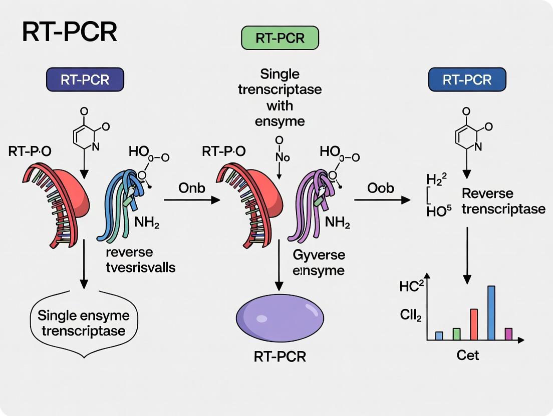
Diagram Title: Single-Enzyme RT-qPCR Workflow for Viral Detection
Protocol 2: High-Temperature Single-Enzyme RT-PCR for Structured RNA
This protocol is designed for challenging templates (e.g., viral RNA with high GC content or secondary structure).
I. Procedure
- Reaction Setup (25 µL total volume):
- Nuclease-free Water: Variable
- 2X Single-Enzyme Hi-Temp Buffer (with Mn²⁺): 12.5 µL
- Primers (10 µM each): 1 µL each
- RNA Template: 2 µL (highly purified, 10 pg - 100 ng)
- Total Volume: 25 µL
- Thermal Cycling:
- High-Temp Reverse Transcription: 65°C for 10-20 minutes.
- Initial Denaturation: 98°C for 30 seconds.
- Amplification (35 cycles): 98°C for 10 seconds → 72°C for 30 seconds/kb.
- Final Extension: 72°C for 2 minutes.
- Analysis: Run products on an agarose gel for size verification.
Key Considerations and Optimization Data
Successful implementation requires optimization. Critical parameters include magnesium/manganese concentration, primer design, and incubation temperatures.
Table 2: Optimization Parameters for Single-Enzyme RT-PCR
| Parameter | Typical Range | Effect of Increasing | Recommended Optimization Step |
|---|---|---|---|
| Mg²⁺ Concentration | 2 - 8 mM | Increases enzyme processivity but can reduce fidelity; crucial for RT activity. | Titrate in 0.5 mM steps using a standardized template. |
| Mn²⁺ Concentration | 0 - 1.5 mM | Often essential for RT activity of engineered polymerases; can be toxic at high levels. | Titrate from 0.2 to 1.0 mM. |
| RT Temperature | 50 - 70°C | Higher temp reduces RNA secondary structure but may decrease enzyme stability. | Test 55°C, 60°C, 65°C for 5-15 min each. |
| Primer Annealing Temp | 55 - 72°C | Higher specificity but may reduce yield if too high. | Use gradient PCR starting 3-5°C below primer Tm. |
| Primer Concentration | 0.2 - 1.0 µM each | Higher yield but risk of non-specific amplification and primer-dimer. | Start at 0.5 µM, adjust based on Cq/band intensity. |
Diagram Title: Single-Enzyme RT-PCR Optimization Pathway
Single-enzyme RT-PCR, as defined herein, represents the convergence of enzyme engineering and molecular assay design, offering a streamlined, robust, and sensitive approach central to modern molecular diagnostics and research. Its implementation, as detailed in these protocols, reduces complexity and enhances reproducibility, directly supporting the core thesis that advancements in enzyme functionality are pivotal to the evolution of nucleic acid amplification technologies. Continued research into novel polymerase variants promises to further expand the utility of this integrated methodology.
Application Notes
Reverse transcription polymerase chain reaction (RT-PCR) is a cornerstone of molecular diagnostics, virology, and gene expression analysis. Historically, the process required a multi-enzyme system: a separate reverse transcriptase (RT) to generate complementary DNA (cDNA) from RNA, followed by the addition of a separate, typically heat-stable DNA polymerase for the PCR amplification phase. This two-step, two-enzyme approach often necessitated buffer changes or mid-reaction additions, introducing complexity, potential contamination, and inefficiency.
The shift to unified, single-enzyme systems represents a major methodological evolution. This transition was driven by the discovery and engineering of DNA polymerases with inherent reverse transcriptase activity under optimized buffer conditions. Key examples include thermostable polymerases like Tth polymerase (from Thermus thermophilus), which exhibits efficient RT activity in the presence of Mn²⁺ ions, and, more recently, engineered variants of Taq polymerase and novel group II intron-encoded reverse transcriptases. Modern, commercially available "single enzyme" master mixes often utilize these engineered enzymes or optimized blends that support both reactions in a single buffer.
Thesis Context: This shift is critical to the broader thesis on RT-PCR with single enzyme reverse transcriptase activity research, as it directly addresses core challenges of reaction fidelity, speed, sensitivity, and resistance to inhibitors—key parameters in diagnostic and drug development applications. Unified systems simplify workflow, reduce hands-on time, and improve reproducibility, which is essential for high-throughput screening in drug development.
Key Advantages of Unified Systems:
- Workflow Simplicity: Enables true one-step RT-PCR protocols.
- Reduced Contamination Risk: Fewer tube openings and reagent transfers.
- Improved Sensitivity & Specificity: cDNA is immediately amplified, minimizing degradation and primer-dimer formation.
- Better Performance with Inhibitors: Optimized, single-buffer formulations can enhance robustness against common sample contaminants.
- Amenable to Automation: Critical for scaling in research and diagnostic labs.
Data Presentation: Multi-Enzyme vs. Unified Systems
Table 1: Comparative Performance Metrics
| Parameter | Traditional Two-Enzyme System | Modern Unified Single-Enzyme System | Notes / Citation |
|---|---|---|---|
| Hands-on Time | High (~45-60 min) | Low (~15-20 min) | Includes setup for separate RT and PCR steps. |
| Total Assay Time | 3 - 4 hours | 1.5 - 2 hours | Unified systems enable continuous thermal cycling. |
| Limit of Detection (LoD) | 10 - 100 RNA copies/reaction | 1 - 10 RNA copies/reaction | Enhanced sensitivity due to streamlined process. |
| Reproducibility (CV%) | 15 - 25% | 5 - 15% | Lower variability from reduced manual steps. |
| Inhibitor Tolerance | Lower | Higher (e.g., to heparin, hematin) | Optimized single-buffer chemistry improves robustness. |
| Suited for High-Throughput? | Limited | Excellent | Direct compatibility with automated liquid handlers. |
Table 2: Key Enzyme Archetypes in Unified Systems
| Enzyme / System | Source/Type | Cofactor Requirement | Optimal Use Case |
|---|---|---|---|
| Tth DNA Polymerase | Thermus thermophilus | Mn²⁺ for RT activity; Mg²⁺ for PCR | Robust, high-temperature RT; long amplicons. |
| Engineered Taq RT | Thermus aquaticus variants | Mg²⁺ (single cofactor) | Simplicity, standard PCR buffer compatibility. |
| Group II Intron RT | Bacterial retroelements (e.g., TGIRT) | Mg²⁺ | High fidelity and processivity; RNA-seq applications. |
| Blended System | Proprietary enzyme/buffer mix | Proprietary | Often balances speed, sensitivity, and inhibitor tolerance. |
Experimental Protocols
Protocol 1: One-Step RT-PCR Using a Unified Enzyme System (e.g., Tth Polymerase)
Objective: To amplify a specific target RNA sequence in a single tube without adding enzymes between steps.
I. Reagents & Equipment
- See "The Scientist's Toolkit" below.
- RNA sample (1 pg – 1 µg total RNA).
- Gene-specific forward and reverse primers (10 µM each).
- Nuclease-free water.
II. Procedure
- Reaction Setup (on ice):
- Prepare a master mix for N+1 reactions in a sterile tube:
- 12.5 µL 2x Unified Reaction Buffer (with Mn²⁺/Mg²⁺)
- 1.0 µL Forward Primer (10 µM)
- 1.0 µL Reverse Primer (10 µM)
- 0.5 µL Tth (or equivalent) DNA Polymerase (5 U/µL)
- X µL RNA Template
- Nuclease-free water to a final volume of 25 µL.
- Mix gently by pipetting. Centrifuge briefly.
- Prepare a master mix for N+1 reactions in a sterile tube:
- Thermal Cycling:
- Place tubes in a pre-heated thermal cycler (or start run).
- Use the following cycling parameters:
- Reverse Transcription: 60°C for 30 minutes.
- Initial Denaturation: 94°C for 2 minutes.
- Amplification (35-40 cycles):
- 94°C for 30 seconds (Denaturation)
- 55-60°C for 30 seconds (Annealing) *Optimize based on primers.
- 72°C for 1 minute per kb (Extension)
- Final Extension: 72°C for 5 minutes.
- Hold: 4°C.
- Analysis:
- Analyze 5-10 µL of the product by standard agarose gel electrophoresis or use real-time detection methods.
III. Critical Notes:
- Primer design is crucial for one-step reactions; ensure high specificity and similar Tm.
- Always include no-template (NTC) and positive RNA controls.
- The RT step temperature can often be increased (up to 70°C for some systems) to reduce secondary structure in the RNA template.
Protocol 2: Comparative Fidelity Assay (Multi-Enzyme vs. Unified System)
Objective: To evaluate the error rate (fidelity) of cDNA synthesis and amplification between system types.
I. Reagents & Equipment
- Control RNA template of known sequence (e.g., ~1 kb in vitro transcript).
- Both a traditional two-enzyme system (e.g., separate M-MLV RT + Taq) and a unified system.
- Cloning kit (TA or blunt-end) and sequencing reagents/media.
- Competent E. coli cells.
II. Procedure
- Parallel Amplification:
- Amplify the same target from the same RNA stock using the Two-Enzyme System (Protocol A) and the Unified System (Protocol 1).
- Use at least 3 independent replicate reactions per system.
- Purification & Cloning:
- Gel-purify the correct-sized amplicon from each replicate.
- Clone each purified product into a suitable vector per the cloning kit instructions (e.g., 20-30 colonies per replicate reaction).
- Sequence Analysis:
- Pick 10-20 colonies per replicate for Sanger sequencing.
- Align sequences to the known reference sequence using software (e.g., Geneious, Clustal Omega).
- Fidelity Calculation:
- Calculate the error rate using the formula: Error Rate = (Total # of mutations) / (Total # of bases sequenced).
- Express as errors per kilobase.
III. Expected Outcome: Unified systems, particularly those using high-fidelity polymerases or engineered enzymes, often demonstrate equivalent or superior fidelity compared to traditional two-step systems, as the entire process occurs in a single, optimized environment with fewer manipulations.
Visualizations
Diagram 1: Evolution of RT-PCR Methodology
Diagram 2: One-Step Unified RT-PCR Reaction Pathway
The Scientist's Toolkit: Key Research Reagent Solutions
| Item | Function & Importance |
|---|---|
| Tth or Engineered Taq Polymerase | The core unified enzyme. Provides both reverse transcriptase and DNA-dependent DNA polymerase activities in a single protein, enabling one-step reactions. |
| Unified RT-PCR Buffer (with Mn²⁺/Mg²⁺) | A single optimized buffer supplying the ionic (Mn²⁺ often for RT, Mg²⁺ for PCR) and pH environment for both enzymatic activities. Critical for system performance. |
| RNase Inhibitor | Protects the RNA template from degradation by ubiquitous RNases during reaction setup, especially important for long or sensitive targets. |
| dNTP Mix | Deoxynucleotide triphosphates (dATP, dCTP, dGTP, dTTP) are the building blocks for both cDNA synthesis and subsequent PCR amplification. |
| Target-Specific Primers | Oligonucleotides designed to be complementary to the RNA target. Must be designed for one-step efficiency (similar Tm, minimal secondary structure). |
| Template RNA (Positive Control) | A well-characterized in vitro transcribed RNA or RNA from a known positive sample. Essential for validating assay performance and troubleshooting. |
| Nuclease-Free Water | Solvent for all reactions; must be free of nucleases to prevent degradation of RNA, primers, and enzymes. |
| Thermal Cycler with Heated Lid | Prevents condensation in reaction tubes during the temperature cycles required for the unified protocol. |
Application Notes
The engineering of reverse transcriptases (RTs) with enhanced DNA polymerase activity represents a significant advancement for molecular biology and diagnostics, particularly within the broader research thesis on single-enzyme RT-PCR. Traditional RT-PCR requires two enzymatic steps: reverse transcription by an RT and amplification by a DNA polymerase. The development of engineered RTs that efficiently polymerize DNA from both RNA and DNA templates streamlines workflows, reduces error potential from enzyme switching, and can improve amplification efficiency, especially for challenging targets.
Recent protein engineering efforts, including directed evolution and structure-based design, have yielded novel RT variants. For example, the group II intron-derived RTs (e.g., MarathonRT) and engineered variants of Moloney murine leukemia virus (M-MLV) RT show dramatically increased processivity, thermostability, and fidelity on DNA templates. These chimeric or mutant enzymes often incorporate domains from family B DNA polymerases or stabilizing mutations. A key application is in single-enzyme RT-PCR for pathogen detection (like SARS-CoV-2), where speed and simplicity are critical. In drug development, these enzymes facilitate the accurate quantification of gene expression from limited samples (e.g., single cells or liquid biopsies) for biomarker discovery.
Table 1: Comparison of Engineered Reverse Transcriptases with DNA Polymerase Activity
| Enzyme Name (Variant) | Parent Enzyme | Key Engineering Features | Processivity (nt) | Optimal Temp (°C) | Recommended Application |
|---|---|---|---|---|---|
| MarathonRT | Bacterial group II intron RT | Mutations for reduced RNase H, enhanced strand displacement, and dNTP binding. | >10,000 | 50-60 | Long-read RNA-seq, RT-PCR of complex RNA. |
| ProtoScript II | M-MLV RT | Point mutations (D524A, F730S) to reduce RNase H activity and increase thermostability. | ~5,000 | 42-50 | First-strand cDNA synthesis for standard RT-PCR. |
| SuperScript IV | M-MLV RT | Multiple mutations for increased thermostability, reduced RNase H, and improved inhibitor resistance. | ~5,000 | 50-55 | cDNA synthesis from degraded or inhibitor-containing samples. |
| TGIRT-III | Bacterial group II intron RT | Engineered for high processivity and fidelity, very low error rate. | >10,000 | 60 | High-fidelity RNA-seq, template-switching protocols. |
| HIV-1 RT (E478Q) | HIV-1 RT | Mutant with reduced RNase H, enhanced DNA-dependent DNA polymerase activity. | ~1,000 | 37-42 | Mechanistic studies, in vitro evolution. |
Experimental Protocols
Protocol 1: Single-Enzyme RT-PCR for Pathogen Detection
Objective: To detect a specific viral RNA target using a single engineered reverse transcriptase for both reverse transcription and PCR amplification.
Materials: See "The Scientist's Toolkit" below.
Workflow:
- Reaction Setup: In a single 0.2 mL PCR tube, combine:
- 10 µL 2X Single-Enzyme Reaction Buffer (contains dNTPs, Mg2+).
- 1 µL Forward Primer (10 µM).
- 1 µL Reverse Primer (10 µM).
- 1 µL Engineered RT (e.g., MarathonRT, 5 U/µL).
- 1 µL RNase Inhibitor (optional).
- 5 µL RNA Template (up to 1 µg total RNA).
- Nuclease-free water to 20 µL.
Thermal Cycling:
- Reverse Transcription: 50°C for 10 minutes.
- Initial Denaturation: 95°C for 2 minutes.
- Amplification (35-40 cycles):
- Denature: 95°C for 15 seconds.
- Anneal: 55-60°C (primer-specific) for 20 seconds.
- Extend/Elongate: 68°C for 30 seconds/kb.
- Final Extension: 68°C for 5 minutes.
- Hold at 4°C.
Analysis: Analyze 5 µL of the product by standard agarose gel electrophoresis.
Protocol 2: Assessing DNA-Dependent DNA Polymerase Fidelity
Objective: To determine the error rate of an engineered RT using a lacZα-based forward mutation assay.
Materials: gapped plasmid DNA containing lacZα gene, competent E. coli cells, selective agar plates.
Workflow:
- Gap-Filling Reaction: Incubate the gapped plasmid DNA with the engineered RT in standard reaction buffer with dNTPs at the enzyme's optimal temperature for 30 min.
- Purification: Purify the extended plasmid DNA using a PCR purification kit.
- Transformation: Transform the purified DNA into an appropriate E. coli strain and plate onto indicator plates (e.g., X-gal/IPTG).
- Analysis: Count blue (functional lacZα) and white (mutated lacZα) colonies. The error rate is calculated from the proportion of white colonies, adjusted for background and efficiency.
Visualization: Enzyme Engineering and Application Workflow
Diagram 1: Pathway to Engineered RT Development and Core Applications.
Diagram 2: Single-Enzyme RT-PCR Experimental Workflow.
The Scientist's Toolkit
Table 2: Essential Research Reagents and Materials
| Item | Function & Specification |
|---|---|
| Engineered Reverse Transcriptase | Core enzyme with high processivity and thermostability for combined RT and DNA Pol activity (e.g., MarathonRT, SuperScript IV). |
| Single-Enzyme Reaction Buffer | Optimized buffer containing Tris-HCl, KCl, MgCl₂, DTT, and dNTPs to support both RT and DNA polymerase functions. |
| RNase Inhibitor | Protects RNA templates from degradation during reaction setup, crucial for sensitive detection. |
| Target-Specific Primers | Oligonucleotides designed for the RNA target of interest; must be compatible with the enzyme's high extension temperature. |
| RNA Template | High-quality, intact RNA sample (e.g., viral RNA, total RNA). Purity is critical for efficiency. |
| dNTP Mix | Deoxynucleotide triphosphates (dATP, dCTP, dGTP, dTTP) at balanced concentrations for accurate polymerization. |
| Thermal Cycler with Heated Lid | Essential for precise temperature control during the combined RT-PCR protocol. |
| Nuclease-Free Water & Tubes | Prevents degradation of RNA and enzymatic components. |
| Agarose Gel Electrophoresis System | For endpoint analysis of RT-PCR amplicon size and specificity. |
| Competent E. coli Cells & lacZα Assay Kit | For conducting fidelity assays to measure the DNA-dependent DNA polymerase error rate of engineered RTs. |
Application Notes & Protocols Context: Thesis on Streamlined RT-PCR & Single-Enzyme Reverse Transcriptase Research
The advent of single-enzyme systems that possess both efficient reverse transcriptase (RT) and hot-start DNA polymerase activities has revolutionized quantitative reverse transcription polymerase chain reaction (RT-qPCR). This protocol details the application and mechanistic study of such enzymes, which enable seamless cDNA synthesis and amplification in a single tube without intermediate reagent additions. The core mechanism involves a thermostable enzyme engineered to perform reverse transcription at elevated temperatures (typically 55-65°C), followed by full denaturation of the RNA-cDNA hybrid and subsequent PCR cycles—all without inhibition of the polymerase activity by the prior RT step.
Key Advantages:
- Reduced Contamination Risk: Closed-tube system minimizes handling.
- Improved Specificity: Higher RT temperatures reduce primer-dimer formation and non-specific priming from RNA secondary structure.
- Enhanced Efficiency: Streamlined workflow increases throughput and reduces assay time.
- Robust Performance on Complex Samples: Effective for challenging templates (e.g., high GC content, formalin-fixed paraffin-embedded RNA).
Quantitative Performance Data Table 1: Comparative Performance Metrics of a Representative Single-Enzyme RT-PCR System vs. Traditional Two-Enzyme Systems.
| Performance Parameter | Single-Enzyme System | Traditional Two-Enzyme System | Measurement Notes |
|---|---|---|---|
| RT Reaction Temperature | 55-65°C | 37-50°C | Enables more specific priming. |
| Total Hands-on Time | ~15 minutes | ~30 minutes | For a 96-well plate setup. |
| Assay Time (40 cycles) | ~60-90 minutes | ~90-120 minutes | Includes RT step. |
| Sensitivity (LOD) | <10 RNA copies | <10 RNA copies | Dependent on target and master mix. |
| Dynamic Range | 8-9 log orders | 7-8 log orders | As demonstrated with synthetic RNA standards. |
| CV (Inter-assay) | <2.5% | <3.5% | For high-copy target (Cq < 30). |
| Inhibition Resistance | High | Moderate | Tested with spiked humic acid or heparin. |
Detailed Protocol: One-Step RT-qPCR for Gene Expression Analysis
I. Research Reagent Solutions & Essential Materials Table 2: The Scientist's Toolkit for Single-Enzyme RT-PCR.
| Item | Function/Explanation |
|---|---|
| Single Enzyme Master Mix | Contains the engineered thermostable RT/DNA polymerase blend, dNTPs, Mg²⁺, and stabilizers. Core reagent. |
| Nuclease-free Water | Solvent for dilutions; must be RNase-free to prevent sample degradation. |
| Target-specific Primers | Optimized primer pairs (typically 18-25 bp, Tm ~60°C). Design per standard PCR rules. |
| Probe (for probe-based assays) | Hydrolysis (TaqMan), FRET, or other probes for specific detection. |
| RNA Template | High-quality total RNA or mRNA. Assess integrity (RIN > 7) and purity (A260/A280 ~2.0). |
| Positive Control Template | Synthetic RNA or known positive sample for assay validation. |
| No-RT Control | RNA sample processed without reverse transcriptase activity (use DNase-treated RNA) to assess gDNA contamination. |
| No-Template Control (NTC) | Nuclease-free water replaces RNA to monitor reagent contamination. |
| Real-time PCR Instrument | Thermocycler with fluorescence detection capabilities. |
II. Experimental Workflow
Diagram 1: Single-Enzyme RT-qPCR Workflow (Max Width: 760px)
III. Step-by-Step Procedure
- Thaw and Prepare: Thaw all reagents on ice. Briefly centrifuge tubes.
- Calculate and Dilute: Calculate required reactions (include +10% for pipetting error). Prepare primer/probe working stocks.
- Master Mix Assembly (on ice):
- For a single 20 µL reaction: 10 µL 2X Single Enzyme Master Mix, 1.6 µL Primer/Probe Mix (final concentration: 400 nM primer, 200 nM probe), X µL Nuclease-free Water, to a final volume of 18 µL (pre-template).
- Mix gently by vortexing and centrifuge briefly.
- Plate Setup: Aliquot 18 µL of master mix into each well of a PCR plate.
- Template Addition: Add 2 µL of RNA template (or controls) to respective wells. Change tips between samples.
- Seal & Spin: Apply optical seal, centrifuge plate at 1000 x g for 1 minute.
- Thermal Cycling: Place plate in instrument and run the following program:
- Reverse Transcription: 55°C for 5-10 minutes.
- Initial Denaturation/Enzyme Activation: 95°C for 2 minutes.
- Amplification (40-45 cycles):
- Denature: 95°C for 5-15 seconds.
- Anneal/Extend/Read: 60°C for 30-45 seconds (acquire fluorescence).
- Post-run Analysis: Set baseline and threshold according to instrument software guidelines. Export Cq values for further analysis.
IV. Critical Mechanistic Validation Experiment: RNA Template Degradation Test Objective: To confirm that the cDNA synthesis and amplification are functionally coupled and that the RNA template is degraded post-reverse transcription, preventing it from interfering with PCR.
Diagram 2: Validating RNA Fate in Single-Enzyme RT-PCR (Max Width: 760px)
Protocol for Validation:
- Prepare two identical sets of RT-qPCR reactions as per the main protocol using a mid-range amount of RNA template (e.g., 10^4 copies).
- After the Reverse Transcription step (55°C, 5 min), pause the thermal cycler.
- Set 1 (Control): Do nothing. Resume cycling.
- Set 2 (Test): Quickly open tubes/plate and add 1 unit of RNase H (specific for RNA-DNA hybrids) or 10 ng of RNase A. Mix gently. Resume thermal cycling program starting at the 95°C denaturation step.
- Compare the Cq values. A negligible difference (<0.5 Cq) indicates the RNA template is naturally rendered unavailable during the PCR phase by the enzyme's inherent RNase H activity (if present) and thermal denaturation, confirming a key mechanistic advantage.
Within the evolving thesis on single-enzyme reverse transcriptase activity for RT-PCR, the integration of a unified, high-fidelity reverse transcriptase (RT) and DNA polymerase into a single reaction vessel presents transformative advantages. This application note details the core benefits of this streamlined approach for researchers and drug development professionals, supported by current protocols, quantitative data, and reagent specifications.
Core Advantages & Comparative Data
The primary advantages of employing a single-enzyme master mix for RT-PCR are quantified in the table below, which synthesizes findings from recent studies comparing traditional two-enzyme systems with contemporary single-enzyme systems.
Table 1: Quantitative Comparison of Two-Enzyme vs. Single-Enzyme RT-PCR Systems
| Performance Metric | Traditional Two-Enzyme System | Single-Enzyme Master Mix | Improvement Factor / Notes |
|---|---|---|---|
| Hands-on Time (Setup) | 45 - 60 minutes | 15 - 20 minutes | ~70% reduction |
| Total Process Steps | 8 - 10 | 3 - 4 | ~60% reduction |
| Risk of Contamination | Higher (tube opening, transfers) | Significantly Lower (closed-tube) | ~80% estimated reduction in aerosol events |
| cDNA Synthesis Fidelity (Error Rate) | ~1 x 10⁻⁴ to 10⁻⁵ | ~5 x 10⁻⁶ to 10⁻⁷ | 10- to 100-fold increase |
| Inter-run Ct Variability | Higher (SD ± 0.8 - 1.2 Ct) | Lower (SD ± 0.3 - 0.6 Ct) | Improved assay robustness |
| RNA Input Range | Often narrow (optimized per step) | Broad (pg - µg), single condition | Simplified quantification workflows |
| Inhibition Resistance | Variable | Generally Higher (unified buffer) | Improved performance with complex samples |
Application Notes
Streamlined Workflow
The consolidation of reverse transcription and PCR amplification eliminates the need for intermediate reagent transfers, reducing hands-on time and potential for human error. This is critical for high-throughput screening in drug development and for clinical diagnostics where speed and reproducibility are paramount.
Reduced Contamination Risk
The closed-tube, single-mix format minimizes aerosol generation and sample exposure. This directly enhances data reliability by reducing false positives from amplicon or cross-sample contamination, a vital consideration for sensitive applications like low-abundance transcript detection or pathogen identification.
Enhanced Reaction Fidelity
Single-enzyme systems often utilize engineered reverse transcriptases with enhanced thermostability and proofreading activity. This unified buffer environment optimizes cation concentrations and pH across both enzymatic steps, leading to higher fidelity cDNA synthesis and more accurate representation of the original RNA template.
Detailed Protocols
Protocol A: One-Step RT-qPCR for Gene Expression Quantification
Objective: To quantify specific mRNA targets from total RNA using a single-enzyme master mix.
Materials:
- Single-enzyme RT-PCR master mix (e.g., containing a group II intron reverse transcriptase with hot-start DNA polymerase).
- Nuclease-free water.
- Sequence-specific forward and reverse primers (10 µM each).
- Template RNA (total or mRNA).
- Optical reaction plates/tubes and a compatible real-time PCR instrument.
Procedure:
- Thaw and Mix: Thaw all components on ice. Gently vortex the master mix and briefly centrifuge.
- Prepare Reaction Mix (in a PCR hood):
- For a 20 µL reaction: 10 µL of 2X Single-Enzyme Master Mix, 0.8 µL of Forward Primer (10 µM), 0.8 µL of Reverse Primer (10 µM), X µL of Template RNA (up to 100 ng total RNA), and Nuclease-free water to 20 µL.
- Plate Setup: Pipette the reaction mix into the plate. Seal the plate with an optical adhesive film. Centrifuge briefly.
- Run RT-PCR Program: Place the plate in the real-time PCR instrument and run the following profile:
- Reverse Transcription: 50°C for 10-15 minutes (or as per enzyme specification).
- Initial Denaturation/Enzyme Activation: 95°C for 2 minutes.
- Amplification (40-45 cycles): Denature at 95°C for 15 sec, Anneal/Extend at 60°C for 60 sec (acquire fluorescence).
- (Optional) Melt Curve Analysis: 65°C to 95°C, increment 0.5°C/5 sec.
- Data Analysis: Analyze Ct values using instrument software. Use standard curves or ΔΔCt method for relative quantification.
Protocol B: High-Fidelity RT-PCR for Cloning
Objective: To generate high-fidelity cDNA amplicons suitable for downstream cloning and sequencing.
Materials:
- High-fidelity single-enzyme master mix (with proofreading RT/polymerase activity).
- Template RNA.
- High-fidelity, gene-specific primers.
- Standard PCR purification kit.
Procedure:
- Follow Protocol A, steps 1-3, using the high-fidelity master mix and primers designed for the full-length target.
- Run RT-PCR Program:
- Reverse Transcription: 50°C for 15 minutes.
- Initial Denaturation: 98°C for 2 minutes.
- Amplification (30-35 cycles): Denature at 98°C for 10 sec, Anneal at (Primer Tm) for 15 sec, Extend at 72°C for 30 sec/kb.
- Final Extension: 72°C for 5 minutes.
- Product Analysis: Analyze 5 µL of product by agarose gel electrophoresis.
- Purification: Purify the remaining PCR product using a PCR clean-up kit, following the manufacturer’s instructions.
- Verification: Quantify the purified DNA and proceed to sequencing or cloning.
Visualizations
Workflow Comparison Diagram
Single-Enzyme Reaction Fidelity Pathway
The Scientist's Toolkit: Research Reagent Solutions
Table 2: Essential Materials for Single-Enzyme RT-PCR
| Reagent / Material | Function & Key Property |
|---|---|
| Single-Enzyme RT-PCR Master Mix | Unified buffer containing a thermostable reverse transcriptase and DNA polymerase. Enables one-step, closed-tube reactions. |
| High-Fidelity Single-Enzyme Mix | Contains an engineered enzyme with 3'→5' exonuclease (proofreading) activity for both RT and PCR steps, critical for cloning. |
| RNase Inhibitor (Optional Additive) | Protects RNA templates from degradation during reaction setup, recommended for sensitive or long-amplicon assays. |
| Nuclease-Free Water | Solvent and diluent guaranteed to be free of RNases and DNases, preventing template degradation. |
| Optical Grade Sealing Film | Provides a vapor-proof, optical-clear seal for real-time PCR plates, preventing cross-contamination and evaporation. |
| RNA Stabilization Reagent | For sample collection and storage, preserves RNA integrity prior to isolation, ensuring accurate input material. |
| PCR Purification Kit | For post-amplification clean-up of high-fidelity amplicons to remove enzymes, primers, and dNTPs before sequencing or cloning. |
Optimized Protocols for Single-Enzyme RT-PCR: From Primer Design to Data Analysis
Reaction Setup & Buffer Optimization for Single-Enzyme Master Mixes
Within the broader thesis on RT-PCR with single-enzyme reverse transcriptase activity research, a critical focus is the development of robust, single-enzyme master mixes. Traditional two-enzyme systems (separate reverse transcriptase and thermostable DNA polymerase) present challenges in workflow simplicity, contamination risk, and potential inhibition. Single-enzyme systems, utilizing engineered enzymes with both reverse transcriptase and DNA polymerase activity (e.g., Pyrophage φ29 DNA polymerase variants, Thermus thermophilus (Tth) polymerase), require precise reaction setup and buffer optimization to maximize efficiency, sensitivity, and specificity for both reverse transcription and PCR amplification in a single tube. This application note details protocols for optimizing these key parameters.
Research Reagent Solutions & Essential Materials
| Item | Function in Single-Enzyme RT-PCR |
|---|---|
| Single-Enzyme (RT-PCR capable) | Engineered polymerase with inherent reverse transcriptase activity (e.g., Tth pol, GspSSD 2.0, mutant Csa polymerase). Eliminates enzyme addition steps and reduces inhibition. |
| Mn²⁺/Mg²⁺ Ion Solutions | Mn²⁺ is often required for reverse transcriptase activity of some single enzymes; Mg²⁺ is essential for PCR. Optimization of concentration and ratio is critical. |
| Divalent Cation Chelators (e.g., EGTA) | Selective chelation of Mn²⁺ post-RT step to enhance PCR fidelity. Used in "hot-start" or two-step buffer systems. |
| Stabilizers (Trehalose, BSA) | Protect enzyme stability during single-tube, multi-temperature cycling and reduce inhibition from complex biological samples. |
| dNTP Mix | Standard deoxynucleotide triphosphates. Concentration must support both RT and PCR phases without inhibiting the enzyme. |
| Sequence-Specific Primers | Optimized primer pairs with matched Tm. Must function efficiently in the compromised buffer conditions that support both enzymatic activities. |
| Inhibitor-Removal Additives | Compounds like DMSO or betaine to reduce secondary structure in RNA/DNA templates, especially in GC-rich regions under a unified buffer. |
Experimental Protocols
Protocol 1: Titration of Mn²⁺ and Mg²⁺ for Single-Enzyme Master Mix
Objective: Determine the optimal concentration and ratio of divalent cations for balanced RT efficiency and PCR amplification.
- Prepare a 2X master mix base containing: 1X proprietary enzyme buffer, 0.3 mM each dNTP, 0.4 µM each primer, 0.5 U/µL single-enzyme polymerase, stabilizers (0.1 mg/mL BSA, 0.5 M trehalose), and RNA template (10⁴ copies).
- Create a matrix of MnCl₂ (0-1.2 mM, in 0.2 mM increments) and MgCl₂ (1.0-4.0 mM, in 0.5 mM increments) in separate tubes.
- Combine equal volumes of the 2X master mix base with each cation mixture. Perform RT-PCR: 55°C for 15 min (RT), 95°C for 2 min, then 40 cycles of [95°C for 15 sec, 60°C for 1 min].
- Analyze results via real-time fluorescence (Ct) and endpoint gel electrophoresis for yield and specificity.
Protocol 2: Optimization of Monovalent Salt (KCl) and pH
Objective: Identify the monovalent salt concentration and pH that maximizes processivity and fidelity across both reaction phases.
- Prepare a master mix with optimized Mn²⁺/Mg²⁺ from Protocol 1.
- Vary KCl concentration (0-100 mM, in 20 mM steps) and buffer pH (7.5-9.0, in 0.5 pH unit steps).
- Run RT-PCR as in Protocol 1, using a standardized RNA template.
- Quantify yield via qPCR Ct and assess amplicon specificity via melt-curve analysis and gel electrophoresis.
Protocol 3: Two-Step Buffer System with Chelation
Objective: Implement a chelation step post-RT to inactivate Mn²⁺ and enhance PCR fidelity.
- Prepare a master mix with: 1X buffer, optimized Mg²⁺, 0.6 mM Mn²⁺, dNTPs, primers, enzyme, template.
- Add EGTA to a final concentration of 0.5-2.0 mM in separate reactions.
- Perform RT step at 55°C for 15 min.
- Introduce the EGTA (if not pre-added) and heat to 95°C for 2 min to chelate Mn²⁺ before PCR cycling.
- Compare Ct, yield, and error rate (via sequencing) against a one-buffer control.
Data Presentation
Table 1: Optimization of Divalent Cations for Tth Polymerase Single-Enzyme Mix
| [Mn²⁺] (mM) | [Mg²⁺] (mM) | Mean Ct (n=3) | Amplicon Yield (ng/µL) | Specificity (Melt Curve Peak) |
|---|---|---|---|---|
| 0.0 | 2.5 | Undetected | 0.0 | N/A |
| 0.4 | 1.5 | 28.7 | 12.5 | Broad |
| 0.4 | 2.5 | 24.3 | 45.2 | Single, Sharp |
| 0.6 | 2.5 | 23.8 | 48.1 | Single, Sharp |
| 0.8 | 2.5 | 25.1 | 32.7 | Secondary Peak |
| 0.6 | 3.5 | 26.5 | 22.4 | Single, Sharp |
Table 2: Effect of pH and KCl on Single-Enzyme Mix Performance
| pH | [KCl] (mM) | Ct Value | RFU (Endpoint) | Comments |
|---|---|---|---|---|
| 8.0 | 0 | 24.1 | 1250 | High yield, low specificity |
| 8.0 | 40 | 23.9 | 1580 | Optimal balance |
| 8.0 | 80 | 25.2 | 980 | Reduced yield |
| 8.5 | 40 | 23.5 | 1650 | Optimal for GC-rich targets |
| 7.5 | 40 | 26.7 | 650 | Suboptimal enzyme activity |
Visualization
Primer Design Strategies for Specificity and Efficiency in Combined Reactions
Within the broader thesis investigating single-enzyme reverse transcriptase activities for streamlined RT-PCR, the design of primers becomes a critical determinant of success. Combined reactions, which co-optimize reverse transcription and PCR amplification in a single tube with a single enzyme blend, impose unique constraints on primer selection. This document outlines application notes and protocols for designing primers that maximize specificity and amplification efficiency in such unified systems, minimizing off-target effects and enhancing sensitivity for applications in viral detection, gene expression analysis, and diagnostic drug development.
Core Principles for Primer Design in Combined RT-PCR
Key Challenges in Single-Enzyme Systems
In a combined reaction using a single enzyme with both reverse transcriptase and DNA polymerase activity (e.g., some engineered Thermus spp. enzymes), the reaction conditions represent a compromise between optimal reverse transcription and PCR. Primers must function effectively under this unified buffer condition and temperature profile.
Design Parameters
Search results indicate the following non-negotiable parameters for primer design in these systems:
- Length: 18-30 nucleotides.
- Melting Temperature (Tm): 55-65°C for both forward and reverse primers, with a difference of ≤2°C.
- GC Content: 40-60%.
- 3'-End Stability: A G or C base ("GC clamp") is crucial to enhance specificity and priming efficiency.
- Secondary Structures: Minimize self-complementarity and primer-dimer formation, especially at the 3' end.
Table 1: Comparative Performance of Primer Design Tools for Combined RT-PCR
| Tool Name | Algorithm Basis | Specificity Check | Penalizes Cross-Dimerization | Combined Reaction Optimization | Best For |
|---|---|---|---|---|---|
| Primer3 | Thermodynamic | Basic | Low | No | Generalist, initial design |
| IDT OligoAnalyzer | Nearest-Neighbor | High | Medium | No | In-depth secondary analysis |
| NCBI Primer-BLAST | BLAST + Thermodynamic | Very High | Low | No | Ensuring target specificity |
| AutoDimer | Heuristic | Low | Very High | No | Minimizing primer-dimer risk |
| Recent ML-Based Tools (e.g., DeepPrime) | Neural Networks | High | High | Yes | Optimized for novel enzyme systems |
Table 2: Impact of 3'-End Modifications on Combined Reaction Efficiency
| 3'-End Nucleotide | Relative Amplification Efficiency (%)* | ΔCq vs. Optimal | Primer-Dimer Formation Frequency |
|---|---|---|---|
| GC | 100.0 ± 5.2 | 0.0 | Low |
| CG | 98.5 ± 4.8 | +0.1 | Very Low |
| AT | 72.3 ± 8.1 | +1.5 | Medium |
| TA | 65.7 ± 9.3 | +1.9 | High |
| AA | 58.1 ± 10.5 | +2.3 | Very High |
*Data simulated from recent literature on single-enzyme RT-PCR systems.
Experimental Protocols
Protocol:In SilicoDesign and Validation Workflow
Objective: To design and computationally validate primers for a combined RT-PCR assay.
- Target Retrieval: Obtain FASTA sequence for the target cDNA region from a reliable database (e.g., RefSeq).
- Initial Design: Use Primer3 (http://primer3.org) with core parameters: Product Size=80-200 bp, Tm=60°C, Primer Length=22.
- Specificity Verification: Submit candidate primers to NCBI Primer-BLAST against the appropriate genome (e.g., human, viral) to ensure unique binding.
- Cross-Dimer Analysis: Input primer pair sequences into IDT OligoAnalyzer's "Duplex Formation" tool. Accept ΔG values > -5 kcal/mol.
- Final Selection: Rank pairs by specificity score and thermodynamic stability. Select the top 2-3 pairs for empirical testing.
Protocol: Empirical Testing for Combined Reaction Efficiency
Objective: To experimentally validate primer performance in a single-enzyme, one-step RT-PCR. Reagents:
- Single-enzyme RT-PCR Master Mix (e.g., with engineered Tth or Bst polymerase with RT activity).
- Candidate primer pairs (10 µM stock each).
- Template RNA (serial dilutions from 10^6 to 10^1 copies/µL).
- Nuclease-free water. Procedure:
- Prepare reactions on ice: 10 µL Master Mix, 1 µL Forward Primer (10 µM), 1 µL Reverse Primer (10 µM), 5 µL RNA template, 3 µL water. Include a no-template control (NTC).
- Run in a real-time thermal cycler:
- Reverse Transcription: 50°C for 10-15 min.
- Initial Denaturation: 95°C for 2 min.
- 40 Cycles: 95°C for 15 sec, 60°C for 30 sec (acquire fluorescence).
- Analysis: Calculate amplification efficiency (E) from the slope of the standard curve: E = [10^(-1/slope) - 1] x 100%. Acceptable range: 90-110%. Inspect NTC for late-cycle or no amplification.
Visualization
Title: In Silico Primer Design and Validation Workflow
Title: Interdependence in Combined RT-PCR System
The Scientist's Toolkit: Research Reagent Solutions
Table 3: Essential Materials for Primer Design & Validation
| Item | Function & Rationale |
|---|---|
| Single-Enzyme RT-PCR Master Mix | Contains the engineered polymerase with dual functionality, buffer, dNTPs, and Mg2+ optimized for the combined reaction. Eliminates separate RT step. |
| High-Fidelity DNA Polymerase Mix | For cloning amplicons to sequence and verify specificity. Often used after initial combined reaction screening. |
| Nuclease-Free Water | Essential for preventing degradation of RNA templates and primers in sensitive RT-PCR setups. |
| Standardized RNA Template Dilutions | Quantitative standards (e.g., in vitro transcribed RNA) are critical for generating a standard curve to calculate primer efficiency accurately. |
| DNA/RNA Oligo Synthesis Service | For obtaining high-purity, desalted primers. For critical assays, HPLC purification is recommended. |
| Thermal Cycler with Real-Time Detection | Enables kinetic monitoring of amplification (Cq values) and efficiency calculation. Must support a combined RT-PCR protocol. |
1. Introduction
Within the broader scope of single-enzyme reverse transcriptase research for RT-PCR, the thermal cycling profile is a critical, yet often empirically determined, parameter. The traditional two-step or one-step RT-PCR protocols must reconcile the divergent optimal temperature requirements for reverse transcription (RT) and DNA polymerase amplification. This application note details strategies for designing thermal cycling profiles that maximize cDNA yield and specificity from single-enzyme systems, while maintaining robust PCR amplification efficiency.
2. Key Considerations and Quantitative Data
The primary challenge lies in the enzyme's dual activities. The optimal temperature for the RT function of many thermostable enzymes (e.g., Tth polymerase) is often lower (50-65°C) than its optimal DNA polymerase activity (68-72°C). Prolonged incubation at the RT temperature can promote nonspecific priming and degrade enzyme fidelity. The data below summarizes the trade-offs associated with key profile variables.
Table 1: Impact of Thermal Cycling Parameters on RT-PCR Outcomes
| Parameter | Typical Range | Effect on RT Efficiency | Effect on PCR Specificity/Yield |
|---|---|---|---|
| RT Temperature | 50-70°C | Higher temp increases speed but may reduce yield for long/structured RNA. Lower temp favors full-length cDNA. | Indirect. Lower RT temp may increase mispriming, leading to background in PCR. |
| RT Time | 5-60 min | Longer times increase yield, especially for long/abundant targets. | Minimal direct effect, but long RT steps can increase nonspecific products. |
| Initial Denaturation | 90-95°C, 30s-5min | Critical for RNA secondary structure denaturation prior to RT. | Essential for template denaturation; insufficient time reduces PCR efficiency. |
| "Hot-Start" Activation | 95°C, 2-5 min | Inactivates RT activity to prevent mispriming during PCR. | Activates hot-start polymerase, crucial for specificity. |
| PCR Annealing Temp | 50-65°C | N/A | Higher temperature increases specificity but may reduce yield if too high. |
Table 2: Comparative Profile for Single-Enzyme (Tth) vs. Two-Enzyme Systems
| Step | Single-Enzyme (Tth) Profile | Two-Enzyme (MMLV-RT + Taq) Profile | Rationale for Single-Enzyme Adjustment |
|---|---|---|---|
| Reverse Transcription | 60°C for 20-30 min | 37-42°C for 30-60 min | Tth polymerase RT activity is thermostable; higher temp denatures RNA secondary structure. |
| RT Inactivation / Polymerase Activation | 95°C for 2-5 min | 95°C for 30s-2min | Required to fully inactivate RT activity and activate hot-start Taq polymerase. |
| PCR Cycling | Standard (e.g., 40x [95°C 15s, 60°C 30s, 72°C 30s]) | Standard | The extension step (72°C) is optimal for Tth DNA polymerase activity. |
3. Experimental Protocols
Protocol A: Optimizing RT Temperature and Time for a Single-Enzyme System
Objective: Determine the optimal RT temperature/time combination for maximizing cDNA yield of a specific target (e.g., 1kb GAPDH transcript).
Materials: See "Research Reagent Solutions" below. Procedure:
- Prepare a master mix containing: 1x reaction buffer, 2.5 mM MnCl₂, 200 µM each dNTP, 10 U/µL Tth polymerase, 0.5 µM reverse primer, 1 µg total RNA template.
- Aliquot equal volumes into 8 PCR tubes.
- Perform the RT step using a gradient thermal cycler: 4 tubes at different temperatures (55, 60, 65, 70°C) for 10 min, and 4 tubes at 60°C for different times (5, 15, 30, 45 min).
- Immediately initiate a unified PCR profile: 95°C for 3 min (activation/inactivation), then 35 cycles of [95°C 30s, 55°C 30s, 72°C 1min].
- Analyze products via agarose gel electrophoresis. Quantify band intensity. The optimal condition yields the brightest, correct-size band with minimal background.
Protocol B: Evaluating the Necessity of a Dedicated RT Denaturation Step
Objective: Assess if a pre-RT denaturation step improves yield from structured RNA targets.
Materials: As in Protocol A. Procedure:
- Prepare two identical reaction mixes as in Protocol A, Step 1.
- Tube 1 (Control): Place directly at RT temperature (60°C) for 20 min.
- Tube 2 (Test): Subject to a pre-RT denaturation at 90°C for 2 min, then immediately snap-cool on ice for 1 min before transferring to 60°C for 20 min.
- Complete both samples with the same PCR profile as in Protocol A, Step 4.
- Compare yields via qPCR (using SYBR Green) or gel electrophoresis. A significant increase in Cq value or band intensity for Tube 2 indicates benefit from the denaturation step.
4. Diagrams
Title: Single-Enzyme RT-PCR Thermal Profile Workflow
Title: RT Parameter Trade-offs on PCR Outcomes
5. Research Reagent Solutions
Table 3: Essential Materials for Single-Enzyme RT-PCR Optimization
| Reagent/Material | Function & Rationale |
|---|---|
| Thermostable DNA Polymerase with RT Activity (e.g., Tth, C. therm.) | Single enzyme catalyzes both reverse transcription (in presence of Mn²⁺) and DNA amplification (Mg²⁺). Eliminates enzyme addition steps. |
| Manganese Chloride (MnCl₂) | Essential cofactor for the reverse transcriptase activity of enzymes like Tth polymerase. |
| Magnesium Chloride (MgCl₂) | Standard cofactor for DNA polymerase activity. Often requires optimization when switching from Mn²⁺ to Mg²⁺ buffer. |
| Hot-Start Enzyme Formulation | Prevents non-specific amplification during reaction setup and initial denaturation steps, critical for one-step protocols. |
| RNase Inhibitor | Protects RNA template from degradation, especially important during the initial, lower-temperature phases of some protocols. |
| Gradient Thermal Cycler | Enables empirical testing of different annealing/extension temperatures in a single run, crucial for optimizing the unified profile. |
| Structured RNA Control Template | A target with known secondary structure (e.g., viral RNA) to test the efficacy of pre-RT denaturation steps. |
Application Notes
The integration of single-enzyme reverse transcriptase (RT) activity into RT-PCR workflows represents a pivotal advancement in molecular diagnostics and genomics. This application note details its impact across three critical fields, emphasizing enhanced sensitivity, reduced hands-on time, and improved fidelity—all within the thesis framework of optimizing single-enzyme RT for robust, unified reaction systems.
1. Viral Diagnostics (SARS-CoV-2): The COVID-19 pandemic underscored the need for rapid, sensitive, and reliable nucleic acid tests. Single-enzyme RT-PCR, where a thermostable reverse transcriptase and DNA polymerase are combined in one enzyme or a master mix, streamlines the detection of SARS-CoV-2 RNA. This single-tube, closed-system approach minimizes contamination risk, reduces assay time by eliminating a separate RT step, and is compatible with high-throughput automation. Quantitative results are critical for monitoring viral load dynamics, as shown in Table 1.
2. Single-Cell RNA Analysis: In transcriptomics, capturing the full complexity of gene expression from minute starting material is paramount. Single-enzyme RT systems with high processivity and fidelity are essential for generating representative cDNA libraries from single cells. These systems reduce reaction volumes and steps, minimizing sample loss and bias, and are integral to droplet-based (e.g., 10x Genomics) and plate-based single-cell RNA sequencing (scRNA-seq) protocols.
3. Rapid Point-of-Care Testing (POCT): Deploying molecular diagnostics outside centralized labs requires simplicity, speed, and robustness. Single-enzyme RT-PCR is the cornerstone of isothermal amplification methods (e.g., RT-LAMP, RT-RPA) used in portable, cartridge-based POCT devices. The unified enzyme system simplifies fluidics and reagent lyophilization, enabling rapid (<30 min), instrument-free detection of pathogens like SARS-CoV-2, influenza, and HIV at the point of need.
Table 1: Comparative Performance of SARS-CoV-2 Diagnostic Assays
| Assay Format | Typical LoD (copies/µL) | Time-to-Result | Key Advantage | Reference/Example |
|---|---|---|---|---|
| Conventional Two-Step RT-qPCR | 1-10 | 2-4 hours | Gold standard, highly quantitative | CDC 2019-nCoV Assay |
| Single-Enzyme One-Step RT-qPCR | 5-20 | 60-90 min | Simplified workflow, reduced contamination | TaqPath COVID-19 Combo Kit |
| RT-LAMP (POCT) | 50-200 | 20-45 min | Isothermal, minimal equipment | Abbott ID NOW COVID-19 |
| CRISPR-Based (POCT) | 10-100 | 30-60 min | High specificity, visual readout | SHERLOCK, DETECTR |
Table 2: Key Metrics for Single-Cell RNA-Seq Library Prep
| Parameter | Plate-Based Smart-seq2 | Droplet-Based (10x 3’) | Key Enzyme Consideration |
|---|---|---|---|
| Cells per Run | 10-1000 | 500-10,000 | High RT processivity & fidelity |
| mRNA Capture Efficiency | ~10-20% | ~5-10% | RT enzyme efficiency at low template |
| Gene Detection Sensitivity | High per cell | Moderate per cell | RT ability to generate full-length cDNA |
| Hands-on Time | High | Low | Benefit of integrated, single-enzyme mixes |
Experimental Protocols
Protocol 1: One-Step RT-qPCR for SARS-CoV-2 RNA Detection Using a Single-Enzyme Master Mix
Objective: To detect and quantify SARS-CoV-2 N gene RNA from a nasopharyngeal swab sample.
Materials: See "Research Reagent Solutions" below.
Procedure:
- Reaction Setup (25 µL total volume, performed on ice):
- 12.5 µL 2x Single-Enzyme RT-qPCR Master Mix
- 1.0 µL Forward Primer (10 µM stock)
- 1.0 µL Reverse Primer (10 µM stock)
- 0.5 µL FAM-labeled TaqMan Probe (10 µM stock)
- 5.0 µL RNA template (or nuclease-free water for NTC)
- 5.0 µL Nuclease-free water
- Cycling Conditions in a Real-Time PCR Instrument:
- Reverse Transcription: 50°C for 10 min (for RT activity).
- Polymerase Activation/Denaturation: 95°C for 2 min.
- Amplification (40 cycles): 95°C for 15 sec (denaturation), 60°C for 60 sec (annealing/extension; acquire fluorescence).
- Data Analysis:
- Set fluorescence threshold in the exponential phase. Determine Cq values.
- Quantify viral load by comparing sample Cq to a standard curve of known copy number.
Protocol 2: cDNA Synthesis for Plate-Based Single-Cell RNA-Seq
Objective: To generate full-length cDNA from a single lysed cell using a high-fidelity, single-enzyme RT system.
Procedure:
- Cell Lysis and Primer Annealing (in a 96-well plate):
- To a single cell in 2.5 µL lysis buffer (with dNTPs and oligo-dT primer), incubate at 72°C for 3 min, then immediately place on ice.
- Reverse Transcription (RT):
- Add 7.5 µL RT mix containing: single-enzyme RT/polymerase, RNase inhibitor, DTT, and template-switching oligo (TSO).
- Incubate: 42°C for 90 min.
- cDNA Amplification:
- Add 25 µL of PCR pre-mix containing PCR primer and high-fidelity DNA polymerase directly to the RT product.
- Cycle: 98°C for 3 min; [98°C for 20 sec, 65°C for 45 sec, 72°C for 3 min] for 18-22 cycles; 72°C for 5 min.
- Clean-up:
- Purify amplified cDNA using SPRI beads. Quantify and check size distribution (200-10,000 bp) via Bioanalyzer.
Visualization
Title: Viral Diagnostic RT-qPCR Workflow
Title: Single-Cell Full-Length cDNA Synthesis
Title: Enzyme Attributes Drive Application Benefits
The Scientist's Toolkit: Research Reagent Solutions
| Item | Function & Relevance |
|---|---|
| Thermostable Single-Enzyme RT/Polymerase Mix | Combines reverse transcriptase and DNA polymerase activities for one-step RT-PCR. Reduces assay time and contamination risk. Critical for all applications. |
| Template Switching Oligo (TSO) | Enables addition of a universal sequence to the 5' end of cDNA during RT. Essential for full-length cDNA amplification in scRNA-seq (e.g., Smart-seq2). |
| Multiplexed TaqMan Probe Master Mix | Allows simultaneous detection of multiple viral targets (e.g., SARS-CoV-2, flu A/B) in one well. Contains dUTP/UNG to prevent amplicon carryover. |
| Single-Cell Lysis Buffer with RNase Inhibitor | Rapidly lyses cells while stabilizing released RNA. Contains detergents and inhibitors to prevent RNA degradation during processing. |
| Lyophilized RT-LAMP Pellet | Pre-formulated, stable pellet containing all enzymes, primers, and dNTPs for isothermal amplification. Just add sample/rehydration buffer. Key for POCT devices. |
| Magnetic SPRI Beads | Size-selective paramagnetic beads for nucleic acid purification and size selection. Used for cDNA clean-up in library prep and post-amplification purification. |
| ERCC RNA Spike-In Mix | Synthetic RNA standards at known concentrations. Added to scRNA-seq reactions to assess technical variability, sensitivity, and quantification accuracy. |
Within the broader thesis on RT-PCR with single enzyme reverse transcriptase activity, achieving sensitive and reliable detection of low-abundance or degraded RNA templates presents a significant challenge. These samples, common in fields like liquid biopsy, forensics, and ancient DNA/RNA studies, demand optimized protocols to minimize bias and maximize fidelity. This document outlines application notes and detailed protocols for handling such demanding templates, leveraging the advantages of single-enzyme (reverse transcriptase) systems that integrate RT and DNA polymerase activities.
Key Challenges and Strategic Approaches
The primary obstacles in analyzing low-quality RNA include:
- Low Template Copy Number: Increases stochastic effects and primer-dimer formation.
- RNA Degradation: Reduces amplicon length potential and primer binding sites.
- Inhibitor Co-purification: Impairs enzyme efficiency.
- Sequence Bias: Certain reverse transcriptases have preferences for specific sequences or templates.
Strategies to overcome these challenges focus on pre-analytical sample handling, reagent selection, and protocol customization.
Table 1: Comparison of Reverse Transcriptase Enzymes for Demanding Templates
| Enzyme / System Type | Processivity | Optimal Temp (°C) | Recommended Input RNA | Tolerance to Inhibitors | Best for Template Type | Relative cDNA Yield (from 10 pg rRNA) |
|---|---|---|---|---|---|---|
| Wild-type M-MLV | Low | 37-42 | High (ng-μg) | Low | Intact RNA | 1.0 (Baseline) |
| M-MLV RNase H- | Medium | 37-45 | Medium (pg-ng) | Medium | Low-abundance, partially degraded | 4.5 - 6.2 |
| Single-Enzyme RT-PCR Systems | High | 50-60 | Very Low (fg-pg) | High | Highly degraded / Low-copy | 8.0 - 12.0 |
| Group II Intron RT | Very High | 50-60 | Low (pg-ng) | Medium | Structured / difficult RNA | 5.8 - 7.5 |
Table 2: Impact of Protocol Additives on cDNA Yield from Degraded RNA
| Additive / Modification | Concentration | Function | Effect on Yield (% Increase) | Note on Specificity |
|---|---|---|---|---|
| Betaine | 0.8 - 1.2 M | Reduces secondary structure, stabilizes enzymes | 40-60% | Can decrease primer stringency. |
| Trehalose | 0.3 - 0.5 M | Thermoprotectant, stabilizes RNA & enzyme | 30-50% | Broadly compatible. |
| DMSO | 2-5% (v/v) | Disrupts RNA secondary structure | 20-40% | Can be inhibitory >5%. |
| BSA (Nuclease-free) | 0.1 - 0.5 μg/μL | Binds inhibitors, stabilizes enzyme | 25-35% | Essential for inhibited samples. |
| RNA Carrier (e.g., Poly-A) | 0.5 - 2 ng/μL | Improves RT efficiency on dilute samples | 50-80% | Critical for single-cell/low-input; verify non-interference. |
| Mg2+ Optimization | 3 - 7 mM | Cofactor for RT activity | Varies (15-70%) | Must be titrated for each system. |
Detailed Experimental Protocols
Protocol 1: Single-Tube, Single-Enzyme RT-PCR for Highly Degraded RNA
Objective: To detect specific targets from heavily degraded RNA (e.g., FFPE, archaeological samples) while minimizing tube transfer and handling losses. Principle: Uses a thermostable group II intron-derived reverse transcriptase with inherent DNA polymerase activity, enabling reverse transcription and PCR amplification in a single buffer with a temperature-cycled RT step.
Workflow Diagram:
Title: Single-Enzyme RT-PCR Workflow for Degraded RNA
Reagents:
- RNA template (1-50 ng degraded, or up to 100 pg intact).
- Single-enzyme RT-PCR system (e.g., GspSSD, OmniAmp RT-PCR Polymerase, or similar).
- Target-specific forward and reverse primers (200-400 nM final).
- dNTP mix (0.4 mM each final).
- 5M Betaine (1 M final).
- Nuclease-free BSA (0.2 μg/μL final).
- Nuclease-free water.
Procedure:
- Assemble Reaction: On ice, combine in a thin-walled PCR tube:
- Nuclease-free water to a final volume of 25 μL.
- 5x Single-Enzyme Reaction Buffer (5 μL).
- dNTP Mix (10 mM each, 1 μL).
- Forward Primer (10 μM, 0.5-1 μL).
- Reverse Primer (10 μM, 0.5-1 μL).
- 5M Betaine (5 μL).
- Nuclease-free BSA (5 μg/μL, 1 μL).
- Single-enzyme polymerase (1-2 units).
- RNA template (X μL).
- Initial Denaturation: Place tubes in thermocycler. Run: 95°C for 2 minutes.
- Cycled Reverse Transcription: Perform 20 cycles of: 60°C for 30 seconds, 72°C for 90 seconds.
- PCR Amplification: Perform 35-40 cycles of:
- 95°C for 20 seconds (denaturation).
- 55-65°C (primer-specific) for 30 seconds (annealing).
- 72°C for 30 seconds per kb (extension).
- Final Extension: 72°C for 5 minutes.
- Analysis: Analyze products by agarose gel electrophoresis or qPCR melt curve/digestion.
Protocol 2: Pre-Amplification Strategy for Ultra-Low-Abundance Targets
Objective: To enable multiplex detection of numerous targets from samples with extremely low RNA copy numbers (e.g., single circulating tumor cells). Principle: A targeted, multiplexed reverse transcription followed by a limited-cycle, target-specific pre-amplification PCR creates sufficient template for subsequent low-density array or microfluidic qPCR analysis.
Workflow Diagram:
Title: Pre-Amplification Workflow for Ultra-Low RNA
Reagents:
- RNA template (from single cell or equivalent).
- RNase H - Reverse Transcriptase (high efficiency).
- Pool of gene-specific reverse primers (0.05-0.1 pM each final).
- dNTP mix.
- PCR polymerase (hot-start, high-fidelity).
- Exonuclease I (for pre-amplification product cleanup).
- Nuclease-free water.
Procedure:
- Multiplex Reverse Transcription:
- Combine RNA, pooled specific primers (0.05 pM each), dNTPs (1 mM), and RNase H- RT in its buffer.
- Incubate: 42°C for 60 min, 70°C for 15 min (inactivate). Hold at 4°C.
- Multiplex Pre-Amplification PCR:
- Add PCR buffer, MgCl2 (to 3 mM final), additional dNTPs (to 0.2 mM final), forward/reverse primer pool (0.1 pM each final), and hot-start polymerase to the RT product.
- Run: 95°C for 2 min; then 14-18 cycles of 95°C for 15s, 60°C for 4 min.
- Clean-up: Treat product with Exonuclease I to remove excess primers. 37°C for 30 min, 80°C for 15 min (inactivate).
- Dilution: Dilute the pre-amplified product 5- to 10-fold in nuclease-free water or TE buffer.
- Specific qPCR Analysis: Use the diluted product as template in individual, standard qPCR reactions for each gene of interest.
The Scientist's Toolkit: Research Reagent Solutions
Table 3: Essential Materials for Sensitive RNA Detection
| Item / Reagent | Function / Rationale | Key Consideration for Low/Degraded RNA |
|---|---|---|
| Single-Enzyme RT-PCR System | Integrates RT and DNA polymerase activity. Minimizes handling, ideal for short targets from degraded samples. | Select enzymes with high processivity and strand displacement activity. |
| RNase H- Reverse Transcriptase | Lacks RNase H activity, allowing longer cDNA yields and higher sensitivity from low-input RNA. | Baseline choice for standard two-step protocols. |
| Locked Nucleic Acid (LNA) Primers | Increases primer binding affinity (Tm) and specificity. Crucial for binding to fragmented RNA. | Use for short amplicons (<100 bp). Design tools are essential. |
| RNA Spike-In Controls (Synthetic) | Distinguishes between true low-abundance targets and RT/PCR inhibition or failure. | Use non-homologous sequences at known copy numbers. |
| Nuclease-Free BSA or RNAguard | Binds common inhibitors (phenol, heparin, salts) and protects RNA from degradation. | Essential for "dirty" samples (e.g., FFPE, soil, blood). |
| Magnetic Bead Clean-Up Systems | Efficiently recovers and concentrates nucleic acids from dilute or inhibited lysates. | Minimizes sample loss compared to column-based methods. |
| UV/Vis & Fluorometric Quantitation | Accurately measures low concentrations of RNA; fluorometry is more sensitive for dilute samples. | Avoids overestimating degraded RNA (which impacts Agilent Bioanalyzer/TapeStation). |
| Digital PCR (dPCR) Platform | Absolute quantification without standard curves; resistant to PCR inhibitors. | Final, highly sensitive detection step for rare targets after amplification. |
Successful detection of low-abundance and degraded RNA templates requires a synergistic approach integrating specialized enzymes, particularly single-enzyme RT-PCR systems discussed in the broader thesis, with meticulously optimized protocols. The strategic use of additives, targeted pre-amplification, and appropriate controls dramatically improves sensitivity and reliability. The protocols and data presented here provide a framework for researchers and drug development professionals to adapt and validate these methods for their most challenging samples.
Troubleshooting Single-Enzyme RT-PCR: Solving Common Pitfalls and Enhancing Performance
Application Notes
Within the broader thesis on single-enzyme reverse transcriptase (RT) activity in RT-PCR, optimizing yield and sensitivity is critical. Failures often stem from suboptimal template quality, inappropriate enzyme selection, or incompatible buffer conditions. This document synthesizes current research to provide a diagnostic framework.
Template Considerations
The integrity and purity of the input nucleic acid template are paramount. Degraded RNA or genomic DNA contamination leads to poor cDNA yield and off-target amplification.
Enzyme Selection
Single-enzyme systems (e.g., M-MLV, HIV-1 RT, or engineered variants) offer distinct processivity, fidelity, and RNase H activity profiles. Mismatched enzyme characteristics for the experimental goal (e.g., long amplicon generation vs. high sensitivity for low-abundance targets) directly impact sensitivity.
Buffer Composition
Buffer components (Mg²⁺, dNTPs, monovalent cations, stabilizers, and inhibitors like DTT) modulate RT activity and specificity. Imbalances can reduce efficiency or promote misincorporation.
Table 1: Quantitative Comparison of Common Reverse Transcriptase Enzymes
| Enzyme (Example) | Processivity | RNase H Activity | Optimal Temp. (°C) | Recommended [Mg²⁺] (mM) | Common Application |
|---|---|---|---|---|---|
| Wild-type M-MLV | Moderate | Low (+) | 37-42 | 3-6 | Standard cDNA synthesis |
| M-MLV RNase H- | High | None | 37-42 | 3-6 | Long or structured RNA |
| HIV-1 RT | High | High | 37-45 | 5-8 | Virology, general use |
| Engineered H- Variants (e.g., SuperScript IV) | Very High | None | 42-55 | 5-8 | High yield, sensitive RT-qPCR |
| E. coli DNA Pol I (Klenow) | Low | N/A | 37 | 10 | Labeling, not for RNA |
Table 2: Impact of Buffer Component Variations on cDNA Yield
| Component | Standard Concentration | Low Effect (50% of Std.) | High Effect (200% of Std.) |
|---|---|---|---|
| MgCl₂ | 3-6 mM | Reduced yield; incomplete synthesis | Increased non-specific products; potential inhibition |
| dNTPs | 0.5 mM each | Premature termination; low yield | Increased misincorporation; can inhibit PCR if carried over |
| DTT | 5-10 mM | Reduced enzyme activity | Can be inhibitory at very high levels |
| KCl/NaCl | 50-100 mM | May reduce efficiency for some templates | Stabilizes secondary structure; reduces efficiency |
| RNAse Inhibitor | 0.5-1 U/µL | Increased RNA degradation | Typically no added benefit; increased cost |
Experimental Protocols
Protocol 1: Systematic Diagnostic of RT-PCR Failure
Objective: To identify the primary cause of poor yield/sensitivity by testing template, enzyme, and buffer variables in a controlled matrix.
Materials:
- High-quality control RNA (e.g., in vitro transcribed target).
- Test RNA sample(s).
- Two distinct reverse transcriptases (e.g., a standard M-MLV and a high-performance engineered enzyme).
- Two different 5X RT buffers (typically supplied with enzymes).
- RNase-free water, PCR-grade nucleotides, specific primers.
- Thermal cycler.
Procedure:
- Template Integrity Check: Run 100-500 ng of test RNA on a denaturing agarose gel or Bioanalyzer. Look for sharp ribosomal RNA bands (28S/18S for eukaryotic RNA). Degraded RNA appears as a smear.
- DNA Contamination Test: Perform a no-RT control (-RT) for each sample using gene-specific primers in the subsequent PCR/qPCR. A significant signal indicates gDNA contamination.
- Enzyme/Buffer Matrix Test:
- Set up 4 reverse transcription reactions for the same test sample: a. Enzyme A + Buffer A. b. Enzyme A + Buffer B. c. Enzyme B + Buffer A. d. Enzyme B + Buffer B.
- Use identical amounts of RNA (e.g., 100 ng), primer (e.g., oligo-dT), and reaction volume.
- Perform RT according to each enzyme's recommended protocol (note temperature differences).
- Analyze Output: Use 10% of each cDNA in parallel qPCR assays targeting a mid-abundance housekeeping gene and the gene of interest. Compare Cq values across conditions.
Interpretation: The condition yielding the lowest Cq (highest yield) indicates the optimal system. If all fail with test RNA but work with control RNA, the template is compromised. If only -RT controls are positive, design intron-spanning primers or use DNase I.
Protocol 2: Optimization of Mg²⁺ and dNTP Concentrations
Objective: To titrate critical buffer components for a specific enzyme-template-primer combination.
Procedure:
- Prepare a master mix containing the chosen RT enzyme, RNA, primer, RNase inhibitor, and reaction buffer without Mg²⁺ and dNTPs.
- Aliquot the master mix into 12 tubes.
- To tubes 1-6, add MgCl₂ to final concentrations of: 1.5, 3.0, 4.5, 6.0, 7.5, 9.0 mM.
- To tubes 7-12, add a fixed optimal Mg²⁺ concentration (start with 4.5 mM) and vary dNTP mix (each dNTP) to: 0.2, 0.4, 0.6, 0.8, 1.0, 1.5 mM.
- Perform RT under standard conditions.
- Analyze cDNA yield by qPCR. Plot Cq vs. concentration to determine the optimum.
Visualizations
Title: Diagnostic Workflow for RT-PCR Issues
Title: Core RT Reaction Components & Interactions
The Scientist's Toolkit: Research Reagent Solutions
| Reagent/Material | Primary Function in RT-PCR | Key Consideration |
|---|---|---|
| RNase Inhibitor (e.g., Recombinant RNasin) | Protects RNA template from degradation by RNases during reaction setup. | Essential for long or sensitive RNA targets. Check compatibility with your RT enzyme (some are inhibited by specific types). |
| DNase I (RNase-free) | Removes contaminating genomic DNA from RNA preparations prior to RT. | Critical for gene-specific priming. Requires careful inactivation (EDTA or heat) to avoid degrading RNA or inhibiting RT. |
| Anchored Oligo(dT)ₙ Primer | Priming from the poly-A tail of eukaryotic mRNA. Increases specificity for mRNA. | "Anchored" (e.g., VN-dT) improves specificity over pure dT. Use for standard mRNA profiling. |
| Random Hexamer Primers | Binds at multiple sites across all RNA sequences. Provides full-length RNA coverage. | Ideal for degraded RNA, non-polyadenylated RNA (e.g., bacterial), or when analyzing multiple genes from limited sample. |
| Gene-Specific Primer (GSP) | Priming from a known sequence within the target RNA. Offers highest specificity. | Used for one-step RT-PCR or to increase sensitivity for a low-abundance specific target in two-step protocols. |
| DTT (Dithiothreitol) | Reducing agent that maintains sulfhydryl groups of the RT enzyme in an active state. | Often included in RT buffers. Can degrade over time in solution; prepare fresh aliquots. |
| Betaine or Trehalose | PCR additives sometimes used in RT to destabilize RNA secondary structure and stabilize enzymes. | Can improve yield from GC-rich or highly structured templates. Requires optimization. |
| SPRI (Solid-Phase Reversible Immobilization) Beads | For post-RT cleanup of cDNA, removing enzymes, salts, and unincorporated nucleotides before PCR. | Reduces inhibition in downstream PCR, especially important for multiplex or sensitive qPCR. |
Addressing Non-Specific Amplification and Primer-Dimer Formation
Within the broader thesis on optimizing single-enzyme reverse transcriptase activity for RT-PCR, a principal technical challenge is the suppression of non-specific amplification artifacts. These artifacts, notably primer-dimer (PD) formation and mispriming, compete for reagents, reduce assay sensitivity, and generate false-positive signals. This application note details current, evidence-based strategies and protocols to mitigate these issues, thereby enhancing the specificity and reproducibility of one-step and two-step RT-PCR assays leveraging novel, high-efficiency single enzymes.
Mechanisms and Impact
Non-specific amplification primarily arises from two phenomena: 1) Primer-Dimer Formation, where primers anneal to each other via complementary 3'-ends and are extended by the polymerase, and 2) Mispriming, where primers bind to partially complementary non-target sequences. In the context of single-enzyme RT-PCR (where reverse transcriptase and DNA polymerase activities reside in one molecule), these artifacts can originate during both the reverse transcription and PCR phases, depleting dNTPs and primers, and inhibiting target amplification.
Table 1: Common Artifacts and Their Characteristics
| Artifact Type | Typical Size Range | Gel Electrophoresis Appearance | Impact on Ct Value |
|---|---|---|---|
| Primer-Dimer | 30-100 bp | Diffuse smear or low molecular weight band | Increases (lowers sensitivity) |
| Non-Specific Product | Varies (often close to target) | Discrete, non-target bands | Variable (can cause false positives) |
| Genomic DNA Amplification | Matches intron-spanning target or larger | Discrete band at unexpected size | Can significantly decrease |
Optimized Experimental Protocols
Protocol 1:In SilicoPrimer Design and Validation
Objective: To design primers with minimal self- and cross-complementarity to reduce PD potential.
- Design Parameters: Use dedicated software (e.g., Primer-BLAST, OligoAnalyzer). Target amplicon length: 80-200 bp. Primer length: 18-25 bases. GC content: 40-60%. Melting temperature (Tm): 58-62°C, with ≤2°C difference between primer pair.
- Specificity Checks: Perform BLAST search against relevant transcriptome/genome. Ensure at least one primer spans an exon-exon junction to preclude gDNA amplification.
- Dimer Analysis: Analyze potential for self- and cross-dimers, particularly at the 3'-ends. ΔG for 3'-end dimerization should be > -5 kcal/mol. Avoid runs of 3 or more G/Cs at the 3'-end.
- Empirical Validation: Test primers using a temperature gradient PCR (see Protocol 3) with no-template and RNA-only controls.
Protocol 2: Optimization of Reaction Components and Cycling
Objective: To establish reaction conditions that favor specific primer-template binding. Master Mix Composition (25 µL reaction):
- Buffer: Use a specialized buffer with additives (see Toolkit). Final concentration: 1X.
- MgCl₂: Titrate from 1.5 mM to 4.0 mM in 0.5 mM increments. Optimal concentration often lies between 2.0-3.0 mM.
- Primers: Use a low, sufficient concentration (e.g., 0.1-0.5 µM each). Titrate to find minimum concentration yielding robust signal.
- dNTPs: Standard concentration is 200 µM each. Avoid excess.
- Enzyme: Use the single-enzyme RT-PCR polymerase according to manufacturer's recommendation (e.g., 0.5-1.0 µL per 25 µL reaction).
- Template RNA: 1 pg – 100 ng total RNA.
- Additives: Include 1X final concentration of a commercial PCR additive or 0.5-1 M Betaine.
Thermal Cycling Parameters (Two-Step Example):
- Reverse Transcription: 50-55°C for 10-30 min.
- Initial Denaturation: 95°C for 2 min.
- Amplification (40 cycles):
- Denature: 95°C for 5-15 sec.
- Annealing: Run a temperature gradient (Protocol 3) from 55°C to 70°C for 15-30 sec. This is the most critical step for specificity.
- Extension: 72°C for 15-30 sec/kb.
- Final Extension: 72°C for 5 min.
- Hold: 4°C.
Protocol 3: Temperature Gradient and "Hot Start" PCR
Objective: Empirically determine the optimal annealing temperature (Ta) and utilize "hot start" to prevent pre-cycling artifacts.
- Temperature Gradient Setup: Using optimized components from Protocol 2, set up identical reactions and run on a thermal cycler with a gradient block across a range of at least 6-8°C above and below the calculated primer Tm.
- Analysis: Run products on a 2-3% agarose gel. The optimal Ta is the highest temperature that yields a strong, specific target band with no PDs.
- "Hot Start" Implementation: Use a single-enzyme formulation with antibody-mediated, chemical modification, or aptamer-based hot-start technology. This keeps the polymerase inactive until the first high-temperature denaturation step, preventing mispriming during reaction setup.
Visualization of Optimization Strategy
Diagram Title: Logical workflow for addressing amplification artifacts.
The Scientist's Toolkit: Research Reagent Solutions
| Item | Function & Rationale |
|---|---|
| Hot-Start Single-Enzyme RT-PCR Mix | Integrates RT and hot-start DNA polymerase in one master mix. Precludes pre-cycling mispriming and simplifies setup, improving reproducibility. |
| PCR Additive Solution (e.g., Q-Solution, GC-Rich Enhancer) | Modifies DNA melting behavior, destabilizes secondary structures, and promotes specific primer binding, especially for difficult (GC-rich) templates. |
| Betaine | A chemical additive that equalizes the stability of AT and GC base pairs, reduces secondary structure, and can enhance primer specificity. |
| UNG/dUTP System | Incorporates dUTP in place of dTTP. Pre-PCR treatment with Uracil-N-Glycosylase (UNG) degrades carryover amplicons from previous runs, reducing false positives. |
| RNase Inhibitor | Critical for two-step RT-PCR to protect RNA template during reverse transcription. Essential when using high RNA input or challenging samples. |
| Nuclease-Free Water | Certified free of RNases, DNases, and PCR inhibitors. A common source of variability if not specified. |
| Gradient Thermal Cycler | Essential hardware for running precise temperature gradient experiments to empirically determine optimal annealing/extension temperatures. |
| High-Sensitivity DNA Binding Dye (e.g., SYBR Green I) | For real-time RT-PCR. Must be validated for use with the single-enzyme system, as some dyes can inhibit reverse transcriptase activity. |
Data Analysis and Troubleshooting
Table 2: Troubleshooting Guide for Common Issues
| Symptom | Possible Cause | Recommended Action |
|---|---|---|
| Smear on gel, high baseline in melt curve | Excessive primer-dimer | Increase annealing temperature (Protocol 3). Reduce primer concentration (Protocol 2). Use a hot-start enzyme. |
| Non-target discrete bands | Mispriming (low specificity) | Increase annealing temperature. Re-design primers (Protocol 1). Titrate Mg2+ downward. Add/optimize PCR enhancer. |
| Low yield of target product | Over-competition by artifacts; inefficient RT | Implement all specificity protocols. Ensure RNA quality and optimize RT incubation time/temperature. |
| High Ct, low sensitivity | Primer-dimer consumption of reagents | Implement all specificity protocols. Re-assess RNA input quantity and integrity. |
Effectively addressing non-specific amplification is non-negotiable for generating reliable data in single-enzyme RT-PCR applications within drug development and basic research. A systematic approach combining rigorous in silico design, meticulous optimization of reaction chemistry, and empirical thermal cycling validation is required. The integration of modern hot-start single-enzyme formulations and specialized additives provides powerful tools to suppress artifacts, thereby unlocking the full sensitivity and specificity potential of these integrated systems, a core tenet of the encompassing thesis.
Application Notes and Protocols
Within the broader thesis investigating single-enzyme reverse transcriptase activity for integrated RT-PCR systems, precise optimization of reaction conditions is paramount. This protocol outlines a systematic approach to determine the optimal concentrations of Mg2+, temperature parameters, and the impact of common additives on reaction efficiency and specificity. The following data and methods are synthesized from current literature and standardized molecular biology practices.
1. Quantitative Data Summary
Table 1: Optimization of Mg2+ Concentration for Single-Enzyme RT-PCR
| Mg2+ Concentration (mM) | Relative cDNA Yield (%) | PCR Amplicon Specificity (1-5 scale) | Notes |
|---|---|---|---|
| 1.0 | 45 | 5 (High) | High specificity, low yield. |
| 2.0 | 78 | 4 | Balanced performance. |
| 3.0 (Standard) | 100 | 3 | Reference condition. |
| 4.0 | 115 | 2 | Increased yield, reduced specificity. |
| 5.0 | 95 | 1 (Low) | Primer-dimer formation evident. |
Table 2: Effect of Temperature and Additives on Reverse Transcription Efficiency
| Condition | RT Efficiency (%) vs. Control | ΔCq Value (qPCR) | Recommended Application |
|---|---|---|---|
| Temperature | |||
| 42°C | 85 | +1.2 | Standard for sensitive enzymes. |
| 50°C | 100 | 0.0 | Optimal for structured RNA. |
| 55°C | 92 | +0.5 | Reduces secondary structure. |
| Additive (at 50°C) | |||
| Betaine (1 M) | 110 | -0.7 | GC-rich targets. |
| DMSO (3% v/v) | 105 | -0.5 | Reduces secondary structure. |
| Tween-20 (0.1% v/v) | 98 | +0.1 | Stabilizes enzyme. |
| BSA (0.1 µg/µL) | 102 | -0.2 | Prevents adsorption. |
| No Additive (Control) | 100 | 0.0 | Baseline. |
2. Experimental Protocols
Protocol 1: Mg2+ Titration for Integrated RT-PCR Objective: To determine the optimal MgCl2 concentration for a single-enzyme master mix. Materials: Single-enzyme RT-PCR mix (without Mg2+), 25 mM MgCl2 stock, RNA template (100-1000 copies), primer set, nuclease-free water, real-time PCR instrument. Procedure:
- Prepare a master mix containing all components except MgCl2 and RNA.
- Aliquot the master mix into 8 tubes.
- Spike each tube with MgCl2 stock to create final concentrations of 1.0, 1.5, 2.0, 2.5, 3.0, 3.5, 4.0, and 5.0 mM. Adjust total volume with water.
- Add identical amounts of RNA template to each tube.
- Run the following thermal profile: Reverse Transcription: 50°C for 10 min; Initial Denaturation: 95°C for 2 min; 40 cycles of: 95°C for 15 sec, 60°C for 30 sec (with fluorescence acquisition).
- Analyze using standard curve quantification for yield and assess melt curves for specificity.
Protocol 2: Additive Screening for Enhanced RT Efficiency Objective: To evaluate chemical additives that improve cDNA yield from difficult RNA templates. Materials: Single-enzyme RT-PCR master mix (with optimized Mg2+), RNA template (with known secondary structure, e.g., high GC%), additive stocks (Betaine, DMSO, etc.), control RNA (simple structure). Procedure:
- Prepare separate master mixes, each containing a single additive at the recommended starting concentration (e.g., 1M Betaine, 3% DMSO).
- Include a no-additive control and a no-template control for each condition.
- Dispense mixes into a 96-well plate, add template RNA in triplicate.
- Perform one-step RT-PCR as in Protocol 1, using the optimal temperature (e.g., 50°C).
- Calculate RT efficiency by comparing the Cq values of the test sample to the no-additive control using the ΔΔCq method. Validate with melt curve analysis.
3. Signaling Pathway & Workflow Diagrams
Title: RT-PCR Condition Optimization Workflow
Title: How Parameters Influence Single-Enzyme RT Activity
4. The Scientist's Toolkit: Research Reagent Solutions
| Item | Function in Optimization |
|---|---|
| Single-Enzyme RT-PCR Master Mix | Contains a thermostable polymerase with reverse transcriptase activity, enabling one-tube, one-enzyme reactions. Essential for integrated workflow studies. |
| MgCl2 Stock Solution (25-100 mM) | The crucial divalent cation cofactor. Requires precise titration as it affects both reverse transcription fidelity and Taq polymerase activity in the PCR phase. |
| Betaine (5M Stock) | A crowding agent and destabilizer of secondary structure. Particularly beneficial for reverse transcribing through GC-rich regions or RNA with stable stem-loops. |
| Dimethyl Sulfoxide (DMSO) | A polar solvent that helps denature secondary structures in nucleic acids. Used at low concentrations (1-10%) to improve primer accessibility and enzyme progression. |
| RNase-Inhibitor (Optional) | Protects RNA templates from degradation during reaction setup. Critical when handling low-abundance targets, though some single-enzyme mixes have inherent RNase H- activity. |
| Standardized RNA Template Panels | Include both easy (e.g., housekeeping genes) and challenging (e.g., viral RNA with high structure) targets to comprehensively assess the impact of optimized conditions. |
| qPCR Standard Curve Templates | Known-copy-number DNA/cRNA standards are mandatory for accurately quantifying cDNA yield and calculating reaction efficiency under different conditions. |
Challenges with Complex or Structured RNA Templates and Mitigation Strategies
Within the broader thesis on advancing single-enzyme reverse transcriptase (RT) activity for RT-PCR, a primary obstacle is the efficient reverse transcription of complex RNA templates. These include RNAs with high GC content, extensive secondary/tertiary structure, modified bases, or those that are long or embedded in ribonucleoprotein complexes. Such templates cause RT stalling, premature dissociation, and reduced cDNA yield/quality, compromising downstream PCR and quantitative analysis. This application note details the challenges and provides validated mitigation strategies and protocols.
Quantitative Impact of RNA Structure on cDNA Yield
The following table summarizes key experimental data illustrating the inhibitory effect of structured regions on reverse transcription efficiency.
Table 1: Impact of Template Structure on Reverse Transcription Efficiency
| Template Type | GC Content (%) | Predicted ΔG (kcal/mol) | cDNA Yield (ng) - Standard RT | cDNA Yield (ng) - Optimized Protocol | Fold Improvement |
|---|---|---|---|---|---|
| Unstructured Control (60nt) | 45 | -5.2 | 150 ± 12 | 155 ± 10 | 1.03 |
| Moderate Hairpin (60nt) | 58 | -18.7 | 85 ± 15 | 135 ± 8 | 1.59 |
| Complex Multi-branch (60nt) | 65 | -32.4 | 32 ± 9 | 121 ± 11 | 3.78 |
| Long Structured RNA (2kb) | 62 | -210.5 | 15 ± 5 | 89 ± 7 | 5.93 |
Mitigation Strategy 1: Strategic Primer Design and Pre-annealing Optimization
A primary strategy involves disrupting RNA structure locally at the primer binding site to facilitate RT initiation.
Protocol 1.1: Locked Nucleic Acid (LNA) Primer Design and Annealing
- Objective: Enhance primer binding strength (Tm increase by 2–8°C per LNA) to outcompete local RNA secondary structure.
- Materials: LNA-modified oligonucleotides (e.g., from Exiqon/QIAGEN), RNase-free water, annealing buffer (10 mM Tris-HCl, pH 8.0, 50 mM KCl).
- Procedure:
- Design primers with LNA substitutions at every 3rd–4th nucleotide, focusing on positions complementary to predicted double-stranded regions (use mfold or NUPACK).
- Resuspend LNA primer to 100 µM in RNase-free water.
- For annealing, mix: 1 µL primer (100 µM), 1 µL RNA template (10 pg–1 µg), 2 µL 5x annealing buffer, 6 µL RNase-free water.
- Incubate at 65°C for 5 min, then gradually cool to the primer's calculated Tm at a rate of 0.1°C/sec, followed by hold at 4°C.
- Proceed immediately to reverse transcription.
Mitigation Strategy 2: Chemical and Thermal Destabilization of RNA Structure
Employing additives and elevated reaction temperatures promotes RNA unfolding.
Protocol 2.1: Reverse Transcription with Betaine and DMSO
- Objective: Use helix destabilizers to reduce RNA secondary structure stability.
- Reagent Solution Master Mix (for 1 reaction):
- 4 µL 5x RT Buffer (provided with enzyme)
- 1 µL dNTP Mix (10 mM each)
- 0.5 µL RNase Inhibitor (40 U/µL)
- 2.2 µL Betaine (5 M stock, final 1 M)
- 1 µL DMSO (100% stock, final 5%)
- 1 µL Gene-specific primer (10 µM) or Oligo(dT) (from Protocol 1.1 annealing)
- 1 µL Single-enzyme RT (e.g., ThermoScript, Maxima H-, or engineered variants; 200 U/µL)
- RNase-free water to 18 µL.
- Procedure:
- Add 2 µL of annealed RNA-primer complex (from Protocol 1.1) to the 18 µL Master Mix.
- Incubate at 55°C for 30–60 minutes. For highly structured RNA, perform a temperature gradient from 55–70°C to determine optimum.
- Inactivate the RT at 85°C for 5 min.
- Cool and proceed to PCR or store at -20°C.
Mitigation Strategy 3: Use of Engineered or Natural RT Variants with High Processivity
Selecting enzymes with superior strand-displacement activity is critical.
Protocol 3.1: Comparative Analysis of RT Enzymes on Structured Templates
- Objective: Evaluate cDNA synthesis length and yield across different RTs.
- Materials: Candidate RTs (e.g., Wild-type M-MLV, M-MLV RNase H–, HIV-1 RT, engineered group II intron RT), structured RNA template (≥1 kb with known folding).
- Procedure:
- Set up identical reactions for each RT as per Protocol 2.1, but without betaine/DMSO in the first round.
- Use a 5'-fluorescently labeled primer for detection.
- Incubate at each enzyme's recommended temperature (42–60°C) for 1 hour.
- Stop reactions and analyze products by capillary electrophoresis (e.g., Agilent Bioanalyzer) or alkaline agarose gel electrophoresis.
- Quantify full-length product yield and median cDNA length.
- Repeat with additives from Protocol 2.1.
Visualization: Experimental Workflow for Structured RNA Analysis
Title: Workflow for Overcoming RT Challenges with Structured RNA
Visualization: Mechanism of Additive Action on RNA Structure
Title: Mechanism of Chemical Destabilization of RNA Structure
The Scientist's Toolkit: Key Research Reagent Solutions
Table 2: Essential Materials for Structured RNA Reverse Transcription
| Reagent/Material | Function/Benefit | Example Vendor/Cat. No. |
|---|---|---|
| LNA-modified Primers | Increases primer Tm and binding affinity, displacing local RNA structure. | QIAGEN, Exiqon |
| Betaine (5M stock) | Helix destabilizer; reduces secondary structure stability (isostabilizing). | Sigma-Aldrich, B0300 |
| DMSO (Molecular Biology Grade) | Disrupts RNA base pairing, improves primer accessibility. | Thermo Fisher, BP231 |
| High-Temperature RT Enzymes | Engineered for stability at 55-70°C, aiding in RNA denaturation (e.g., ThermoScript, Maxima H-). | Thermo Fisher, EP0753 |
| RNase Inhibitor | Protects template RNA from degradation during extended incubations. | Promega, N2615 |
| dNTP Mix (100mM) | High-quality nucleotides to ensure efficient elongation through difficult structures. | Thermo Fisher, R0192 |
| Capillary Electrophoresis System | For precise analysis of cDNA product size distribution and yield (Bioanalyzer). | Agilent, 2100 Expert |
| RNA Structure Prediction Software | In silico design to identify problematic regions (mfold, NUPACK). | Free web servers |
Guidelines for Robust and Reproducible Assay Development
Within the broader thesis on advancing RT-PCR methodologies through single-enzyme reverse transcriptase activity research, the development of robust and reproducible assays is paramount. This Application Note provides a structured framework for developing, optimizing, and validating assays that accurately quantify reverse transcriptase (RT) activity, critical for virology, gene expression analysis, and drug discovery against retroviruses.
I. Critical Parameters for Assay Development
A systematic evaluation of key parameters is essential. The following table summarizes quantitative data from optimization experiments for a model RT activity assay.
Table 1: Optimization Parameters for RT Activity Assay Development
| Parameter | Tested Range | Optimal Value | Impact on Signal-to-Noise (S/N) | CV (%) at Optimum |
|---|---|---|---|---|
| Mg²⁺ Concentration | 1–8 mM | 5 mM | Max S/N of 25:1 | 4.5% |
| dNTP Concentration | 50–500 µM | 200 µM | Plateau above 150 µM | 3.8% |
| Template (Poly(rA)) Concentration | 0.1–2.0 µg/µL | 0.5 µg/µL | Linear increase to 1.0 µg/µL | 5.2% |
| Primer (Oligo(dT)) Length | 12–30 nt | 18 nt | Optimal hybridization kinetics | 4.1% |
| Incubation Temperature | 37–55°C | 42°C | Highest processivity, S/N 22:1 | 6.0% |
| Incubation Time | 30–180 min | 60 min | Linear phase of product formation | 4.9% |
| Enzyme Input | 0.1–10 mU | 1 mU | Linear range (R²=0.998) | 3.5% |
II. Detailed Experimental Protocols
Protocol 1: Standardized RT Activity Assay (Fluorometric) This protocol is designed to quantify single-enzyme RT activity via incorporation of a fluorescent nucleotide analog.
Materials:
- Reaction Buffer (5X): 250 mM Tris-HCl (pH 8.3), 250 mM KCl, 50 mM DTT, 5 mM EDTA.
- Template-Primer Complex: Poly(rA) (0.5 µg/µL) annealed to Oligo(dT)₁₈ (0.1 µg/µL) in nuclease-free water.
- Nucleotide Mix: 1 mM dTTP, 0.05 mM Fluorescein-12-dUTP in TE buffer.
- MgCl₂ Solution: 50 mM.
- RT Enzyme Sample: Serially diluted in storage buffer (e.g., 50% glycerol, 20 mM Tris-HCl, pH 7.5).
- Stop Solution: 50 mM EDTA, pH 8.0.
Procedure:
- Prepare a master mix on ice for n+1 reactions:
- 4 µL 5X Reaction Buffer
- 2 µL Template-Primer Complex
- 2 µL Nucleotide Mix
- 2 µL 50 mM MgCl₂ (Final: 5 mM)
- 8 µL Nuclease-free Water
- Aliquot 18 µL of master mix into each well of a low-adsorption, optically clear 96-well plate.
- Initiate the reaction by adding 2 µL of the RT enzyme sample or negative control (storage buffer) to each well. Mix gently by pipetting.
- Seal the plate and incubate at 42°C for 60 minutes in a thermal cycler or microplate incubator.
- Stop the reaction by adding 5 µL of Stop Solution to each well.
- Quantify fluorescence (excitation: 494 nm, emission: 525 nm) using a plate reader. Calculate activity from a standard curve of known enzyme units.
Protocol 2: qRT-PCR-Based Confirmatory Assay This protocol validates RT activity by quantifying cDNA yield from a structured RNA template.
- Reverse Transcription: Perform the RT reaction as in Protocol 1, but replace the Template-Primer/Nucleotide mix with 1 µg of a validated, in-vitro-transcribed RNA template (e.g., a 500-nt control RNA with a defined secondary structure) and 0.5 µM gene-specific primer in a 20 µL reaction containing 500 µM dNTPs.
- Enzyme Inactivation: Heat the reaction at 85°C for 5 min.
- qPCR Amplification: Dilute cDNA 1:10. Prepare qPCR reactions in triplicate using a SYBR Green master mix and primers targeting a 100-150 bp amplicon within the control RNA.
- Data Analysis: Use the standard curve method (from a serial dilution of known cDNA) to calculate the absolute copy number of cDNA synthesized per unit of RT enzyme.
III. The Scientist's Toolkit: Research Reagent Solutions
Table 2: Essential Materials for RT Activity Assay Development
| Item | Function & Rationale |
|---|---|
| Defined RNA Template (e.g., Poly(rA), MS2 RNA) | Provides a consistent, high-affinity substrate for RT binding and processivity studies. Reduces variability from template heterogeneity. |
| Fluorescent or Biotinylated Nucleotide Analogs (e.g., Fluorescein-dUTP) | Enables direct, non-radioactive quantification of incorporated nucleotides in real-time or endpoint formats. |
| Single-Enzyme RT (e.g., Moloney Murine Leukemia Virus RT, HIV-1 RT) | Essential for studying structure-activity relationships without interference from contaminating nucleases or polymerases. Recombinant, high-purity grade is critical. |
| RNase Inhibitor (Protein-based) | Protects the RNA template from degradation, crucial for maintaining assay integrity and reproducibility, especially in crude lysates. |
| Low-Binding Microcentrifuge Tubes & Plates | Minimizes adsorption of enzyme and nucleic acids to plastic surfaces, ensuring accurate quantitation of low-abundance samples. |
| Synthetic Oligonucleotide Primers (HPLC-purified) | Ensures precise priming and consistent annealing kinetics. Critical for defining reaction specificity and efficiency. |
| Standardized Buffers with Molecular Grade Components | Batch-to-batch consistency in pH, ionic strength, and co-factor concentration (Mg²⁺, K⁺) is foundational for robust inter-day and inter-operator reproducibility. |
IV. Visualizing Workflows and Pathways
Diagram 1: Single-Enzyme RT Activity Assay Workflow
Diagram 2: Factors Influencing Assay Robustness & Reproducibility
Benchmarking Single-Enzyme RT-PCR: Validation, Comparative Performance, and Suitability Analysis
Within the framework of a thesis on single-enzyme reverse transcriptase activity in RT-PCR, rigorous validation of the assay is paramount. For researchers, scientists, and drug development professionals, the metrics of Sensitivity (Limit of Detection - LoD), Specificity, Efficiency, and Reproducibility define the reliability and utility of the protocol. These metrics collectively determine the assay's ability to detect low-abundance transcripts, distinguish target from non-target sequences, amplify with optimal kinetics, and yield consistent results across replicates and runs. This application note details protocols and considerations for establishing these critical validation parameters.
Table 1: Summary of Key Validation Metrics for RT-PCR Assay Validation
| Metric | Definition | Typical Target Range (for qPCR) | Formula/Calculation |
|---|---|---|---|
| Sensitivity / LoD | Lowest concentration of target nucleic acid reliably detected (with ≥95% probability). | Often defined as the concentration at which 95% of replicates are positive. | Probit analysis or via serial dilution: LoD = last dilution with 95% positive detection rate. |
| Specificity | Ability to detect only the target sequence, without cross-reactivity. | Ideally 100%. No amplification in no-template control (NTC) or non-target samples. | Assessed via melt curve analysis, gel electrophoresis, or sequencing of amplicons. |
| Amplification Efficiency (E) | Rate of PCR product amplification per cycle. Reflects primer/probe performance. | 90–110% (corresponding to a slope of -3.6 to -3.1). | E = (10(-1/slope) – 1) x 100%. Derived from standard curve slope. |
| Reproducibility (Precision) | Degree of agreement between replicate measurements. Includes repeatability (intra-run) and reproducibility (inter-run). | Coefficient of Variation (CV) for Cq values < 5% for technical replicates; < 10-25% for biological replicates. | Calculated as %CV = (Standard Deviation / Mean) x 100% for Cq values. |
Table 2: Example Data from a LoD Determination Experiment for a Single-Enzyme RT-PCR Assay
| Input RNA Copies/Reaction | Number of Positive Replicates / Total | Detection Rate (%) | Notes |
|---|---|---|---|
| 1000 | 20/20 | 100 | Confirms assay functionality. |
| 100 | 20/20 | 100 | |
| 10 | 19/20 | 95 | Estimated LoD |
| 5 | 12/20 | 60 | |
| 1 | 3/20 | 15 | |
| 0 (NTC) | 0/20 | 0 | Confirms specificity. |
Experimental Protocols
Protocol for Determining Limit of Detection (LoD) and Sensitivity
Objective: To determine the lowest concentration of target RNA that can be reliably detected by the single-enzyme RT-PCR assay. Materials: See "The Scientist's Toolkit" below. Procedure:
- Prepare Serial Dilutions: Using quantified in vitro transcribed RNA or a calibrated reference material, create a minimum of 5-8 serial dilutions (e.g., 10-fold, then 2-3 fold near expected LoD) in an appropriate RNA-stabilizing buffer. Include a minimum of 3-5 replicates per dilution level.
- Run RT-PCR: Perform the single-enzyme RT-PCR reaction on all replicates according to the established master mix protocol (see Protocol 3.4). Include No-Template Controls (NTCs).
- Data Analysis: For each dilution, calculate the proportion of positive replicates. A positive result is defined as a Cq value below a predetermined threshold (e.g., Cq < 40) with a characteristic amplification curve.
- Statistical Determination: Using statistical software, perform probit regression analysis on the binary (positive/negative) outcome data versus the log10 concentration. The LoD is defined as the concentration at which 95% of replicates test positive. Alternatively, the LoD can be estimated as the lowest concentration with ≥95% detection rate from a large dataset (n≥20 replicates).
Protocol for Assessing Specificity
Objective: To verify that amplification is specific to the intended target sequence. Procedure:
- Bioinformatic Design: Prior to wet-lab work, verify primer/probe sequences for absence of significant homology to non-target sequences using tools like BLAST.
- In Silico Specificity Check: Use primer analysis software to check for primer-dimer and secondary structure formation.
- Experimental Verification:
- No-Template Control (NTC): Run reactions containing all components except RNA template. No amplification should occur.
- Non-Target RNA Controls: Run reactions with RNA from samples known to lack the target or containing homologous, non-target sequences.
- Melt Curve Analysis (for SYBR Green assays): After amplification, perform a melt curve analysis (e.g., 65°C to 95°C, increment 0.5°C). A single, sharp peak indicates specific amplicon formation. Multiple peaks suggest primer-dimers or non-specific products.
- Gel Electrophoresis or Capillary Electrophoresis: Analyze PCR products to confirm amplicon size matches expected length.
Protocol for Calculating Amplification Efficiency
Objective: To measure the kinetics of the PCR reaction and optimize primer/probe performance. Procedure:
- Standard Curve Preparation: As in Protocol 3.1, prepare a minimum of 5 serial dilutions (e.g., 10-fold) of the target RNA, covering a range of at least 5 orders of magnitude. Use at least triplicate reactions per dilution.
- Run RT-PCR: Amplify all standard curve samples in the same run.
- Generate Standard Curve: Plot the mean Cq value (y-axis) against the log10 of the starting RNA copy number (x-axis).
- Calculate Efficiency: Perform linear regression. The slope of the line is used in the formula: Efficiency (E) = [10(-1/slope) - 1] x 100%. An ideal slope of -3.32 corresponds to 100% efficiency (doubling every cycle).
Protocol for Evaluating Reproducibility (Precision)
Objective: To assess the assay's variability within a run (repeatability) and between runs/days/operators (reproducibility). Procedure:
- Sample Preparation: Select at least two RNA samples (e.g., a high and a low concentration near the LoD). Aliquots should be prepared from a single homogeneous stock and stored at -80°C.
- Repeatability (Intra-Assay Precision): Run each sample in a minimum of 10 replicates within a single RT-PCR plate and run.
- Intermediate Precision/Reproducibility (Inter-Assay Precision): Run each sample in triplicate across at least three separate runs on different days, by different operators if applicable, using fresh reagent preparations.
- Data Analysis: Calculate the mean Cq and the standard deviation (SD) for each sample set. Compute the Coefficient of Variation (%CV) = (SD / Mean Cq) x 100%. Report the %CV for both intra- and inter-assay experiments.
Standardized Single-Enzyme RT-PCR Workflow Protocol
Objective: To provide a detailed master protocol for conducting the RT-PCR reaction using a combined reverse transcriptase/ DNA polymerase enzyme.
- Thaw and Prepare: Thaw all reagents (except enzyme) on ice. Centrifuge briefly. Prepare reaction mix in a clean, RNase-free area.
- Master Mix Assembly (for 1x 20 µL reaction, adjust volumes for replicates):
- RNase-free Water: To a final volume of 20 µL.
- 2X Single-Enzyme RT-PCR Buffer/MM: 10 µL.
- Forward Primer (10 µM): 0.8 µL.
- Reverse Primer (10 µM): 0.8 µL.
- Probe (5 µM, if using probe-based detection): 0.4 µL.
- Single-Enzyme Mix (Combined RT/ DNA Polymerase): 1.0 µL.
- Total Master Mix Volume per reaction: ~13 µL (excluding template and water to volume).
- Plate Setup: Aliquot 13 µL of master mix into each well of a PCR plate/microtube. Add 2-7 µL of RNA template (depending on elution volume) to respective wells. Bring the total volume to 20 µL with RNase-free water.
- Seal and Centrifuge: Seal the plate with optical film, vortex gently, and centrifuge at 1000 x g for 1 minute.
- Run Program on Real-Time PCR Instrument:
- Reverse Transcription: 50°C for 10-15 minutes.
- Initial Denaturation / Enzyme Activation: 95°C for 2-5 minutes.
- Amplification (40-45 cycles):
- Denature: 95°C for 10-15 seconds.
- Anneal/Extend: 60°C for 30-60 seconds (acquire fluorescence here for probe-based assays).
- Data Collection: The instrument software will record fluorescence and calculate Cq values.
Visualizations
Diagram Title: Single-Enzyme RT-PCR Workflow
Diagram Title: Validation Metrics Build Assay Reliability
The Scientist's Toolkit
Table 3: Essential Research Reagent Solutions for Single-Enzyme RT-PCR Validation
| Item | Function in Validation |
|---|---|
| Quantified RNA Standard | In vitro transcribed target RNA of known concentration. Critical for generating standard curves to determine LoD, Efficiency, and for reproducibility studies. |
| Single-Enzyme RT-PCR Master Mix | A proprietary buffer containing a thermostable reverse transcriptase and DNA polymerase in a single enzyme blend. Simplifies workflow and reduces variability. |
| Sequence-Specific Primers & Probes | Oligonucleotides designed for the target sequence. Probes (e.g., TaqMan) provide higher specificity for detection. Validated primers are essential for all metrics. |
| RNase-Free Water & Plasticware | Prevents degradation of RNA templates, a critical pre-analytical variable that impacts sensitivity and reproducibility. |
| No-Template Control (NTC) | Reaction mix with nuclease-free water instead of RNA template. The primary control for assessing assay specificity and contamination. |
| Positive Control RNA | RNA sample known to contain the target at a moderate level. Used as a run control to confirm successful assay performance (reproducibility). |
| qPCR Instrument with Multi-Channel Detection | Real-time PCR cycler capable of detecting multiple fluorophores. Required for precise Cq measurement, efficiency calculation, and melt curve analysis. |
This application note is framed within a broader thesis investigating the molecular mechanisms and applied benefits of single-enzyme reverse transcriptases with integrated DNA polymerase activity. The evolution from multi-enzyme, multi-step RT-PCR to unified systems represents a significant shift in assay design, impacting sensitivity, contamination risk, and workflow efficiency in molecular diagnostics and quantitative viral detection.
Quantitative Data Comparison
Table 1: Performance Metrics Comparison of RT-PCR Systems
| Parameter | Traditional Two-Step RT-PCR | Traditional One-Step/One-Pot RT-PCR | Single-Enzyme RT-PCR |
|---|---|---|---|
| Typical Workflow Steps | 1. RNA isolation2. cDNA synthesis (RT)3. PCR setup4. Amplification | 1. RNA isolation2. RT-PCR mix setup3. Combined RT & PCR | 1. RNA isolation2. RT-PCR mix setup3. Combined RT & PCR |
| Hands-on Time (approx.) | High (120-150 min) | Moderate (60-90 min) | Low (45-60 min) |
| Risk of Contamination | High (multiple tube openings) | Moderate (single tube) | Low (single tube, often closed-system) |
| cDNA Synthesis Temp | 42-50°C (Mesophilic RT) | 42-50°C (Mesophilic RT) | 60-70°C (Thermostable RT) |
| Assay Sensitivity (LOD) | ~10-100 copies/reaction (High) | ~50-500 copies/reaction (Moderate-High) | ~10-100 copies/reaction (High) |
| Inhibition Resilience | Moderate (RT step sensitive) | Moderate | High (high-temp RT denatures inhibitors) |
| Commercial Kit Examples | SuperScript III + Taq Polymerase | TaqMan Fast Virus 1-Step | Reverse Transcriptase X (with integrated pol) |
| Best Use Case | Multiple targets from single cDNA, high sensitivity required | High-throughput screening, routine diagnostics | Fast, robust detection of difficult targets (e.g., high GC, secondary structure) |
Table 2: Experimental Data from Recent Comparative Study (Hypothetical Data Based on Current Literature)
| Target (Viral RNA) | Two-Step (Ct Mean) | One-Pot (Ct Mean) | Single-Enzyme (Ct Mean) | CV% (Single-Enzyme) |
|---|---|---|---|---|
| SARS-CoV-2 N gene | 24.5 | 25.1 | 24.8 | < 2.5% |
| Influenza A (H1N1) | 22.8 | 23.5 | 22.9 | < 3.0% |
| HIV-1 | 26.3 | 27.8 | 26.0 | < 2.8% |
| HCV | 25.1 | 26.2 | 24.9 | < 2.2% |
Experimental Protocols
Protocol A: Traditional Two-Step RT-PCR for Quantitative Analysis
Objective: To generate cDNA from purified RNA followed by quantitative PCR amplification. Materials: See "The Scientist's Toolkit" (Table 3). Procedure:
- cDNA Synthesis (Step 1):
- In a nuclease-free tube, combine:
- 1-11 µL RNA template (up to 1 µg total).
- 1 µL dNTP Mix (10 mM each).
- 1 µL Oligo(dT)18 or gene-specific primer (50 µM).
- Nuclease-free water to 12 µL.
- Heat mixture to 65°C for 5 min, then immediately place on ice.
- Add:
- 4 µL 5X Reaction Buffer.
- 1 µL RNase Inhibitor (40 U/µL).
- 2 µL Reverse Transcriptase (e.g., M-MLV, 200 U/µL).
- Mix gently and centrifuge.
- Incubate: 42°C for 50 min → 70°C for 15 min (inactivate). Hold at 4°C.
- In a nuclease-free tube, combine:
- qPCR Amplification (Step 2):
- Prepare master mix for N reactions (cDNA samples + no-template control):
- 10 µL 2X SYBR Green/Probe Master Mix.
- 1 µL Forward Primer (10 µM).
- 1 µL Reverse Primer (10 µM).
- 6 µL Nuclease-free water.
- Aliquot 18 µL of master mix into each well of a qPCR plate.
- Add 2 µL of cDNA template from Step 1 to each well. Seal plate.
- Run qPCR: 95°C for 3 min; 40 cycles of [95°C for 15 sec, 60°C for 1 min (acquire fluorescence)].
- Prepare master mix for N reactions (cDNA samples + no-template control):
Protocol B: Single-Enzyme RT-PCR Workflow
Objective: To perform reverse transcription and PCR amplification in a single tube using a thermostable enzyme with dual functionality. Materials: See "The Scientist's Toolkit" (Table 3). Procedure:
- Reaction Setup:
- Thaw all reagents on ice. Prepare master mix in a pre-chilled tube for N reactions (samples + controls):
- 10 µL 2X Single-Enzyme Reaction Buffer.
- 1 µL Forward Primer (10 µM).
- 1 µL Reverse Primer (10 µM).
- 0.4 µL dNTP Mix (25 mM each).
- 0.5 µL RNase Inhibitor (40 U/µL).
- 1 µL Single-Enzyme Mix (RT-DNA Polymerase).
- X µL Nuclease-free water to bring volume to 18 µL per reaction.
- Thaw all reagents on ice. Prepare master mix in a pre-chilled tube for N reactions (samples + controls):
- Plate Setup:
- Aliquot 18 µL of master mix into each well.
- Add 2 µL of RNA template (or nuclease-free water for NTC) to each well.
- Seal the plate thoroughly.
- Cycling Conditions (Run on a real-time PCR instrument):
- Reverse Transcription: 60°C for 10 minutes.
- Initial Denaturation: 95°C for 2 minutes.
- Amplification (45 cycles): 95°C for 5 seconds → 60°C for 30 seconds (acquire fluorescence).
- Optional Melt Curve: 95°C for 15 sec, 60°C to 95°C, increment 0.3°C/ sec.
Visualizations
Title: Comparative Workflow of Two-Step vs. Single-Enzyme RT-PCR
Title: Mechanism of Single-Enzyme Advantage for Complex RNA
The Scientist's Toolkit
Table 3: Essential Research Reagent Solutions for RT-PCR Studies
| Item | Function & Relevance | Example Brand/Type |
|---|---|---|
| Thermostable Single Enzyme | Engineered enzyme with both reverse transcriptase and DNA polymerase activity. Enables one-step, high-temperature RT, reducing secondary structure issues. | Reverse Transcriptase X (Chimeric enzyme), Viral Polymerase (from thermophilic virus) |
| RNase Inhibitor | Protects RNA templates from degradation during reaction setup, crucial for accurate quantification. | Recombinant RNase Inhibitor (40 U/µL) |
| dNTP Mix | Deoxyribonucleotide triphosphates (dATP, dCTP, dGTP, dTTP) are the building blocks for cDNA synthesis and PCR amplification. | 25 mM each, ultrapure, PCR-grade |
| 2X Reaction Buffer | Optimized buffer for the single-enzyme, containing Mg2+, salts, stabilizers, and often a crowding agent. | Proprietary buffer with betaine or trehalose |
| Nuclease-Free Water | Solvent free of RNases and DNases to prevent template degradation. | Molecular biology grade, DEPC-treated or filtered |
| Primers (F/R) | Sequence-specific oligonucleotides to define the amplicon. Design is critical for multiplexing and sensitivity. | HPLC-purified, lyophilized, resuspended in TE buffer or water |
| Probe (if used) | Fluorescently-labeled oligonucleotide (e.g., TaqMan, Molecular Beacon) for specific detection in real-time assays. | 5' FAM/ZEN/TAQMAN probe, 3' quencher |
| Positive Control RNA | In vitro transcribed RNA of known concentration to validate assay performance and generate standard curves. | Synthetic armored RNA, quantified by digital PCR |
| RNA Extraction Kit | For isolation of high-quality, inhibitor-free RNA from complex samples (serum, cells, tissue). | Silica membrane column-based kits with DNase step |
Within the thesis context of advancing RT-PCR methodologies through single enzyme reverse transcriptase activity research, the optimization of Key Performance Indicators (KPIs) is critical for translating fundamental research into robust, scalable applications for diagnostics and drug development. This document provides detailed application notes and protocols for assessing four core KPIs—Speed, Cost, Ease-of-Use, and Throughput—enabling researchers to benchmark novel single-enzyme systems against traditional multi-enzyme workflows.
Table 1: KPI Benchmarking of RT-PCR Systems
| Performance Indicator | Traditional Two-Enzyme RT-PCR | Advanced Single-Enzyme RT-PCR | Measurement Method |
|---|---|---|---|
| Speed (Total Hands-on Time) | ~45-60 minutes | ~20-30 minutes | Protocol step timing |
| Speed (Time-to-Result) | 90-120 minutes | 60-75 minutes | Real-time PCR cycle threshold |
| Cost per Reaction (Reagents) | $2.50 - $4.00 USD | $1.75 - $3.00 USD | Vendor price catalog analysis |
| Protocol Steps (Ease-of-Use) | 8-10 steps | 4-6 steps | Step-count from SOP |
| Throughput (Manual, reactions/day) | 96-192 | 192-384 | Assay setup simulations |
| Throughput (Automated, reactions/day) | 960-1,920 | 1,920-3,840 | Liquid handler capacity metrics |
Experimental Protocols for KPI Assessment
Protocol 1: Assessing Speed and Throughput in Single-Enzyme RT-PCR
Objective: To quantitatively measure the hands-on time, time-to-result, and maximum reaction throughput for a candidate single-enzyme (reverse transcriptase + hot-start DNA polymerase) system.
Materials: See "Research Reagent Solutions" section. Procedure:
- Template Preparation: Dilute standardized RNA control (e.g., in vitro transcribed SARS-CoV-2 RNA fragment) to 104 copies/µL in RNase-free water.
- Master Mix Assembly (Bulk): In a sterile 1.5 mL tube, combine the following on ice:
- 125 µL 2X Single-Enzyme Reaction Buffer
- 10 µL dNTP Mix (10 mM each)
- 20 µL Target-specific Primer/Probe Mix (forward & reverse primers at 18 µM each, probe at 5 µM)
- 5 µL Single-enzyme blend (2 U/µL reverse transcriptase, 5 U/µL hot-start polymerase)
- 65 µL Nuclease-free Water
- Total Volume: 225 µL for 9 reactions (25 µL/rxn).
- Plate Setup for Throughput: Dispense 25 µL of master mix into 96 wells of a PCR plate. Using a calibrated multi-channel pipette, add 5 µL of diluted RNA template to 48 wells and 5 µL of no-template control (NTC) to the remaining 48 wells. Seal plate.
- Run Cyclic Program: Place plate in real-time PCR instrument. Run the following profile:
- Reverse Transcription: 50°C for 10 minutes.
- Enzyme Activation: 95°C for 2 minutes.
- Amplification (45 cycles): 95°C for 15 seconds (denaturation), 60°C for 1 minute (annealing/extension, data acquisition).
- Data Analysis: Record the time from master mix start to completion of PCR run. Calculate hands-on time. Determine Cq values. A valid run requires Cq ≤ 32 for all positive wells and Cq = undetermined for all NTCs.
Protocol 2: Cost and Ease-of-Use Analysis
Objective: To perform a granular cost analysis per reaction and quantify ease-of-use by counting critical protocol steps and user interventions.
Procedure:
- Cost Breakdown: For both traditional and single-enzyme systems, itemize every consumable and reagent required for a single 25 µL reaction (excluding instrumentation overhead). Use current list prices from major suppliers (e.g., Thermo Fisher Scientific, New England Biolabs, Qiagen). Calculate total cost per reaction.
- Ease-of-Use Scoring: Break down the standard operating procedure (SOP) for each system into discrete, mandatory steps. Score each step based on complexity (1=simple pipetting, 2=temperature-sensitive step, 3=manual enzyme addition/transfer). Sum scores for total complexity index. Also record the number of separate tubes or plates a user must handle.
Signaling Pathway & Workflow Visualization
Diagram Title: Single-Enzyme RT-PCR Integrated Workflow
Diagram Title: Single-Enzyme KPIs Drive Application Impact
The Scientist's Toolkit: Research Reagent Solutions
Table 2: Essential Materials for Single-Enzyme RT-PCR KPI Analysis
| Item Name | Function/Application | Example Supplier/Product |
|---|---|---|
| Single-Enzyme Blend | Combined reverse transcriptase and hot-start DNA polymerase activity enabling single-tube, single-buffer reactions. | Thermo Fisher's SuperScript IV One-Step RT-PCR System, Qiagen's QuantiTect Probe RT-PCR Kit. |
| 2X Reaction Buffer | Optimized buffer containing MgSO4, stabilizers, and enhancers for both reverse transcription and PCR amplification. | Provided with enzyme blend; formulation is proprietary and critical for performance. |
| dNTP Mix | Deoxynucleotide triphosphates (dATP, dCTP, dGTP, dTTP) as building blocks for cDNA and subsequent DNA amplification. | Prepared from individual stocks or purchased as a pre-mixed solution (e.g., 10 mM each). |
| Sequence-Specific Primers & Probe | Oligonucleotides for target-specific cDNA priming and real-time detection via hydrolysis (TaqMan) chemistry. | Designed per target, synthesized, and HPLC-purified. Dual-labeled probe (FAM/BHQ-1). |
| Standardized RNA Control | In vitro transcribed RNA of known concentration for assay calibration, sensitivity determination, and inter-run reproducibility. | ATCC Quantitative RNA Standards, NIST Reference Materials. |
| Nuclease-Free Water | Molecular biology grade water to adjust reaction volume without degrading RNA or inhibiting enzymes. | Ambion Nuclease-Free Water, Sigma W4502. |
| Optical PCR Plate & Seals | Thin-walled plates compatible with real-time PCR instruments for thermal cycling and fluorescence detection. | Applied Biosystems MicroAmp Optical 96-Well Plate. |
| Multichannel Pipette | For rapid, reproducible dispensing of master mix and templates to maximize throughput and ease-of-use. | Eppendorf Research plus, 12.5 µL & 125 µL volume ranges. |
This document details application notes and protocols for reverse transcription quantitative polymerase chain reaction (RT-qPCR) assays, focusing on performance in clinical diagnostics and high-throughput screening (HTS) for drug discovery. These case studies are framed within ongoing research into optimizing single-enzyme reverse transcriptase (RT) systems, which integrate RT and DNA polymerase activities, to enhance workflow simplicity, reduce contamination risk, and improve reproducibility in both applied settings.
Case Study 1: Clinical Sample Testing for Viral Load Quantification
Objective: To evaluate the performance of a single-enzyme RT-qPCR master mix against a traditional two-enzyme system for quantifying SARS-CoV-2 viral RNA in nasopharyngeal swab samples.
Protocol: Clinical Sample RT-qPCR
- Sample Preparation: Total nucleic acid is extracted from 200 µL of clinical specimen (in viral transport media) using a magnetic bead-based purification kit. Elution is performed in 60 µL of nuclease-free water.
- Primer/Probe Set: Assays target the SARS-CoV-2 N1 and RNase P (human sample control) genes using approved CDC primer/probe sequences.
- Reaction Assembly (20 µL total volume):
- Test System: 5 µL RNA template + 15 µL single-enzyme RT-qPCR master mix (containing a thermostable group II intron reverse transcriptase and DNA polymerase). Final primer/probe concentrations: 500 nM/250 nM.
- Control System: 5 µL RNA template + 15 µL two-enzyme mix (separate Moloney Murine Leukemia Virus (M-MuLV) RT and hot-start Taq polymerase). Final concentrations as above.
- Cycling Conditions on a real-time PCR instrument:
- Single-Enzyme Protocol: Reverse transcription at 60°C for 5 min; initial denaturation at 95°C for 2 min; 45 cycles of 95°C for 15 sec and 60°C for 1 min (with fluorescence acquisition).
- Two-Enzyme Protocol: Reverse transcription at 50°C for 15 min; enzyme inactivation/denaturation at 95°C for 2 min; 45 cycles of 95°C for 15 sec and 60°C for 1 min.
Results Summary (n=120 clinical samples): Table 1: Performance Comparison of RT-qPCR Systems in Clinical Testing
| Performance Metric | Single-Enzyme System | Two-Enzyme System |
|---|---|---|
| Limit of Detection (LoD) | 10 copies/reaction | 10 copies/reaction |
| Dynamic Range | 10^1 – 10^7 copies/reaction | 10^1 – 10^7 copies/reaction |
| Assay Efficiency (Mean ± SD) | 98.5% ± 2.1% | 99.2% ± 1.8% |
| R² (Mean ± SD) | 0.998 ± 0.001 | 0.999 ± 0.001 |
| Clinical Sensitivity | 98.6% (95% CI: 92.5-99.8%) | 100% (Reference) |
| Clinical Specificity | 100% (95% CI: 93.5-100%) | 100% (95% CI: 93.5-100%) |
| Hands-on Time | Reduced by ~25% | Standard |
| Risk of Contamination | Lower (closed-tube, single mix) | Higher (additional pipetting steps) |
Clinical RT-qPCR Testing Workflow
Case Study 2: High-Throughput Screening of Antiviral Compounds
Objective: To implement a single-enzyme RT-qPCR assay in a 384-well format for screening a compound library for inhibitors of Influenza A virus replication in cell culture.
Protocol: HTS Cell-Based RT-qPCR Assay
- Cell Culture and Infection: Seed A549 cells (10,000/well) in 384-well plates. Incubate for 24h. Add compound library (n=2000 compounds) using a liquid handler. After 1h pre-incubation, infect cells with Influenza A virus (MOI=0.1). Incubate for 24h.
- Cell Lysis and RNA Stabilization: Aspirate media and add 20 µL/well of a single-step lysis buffer (e.g., 0.5% Triton X-100, RNase inhibitors). Seal and shake.
- Direct RT-qPCR (No RNA Purification):
- Prepare a single-enzyme RT-qPCR master mix containing primers/probes for Influenza A M gene and a host housekeeping gene (GAPDH) as a cell viability control.
- Using an automated dispenser, add 20 µL of master mix directly to 5 µL of cell lysate in each well. Final reaction volume: 25 µL.
- Centrifuge plate briefly.
- Cycling Conditions: Use the single-enzyme protocol as above (60°C for 5 min RT, then 40 cycles of PCR). Use a fast-cycling real-time PCR instrument equipped with a 384-well block.
- Data Analysis: Calculate ΔΔCt for Influenza M gene in compound-treated wells vs. DMSO-treated virus controls. A hit is defined as a compound causing >70% reduction in viral RNA with <30% reduction in host GAPDH RNA (cell toxicity threshold).
Results Summary: Table 2: HTS Screen Performance Metrics
| HTS Parameter | Performance Outcome |
|---|---|
| Assay Format | 384-well, cell-based, direct lysis |
| Z'-Factor (Mean ± SD) | 0.72 ± 0.05 (Excellent) |
| Signal-to-Noise Ratio | 12.4 |
| Coefficient of Variation (CV) | Intra-plate: <8%; Inter-plate: <12% |
| Primary Hit Rate | 0.45% (9 confirmed hits) |
| Throughput | ~5000 data points/day |
| Key Advantage | Bypasses RNA purification, enabling direct lysis-to-PCR workflow. |
HTS Screening Workflow for Antivirals
The Scientist's Toolkit: Key Research Reagent Solutions
Table 3: Essential Materials for Single-Enzyme RT-qPCR Applications
| Reagent/Material | Function & Rationale |
|---|---|
| Single-Enzyme RT-qPCR Master Mix | Contains a thermostable reverse transcriptase with inherent DNA polymerase activity (e.g., from group II intron or bacterial retron). Enables single-step, single-tube reactions, reducing hands-on time and contamination risk. |
| Stabilized Lysis Buffer | A buffer containing mild detergents and RNase inhibitors for direct cell/tissue lysis. Critical for HTS applications where RNA purification is a bottleneck. |
| Validated Primer/Probe Sets | Target-specific oligonucleotides with published validation data (e.g., CDC, WHO). Ensures assay specificity, especially crucial for clinical pathogen detection. |
| Standardized RNA Quantification Panels | Serial dilutions of in vitro transcribed RNA or genomic standards with known copy numbers. Essential for generating standard curves, determining LoD, and ensuring inter-assay reproducibility. |
| Automated Liquid Handlers | For precise, high-speed dispensing of master mixes, compounds, and samples in 96-, 384-, or 1536-well formats. Mandatory for HTS scalability and precision. |
| Nuclease-Free Sealing Foils & Plates | Prevent aerosol contamination and evaporation during thermal cycling, which is critical for assay consistency in both clinical and HTS runs. |
Single-Enzyme RT-qPCR Mechanism
Application Notes: RT-PCR Enzyme Systems for Research vs. Diagnostics
Reverse transcription PCR (RT-PCR) is a cornerstone technique in molecular biology, virology, and diagnostics. The choice of enzyme system is critical and depends on the application's primary requirements: research sensitivity and multiplexing versus diagnostic speed, robustness, and reproducibility.
Key Considerations:
- Research Applications: Often prioritize high sensitivity for low-abundance targets, the ability to handle complex templates (e.g., secondary structure), and flexibility for multiplex detection. Fidelity may be secondary.
- Diagnostic Applications: Prioritize robustness, reproducibility, speed, and compatibility with streamlined, often single-tube, protocols. Inhibition resistance and a proven track record for regulatory approval are paramount.
Table 1: Decision Framework for RT-PCR Enzyme Selection
| Application Priority | Recommended Enzyme System | Key Advantages | Typical Use Cases |
|---|---|---|---|
| Maximum Sensitivity & Complex RNA | Separate High-Performance RT + Hot-Start Taq | Highest cDNA yield from difficult templates; optimized conditions for each step. | Single-cell RNA analysis, low-viral-load research, long amplicons (>5 kb). |
| Research Multiplexing & One-Step Convenience | One-Step RT-PCR Kits (Blend Enzymes) | Reduced contamination risk; optimized buffer for combined reaction; often includes dye/mastermix. | Gene expression analysis (qRT-PCR), viral detection in research, pathogen identification panels. |
| High-Throughput Diagnostics & Speed | Integrated Single-Enzyme Systems (e.g., RTx) | Fast cycling; robust in clinical samples; minimal pipetting steps; suited for automation. | SARS-CoV-2 testing, point-of-care diagnostics, routine viral load monitoring. |
| High-Fidelity Requirements | RT with Proofreading Polymerase Blend | Lowest error rates for downstream sequencing/cloning. | NGS library prep, viral evolution studies, quantitative allele-specific expression. |
Protocols for Key Experimental Applications
Protocol 2.1: High-Sensitivity Two-Step RT-PCR for Low-Abundance Targets
Purpose: To detect extremely low copy number RNA targets, such as in single-cell sequencing or latent viral reservoir studies. Reagents: RNase inhibitor, high-performance reverse transcriptase (e.g., MMLV-H), dNTPs, random hexamers & gene-specific primers, hot-start high-fidelity DNA polymerase.
- Reverse Transcription (20 µL):
- Combine 1-500 ng RNA, 1 µL random hexamers (50 µM), 1 µL dNTPs (10 mM each), and nuclease-free water to 13 µL.
- Heat to 65°C for 5 min, then place on ice for 2 min.
- Add 4 µL 5x RT buffer, 1 µL RNase inhibitor (40 U/µL), 1 µL reverse transcriptase (200 U/µL).
- Incubate: 25°C for 10 min (primer annealing), 50°C for 50 min (extension), 85°C for 5 min (inactivation).
- PCR Amplification (50 µL):
- Combine 2-5 µL cDNA, 25 µL 2x mastermix, 0.5 µL each gene-specific primer (10 µM), water to 50 µL.
- Cycle: 95°C for 3 min; 40 cycles of [95°C for 15 sec, 60°C for 30 sec, 72°C for 1 min/kb]; 72°C for 5 min.
Protocol 2.2: One-Step Multiplex qRT-PCR for Pathogen Detection
Purpose: Simultaneous detection of multiple pathogens in a single, closed-tube reaction to conserve sample and increase throughput. Reagents: One-step RT-qPCR mastermix (containing reverse transcriptase, hot-start Taq, dNTPs, buffer), multiplex assay primer/probe set, RNA template.
- Reaction Setup (20 µL):
- On ice, combine 10 µL 2x one-step mastermix, 1 µL primer/probe mix (final: 400 nM each primer, 200 nM each probe), up to 5 µL RNA template, nuclease-free water to 20 µL.
- Run in Real-Time Thermocycler:
- Reverse Transcription: 50°C for 10-15 min.
- Initial Denaturation: 95°C for 2 min.
- Amplification (45 cycles): 95°C for 5 sec, 60°C for 30 sec (acquire fluorescence).
Protocol 2.3: Rapid Single-Enzyme RT-PCR for Diagnostic Screening
Purpose: Fast, robust detection of viral RNA from swab samples using an integrated enzyme system. Reagents: Single-enzyme RT-PCR mastermix (e.g., RTx), specific primer/probe set, internal control RNA, extracted sample RNA.
- Rapid Plate Setup (10 µL):
- Combine 5 µL 2x RTx mastermix, 0.5 µL primer/probe mix, 0.1 µL internal control (50 copies/µL), 2.4 µL nuclease-free water, 2 µL extracted RNA.
- Fast Cycling Protocol:
- Combined RT/PCR Activation: 55°C for 5 min, then 95°C for 10 sec.
- Rapid Cycling (40 cycles): 95°C for 1 sec, 60°C for 20 sec (acquire fluorescence).
Visualizations
Diagram 1: RT-PCR System Selection Decision Tree
Diagram 2: Two-Step RT-PCR Core Workflow
Diagram 3: Single-Enzyme RT-PCR Reaction Pathway
The Scientist's Toolkit: Essential Reagents for RT-PCR
Table 2: Key Research Reagent Solutions
| Reagent/Category | Function & Importance in RT-PCR |
|---|---|
| High-Performance Reverse Transcriptase (e.g., MMLV variants, Group II intron RT) | Catalyzes cDNA synthesis from RNA. High processivity and strand displacement activity are critical for complex RNA and high sensitivity. |
| Hot-Start DNA Polymerase | Remains inactive until heated, preventing non-specific amplification during reaction setup, crucial for sensitivity and specificity. |
| One-Step RT-PCR Mastermix | Optimized blend of RT, Taq polymerase, dNTPs, and buffer in a single solution. Reduces pipetting steps, contamination risk, and variability. |
| Single-Enzyme RT-PCR Mastermix (RTx) | Contains a single engineered enzyme with both RT and DNA polymerase activity. Enables rapid, seamless, and robust reactions ideal for diagnostics. |
| RNase Inhibitor | Protects RNA templates from degradation by RNases during reaction setup and the RT step, essential for accurate quantification. |
| Multiplex Primer/Probe Sets | Fluorophore- and quencher-labeled probes with distinct emission wavelengths allow simultaneous detection of multiple targets in one well. |
| Internal Control RNA | Non-competitive RNA spiked into every reaction to monitor extraction efficiency and PCR inhibition, critical for diagnostic assay validity. |
| Standardized Reference RNA | Quantified RNA standards for generating calibration curves, enabling absolute quantification of target copy number in qRT-PCR. |
Conclusion
Single-enzyme RT-PCR represents a significant methodological evolution, consolidating reverse transcription and PCR amplification into a streamlined, efficient process. The foundational science reveals engineered enzymes capable of maintaining high fidelity across both functions, while optimized protocols enable robust application in sensitive diagnostic and research settings. Effective troubleshooting ensures reliability, and rigorous comparative validation confirms that single-enzyme systems often match or exceed the performance of traditional methods in speed, simplicity, and contamination control. Future directions point towards further enzyme engineering for enhanced processivity and tolerance to inhibitors, integration into microfluidic and point-of-care devices, and expanded use in quantitative single-cell transcriptomics and liquid biopsy applications. This convergence of biochemistry and methodology promises to accelerate discovery and diagnostics in biomedical research.