The Double Life of Tissue Factor
Blood's Master Switch and Cellular Messenger
Introduction: More Than Just a Clotting Factor
Tissue Factor (TF), also known as coagulation factor III or CD142, is far more than a simple trigger for blood clotting. Once viewed solely as the ignition switch for the coagulation cascade, groundbreaking research now reveals TF as a multifunctional receptor involved in cancer progression, inflammation, and cellular signaling 1 9 . Its dual identity—as both an enzyme cofactor for clotting factors and a signaling molecule—makes it a fascinating pivot point between survival and disease.
Anatomy of a Master Regulator
TF's structure holds the key to its versatility:
Extracellular Domain
Two fibronectin-type regions bind Factor VIIa, forming the TF:VIIa complex that jumpstarts coagulation 5 .
Transmembrane Domain
Anchors TF to cell membranes, positioning it to respond to vascular injury 5 .
Cytoplasmic Tail
Phosphorylation sites enable signal transduction, influencing cell migration and gene expression 9 .
| Domain | Key Features | Biological Role |
|---|---|---|
| Extracellular | Fibronectin III folds; Factor VIIa binding site | Coagulation initiation |
| Transmembrane | Hydrophobic anchor | Membrane stabilization |
| Cytoplasmic | Ser253/Ser258 phosphorylation sites | Intracellular signaling; cancer metastasis |
Structurally, TF resembles cytokine receptors, hinting at roles beyond hemostasis 5 . This duality allows it to act as both a coagulation catalyst and a cellular communicator.
TF in Action: The Thrombin Generation Experiment
A landmark 2023 study developed a breakthrough method to measure TF activity in cancer patients' blood—revealing how "hidden" TF drives thrombosis 2 .
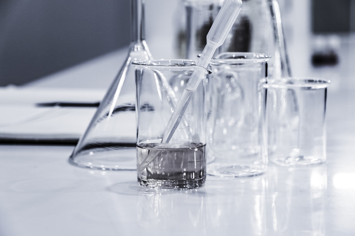
Methodology: Tracking TF's Stealth Mode
1 Sample Prep
Blood from healthy donors and cancer patients was collected in tubes containing Corn Trypsin Inhibitor (CTI) to block contact activation.
2 EV Isolation
Extracellular vesicles (EVs)—TF-rich particles shed by cells—were spun down via ultracentrifugation (20,000 × g for 1 hour).
3 Thrombin Detection
EVs were added to TF-depleted plasma, and thrombin generation was measured using Calibrated Automated Thrombography (CAT).
| TF Concentration | Mean Lag Time (min) | Clinical Implication |
|---|---|---|
| 10 fM | 12.4 ± 0.8 | Baseline in healthy individuals |
| 30 fM | 8.2 ± 0.5 | Early cancer/Thrombosis risk |
| 100 fM | 4.1 ± 0.3 | High-risk metastatic disease |
The Scientist's Toolkit: Decoding TF Activity
Key reagents used in the CAT assay and their roles:
| Reagent | Function | Key Insight |
|---|---|---|
| Anti-TFPI Antibodies | Neutralize TF inhibitor (TFPI) | Unmasks full TF activity |
| CTI (Corn Trypsin Inhibitor) | Blocks contact activation | Prevents false clotting signals |
| Fluorescent Substrates | Detect thrombin (e.g., Z-Gly-Gly-Arg-AMC) | Enables real-time thrombin tracking |
| TF-Depleted Plasma | "Blank canvas" for adding test EVs | Eliminates background noise |
TF's Dark Side: Beyond Clotting to Cancer
TF's signaling function makes it a dangerous accomplice in disease:
Cancer Metastasis
Tumors overexpress TF, fueling angiogenesis and invasion. The alternative-spliced TF (asTF) variant binds integrins, promoting cell migration independent of clotting 3 .
Inflammation
TF activates protease-activated receptors (PARs), triggering cytokine storms in sepsis or atherosclerosis 9 .
Therapeutic Target
Drugs like tisotumab vedotin (FDA-approved for cervical cancer) use anti-TF antibodies to deliver toxins directly to tumor cells 3 .
Controversies and Cutting-Edge Debates
Future Frontiers: Precision Tools and Therapies
Spatial Mapping
Advanced AAV vectors now target TF-expressing cells in specific brain regions, hinting at applications for coagulation disorders 4 .
Nanoparticle Redeployment
TF-conjugated nanoparticles could deliver clot-busting drugs or cancer therapeutics directly to disease sites 3 .
Aging Insights
Spatial transcriptomics reveal aged bone marrow reorganizes TF-expressing stem cell niches, linking hemostasis to aging 6 .
Conclusion: The Uncharted Territory of a Cellular Chameleon
Tissue Factor exemplifies biology's elegance: a single molecule that stitches wounds yet can unravel health when dysregulated. As research unpacks its roles in neurobiology, immunity, and metastasis, TF emerges as a linchpin for next-generation therapies. The goal? To harness its power for healing while curbing its dark potential—a balancing act as precise as coagulation itself.
"In TF, we see the paradox of life: the same force that protects can destroy. Decoding its duality may rewrite medicine's playbook for strokes, cancer, and beyond."