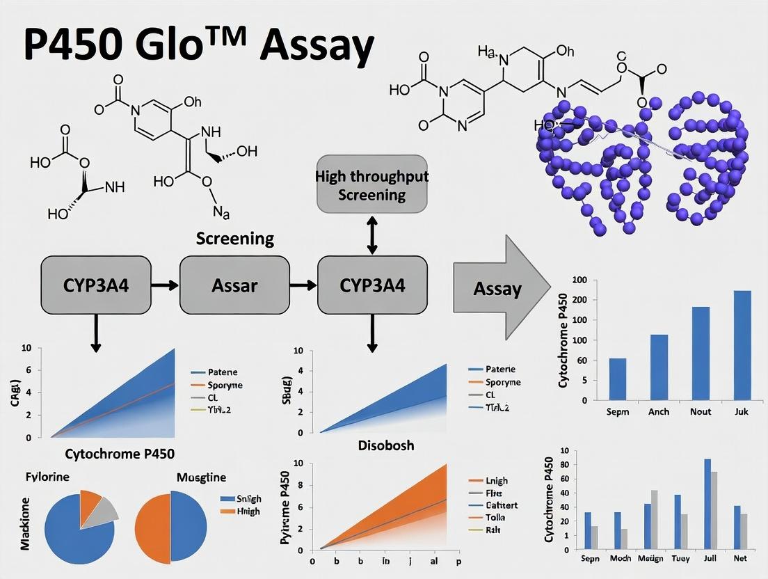
The Ultimate Guide to P450-Glo Assays: High-Throughput Cytochrome P450 Screening for Drug Discovery
This comprehensive guide provides researchers and drug development professionals with an in-depth exploration of Cytochrome P450-Glo™ luciferase reporter assays for high-throughput screening (HTS).
The Ultimate Guide to P450-Glo Assays: High-Throughput Cytochrome P450 Screening for Drug Discovery
Abstract
This comprehensive guide provides researchers and drug development professionals with an in-depth exploration of Cytochrome P450-Glo™ luciferase reporter assays for high-throughput screening (HTS). The article begins with foundational knowledge on the critical role of CYP450 enzymes in drug metabolism and toxicity. It details the step-by-step methodology, from assay setup to data analysis, enabling robust implementation in HTS workflows. Practical troubleshooting and optimization strategies are provided to address common challenges and enhance assay performance. Finally, the guide compares P450-Glo technology to alternative methods, validating its advantages in sensitivity, specificity, and adaptability. This resource equips scientists with the knowledge to effectively integrate this powerful tool into preclinical drug development.
Understanding Cytochrome P450 Screening: Why P450-Glo Assays are Essential for Modern Drug Development
Cytochrome P450 (CYP) enzymes are a superfamily of hemeproteins primarily located in the endoplasmic reticulum of hepatocytes, responsible for the oxidative metabolism of a vast array of endogenous compounds and xenobiotics, including approximately 70-80% of all clinically used drugs. The most significant isoforms in human drug metabolism are CYP3A4, CYP2D6, CYP2C9, CYP2C19, and CYP1A2. Drug-Drug Interactions (DDIs) occur when one drug alters the metabolic clearance of another, primarily via induction or inhibition of these CYP enzymes, leading to potentially toxic drug accumulation or therapeutic failure.
Quantitative Data on Major Human CYP Enzymes
Table 1: Major Human Hepatic CYP Enzymes: Substrate Prevalence and Polymorphism Impact
| CYP Isoform | Approx. % of Drugs Metabolized | Notable Genetic Polymorphism | Clinical Impact of Polymorphism |
|---|---|---|---|
| CYP3A4/5 | ~45-50% | Low for 3A4, significant for 3A5 | Altered dose requirements for tacrolimus, midazolam. |
| CYP2D6 | ~20-25% | Extensive (Over 100 alleles) | Poor vs. Ultrarapid metabolizer status affects efficacy/toxicity of opioids, antidepressants. |
| CYP2C9 | ~15% | Significant (*2, *3 alleles) | Warfarin sensitivity; reduced NSAID metabolism. |
| CYP2C19 | ~10% | Significant (*2, *17 alleles) | Altered clopidogrel efficacy; PPIs, antidepressants dosing. |
| CYP1A2 | ~8-10% | Moderate | Variable metabolism of clozapine, theophylline. |
Table 2: Common CYP Inhibitors and Inducers and Their DDI Risk
| Compound/Drug | Target CYP(s) | Mechanism | Clinical DDI Example (Victim Drug) | Risk Level |
|---|---|---|---|---|
| Ketoconazole | CYP3A4 | Reversible Inhibition | Increased statin (simvastatin) exposure → myopathy. | High |
| Ritonavir | CYP3A4, others | Mechanism-based Inactivation | Profound, long-lasting inhibition of many co-administered drugs. | High |
| Rifampin | CYP3A4, 2C9, others | Induction (PXR activation) | Reduced efficacy of oral contraceptives, warfarin. | High |
| Fluoxetine | CYP2D6 | Reversible Inhibition | Increased TCA (e.g., nortriptyline) levels → toxicity. | Moderate |
| Omeprazole | CYP2C19 | Competitive Inhibition | Reduced clopidogrel activation → reduced antiplatelet effect. | Moderate |
Detailed Experimental Protocol: P450-Glo Luminescent Assay for CYP Inhibition Screening
Protocol Title: High-Throughput Screening for Direct CYP Inhibition Using Recombinant Enzymes and Luciferin- Derived Probe Substrates
Objective: To determine the half-maximal inhibitory concentration (IC50) of a test compound against a specific human recombinant CYP isoform in a 96- or 384-well plate format.
Principle: The assay uses a proprietary luminogenic probe substrate (e.g., Luciferin-IPA for CYP3A4). CYP metabolism converts the probe to a luciferin product, which is detected in a subsequent luciferase reaction, generating light. Inhibitors reduce light output in a concentration-dependent manner.
Materials (The Scientist's Toolkit):
Table 3: Key Research Reagent Solutions for P450-Glo Assay
| Reagent/Material | Function & Specification |
|---|---|
| Recombinant Human CYP Enzyme (e.g., CYP3A4 + P450 Reductase in membranes) | Catalytic source for the specific reaction. |
| P450-Glo Assay Buffer (1X) | Provides optimal pH and ionic strength for enzyme activity. |
| Luciferin-based Probe Substrate (e.g., Luciferin-IPA) | Isoform-specific, non-fluorescent probe metabolized to D-luciferin. |
| NADPH Regeneration System (or cofactor solution) | Supplies reducing equivalents (NADPH) required for CYP oxidation. |
| Luciferin Detection Reagent | Contains luciferase to convert generated D-luciferin to luminescent signal. |
| Reference Inhibitor (e.g., Ketoconazole for CYP3A4) | Positive control for inhibition. |
| Test Compounds in DMSO | Compounds for screening, typically in 10-point serial dilution. |
| White, Solid-Bottom Microplates | Optimal for luminescence signal detection with minimal cross-talk. |
| Plate Luminometer | Instrument to measure relative light units (RLU). |
Procedure:
- Plate Preparation: Dilute test and control inhibitors in assay buffer. Prepare a 2X NADPH regeneration system.
- Reaction Assembly (10 µL final volume in well):
- Add 2.5 µL of test compound (or control/blank) in buffer to the plate.
- Add 5 µL of recombinant CYP enzyme (diluted in buffer to predetermined concentration).
- Add 2.5 µL of 2X probe substrate solution.
- Pre-incubation & Reaction Initiation:
- Pre-incubate plate for 10 minutes at room temperature.
- Initiate reaction by adding 10 µL of 2X NADPH regeneration system. Final concentrations: [CYP], [Probe] at ~Km, [NADPH] ~1mM, [DMSO] ≤0.5%.
- Incubate for a linear time period (e.g., 30 min for CYP3A4) at 37°C.
- Detection:
- Terminate reaction by adding 20 µL of Luciferin Detection Reagent.
- Incubate at room temperature for 20 minutes to stabilize luminescent signal.
- Measure luminescence (RLU) on a plate-reading luminometer.
- Data Analysis:
- Calculate % Inhibition:
[1 - (RLU_compound - RLU_blank)/(RLU_control - RLU_blank)] * 100. - Plot % Inhibition vs. log[Compound]. Fit sigmoidal dose-response curve to determine IC50.
- Calculate % Inhibition:
Visualizations
CYP-Mediated DDI Mechanism
P450-Glo Assay Workflow
Application Notes
Drug-drug interactions (DDIs) mediated by the inhibition of cytochrome P450 (CYP450) enzymes remain a leading cause of late-stage clinical trial failures and post-market drug withdrawals. Early identification of potent CYP inhibitors is therefore paramount for de-risking drug discovery pipelines. High-throughput screening (HTS) using luminescence-based assays, such as the P450-Glo platform, provides a robust solution for generating critical data on compound liability against key CYP isoforms (CYP1A2, 2C9, 2C19, 2D6, 3A4) during the lead identification and optimization phases. Integrating this data into structure-activity relationship (SAR) analyses allows medicinal chemists to steer away from problematic chemotypes, thereby improving the safety profile and developmental success rate of clinical candidates.
Table 1: Key CYP450 Isoforms, Their Proportion of Drug Metabolism, and Common Probe Substrates for HTS
| CYP Isoform | % of Drugs Metabolized | Primary Role | Example Probe Substrate (P450-Glo Assay) |
|---|---|---|---|
| CYP3A4 | ~30-50% | Metabolism of largest range of drugs | Luciferin-IPA (Luciferin isopropyl acetal) |
| CYP2D6 | ~20-25% | Metabolism of many CNS and CV drugs | Luciferin-ME EGE (Luciferin methyl ether) |
| CYP2C9 | ~10-15% | Metabolism of NSAIDs, oral anticoagulants | Luciferin-H (Luciferin H) |
| CYP2C19 | ~5-10% | Metabolism of proton pump inhibitors, antidepressants | Luciferin-H EGE (Luciferin H ethyl ether) |
| CYP1A2 | ~5-10% | Metabolism of caffeine, polycyclic aromatics | Luciferin-CEE (Luciferin chloroethyl ether) |
Table 2: Advantages of Luminescent (P450-Glo) vs. Traditional Fluorescent & LC-MS/MS CYP Inhibition Assays
| Assay Parameter | P450-Glo (Luminescent) | Fluorescent Probes | LC-MS/MS (Gold Standard) |
|---|---|---|---|
| Throughput | Very High (384/1536-well) | Very High | Low to Medium |
| Sensitivity | Very High (low enzyme consumption) | Moderate | Highest |
| Specificity | High (isoform-specific proluciferins) | Low (probe cross-reactivity) | Very High |
| Assay Complexity | Simple, "add-mix-read" | Simple | Complex (sample prep, separation) |
| Cost per Data Point | Low | Lowest | High |
| Primary Use | Early HTS & SAR | Preliminary Screening | Definitive Kinetics & Regulatory |
Experimental Protocols
Protocol 1: High-Throughput Screening for CYP450 Inhibition Using P450-Glo Assays
Objective: To identify potential inhibitors of a specific CYP450 isoform (e.g., CYP3A4) from a compound library in a 384-well format.
Materials: See "Research Reagent Solutions" table below.
Workflow:
- Plate Preparation: Dilute test compounds in DMSO to a 100X final desired concentration (e.g., 10 µM final). Transfer 0.1 µL to corresponding wells of a white, opaque 384-well plate.
- Reaction Mixture Assembly: Prepare a master mix on ice containing:
- Recombinant Human CYP450 enzyme (e.g., CYP3A4, pre-mixed with NADPH-P450 reductase).
- NADP⁺ Regeneration System (glucose-6-phosphate and G6PDH).
- Luciferin-Probe substrate (e.g., Luciferin-IPA for CYP3A4) at a concentration near its apparent Km.
- Initiate Reaction: Dispense 9.9 µL of the master mix into each well containing compound. Include controls: No-Inhibition Control (DMSO only), Background Control (no enzyme), and a known potent inhibitor (e.g., Ketoconazole for CYP3A4) as a positive control.
- Incubation: Seal the plate and incubate at 37°C for a predetermined time (typically 10-30 minutes) to allow the CYP enzyme to convert the probe substrate to luciferin.
- Signal Development: Add 10 µL of Luciferin Detection Reagent to each well. This reagent simultaneously stops the CYP reaction and initiates the luciferase reaction, producing a stable, "glow-type" luminescent signal proportional to the amount of luciferin generated.
- Signal Measurement: Read luminescence on a plate reader after a 20-minute incubation at room temperature.
- Data Analysis: Calculate % Inhibition:
[1 - (Signal_Compound - Signal_Background) / (Signal_No_Inhibition - Signal_Background)] * 100. Compounds showing >50% inhibition at the test concentration are flagged for follow-up IC₅₀ determination.
Protocol 2: Determination of IC₅₀ Values for Hit Confirmation
Objective: To generate a concentration-response curve and calculate the half-maximal inhibitory concentration (IC₅₀) for confirmed hits.
Workflow:
- Prepare a 3-fold serial dilution of test compounds (typically 10 concentrations, e.g., from 30 µM to 0.05 nM) in DMSO.
- Transfer 0.1 µL of each dilution to triplicate wells.
- Repeat steps 2-6 from Protocol 1.
- Data Analysis: Plot % Inhibition vs. log[Compound]. Fit the data using a four-parameter logistic (sigmoidal) curve-fitting model (e.g., in GraphPad Prism) to calculate the IC₅₀ value.
Diagrams
Title: P450-Glo HTS Inhibition Screening Workflow
Title: CYP Inhibition Assay Signaling Pathway
The Scientist's Toolkit: Research Reagent Solutions
| Reagent / Material | Function & Role in Assay |
|---|---|
| P450-Glo Assay Kits | Complete, optimized systems for specific isoforms. Contain recombinant CYP enzyme, luciferin-probe substrate, NADP⁺ regeneration system, detection buffer, and positive control inhibitor. |
| Recombinant Human CYP Enzymes (e.g., Supersomes, Baculosomes) | Provide consistent, isoform-specific CYP activity without interference from other cellular components. Essential for clean, interpretable data. |
| Isoform-Specific Luciferin Probes (e.g., Luciferin-IPA) | Non-luminescent proluciferin substrates. CYP metabolism cleaves the ether bond to release free D-luciferin, the substrate for the final luciferase reaction. |
| NADP⁺ Regeneration System | Continuously supplies NADPH, the essential electron donor for CYP catalytic activity. Typically includes Glucose-6-Phosphate and Glucose-6-Phosphate Dehydrogenase. |
| Luciferin Detection Reagent | Contains luciferase and ATP in a stabilizing buffer. Stops the CYP reaction and initiates the luminescent signal, producing a stable "glow" for high-throughput reading. |
| White, Opaque 384-Well Plates | Maximize luminescence signal collection and minimize cross-talk between wells during plate reading. |
| DMSO (Cell Culture Grade) | Universal solvent for compound libraries. Must be of high purity and used at low final concentration (<1% v/v) to avoid enzyme inhibition. |
| Positive Control Inhibitors (e.g., Ketoconazole for CYP3A4) | Validates assay performance in each run by demonstrating expected maximum inhibition, used for Z'-factor calculation. |
Within the context of high-throughput screening (HTS) for drug metabolism and drug-drug interaction studies, the P450-Glo assay platform provides a luminescent, homogeneous method for measuring cytochrome P450 (CYP) enzyme activity. This technology is central to modern research, enabling rapid, sensitive, and convenient screening of new chemical entities for their potential to inhibit or induce specific CYP isoforms, which is critical for predicting metabolic stability and toxicity.
Core Principle and Mechanism
The P450-Glo assays are based on proluciferin substrates, which are derivatives of beetle luciferin. Each proluciferin substrate is specifically tailored to be metabolized by a single, recombinant human CYP isoform (e.g., CYP3A4, CYP2D6). The core principle involves a two-step reaction:
- CYP Enzymatic Reaction: The recombinant CYP enzyme, in the presence of NADPH, catalyzes the conversion of its specific proluciferin substrate into luciferin.
- Luminescent Detection: The luciferin product is subsequently detected by adding a proprietary "Luciferin Detection Reagent." This reagent contains luciferase, which converts luciferin to oxyluciferin in an ATP-dependent reaction, producing a stable, sustained "glow-type" luminescent signal. The intensity of the light produced is proportional to the amount of luciferin generated, which is directly proportional to the CYP enzyme activity.
Diagram: P450-Glo Two-Step Reaction Principle
Application Notes and Key Quantitative Data
P450-Glo assays are validated for HTS applications. Key performance metrics include:
Table 1: Representative Performance Metrics for P450-Glo CYP3A4 Assay
| Parameter | Value | Notes |
|---|---|---|
| Signal-to-Background | >100 | High dynamic range. |
| Z'-Factor | >0.7 | Excellent for HTS robustness. |
| Assay Format | 384- and 1536-well | Low volume, HTS compatible. |
| Incubation Time | 10-30 minutes | Short CYP reaction step. |
| Luminescence Signal Half-life | >3 hours | Stable "glow" signal for batch processing. |
| Recommended [Enzyme] per well | 1-10 nM (rCYP) | Optimized for sensitivity and linearity. |
| Linear Range | Up to 10 pmol luciferin | For standard protocol. |
Table 2: Common CYP Isoforms and Their Proluciferin Substrates
| CYP Isoform | Primary Role in Drug Metabolism | Example Proluciferin Substrate |
|---|---|---|
| CYP3A4 | Metabolizes >50% of clinically used drugs. | Luciferin-IPA (isopropylacetal) |
| CYP2D6 | Polymorphic, involved in ~25% of drugs. | Luciferin-ME EGE (methoxyethyl ether) |
| CYP2C9 | Metabolizes many NSAIDs and anticoagulants. | Luciferin-H (benzyl ether) |
| CYP1A2 | Metabolizes aromatic amines and heterocyclics. | Luciferin-CEE (chloroethyl ether) |
| CYP2C19 | Polymorphic, important for proton pump inhibitors. | Luciferin-H EGE (hydroxyethyl ether) |
Experimental Protocols
Protocol 1: Standard CYP Inhibition Screening Assay
Objective: To determine the inhibitory potential (IC50) of test compounds against a specific CYP isoform.
Materials: (See "The Scientist's Toolkit" below) Procedure:
- Plate Preparation: In a white, opaque-walled assay plate (384-well), add 2.5 µL of test compound (in DMSO, serially diluted) or control (DMSO for no-inhibition, strong CYP inhibitor for background control).
- Enzyme/Substrate Addition: Add 10 µL of a pre-mixed solution containing the recombinant CYP enzyme and its specific proluciferin substrate in a suitable reaction buffer (e.g., PBS, pH 7.4).
- Initiate Reaction: Add 2.5 µL of NADPH regeneration system (or a solution of NADPH itself) to initiate the CYP enzymatic reaction. Final assay volume is 15 µL.
- Incubation: Incubate plate at room temperature for a predetermined time (e.g., 10-30 minutes), protected from light.
- Signal Development: Add 15 µL of the Luciferin Detection Reagent to stop the CYP reaction and initiate the luminescent reaction. Mix gently.
- Detection: Incubate at room temperature for 20 minutes to stabilize the signal. Measure luminescence on a plate-reading luminometer.
- Data Analysis: Calculate % activity relative to no-inhibition control. Fit dose-response data to determine IC50 values.
Diagram: CYP Inhibition Assay Workflow
Protocol 2: CYP Induction Assessment (Using Luminescent Readout)
Objective: To assess the potential of a compound to induce CYP gene expression in a cellular model (e.g., hepatocytes).
Materials: Cultured human hepatocytes, induction medium, test compounds, P450-Glo assay components for target CYP. Procedure:
- Cell Treatment: Seed hepatocytes in culture plates. After attachment, treat cells with test compound, vehicle control, or positive control (e.g., rifampin for CYP3A4) for 48-72 hours, refreshing medium/compound daily.
- Cell Lysis & Substrate Addition: At endpoint, carefully remove treatment medium. Add a buffer containing the specific proluciferin substrate for the CYP of interest and a lytic agent to permeabilize the cells.
- Enzymatic Reaction: Endogenous induced CYP enzymes within the lysed cells metabolize the proluciferin to luciferin. Incubate for a defined period (e.g., 10-60 mins).
- Signal Development & Detection: Add an equal volume of Luciferin Detection Reagent. After signal stabilization (20 mins), measure luminescence.
- Data Analysis: Normalize luminescence to total protein content (via a separate assay). Fold induction is calculated relative to vehicle-treated control cells.
The Scientist's Toolkit: Essential Research Reagents
Table 3: Key Components for P450-Glo Assays
| Item | Function | Notes |
|---|---|---|
| Recombinant Human CYP Enzyme | Catalyzes the conversion of the proluciferin substrate. | Isoform-specific (e.g., CYP3A4, baculosomes). |
| Proluciferin Substrate | CYP isoform-selective probe. Becomes luciferin upon demethylation/dealkylation. | E.g., Luciferin-IPA for CYP3A4. Supplied in buffer. |
| NADPH Regeneration System | Supplies reducing equivalents (NADPH) required for CYP catalytic cycle. | Can use System A (Glucose-6-P + Dehydrogenase) or direct NADPH. |
| Luciferin Detection Reagent | Contains luciferase and ATP to generate light from the luciferin product. | Provides a stable "glow" signal. Stops CYP reaction. |
| Assay Buffer | Provides optimal pH and ionic conditions for CYP activity. | Typically phosphate-based, pH 7.4. |
| Reference Inhibitors | Positive controls for inhibition assays (e.g., Ketoconazole for CYP3A4). | Used to define 100% inhibition baseline. |
| White Opaque Microplates | Plate format for luminescence detection. Minimizes signal crosstalk. | 384-well and 1536-well formats for HTS. |
| Plate-Reading Luminometer | Instrument to detect and quantify the luminescent signal. | Must be compatible with microplate format. |
Key CYP450 Isoforms Screened (CYP3A4, 2D6, 2C9, 2C19, 1A2) and Their Clinical Relevance
Within the context of a thesis on high-throughput screening (HTS) using P450-Glo assays, the evaluation of key Cytochrome P450 (CYP) isoforms is a cornerstone of early-phase drug discovery. CYP3A4, 2D6, 2C9, 2C19, and 1A2 are responsible for metabolizing a vast majority of clinically used drugs. Screening for inhibition or induction of these enzymes is critical to predict and mitigate risks of drug-drug interactions (DDIs), which can lead to therapeutic failure or adverse events. This document details the application notes and experimental protocols for their assessment using luminescent P450-Glo technology.
Clinical Relevance of Key CYP Isoforms
The following table summarizes the clinical relevance, genetic polymorphism, and example substrates for each key isoform.
Table 1: Key CYP450 Isoforms, Polymorphism, and Clinical Relevance
| Isoform | Approx. % of Drug Metabolism | Genetic Polymorphism | Major Clinical Impact & Example Drugs |
|---|---|---|---|
| CYP3A4 | ~50% | Low | Highest DDI risk; metabolizes statins (simvastatin), immunosuppressants (cyclosporine), many opioids. |
| CYP2D6 | ~20-25% | High (PM, IM, EM, UM) | Altered efficacy/toxicity of antidepressants (fluoxetine), antipsychotics, beta-blockers (metoprolol). |
| CYP2C9 | ~15% | High | Warfarin dosing (S-warfarin); phenytoin and NSAID (ibuprofen) metabolism variability. |
| CYP2C19 | ~10% | High | Clopidogrel activation (PMs: therapeutic failure); PPIs (omeprazole) metabolism. |
| CYP1A2 | ~5-10% | Moderate | Inducible by smoking; metabolizes clozapine, theophylline, caffeine. |
P450-Glo Assay Principle & High-Throughput Screening Workflow
The P450-Glo assay is a bioluminescent, cell-free method using recombinant CYP isoforms and a proluciferin substrate specific to each enzyme. CYP activity converts the proluciferin to luciferin, which is detected by a luciferase reaction, generating light proportional to CYP activity. This homogeneous "add-mix-read" format is ideal for HTS.
Detailed Experimental Protocols
Protocol 1: Initial Single-Concentration Inhibition Screening
Objective: To rapidly identify compounds that inhibit a specific CYP isoform at a fixed concentration (e.g., 10 µM).
Materials:
- White, opaque 96- or 384-well plates
- Test compounds (in DMSO, final concentration typically 1-10 µM)
- Recombinant Human CYP Isoform (e.g., CYP3A4, baculosomes)
- NADPH Regeneration System (Solution A: NADP+, Glucose-6-phosphate; Solution B: Glucose-6-phosphate dehydrogenase)
- CYP-specific Luciferin-Proluciferin Substrate (e.g., Luciferin-IPA for CYP3A4)
- P450-Glo Detection Reagent (contains luciferin detection buffer and luciferase)
- Positive Control Inhibitor (e.g., Ketoconazole for CYP3A4)
- Negative Control (0.5-1.0% DMSO vehicle)
- Multichannel pipettes, plate shaker, luminescence microplate reader
Procedure:
- Plate Preparation: Dilute test compounds in assay buffer to 2X final desired concentration. Transfer 25 µL to designated assay plate wells. Include negative (vehicle) and positive control wells.
- Enzyme/Substrate Reaction:
- Prepare the P450 Reaction Mix on ice: Combine recombinant CYP enzyme (at predetermined protein concentration), NADPH Regeneration System, and the specific proluciferin substrate in assay buffer.
- Add 25 µL of the P450 Reaction Mix to each well using a multichannel pipette.
- Seal plate, mix briefly on a plate shaker, and incubate for the optimal time (typically 10-30 minutes at 37°C). This allows metabolism.
- Signal Detection:
- Equilibrate the P450-Glo Detection Reagent to room temperature.
- Add an equal volume of Detection Reagent (e.g., 50 µL) to each well to stop the CYP reaction and initiate the luminescent reaction.
- Mix briefly, incubate at room temperature for 10-20 minutes to stabilize signal.
- Luminescence Measurement: Read luminescence on a compatible plate reader using integration times of 0.1-1 second per well.
- Data Analysis: Calculate percent inhibition relative to controls:
% Inhibition = [1 - (Signal_Compound - Signal_PosCtrl) / (Signal_NegCtrl - Signal_PosCtrl)] * 100.
Protocol 2: Determination of IC50 Values
Objective: To characterize the potency of identified inhibitors by determining the half-maximal inhibitory concentration (IC50).
Materials: As in Protocol 1, with the addition of compound stock solutions for serial dilution.
Procedure:
- Compound Dilution: Prepare a 3- or 4-fold serial dilution of the test compound in DMSO (e.g., from 10 mM to nM range). Further dilute in assay buffer to create a 2X working concentration series, ensuring final DMSO concentration is constant (e.g., ≤1%).
- Assay Execution: Perform steps 1-4 from Protocol 1 using the dilution series. Run in triplicate.
- Data Analysis:
- Plot mean luminescence (or % Activity) against the log10 of compound concentration.
- Fit data to a four-parameter logistic (sigmoidal) curve using software (e.g., GraphPad Prism).
- Determine the IC50 from the curve fit as the concentration yielding 50% inhibition of enzyme activity.
Table 2: Example Assay Conditions for Key Isoforms Using P450-Glo
| Isoform | Recommended Proluciferin Substrate | Typical Incubation Time | Common Positive Control Inhibitor |
|---|---|---|---|
| CYP3A4 | Luciferin-IPA | 10-15 min | Ketoconazole |
| CYP2D6 | Luciferin-ME EGE | 30-45 min | Quinidine |
| CYP2C9 | Luciferin-H | 30 min | Sulfaphenazole |
| CYP2C19 | Luciferin-H | 30 min | (S)-(-)-N-3-Benzylnirvanol |
| CYP1A2 | Luciferin-CEE | 30 min | α-Naphthoflavone |
Data Interpretation and Integration into DDI Risk Assessment
The quantitative data generated feeds directly into regulatory decision-making.
Table 3: Quantitative Decision Criteria for CYP Inhibition Risk (FDA/EMA Guidance)
| Inhibition Potency | [I]/IC50 Ratio* | Clinical DDI Risk & Action |
|---|---|---|
| Strong | ≥ 0.1 (or IC50 < 1 µM) | High Risk. Likely requires clinical DDI study and contraindications. |
| Moderate | 0.01 to < 0.1 | Potential Risk. May require dose adjustment or cautionary labeling. |
| Weak | < 0.01 | Low Risk. Unlikely to be clinically relevant. |
*[I] = maximum total plasma concentration of the inhibitor.
The Scientist's Toolkit: Key Research Reagent Solutions
Table 4: Essential Materials for CYP450 P450-Glo Screening
| Reagent/Material | Function & Rationale | Example Vendor/Product |
|---|---|---|
| Recombinant CYP Isoforms | Consistent, single-isoform source for specific, reproducible reaction kinetics. | Supersomes (Corning), Baculosomes (Thermo Fisher). |
| Isoform-Specific Luciferin-Proluciferins | Highly selective substrates minimize cross-isoform interference, ensuring assay specificity. | P450-Glo Substrates (Promega). |
| NADPH Regeneration System | Provides a constant supply of NADPH, the essential cofactor for CYP catalytic activity. | Promega, Thermo Fisher Scientific. |
| P450-Glo Detection Reagent | Contains luciferase to generate luminescent signal from metabolized luciferin; stops CYP reaction. | Promega P450-Glo Assay Kits. |
| Validated Chemical Inhibitors | Essential assay controls for determining assay window (Z') and validating system performance. | e.g., Ketoconazole (Sigma-Aldrich). |
| Luminescence Plate Reader | High-sensitivity instrument for detecting low-light luminescence signals in HTS format. | GloMax (Promega), EnVision (PerkinElmer). |
Advantages of Luciferin-Based Reporter Assays over Traditional LC-MS Methods
Application Notes
Within the context of high-throughput screening (HTS) for cytochrome P450 enzyme activity, particularly using P450-Glo assays, luciferin-based reporter systems offer distinct advantages over traditional liquid chromatography-mass spectrometry (LC-MS) methods. These advantages are critical for accelerating drug metabolism and pharmacokinetics (DMPK) research in early drug discovery.
The core advantage lies in the conversion of a P450 enzymatic reaction into a bioluminescent readout. A pro-luciferin substrate (e.g., Luciferin-IPA for CYP3A4) is metabolized by the recombinant P450 enzyme to produce D-luciferin. This product is then quantified by a coupled luciferase reaction, generating light proportional to P450 activity. This single-step, homogeneous "add-mix-read" format is inherently suitable for automation and miniaturization.
Key Comparative Advantages:
- Throughput and Speed: Luciferin-based assays enable the screening of >100,000 compounds per day in 1,536-well formats, with signal detection in minutes. LC-MS runs typically require several minutes per sample, creating a bottleneck.
- Simplicity and Cost: Reporter assays require no specialized chromatography or mass spectrometry instrumentation, no complex sample preparation (e.g., protein precipitation, extraction), and reduce reagent consumption. This lowers capital equipment costs and operational expenses.
- Dynamic Range and Sensitivity: Bioluminescence offers a wide dynamic range (often 4-5 orders of magnitude), detecting very low levels of enzyme activity due to the high quantum yield of the luciferase reaction.
- Adaptability: The same luminescence detection platform can be used for a wide array of cytochrome isoforms (CYP1A2, 2C9, 2D6, 3A4) by simply changing the pro-luciferin substrate, streamlining workflow.
Quantitative Comparison of Key Parameters:
Table 1: Comparative Analysis of Assay Methodologies for P450 Screening
| Parameter | Luciferin-Based Reporter Assay (e.g., P450-Glo) | Traditional LC-MS Method |
|---|---|---|
| Throughput | Ultra-High (>100,000 data points/day) | Low-Medium (100s-1,000s/day) |
| Assay Time | ~1 hour (incubation + detection) | Several minutes to hours per sample |
| Sample Prep | Homogeneous, "add-mix-read" | Complex: quenching, extraction, centrifugation |
| Instrumentation | Standard plate reader | HPLC/UPLC, Mass Spectrometer (high capital cost) |
| Data Complexity | Simple, direct activity readout | Complex; requires metabolite identification & quantification |
| Approx. Cost per 1,536-well plate | $500 - $800 | $2,000 - $5,000+ (incl. instrument depreciation) |
| Primary Application | Primary HTS, Inhibition/Phenotyping | Secondary confirmation, Metabolite ID, Kinetic studies |
Experimental Protocols
Protocol 1: P450-Glo CYP3A4 Inhibition Screening Assay (384-Well Format) This protocol details a standard procedure for screening chemical libraries for CYP3A4 inhibitors.
I. Materials & Reagent Preparation
- P450-Glo CYP3A4 Assay Kit (contains Luciferin-IPA, NADP⁺ Regeneration System, Luciferin Detection Reagent, Recombinant CYP3A4 Enzyme).
- Test Compounds: Dissolved in DMSO (typically 10 mM stock). Prepare intermediate dilutions in assay buffer.
- Control Inhibitors: Ketoconazole (strong inhibitor) and solvent (DMSO, negative control).
- Assay Buffer: 100 mM Potassium Phosphate Buffer, pH 7.4.
- White, solid-bottom 384-well assay plates.
- Multichannel pipettes, plate dispenser.
- Luminometer-equipped plate reader.
II. Procedure
- Plate Preparation: Dilute test compounds in buffer to 2X final desired concentration. Transfer 5 µL of each 2X compound or control to assigned wells. Include DMSO-only control wells (0% inhibition) and ketoconazole control wells (100% inhibition).
- Enzyme/Substrate Mixture: Prepare a master mix on ice containing recombinant CYP3A4 enzyme and Luciferin-IPA substrate in assay buffer according to kit instructions. Add NADP⁺ Regeneration System to initiate reaction.
- Reaction Initiation: Immediately add 5 µL of the enzyme/substrate/master mix to each well using a dispenser, bringing the total volume to 10 µL. Final DMSO concentration should be ≤1%.
- Incubation: Cover plate and incubate at room temperature for 30-60 minutes (time-optimized for linear reaction kinetics).
- Detection: Add 10 µL of Luciferin Detection Reagent to each well to stop the P450 reaction and initiate the luminescent reaction. Incubate for 20 minutes at room temperature to stabilize the signal.
- Measurement: Read luminescence on a plate reader with an integration time of 0.5-1 second per well.
III. Data Analysis
- Calculate the percentage of inhibition for each test compound:
% Inhibition = [1 - (LumSample - Lum100%Inh) / (Lum0%Inh - Lum100%Inh)] * 100Where LumSample = compound well, Lum0%Inh = DMSO control average, Lum100%Inh = ketoconazole control average. - Generate dose-response curves from serial compound dilutions to calculate IC₅₀ values.
Protocol 2: LC-MS/MS Method for CYP3A4 Metabolite Detection (Comparative Validation) This protocol is provided for orthogonal validation of hits from the primary HTS.
I. Materials
- LC-MS/MS System: UPLC coupled to a triple quadrupole mass spectrometer.
- Chromatography Column: C18 reverse-phase column (e.g., 2.1 x 50 mm, 1.7 µm).
- Mobile Phases: A: 0.1% Formic acid in water; B: 0.1% Formic acid in acetonitrile.
- Testosterone (substrate) and 6β-Hydroxytestosterone (metabolite) standards.
- Incubation System: Human liver microsomes (HLM) or recombinant CYP3A4, NADPH.
II. Procedure
- Incubation: In a 96-well deep-well plate, incubate testosterone (50 µM) with HLM (0.1 mg/mL) and test compound (at IC₅₀ concentration from Protocol 1) in potassium phosphate buffer (pH 7.4). Start reaction with NADPH (1 mM). Incubate at 37°C for 30 min.
- Quenching & Extraction: Stop reaction with 2 volumes of ice-cold acetonitrile containing an internal standard. Vortex, then centrifuge at 4,000 x g for 15 min to precipitate protein.
- Sample Transfer: Transfer supernatant to a new plate for LC-MS/MS analysis.
- LC Conditions: Inject 5-10 µL. Use a gradient from 20% B to 95% B over 3.5 minutes at 0.4 mL/min.
- MS Conditions: Use electrospray ionization (ESI) in positive mode. Monitor multiple reaction monitoring (MRM) transitions: Testosterone: m/z 289 → 97; 6β-Hydroxytestosterone: m/z 305 → 269.
III. Data Analysis Quantify metabolite formation by integrating peak areas and comparing to a standard curve. Calculate % remaining activity relative to vehicle control to confirm inhibition.
Diagrams
P450 Luciferin-Based Reporter Assay Pathway
P450-Glo HTS Workflow: Add-Mix-Read
The Scientist's Toolkit
Table 2: Essential Research Reagent Solutions for P450-Glo Assays
| Item | Function in the Assay | Key Consideration |
|---|---|---|
| P450-Glo Assay Kits | Provides isoform-specific pro-luciferin substrates, optimized recombinant P450 enzymes, NADPH regeneration system, and luciferin detection reagent in a unified system. | Select kit matched to cytochrome isoform (CYP1A2, 2C9, 2D6, 3A4). |
| NADP⁺ Regeneration System | Supplies a constant level of NADPH, the essential cofactor for P450 enzymatic activity, during the incubation period. | Critical for maintaining linear reaction kinetics. |
| Ultra-Glo Recombinant Luciferase | A stable, engineered luciferase that provides a sustained "glow-type" signal, enabling batch processing of plates. | Superior to "flash" luciferases for HTS. |
| Luciferin-IPA / BE / H / ME | Pro-luciferin substrates selectively metabolized by specific P450s (e.g., Luciferin-IPA for CYP3A4). | Metabolism generates D-luciferin, the luciferase substrate. |
| Quenching/Acquisition Buffer | Stops the P450 reaction and provides optimal pH and conditions for the subsequent luciferase reaction. | Ensures signal stability for up to 3 hours. |
| Control Inhibitors (Ketoconazole, Sulfaphenazole) | Pharmacological tool compounds used to establish baseline (100% inhibition) and validate assay performance for each P450 isoform. | Essential for QC and data normalization. |
| OptiPlate or Similar White Plates | Solid-bottom, white multiwell plates maximize luminescent signal reflection and minimize crosstalk between wells. | Critical for low-volume, high-density (1536-well) formats. |
Step-by-Step Protocol: Implementing P450-Glo Assays in Your HTS Workflow
Within the context of a thesis focused on high-throughput screening (HTS) for cytochrome P450 (CYP) activity and inhibition, the reliability of the P450-Glo assay system is paramount. This luminescent assay converts CYP-dependent activity into a quantifiable luminescent signal via a coupled enzymatic reaction. The core components—specific luminogenic CYP substrates, the cofactor NADPH, and the luciferin detection reagent—must be prepared and handled with precision to ensure data integrity for drug metabolism and toxicity studies. This application note details the preparation, optimization, and use of these critical reagents.
Critical Assay Components: Function and Preparation
Luminogenic CYP Substrates
These are proprietary pro-luciferin compounds designed to be selective for specific CYP isozymes (e.g., CYP3A4, CYP2D6). The CYP enzyme cleaves the substrate to release D-luciferin, the substrate for luciferase.
Preparation Protocol:
- Reconstitution: Add the recommended volume of nuclease-free water or specified buffer (often provided) to the vial to create a concentrated stock solution (typically 1-10 mM).
- Aliquoting: Immediately aliquot the reconstituted substrate into single-use volumes to avoid repeated freeze-thaw cycles.
- Storage: Store aliquots at ≤ -60°C protected from light. Working solutions are diluted in reaction buffer and kept on ice during use.
β-Nicotinamide Adenine Dinucleotide Phosphate (NADPH)
NADPH is the essential redox cofactor required for CYP-mediated monooxygenation. Its stability is a common limiting factor.
Preparation and Handling Protocol:
- Fresh Preparation: NADPH solutions should be prepared immediately before use. Weigh the required amount of NADPH tetrasodium salt.
- Dissolution: Dissolve in cold, neutral pH buffer (e.g., 100 mM potassium phosphate, pH 7.4). Do not use Tris-based buffers as they can accelerate degradation.
- Concentration: A typical working concentration in the final reaction is 1-10 µM for the P450-Glo coupled system, though a higher starting concentration (e.g., 1 mM) is used for the regeneration system.
- Verification: Confirm the concentration spectrophotometrically using the absorbance at 340 nm (ε340 = 6220 M⁻¹cm⁻¹).
Luciferin Detection Reagent
This reagent contains Ultra-Glo Recombinant Luciferase, which converts the D-luciferin generated by the CYP reaction into light. The reagent also contains components to stop the primary CYP reaction and stabilize the luminescent signal.
Preparation Protocol:
- Thawing: Thaw the frozen detection reagent at room temperature or in a refrigerator overnight. Mix gently by inversion. Do not vortex.
- Equilibration: Allow the reagent to equilibrate to room temperature (22-25°C) before use to ensure consistent luminescence kinetics.
- Usage: The reagent is typically used as provided. For large-scale HTS, aliquot into reservoir containers compatible with automated liquid handlers.
Table 1: Common CYP Isozyme Substrates and Typical Assay Conditions
| CYP Isozyme | Representative Luminogenic Substrate | Common Substrate Working Conc. (µM) | Linear Reaction Time Range (mins) |
|---|---|---|---|
| CYP3A4 | Luciferin-IPA | 3 - 50 | 10 - 45 |
| CYP2D6 | Luciferin-ME EGE | 30 - 100 | 15 - 60 |
| CYP2C9 | Luciferin-H | 10 - 100 | 15 - 60 |
| CYP1A2 | Luciferin-CEE | 10 - 100 | 10 - 30 |
Table 2: NADPH Stability in Different Buffers (37°C)
| Buffer System (100 mM, pH 7.4) | NADPH Half-life (t½, minutes) | Recommended for P450-Glo? |
|---|---|---|
| Potassium Phosphate | ~90 - 120 | Yes |
| Tris-HCl | ~30 - 45 | No |
| HEPES | ~60 - 90 | With Caution |
Table 3: Key Properties of the Luciferin Detection Reagent
| Component/Property | Description/Function |
|---|---|
| Ultra-Glo Luciferase | Engineered for high stability and glow-type kinetics (signal half-life > 5 hours). |
| Reaction Stopping Agent | Inhibits CYP activity, halting further substrate conversion. |
| Signal Stabilizer | Components to maintain steady luminescence, enabling batch processing in HTS. |
| Optimal pH | ~7.8 - 8.0 |
| Storage | ≤ -60°C protected from light; stable for 6 months. Thawed reagent stable for 1 month at 4°C. |
Experimental Protocols
Protocol 1: Standard P450-Glo Assay for CYP Inhibition Screening
Objective: To determine the IC₅₀ of a test compound for a specific CYP isozyme.
Materials:
- Recombinant CYP enzyme (e.g., Baculosomes)
- Appropriate luminogenic substrate (Table 1)
- NADPH regeneration system (or 1 mM NADPH)
- Luciferin Detection Reagent
- Test compounds (in DMSO, final DMSO ≤1%)
- Assay Buffer (100 mM Potassium Phosphate, pH 7.4)
- White, opaque 96- or 384-well plates
Method:
- Pre-incubation: In a white plate, dilute test compounds in assay buffer. Add recombinant CYP enzyme. Pre-incubate for 10 minutes at 37°C.
- Reaction Initiation: Initiate the reaction by adding the pre-warmed substrate/NADPH mixture. Typical final reaction volume is 25-50 µL.
- Incubation: Incubate at 37°C for the optimal linear time determined from Table 1 (e.g., 30 minutes for CYP3A4).
- Signal Development: Add an equal volume of room-temperature Luciferin Detection Reagent (e.g., 25 µL to 25 µL reaction). Mix briefly on a plate shaker.
- Signal Measurement: Allow the plate to incubate at room temperature for 20 minutes to stabilize. Measure luminescence using a plate-reading luminometer.
- Data Analysis: Calculate % inhibition relative to controls (vehicle = 0% inhibition, strong inhibitor = 100% inhibition). Fit data to a sigmoidal dose-response model to determine IC₅₀.
Protocol 2: Verification of NADPH Cofactor Integrity
Objective: To confirm the activity of a prepared NADPH solution.
Materials:
- NADPH stock solution
- Assay Buffer
- Spectrophotometer with UV cuvette
Method:
- Dilute 10 µL of the NADPH stock into 990 µL of assay buffer (1:100 dilution) in a quartz cuvette.
- Blank the spectrophotometer with assay buffer.
- Measure the absorbance of the diluted NADPH solution at 340 nm (A₃₄₀).
- Calculate concentration: [NADPH] (M) = (A₃₄₀ / 6220) * Dilution Factor.
- A fresh, active 1 mM NADPH solution should yield an A₃₄₀ of approximately 0.062 for a 1:100 dilution. A significantly lower value indicates degradation.
The Scientist's Toolkit
Table 4: Essential Research Reagent Solutions for P450-Glo Assays
| Reagent/Solution | Function/Explanation |
|---|---|
| Luminogenic CYP Substrate Stocks | Isozyme-specific probes that generate D-luciferin upon CYP metabolism. |
| NADPH Regeneration System (Solution A & B) | Provides a continuous supply of fresh NADPH via glucose-6-phosphate and its dehydrogenase. |
| Potassium Phosphate Buffer (100 mM, pH 7.4) | Optimal buffering system for maintaining CYP activity and NADPH stability. |
| Recombinant CYP Enzymes (Baculosomes) | Membrane-bound, supersomal enzymes providing consistent, isozyme-specific activity without microsomes. |
| Luciferin Detection Reagent | Single-addition reagent that stops the CYP reaction and generates the stable luminescent readout. |
| Control Inhibitors (e.g., Ketoconazole) | Potent, specific CYP inhibitors for validating assay performance and as reference standards. |
| D-Luciferin (free acid) Standard | Used to generate a standard curve for absolute quantification of CYP activity (pmol luciferin formed). |
Visualizations
Title: P450-Glo Assay Luminescence Generation Pathway
Title: P450 Inhibition Screening Protocol Workflow
Optimized Plate Format Setup (384/1536-well) for Maximum Throughput
This application note details optimized protocols for performing cytochrome P450 inhibition and induction screening using the P450-Glo assay system in 384-well and 1536-well microplate formats. The transition from lower-density formats (e.g., 96-well) to high-density plates is a cornerstone of modern drug discovery, enabling the rapid profiling of thousands of compounds against key human P450 enzymes (CYP1A2, 2C9, 2C19, 2D6, 3A4) for early assessment of drug-drug interaction potential. Maximizing throughput without compromising data quality requires meticulous optimization of liquid handling, reagent dispensing, incubation conditions, and signal detection parameters.
Quantitative Comparison of Plate Formats
Table 1: Throughput and Reagent Consumption Analysis for P450-Glo Assay
| Parameter | 96-Well (Standard) | 384-Well (Optimized) | 1536-Well (Optimized) |
|---|---|---|---|
| Total Assay Volume | 50-100 µL | 10-25 µL | 2-8 µL |
| P450 Enzyme Consumption per well | ~10 pmol | ~2.5 pmol | ~0.6 pmol |
| Substrate (Luciferin-based) Consumption | 100% (Baseline) | 25% of 96-well | 6-10% of 96-well |
| Cells/Well (for Induction) | 50,000-100,000 | 10,000-20,000 | 2,500-5,000 |
| Compounds Screened per Plate | 80-320 | 320-1,280 | 1,280-5,120 |
| Estimated Plates per Day (Robotic) | 20-40 | 80-160 | 200-400 |
| Liquid Handling Critical Tolerance | ±5% CV | ±2-3% CV | ±1-2% CV |
Table 2: Signal-to-Noise (S/N) and Z'-Factor Benchmarks for Key CYP Isoforms
| CYP Isoform | 384-Well (S/N) | 384-Well (Z') | 1536-Well (S/N) | 1536-Well (Z') | Recommended Substrate |
|---|---|---|---|---|---|
| 3A4 (Luciferin-IPA) | 120-150 | 0.75-0.85 | 80-110 | 0.65-0.78 | Luciferin-IPA |
| 2D6 (Luciferin-ME EGE) | 90-130 | 0.70-0.82 | 70-100 | 0.60-0.72 | Luciferin-ME EGE |
| 2C9 (Luciferin-H) | 100-140 | 0.72-0.84 | 75-105 | 0.62-0.75 | Luciferin-H |
| 1A2 (Luciferin-CEE) | 80-110 | 0.68-0.80 | 60-90 | 0.58-0.70 | Luciferin-CEE |
Detailed Experimental Protocols
Protocol 3.1: Miniaturized P450 Inhibition Assay in 1536-Well Format
Objective: To screen chemical libraries for direct inhibition of recombinant CYP3A4 activity.
Materials: See "The Scientist's Toolkit" below. Pre-Assay Plate Preparation (Day 1):
- Compound Transfer: Using a non-contact acoustic dispenser or pintool, transfer 23 nL of 1 mM test compound in DMSO (<0.5% final DMSO) to black, solid-bottom 1536-well assay plates. Include controls: 23 nL DMSO (100% activity), 23 nL of 50 µM Ketoconazole in DMSO (0% activity).
- Dilution: Add 2 µL of 50 mM Potassium Phosphate Buffer (pH 7.4) to all wells using a bulk dispenser. Centrifuge briefly (500 rpm, 30 sec).
Enzyme Reaction (Day 1):
- Master Mix Preparation: Prepare on ice: 50 nM recombinant P450 3A4, 5 µM Luciferin-IPA, and 1 mM NADP⁺ in 50 mM Potassium Phosphate Buffer (pH 7.4). Keep on ice.
- Initiation: Using a high-speed dispenser (e.g., Multidrop Combi), add 2 µL of the master mix to all wells of the assay plate. Final well volume is 4 µL.
- Incubation: Seal plate, incubate at 37°C for 30 minutes in a humidified chamber.
Detection (Day 1):
- Stop & Develop: Add 4 µL of P450-Glo Detection Reagent (pre-equilibrated to room temperature) to each well using a bulk dispenser.
- Signal Stabilization: Seal plate, incubate at room temperature for 20 minutes.
- Luminescence Reading: Read luminescence on a plate reader (e.g., ViewLux) with a 1-second integration time per well.
Protocol 3.2: Cell-Based CYP Induction Assay in 384-Well Format
Objective: To assess CYP3A4 induction potential in human hepatocytes (e.g., HepaRG cells).
Materials: See "The Scientist's Toolkit" below. Cell Seeding and Treatment (Day 1):
- Cell Preparation: Seed HepaRG cells at 15,000 cells/well in 25 µL of growth medium into collagen-coated 384-well plates. Incubate for 24-48h at 37°C, 5% CO₂ until 70-80% confluent.
- Compound Treatment: Prepare test compounds and controls (1 µM Rifampicin for max induction, vehicle) in induction medium. Using a liquid handler, remove old medium and add 25 µL of treatment medium to respective wells.
Induction Period (Days 2-4):
- Medium Change: Refresh treatment medium every 24 hours for 72 hours total induction.
Lysis and Measurement (Day 4):
- Cell Lysis: Aspirate treatment medium. Add 20 µL of 1X Passive Lysis Buffer (Promega) to each well. Shake for 5 min.
- Substrate Reaction: Transfer 10 µL of lysate to a new white 384-well plate. Add 10 µL of Luciferin-IPA/NADP⁺ reconstituted substrate mix.
- Incubation & Detection: Incubate for 1h at 37°C. Add 20 µL of Detection Reagent, incubate 20 min at RT, and read luminescence.
- Normalization: Use a parallel CellTiter-Glo assay on remaining lysate to normalize for cell viability.
Diagrams for Experimental Workflow and Pathway
The Scientist's Toolkit: Key Reagent Solutions
Table 3: Essential Materials for High-Throughput P450-Glo Assays
| Item / Reagent Solution | Function in Assay | Key Consideration for Miniaturization |
|---|---|---|
| Recombinant P450 Enzymes (Supersomes) | Catalytic source of specific CYP isoform activity. | Use low-volume, high-concentration stocks to minimize addition volume variance. |
| CYP-Specific Luciferin Prodrugs (e.g., Luciferin-IPA, -H, -CEE) | Isoform-selective substrates. Luminescent upon CYP metabolism. | Ensure complete solubility at high stock concentrations for pintool transfer. |
| NADP⁺ Regeneration System | Provides essential cofactor for CYP enzymatic activity. | Optimize concentration to avoid rate-limiting kinetics in sub-10 µL volumes. |
| P450-Glo Detection Reagent | Contains luciferase to detect generated luciferin, producing stable glow-type signal. | Must be dispensed with high precision; viscosity affects low-volume dispensing. |
| Dimethyl Sulfoxide (DMSO), >99.9% purity | Universal solvent for compound libraries. | Final concentration must be kept low (<0.5%) and consistent to avoid enzyme inhibition/denaturation. |
| Low-Volume, Non-Contact Dispenser (e.g., Acoustic, SPT) | Transfers nanoliters of compound stocks with high precision. | Critical for 1536-well; minimizes cross-contamination and well-to-well variability. |
| Solid-Bottom, Black Microplates (1536/384-well) | Optimal for luminescence signal capture with minimal crosstalk. | Plate quality and well-to-well consistency are paramount for robust Z' factors. |
| Bulk Reagent Dispenser (e.g., Multidrop) | Rapid, precise addition of buffers, master mixes, and detection reagents. | Must have dedicated, low-volume cassettes for 2-10 µL dispensing with low CV%. |
Application Notes
This protocol details the critical steps for the P450-Glo Assay system, a luminescent high-throughput screening (HTS) platform for cytochrome P450 (CYP) activity. The assay measures CYP-mediated conversion of a proluciferin substrate to a luciferin product, which is subsequently detected by a luciferin detection reagent. The workflow is integral to a broader thesis on CYP inhibition/induction profiling in early drug discovery, enabling rapid identification of drug-drug interaction risks.
Detailed Protocols
Protocol 1: Primary Incubation of CYP Reaction
Objective: To facilitate the CYP enzyme-catalyzed conversion of a proluciferin substrate.
- Reagent Preparation: Thaw and gently mix NADP⁺ Regeneration System components (e.g., 1.25 mM NADP⁺, 6.25 mM Glucose-6-phosphate, 0.5 U/mL G6PDH) and P450-Glo Buffer. Keep all reagents on ice.
- Assay Plate Setup: In a white, opaque-walled 96- or 384-well plate, prepare a 2X concentrated mix of test compound (inhibitor/inducer) and human recombinant CYP enzyme (e.g., CYP3A4, 2A6) in P450-Glo Buffer. Include positive control (known inhibitor) and negative control (buffer only) wells.
- Reaction Initiation: Initiate the reaction by adding an equal volume of a 2X concentrated NADP⁺ Regeneration System and proluciferin substrate mix. Final typical reaction volume is 25-50 µL.
- Incubation: Seal the plate and incubate at 37°C for a predetermined time (e.g., 10-60 minutes). This period is critical for linear product formation.
Protocol 2: Reaction Termination and Signal Generation
Objective: To stop the CYP reaction and initiate the luminescent detection reaction.
- Termination: Following primary incubation, add an equal volume of P450-Glo Detection Reagent to each well. This reagent contains Ultra-Glo Recombinant Luciferase, which immediately terminates the CYP reaction (due to proprietary formulation) and initiates the second enzymatic step.
- Signal Development: Seal the plate, mix briefly on an orbital shaker, and incubate at room temperature for 20 minutes to allow for signal stabilization.
- Luminescence Measurement: Read luminescence (relative light units, RLU) using a standard plate-reading luminometer. Integration time is typically 0.5-1 second per well.
Table 1: Typical P450-Glo Assay Performance Parameters (CYP3A4)
| Parameter | Value | Notes |
|---|---|---|
| Z'-Factor | 0.7 - 0.9 | Indicator of assay robustness for HTS. |
| Signal-to-Background (S/B) | > 50-fold | RLU of positive control vs. negative control. |
| Reaction Linearity | Up to 60 min | Time range for linear luciferin production. |
| Enzyme Concentration | 1-10 nM | Recombinant human CYP in final reaction. |
| Substrate (Luciferin-IPA) Km | ~3 µM | For CYP3A4; varies by isoform. |
| IC₅₀ Reference Inhibitor (Ketoconazole) | 0.02 - 0.05 µM | Validates assay sensitivity. |
Visualizations
Title: P450-Glo Assay Core Workflow
Title: Signaling Pathway for Luminescence Generation
The Scientist's Toolkit
Table 2: Key Research Reagent Solutions for P450-Glo Assays
| Item | Function in Assay |
|---|---|
| Recombinant Human CYP Isozymes | Catalytic enzyme source (e.g., CYP3A4, 2D6). Expressed with P450 reductase. |
| Isozyme-Specific Luciferin Prodrug (Proluciferin) | CYP-selective substrate. Cleavage generates D-luciferin. |
| NADP⁺ Regeneration System | Sustains CYP activity by continuously providing the essential cofactor NADPH. |
| P450-Glo Detection Reagent | Contains Ultra-Glo Luciferase, ATP, and stabilizing agents. Terminates CYP reaction and detects luciferin. |
| P450-Glo Buffer | Optimized buffer (pH ~7.4-8.0) for maximal CYP activity and luciferase signal. |
| Reference Inhibitors/Inducers | Pharmacological controls (e.g., Ketoconazole for CYP3A4 inhibition). |
| White Opaque Microplates | Minimizes signal crosstalk and maximizes luminescence collection. |
| Luminometer | Instrument for sensitive detection of relative light units (RLU). |
Data Acquisition and Calculation of Key Parameters (% Inhibition, IC50).
Within the broader thesis on optimizing high-throughput screening (HTS) for cytochrome P450 (CYP) inhibition, this Application Note details the standardized protocols for data acquisition and analysis. Accurate determination of percent inhibition and half-maximal inhibitory concentration (IC50) is critical for identifying and characterizing drug candidates and xenobiotics that may cause CYP-mediated drug-drug interactions. This document provides researchers with robust methodologies to ensure reliable and reproducible results from P450-Glo and similar luminescent assays.
Experimental Protocols
P450-Glo Assay Protocol for CYP Inhibition Screening
Principle: Recombinant CYP enzymes convert a luciferin-derived pro-substrate to a luciferin product. CYP activity is proportional to the luminescence generated in a subsequent Ultra-Glo Luciferase reaction. Inhibitors reduce luminescent signal.
Materials & Reagents:
- White, solid-bottom 96- or 384-well plates.
- Test compounds (typically prepared as 10 mM stock in DMSO).
- P450-Glo Assay Kit (Promega, or equivalent), containing:
- Recombinant CYP enzyme (e.g., CYP3A4, 2D6).
- Luciferin-specific substrate (e.g., Luciferin-6' E for CYP3A4).
- NADPH Regeneration System.
- Luciferin Detection Reagent.
- Positive control inhibitor (e.g., Ketoconazole for CYP3A4).
- Negative control (0.5% DMSO, v/v).
- Plate reader capable of luminescence detection.
Procedure:
- Plate Preparation: Dilute test compounds in assay buffer to desired concentrations (e.g., 0.1 nM – 100 µM). Maintain a constant final DMSO concentration (e.g., ≤0.5%). Dispense 10 µL per well into plate.
- Enzyme Reaction: Thaw and prepare the complete Reaction Mix (CYP enzyme + luciferin substrate + NADPH system in buffer). Add 10 µL of the Reaction Mix to each well. Final reaction volume is 20 µL.
- Incubation: Seal plate and incubate at 37°C for a predetermined time (e.g., 10-30 minutes), linear for product formation.
- Detection: Add 20 µL of Luciferin Detection Reagent to each well to terminate the CYP reaction and initiate the luminescent reaction. Incubate at room temperature for 10-20 minutes.
- Data Acquisition: Measure luminescence (RLU, Relative Light Units) on a plate reader with an integration time of 0.5-1 second/well.
Data Processing Protocol for % Inhibition and IC50
Raw Data Normalization:
- Calculate the mean luminescence for the negative control wells (100% Activity, C_min).
- Calculate the mean luminescence for the positive control wells (0% Activity, C_max).
- For each test well (L), calculate normalized % Activity:
% Activity = [(L - C_max) / (C_min - C_max)] * 100
Percent Inhibition Calculation:
- Calculate % Inhibition for each test compound concentration:
% Inhibition = 100 - % Activity
- Calculate % Inhibition for each test compound concentration:
IC50 Curve Fitting:
- Plot % Inhibition (or % Activity) against the logarithm of the compound concentration.
- Fit the data using a four-parameter logistic (4PL) nonlinear regression model:
Y = Bottom + (Top - Bottom) / (1 + 10^((LogIC50 - X) * HillSlope))Where Y is % Inhibition, X is log[compound], Top and Bottom are the plateaus, and HillSlope describes the curve steepness. - The IC50 is the compound concentration yielding 50% inhibition between the Top and Bottom asymptotes. Perform curve fitting using validated software (e.g., GraphPad Prism, R).
Data Presentation
Table 1: Representative Raw and Processed Data from a CYP3A4 Inhibition Screen
| Compound ID | Conc. (µM) | Mean RLU | % Activity | % Inhibition |
|---|---|---|---|---|
| Vehicle Control | 0 | 1,250,000 | 100.0 | 0.0 |
| Ketoconazole | 100 | 85,000 | 0.8 | 99.2 |
| Test-A | 100 | 131,000 | 4.0 | 96.0 |
| Test-A | 10 | 450,000 | 31.4 | 68.6 |
| Test-A | 1 | 990,000 | 77.7 | 22.3 |
| Test-A | 0.1 | 1,180,000 | 94.0 | 6.0 |
| Test-B | 100 | 1,100,000 | 87.0 | 13.0 |
| Test-B | 10 | 1,220,000 | 97.4 | 2.6 |
Table 2: Calculated IC50 Values from Fitted Curves
| Compound ID | IC50 (µM) | 95% Confidence Interval | R² (Goodness of Fit) |
|---|---|---|---|
| Ketoconazole (Control) | 0.025 | 0.021 – 0.030 | 0.997 |
| Test-A | 3.15 | 2.55 – 3.89 | 0.991 |
| Test-B | >100* | N/A | N/A |
*Compound showed <50% inhibition at highest tested concentration.
Mandatory Visualization
Diagram 1: HTS Workflow for P450 Inhibition Screening
Diagram 2: P450-Glo Assay Signaling Pathway
The Scientist's Toolkit: Research Reagent Solutions
Table 3: Essential Materials for P450 Inhibition Screening
| Item | Function & Application | Example (Supplier) |
|---|---|---|
| P450-Glo Assay Kits | Complete system for specific CYP isoforms; includes recombinant enzyme, pro-luciferin substrate, and detection reagent. Essential for standardized HTS. | CYP3A4, 2D6, 2C9 kits (Promega) |
| NADPH Regeneration System | Supplies reducing equivalents (NADPH) required for CYP catalytic activity. Often included in assay kits. | Component of P450-Glo kits |
| Validated Chemical Inhibitors | Potent, isoform-selective inhibitors used as positive controls for assay validation and data normalization. | Ketoconazole (3A4), Quinidine (2D6) |
| Luciferin Detection Reagent | Contains Ultra-Glo Luciferase to convert the luciferin product to a stable luminescent signal. Enables "add-and-read" simplicity. | Component of P450-Glo kits |
| DMSO (Cell Culture Grade) | Universal solvent for compound libraries. Critical to maintain low, constant concentration (≤0.5%) to avoid enzyme inhibition. | Sigma-Aldrich D8418 |
| Low-Volume Assay Plates | White, solid-bottom plates optimized for luminescence signal collection in 20-50 µL volumes. | 384-well, white plate (Corning 3570) |
| Luminescence Plate Reader | Instrument capable of sensitive, rapid detection of RLU from multi-well plates. | GloMax Discover (Promega) |
| Data Analysis Software | For curve fitting, IC50 calculation, and data management. Uses 4-parameter logistic regression. | GraphPad Prism, Genedata Screener |
Application in Lead Optimization and Early Safety Profiling
Within the broader thesis on P450 Glo assay development for cytochrome P450 high-throughput screening (HTS), this document details its critical application in lead optimization and early safety profiling. The central thesis posits that robust, luminescence-based CYP inhibition and induction assays are indispensable for generating early ADME-Tox data, enabling the efficient deselection of problematic compounds and guiding the synthesis of safer drug candidates. This application note provides the protocols and data interpretation frameworks to operationalize this thesis within a drug discovery pipeline.
Application Notes
Role in Lead Optimization
During lead optimization, the P450 Glo assay platform is used to profile chemical series against major drug-metabolizing CYPs (e.g., 1A2, 2C9, 2C19, 2D6, 3A4). The primary goals are:
- Mitigate Drug-Drug Interaction (DDI) Risk: Identify and eliminate compounds with potent inhibition of key CYPs.
- Optimize Metabolic Stability: Understand which CYP isoforms are responsible for compound clearance.
- Guide Medicinal Chemistry: Use structure-activity relationship (SAR) data to rationalize chemical modifications that reduce CYP inhibition while maintaining potency.
Role in Early Safety Profiling
Early safety assessment focuses on identifying mechanisms-based toxicity risks:
- CYP Induction Screening: Assess the potential of leads to upregulate CYP3A4 via PXR activation, a key indicator of likely clinical DDIs.
- Reactive Metabolite Screening: Couple CYP inhibition assays with glutathione trapping to flag compounds that may form reactive, potentially hepatotoxic intermediates.
- Pan-CYP Inhibition: Evaluate the risk of non-specific, broad-spectrum CYP inhibition, which carries a high DDI liability.
The following tables summarize typical benchmark data and acceptance criteria for P450 Glo assays in this context.
Table 1: Benchmark IC50 Values for Prototypical CYP Inhibitors (P450 Glo Assay)
| CYP Isoform | Prototype Inhibitor | Mean IC50 (nM) ± SD (n=3) | Assay Signal-to-Background |
|---|---|---|---|
| CYP3A4 | Ketoconazole | 25 ± 5 | > 50:1 |
| CYP2D6 | Quinidine | 75 ± 15 | > 40:1 |
| CYP2C9 | Sulfaphenazole | 600 ± 100 | > 30:1 |
| CYP2C19 | (+)-N-3-Benzyl-nirvanol | 150 ± 25 | > 35:1 |
| CYP1A2 | α-Naphthoflavone | 250 ± 50 | > 25:1 |
Table 2: Early Safety Profiling Decision Matrix
| Parameter | Low Risk (Green) | Moderate Risk (Yellow) | High Risk (Red) | Action |
|---|---|---|---|---|
| CYP3A4 Inhibition (IC50) | > 10 µM | 1 - 10 µM | < 1 µM | Red: Prioritize for SAR. Yellow: Monitor in follow-up. |
| CYP2D6 Inhibition (IC50) | > 5 µM | 0.5 - 5 µM | < 0.5 µM | Red: High clinical DDI risk; avoid or mitigate. |
| CYP Induction (Fold Increase) | < 2x | 2 - 4x | > 4x | Red: Progress to mechanistic (PXR) assays. |
| Pan-CYP Inhibition (>3 isoforms @ 10 µM) | < 50% Inhibition | 50-80% Inhibition | > 80% Inhibition | Red: Indicator of non-specific binding; assess selectivity. |
Experimental Protocols
Protocol A: High-Throughput CYP Inhibition Screening (IC50 Determination)
Objective: To determine the half-maximal inhibitory concentration (IC50) of test compounds against recombinant human CYP isoforms.
Materials: See "Scientist's Toolkit" (Section 5). Workflow:
- Plate Preparation: Dispense 20 µL of assay buffer (PBS, pH 7.4) into white, opaque 384-well plates.
- Compound Transfer: Using an acoustic liquid handler, transfer 50 nL of test compound (in DMSO, 10-point, 3-fold serial dilution from 10 mM stock) to assigned wells. Include controls: 100% inhibition (wells with 50 nL of 10 mM strong inhibitor) and 0% inhibition (wells with 50 nL DMSO).
- Enzyme/Substrate Addition: Add 10 µL of CYP enzyme (e.g., Baculosomes) and luminogenic substrate mixture (pre-mixed at manufacturer's recommended concentration in NADP+ regeneration system).
- Incubation: Seal plate and incubate at 37°C for 30 minutes (or time determined to be in linear range).
- Detection: Add 10 µL of Luciferin Detection Reagent. Incubate at room temperature for 20 minutes protected from light.
- Measurement: Read luminescence on a plate reader (integration time: 0.5-1 second/well).
- Data Analysis: Normalize data: % Inhibition = 100 * [1 - (RLUcompound - RLU100%Inhibition)/(RLU0%Inhibition - RLU100%Inhibition)]. Fit normalized dose-response data to a 4-parameter logistic model to calculate IC50.
Protocol B: CYP3A4 Induction Screening (Cell-Based)
Objective: To assess the potential of test compounds to induce CYP3A4 activity in a human hepatocyte model (e.g., HepG2 cells expressing a CYP3A4 promoter-luciferase reporter).
Workflow:
- Cell Seeding: Seed reporter cells in collagen-coated 96-well plates at 30,000 cells/well in growth medium. Incubate for 24 hrs.
- Compound Treatment: Replace medium with treatment medium containing test compound (typically 3-5 concentrations, run in triplicate). Include positive control (10 µM Rifampicin) and vehicle control (0.1% DMSO).
- Induction Period: Incubate cells for 48 hours, with a medium/compound change at 24 hours.
- Luciferase Assay: Aspirate medium, add cell lysis buffer followed by Luciferase Assay Reagent per manufacturer's instructions.
- Measurement & Analysis: Read luminescence. Normalize data to vehicle control. Report results as fold-induction over control. An induction ≥ 2-fold is typically considered positive and triggers secondary mechanistic assays.
Visualization Diagrams
Title: Lead Optimization Workflow with P450 Profiling
Title: P450 Glo Assay Luminescence Signaling Pathway
The Scientist's Toolkit: Research Reagent Solutions
| Item | Function & Rationale |
|---|---|
| P450 Glo Assay Kits (CYP-specific) | Provides optimized, lyophilized luminogenic substrate, NADP+ regeneration system, and detection reagent for a specific CYP isoform. Ensures assay reproducibility and sensitivity. |
| Recombinant Human CYP Enzymes (Baculosomes) | Membrane-prepared recombinant CYPs co-expressed with human P450 reductase. Offers consistent, isoform-specific activity without other interfering metabolizing enzymes. |
| Ultra-Pure DMSO | Standard compound solvent. Must be <0.1% water content to avoid compound precipitation and ensure accurate nanoliter dispensing. |
| Positive Control Inhibitors | Potent, isoform-specific inhibitors (e.g., Ketoconazole for 3A4) for generating 100% inhibition control values and validating assay performance. |
| Luciferin Detection Reagent | Contains luciferase and necessary cofactors to convert the CYP-generated luciferin product into a stable, luminescent signal. |
| CYP3A4 Induction Reporter Cell Line | Stably transfected hepatoma cells (e.g., HepG2) with a CYP3A4 promoter-driven firefly luciferase gene. Gold-standard for screening induction via nuclear receptor activation. |
| NADP+ Regeneration System | Comprises glucose-6-phosphate and dehydrogenase to continuously generate NADPH, the essential cofactor for CYP activity, during incubation. |
| White, Opaque 384-Well Plates | Maximizes luminescence signal collection and minimizes cross-talk between wells during plate reading. |
Within the broader thesis on P450-Glo assay utility in cytochrome P450 high-throughput screening (HTS), this case study demonstrates the integration of its luminescent data into a tiered, quantitative framework for Drug-Drug Interaction (DDI) risk assessment. The P450-Glo assay, based on luminogenic CYP-specific substrates, provides high-sensitivity IC50 and Ki values for time-dependent inhibition (TDI) and reversible inhibition. These in vitro parameters are critical inputs for predicting clinical changes in victim drug exposure via mechanistic static and dynamic models.
Key Experimental Data and Interpretation
Quantitative data from P450-Glo screening are used to calculate key parameters for DDI risk assessment.
Table 1: Example In Vitro CYP Inhibition Data from P450-Glo Assay
| CYP Isoform | Test Compound IC50 (µM) | Ki (µM) | Inhibition Type | TDI Kinact (min⁻¹) | TDI KI (µM) |
|---|---|---|---|---|---|
| CYP3A4 | 0.15 | 0.08 | Competitive | 0.12 | 0.30 |
| CYP2D6 | 5.60 | 3.10 | Mixed | N/A | N/A |
| CYP2C9 | >50 | N/A | No Inhibition | N/A | N/A |
| CYP1A2 | 12.5 | 7.80 | Non-Competitive | N/A | N/A |
Table 2: Calculated DDI Risk Parameters from In Vitro Data
| Parameter | Formula | Example Calculation (CYP3A4) | Value | Risk Threshold |
|---|---|---|---|---|
| Reversible [I]/Ki | [I]max,u / Ki | (10 µM * 0.01) / 0.08 µM | 1.25 | ≥ 0.1 (Potential Risk) |
| TDI Risk Fold Change | (Kinact * [I]) / (KI * kdeg) | (0.12 * 0.1) / (0.3 * 0.0003) | 133.3 | ≥ 1.25 (High Risk) |
| R-value (Static Model) | 1 + ([I]max,u / Ki) | 1 + 1.25 | 2.25 | ≥ 1.02 (Potential Risk) |
[I]max,u: Maximum unbound plasma concentration of inhibitor; kdeg: Degradation rate constant of the enzyme (assumed 0.0003 min⁻¹ for CYP3A4).
Detailed Experimental Protocols
Protocol 3.1: P450-Glo Luminescent Screening for Reversible Inhibition
Objective: Determine IC50 values for reversible inhibition of major CYP isoforms.
- Reagent Preparation: Thaw and dilute P450-Glo Assay Buffer, NADP⁺ Regenerating System, Luciferin Detection Reagent, and human liver microsomes (HLM) or recombinant CYP enzymes.
- Inhibition Reaction Setup: In a white 96- or 384-well plate, mix:
- 25 µL of test compound (serial dilution in DMSO, final DMSO ≤1%).
- 12.5 µL of CYP-specific luminogenic substrate (e.g., Luciferin-6' E for CYP3A4) at Km concentration.
- 12.5 µL of HLM/rCYP (protein concentration pre-optimized for linear kinetics).
- Pre-Incubation: Incubate at 37°C for 10 min.
- Reaction Initiation: Add 25 µL of NADP⁺ Regenerating System to start the reaction. Incubate at 37°C for a predetermined linear time (e.g., 30 min).
- Reaction Termination & Signal Generation: Add 50 µL of Luciferin Detection Reagent, incubate at room temperature for 20 minutes.
- Measurement: Record luminescence on a plate reader.
- Data Analysis: Plot % Activity vs. log[Inhibitor]. Calculate IC50 using a four-parameter logistic curve fit.
Protocol 3.2: Time-Dependent Inhibition (TDI) Assessment
Objective: Determine inactivation parameters Kinact and KI.
- Primary Inactivation: In a pre-incubation plate, mix test compound (varying concentrations), NADP⁺ Regenerating System, and HLM. Incubate at 37°C for 0, 5, 10, 15, and 30 min.
- Dilution & Activity Probe: Dilute the primary mix 1:10 into a secondary plate containing a high concentration of CYP-specific luminogenic substrate in fresh buffer to measure remaining enzyme activity.
- Secondary Reaction: Incubate for a short, linear time (e.g., 5 min) to quantify metabolite formation.
- Detection: Add Luciferin Detection Reagent, incubate, and record luminescence.
- Data Analysis: Calculate residual activity. Plot natural log of % activity remaining vs. pre-incubation time for each [I]. Determine KI (inhibitor concentration for half-maximal inactivation) and Kinact (maximal inactivation rate constant) via nonlinear regression.
Visualization: DDI Risk Assessment Workflow
Title: Tiered DDI Risk Assessment Workflow from P450-Glo Data
Title: P450-Glo Assay Biochemical Pathway and Inhibition
The Scientist's Toolkit: Key Research Reagent Solutions
| Item | Function in P450-Glo DDI Studies |
|---|---|
| P450-Glo CYP-Specific Screening Kits | Provide optimized, luminogenic pro-substrates, detection reagent, and buffer for individual CYP isoforms (e.g., 3A4, 2D6, 2C9). Enable rapid, homogeneous assessment of inhibition. |
| Human Liver Microsomes (HLM) | Pooled, characterized human CYP enzymes for more physiologically relevant inhibition studies compared to recombinant systems. |
| Recombinant CYP Enzymes (rCYP) | Individual, high-purity CYP isoforms expressed in insect cells. Useful for isoform-specific screening without interference from other enzymes. |
| NADP⁺ Regenerating System | Supplies a constant level of NADPH, the essential cofactor for CYP oxidative metabolism, during the enzymatic reaction. |
| Luminometer/Plate Reader | Instrument capable of detecting low-light luminescence signals from 96- or 384-well plates with high sensitivity and dynamic range. |
| PBPK/DDI Simulation Software | (e.g., Simcyp, GastroPlus). Uses in vitro Ki/Kinact data from P450-Glo assays to build mechanistic models and predict clinical AUC changes. |
| Positive Control Inhibitors | Chemical standards with known inhibition profiles (e.g., Ketoconazole for CYP3A4, Quinidine for CYP2D6) for assay validation and quality control. |
Troubleshooting P450-Glo Assays: Solving Common Problems and Enhancing Performance
Addressing Low Signal-to-Noise Ratio (S/N) and High Background
Within the context of high-throughput screening (HTS) for drug metabolism and drug-drug interaction studies using the P450-Glo assay platform, achieving a high signal-to-noise ratio (S/N) is paramount for reliable data. Low S/N and high background fluorescence or luminescence can obscure true enzymatic activity, leading to false negatives or inaccurate IC50/EC50 determinations. This application note details systematic strategies to optimize assay conditions, specifically for cytochrome P450 (CYP) isoforms like CYP3A4, 2D6, and 2C9, to mitigate these issues and ensure robust screening outcomes.
Key Challenges and Quantitative Optimization Data
The primary sources of high background and low S/N in luminescent P450 assays include: endogenous reductase activity, auto-fluorescence of test compounds, non-enzymatic luciferin formation, and imprecise reagent handling. The following table summarizes optimization parameters and their quantitative impact on S/N ratio.
Table 1: Optimization Parameters for P450-Glo Assay Signal-to-Noise Ratio
| Parameter | Sub-Optimal Condition | Optimized Condition | Typical Impact on S/N Ratio | Rationale |
|---|---|---|---|---|
| Cell Lysate/Enzyme Concentration | Too High | Titrated to linear range (e.g., 0.5-2 µg/well) | Increase by 2-5 fold | Reduces non-specific background luminescence from excess enzyme. |
| Incubation Time | Over-incubation | Kinetic determination (e.g., 30-60 mins) | Increase by 1.5-3 fold | Minimizes non-enzymatic degradation of substrate and generation of background signal. |
| Substrate (Luciferin Derivative) Concentration | At or above Km | At Km value (determined empirically) | Increase by 2-4 fold | Maximizes enzyme velocity while reducing substrate-driven background. |
| NADPH Regeneration System | Inconsistent | Freshly prepared or commercial system | Increase by 1.5-2 fold | Ensures steady cofactor supply, preventing stalled reactions that increase variability. |
| Quenching/Background Control | No quench control | Use of specific P450 inhibitor (e.g., 1-ABT) | Defines true background | Allows subtraction of non-P450 related luciferin formation. |
| Plate Type | Non-opaque white | Solid white, opaque plates | Increase by 3-10 fold | Minimizes cross-talk and light scattering. |
| Luminescence Read Delay | Immediate read after Stop/Glo | Consistent delay (e.g., 10-20 minutes) | Increase by 1.5-2 fold | Allows reaction stabilization, improving well-to-well uniformity. |
Detailed Experimental Protocols
Protocol 1: Determining Optimal Enzyme Concentration for Maximal S/N
Objective: To identify the enzyme (recombinant P450, microsomes, or cell lysate) concentration yielding the highest signal (with saturating substrate) relative to background (no-enzyme control).
- Prepare a dilution series of your P450 enzyme source in the recommended assay buffer (e.g., 50 mM Potassium Phosphate, pH 7.4).
- Dispense 25 µL of each dilution in triplicate into a white, opaque 96- or 384-well plate. Include a buffer-only control for background.
- Initiate the reaction by adding 25 µL of the appropriate luciferin-based substrate (e.g., Luciferin-IPA for CYP3A4) prepared at 2X the final desired concentration in buffer containing an NADPH regeneration system.
- Incubate for the predetermined optimal time (e.g., 30 minutes at 37°C).
- Stop the reaction and develop luminescence by adding 50 µL of the P450-Glo Stop & Glo Reagent. Incubate at room temperature for 10-20 minutes.
- Measure luminescence on a plate reader.
- Calculate S/N for each point: (Mean Signal of Enzyme Well) / (Mean Signal of No-Enzyme Control). Plot S/N vs. enzyme concentration. The point before the plateau is optimal.
Protocol 2: Counteracting Compound Interference (Autofluorescence & Quenching)
Objective: To distinguish true P450 inhibition from artificial signal reduction caused by compound interference.
- Perform the standard P450-Glo assay with test compounds at a single high concentration (e.g., 10 µM) alongside controls (high activity, low background, inhibited control).
- Prepare a "Compound-Only Control Plate". This plate contains:
- Column 1: Substrate + NADPH system + Compound + Buffer (No Enzyme).
- Column 2: Luciferin standard (not the probe substrate) + Detection Reagent + Compound + Buffer.
- Run the control plate in parallel with the assay plate.
- Analyze:
- If signal is high in Column 1, the compound may react non-enzymatically with the probe.
- If signal is low in Column 2 compared to a no-compound control, the compound quenches the luminescence reaction.
- Apply correction factors or flag compounds showing significant interference (>20% signal modulation in control wells).
Protocol 3: Validating Assay Quality with Z'-Factor
Objective: To statistically confirm the assay's robustness for HTS in optimized conditions.
- Run a full plate (e.g., 96-well) with two sets of controls: High Signal Controls (enzyme + substrate + NADPH) and Low Signal Controls (enzyme + substrate + NADPH + a potent selective inhibitor, e.g., Ketoconazole for CYP3A4). Use at least 16 wells each.
- Process and measure the plate following the optimized protocol.
- Calculate the Z'-Factor:
- Z' = 1 - [ (3σhigh + 3σlow) / |µhigh - µlow| ] where σ = standard deviation, µ = mean.
- Interpretation: A Z' factor > 0.5 is excellent for HTS. Optimization should target Z' > 0.7.
Signaling Pathway and Workflow Visualizations
Title: P450-Glo Assay Luminescence Generation Pathway
Title: P450-Glo Assay and Optimization Workflow
The Scientist's Toolkit: Essential Research Reagent Solutions
Table 2: Key Reagents and Materials for Optimized P450-Glo Assays
| Item | Function in Assay | Optimization Purpose |
|---|---|---|
| Recombinant P450 Enzymes (Supersomes) | Defined CYP isoform source with reductase. | Ensures consistent, high-specific activity; allows precise concentration titration. |
| P450-Glo Assay Kits (e.g., CYP3A4) | Provides optimized luciferin-substrate, detection reagent, buffers. | Reduces background via proprietary substrate chemistry; standardized protocol. |
| NADPH Regeneration System (A & B) | Provides sustained supply of NADPH cofactor. | Prevents reaction stall, lowering variable background and improving linearity. |
| Selective CYP Inhibitors (e.g., Ketoconazole) | Potent, specific inhibitor for a given CYP isoform. | Serves as a low-signal control for Z'-factor and defines enzyme-specific background. |
| Solid White Opaque Microplates | Platform for reactions and luminescence detection. | Maximizes light output collection and minimizes well-to-well crosstalk (background). |
| 1-Aminobenzotriazole (1-ABT) | Broad-spectrum, irreversible P450 inhibitor. | Used as a quenching control to measure non-P450 related luciferin formation. |
| Luciferin Standard (D-Luciferin) | Free luciferin for control experiments. | Diagnoses signal quenching or enhancement by test compounds in detection step. |
| Dimethyl Sulfoxide (DMSO), Low Grade | Universal solvent for drug libraries. | Using low UV-absorbance, pure DMSO (<0.1% final) minimizes interference. |
Thesis Context: Within the broader thesis on advancing high-throughput screening (HTS) for drug metabolism and toxicity using P450-Glo assays, precise optimization for specific Cytochrome P450 (CYP) isoforms is paramount. This protocol details the systematic approach to determine optimal enzyme (recombinant CYP) and substrate concentrations for accurate, isoform-specific activity measurement in a luminescent format.
Key parameters for common CYP isoforms are summarized based on current literature and manufacturer recommendations.
Table 1: Recommended Initial Optimization Range for Key CYP Isoforms
| CYP Isoform | Recombinant Enzyme Concentration (pmol/mL) | Probe Substrate | Substrate Concentration Range (µM) | Typical Km (µM) |
|---|---|---|---|---|
| CYP3A4 | 5 - 20 | Luciferin-IPA | 2 - 50 | ~10-15 |
| CYP2D6 | 5 - 15 | Luciferin-ME EGE | 1 - 15 | ~3-5 |
| CYP2C9 | 10 - 25 | Luciferin-H | 5 - 50 | ~20-30 |
| CYP1A2 | 5 - 15 | Luciferin-CEE | 10 - 100 | ~40-60 |
| CYP2C19 | 10 - 30 | Luciferin-H EGE | 5 - 40 | ~15-25 |
Table 2: Example Optimization Results for CYP3A4 (Final Protocol)
| Parameter | Optimized Value | Rationale |
|---|---|---|
| Recombinant CYP3A4 | 10 pmol/mL | Linear rate, sufficient signal-to-background (S/B) > 10:1 |
| Luciferin-IPA | 25 µM | Near Km, avoids substrate depletion at initial rates. |
| Incubation Time | 30 minutes | Within linear range of product formation. |
| NADPH Regeneration System | 1X concentration | Provides sustained cofactor supply. |
| Luminescence Signal (RLU) | 750,000 ± 50,000 | Robustly above background (~50,000 RLU). |
Detailed Experimental Protocols
Protocol 1: Determining Optimal Substrate Concentration (Km Apparent) Objective: To determine the apparent Michaelis constant (Km) for the probe substrate with a specific recombinant CYP isoform. Materials: Recombinant CYP isoform + P450 reductase (±b5), NADPH Regeneration System (Solution A & B), P450-Glo Assay Buffer, Luciferin-probe substrate, Luciferin Detection Reagent, white opaque 96- or 384-well plates. Procedure:
- Reaction Setup: In a final volume of 50 µL per well, serially dilute the Luciferin-probe substrate (e.g., 1-100 µM) in assay buffer. Include a no-substrate control.
- Initiate Reaction: Add a fixed, intermediate concentration of recombinant CYP enzyme (e.g., 10 pmol/mL) and NADPH Regeneration System (pre-mixed per manufacturer's instructions) to all wells. Start reaction by adding enzyme/cofactor mix.
- Incubate: Incubate plate at 37°C for a pre-determined time (e.g., 15-30 min) ensuring linear kinetics.
- Stop & Develop: Add 50 µL of Luciferin Detection Reagent to stop the reaction and initiate the luminescent reaction. Incubate at room temperature for 20 minutes.
- Measurement: Record luminescence (RLU) on a plate-reading luminometer.
- Analysis: Plot RLU vs. substrate concentration. Fit data to the Michaelis-Menten equation (v = Vmax*[S]/(Km+[S])) using nonlinear regression software to derive apparent Km and Vmax.
Protocol 2: Titrating Enzyme Concentration for HTS Objective: To identify the minimal enzyme concentration yielding a robust Z'-factor (>0.5) for high-throughput screening. Materials: As in Protocol 1. Procedure:
- Setup: Using the optimized substrate concentration (near Km from Protocol 1), serially dilute the recombinant CYP enzyme (e.g., 2-50 pmol/mL) in a 50 µL reaction volume.
- Controls: Include high-activity controls (full reaction) and low-activity controls (no NADPH or heat-inactivated enzyme). Use n≥8 per control for statistical rigor.
- Reaction & Detection: Incubate at 37°C for the optimized time. Stop and develop with 50 µL Luciferin Detection Reagent.
- Analysis: Calculate Signal-to-Background (S/B = Mean Signal / Mean Low Control) and Signal-to-Noise (S/N = (Mean Signal - Mean Low Control) / SD of Low Control). Calculate Z'-factor: Z' = 1 - [3*(SDhigh + SDlow) / |Meanhigh - Meanlow|]. Select enzyme concentration where Z' > 0.5 and S/B > 10.
Visualization of Workflows and Relationships
Diagram 1: Isoform Optimization Workflow
Diagram 2: P450-Glo Assay Core Principle
The Scientist's Toolkit: Key Research Reagent Solutions
| Item | Function / Explanation |
|---|---|
| Recombinant CYP Isoforms | Purified, individual human CYP enzymes (e.g., CYP3A4, 2D6). Essential for isoform-specific reaction characterization. |
| P450-Glo Assay Buffer | Optimized buffer to maintain CYP enzyme activity and stability during incubation. |
| NADPH Regeneration System | Provides a constant supply of NADPH, the essential electron donor for CYP catalytic cycle. |
| Isoform-Specific Luciferin-Probe Substrates (e.g., Luciferin-IPA) | Non-luminescent proluciferins metabolized specifically by target CYP to yield D-luciferin. |
| Luciferin Detection Reagent | Contains luciferase and other components to convert D-luciferin to a luminescent signal; also stops CYP reaction. |
| White Opaque Multiwell Plates | Maximize luminescent signal collection and minimize cross-talk between wells. |
| Positive Control Inhibitors | Chemical inhibitors specific to each CYP (e.g., Ketoconazole for CYP3A4) to validate assay performance and specificity. |
In high-throughput screening (HTS) for drug discovery using the P450-Glo assay platform, compound interference poses a significant challenge to data integrity. This application note details protocols for identifying and managing three primary interference types: fluorescence interference, quenching of the luminescent signal, and compound solubility limitations. These issues are critical within the broader thesis context of optimizing cytochrome P450 enzyme activity screening for reliable lead compound identification.
Interference Mechanisms & Quantitative Assessment
Fluorescence Interference
Fluorescent compounds can produce false-positive signals by emitting light at the same wavelength as the luciferin product of the P450-Glo assay. This is particularly problematic for CYP3A4, 2C9, and 2D6 isoforms using proluciferin substrates.
Signal Quenching
Compound-mediated quenching reduces the luminescent signal from the luciferase reaction, leading to false-negative results or underestimated enzyme activity. Quenching can occur via absorption of emitted light or chemical inhibition of the luciferase enzyme.
Solubility Issues
Poor aqueous solubility of test compounds can lead to precipitation, non-specific binding, and inaccurate concentration-dependent activity readings, compromising dose-response analyses.
Table 1: Common Interference Thresholds in P450-Glo Assays
| Interference Type | Typical Threshold for Significance | Assay Impact | Primary CYP Isoforms Affected |
|---|---|---|---|
| Fluorescence | Signal > 10% of control luminescence | False positive inhibition/induction | 3A4, 2C9, 2D6 |
| Quenching | Signal reduction > 20% of control | False negative/underestimation | All isoforms |
| Solubility Limit | Precipitation at [Test] > 10 µM | Non-linear kinetics, inaccurate IC50 | Lipophilic compounds, all isoforms |
Experimental Protocols for Interference Testing
Protocol 3.1: Parallel Fluorescence & Luminescence Measurement
Purpose: To distinguish true P450 inhibition from compound fluorescence. Materials: P450-Glo Assay Buffer, NADPH Regeneration System, Luciferin Detection Reagent, test compounds (10 mM in DMSO), white & black 384-well plates. Procedure:
- Control Wells: Add 25 µL of assay buffer + 1 µL of DMSO to designated wells in both plate types.
- Test Compound Wells: Add 25 µL of assay buffer + 1 µL of 10 mM compound (final 400 µM) in both plate types.
- Incubation: Incubate plates at room temperature for 10 min.
- Luminescence Read: Add 25 µL of Luciferin Detection Reagent to the white plate. Incubate for 20 min, read luminescence.
- Fluorescence Read: Read the black plate at Ex/Em appropriate for the luciferin product (e.g., 490/520 nm for Luciferin-IPA).
- Calculation: Calculate fluorescence interference as (Fluorescence of Test Well / Luminescence of Control Well) * 100. A value >10% indicates significant interference.
Protocol 3.2: Luciferase Quenching Control Assay
Purpose: To detect compounds that quench the firefly luciferase signal. Materials: P450-Glo Assay Complete System, rLuciferase standard (optional), test compounds. Procedure:
- Setup: Perform a standard P450-Glo assay with a known, potent inhibitor as a control for 100% inhibition.
- Spike-in: In a parallel plate, after the enzymatic reaction but prior to adding the Luciferin Detection Reagent, add a known quantity of purified luciferase or a luminescence standard.
- Detection: Add Luciferin Detection Reagent and measure luminescence after 20 min.
- Analysis: Compare luminescence in compound wells with luciferase spike to control wells. A reduction >20% indicates direct luciferase quenching.
Protocol 3.3: Solubility Assessment via Nephelometry
Purpose: To identify compounds that precipitate under assay conditions. Materials: Test compounds, assay buffer, 384-well clear plate, plate reader capable of nephelometry (light scattering). Procedure:
- Preparation: Dilute compounds in assay buffer to the highest test concentration (typically 100 µM).
- Measurement: Immediately transfer 50 µL to a clear plate. Measure light scattering at 500-600 nm (no filter).
- Incubation: Incubate plate at assay temperature (e.g., 37°C) for 60 min.
- Final Measurement: Re-measure light scattering.
- Interpretation: An increase in scattering signal > 3x baseline indicates precipitation. Test concentrations should be kept below this threshold.
Mitigation Strategies & Data Correction
Table 2: Mitigation Strategies for Compound Interference
| Interference Type | Strategy 1 | Strategy 2 | Data Correction Method |
|---|---|---|---|
| Fluorescence | Use a time-resolved luminescence read | Pre-read fluorescence plate & subtract | Signal Subtraction: Corrected Lum = Raw Lum - (Fluor * K) |
| Quenching | Use an internal luciferase standard | Dilute compound to sub-quenching levels | Signal Normalization: % Activity = (Sample/Spiked Control) * 100 |
| Solubility | Reduce final DMSO concentration (<0.5%) | Use co-solvents (e.g., low % acetonitrile) | Limit analysis to soluble concentration range; report apparent IC50 with note. |
The Scientist's Toolkit: Key Reagent Solutions
Table 3: Essential Research Reagents for Managing Interference
| Item | Function | Example Product/Catalog |
|---|---|---|
| P450-Glo Assay Kits | Provides optimized buffers, enzymes, and substrates for specific CYP isoforms. | Promega P450-Glo CYP3A4 (V9001) |
| NADPH Regeneration System | Supplies constant NADPH for CYP enzymatic reactions. | Promega NADP+/NADPH Regeneration System (V9510) |
| Ultra-Pure DMSO | Ensures compound solubility without introducing interfering contaminants. | Sigma D8418 |
| Luciferase Control | Quantifies quenching independently of CYP activity. | Promega Recombinant Luciferase (E1701) |
| Detergent Supplement | Improves solubility of hydrophobic compounds. | 0.01% CHAPS in assay buffer |
| Quenching Standard | Validates luminescence signal linearity and detector function. | Promega QuantiLum Recombinant Luciferase (E170A) |
Diagram Title: P450-Glo Assay Interference Pathways
Diagram Title: Interference Diagnosis & Mitigation Workflow
In high-throughput screening (HTS) for drug discovery, particularly in cytochrome P450 (C450) inhibition assays like the P450-Glo assay, rigorous assay validation is paramount. This document outlines the essential application notes and protocols for implementing critical controls—positive inhibitors and negative controls—and calculating the Z'-factor, a key statistical metric for assay quality assessment. This content is framed within the broader thesis of optimizing P450-Glo assays for robust, reliable cytochrome P450 inhibition screening in early-phase drug development.
Core Concepts & Definitions
Positive Inhibitors (Control Compounds): Known, potent inhibitors of a specific CYP isoform. They are used to confirm assay functionality and sensitivity by generating a maximal inhibition signal. Negative Controls (Vehicle Controls): Samples containing only the assay buffer or solvent (e.g., DMSO) without any test compound. They define the baseline enzymatic activity (0% inhibition). Z'-Factor: A statistical parameter that reflects the assay window and data variation, assessing the suitability of an assay for HTS. It incorporates both the dynamic range between positive and negative controls and the variability associated with those controls.
Table 1: Representative Positive Inhibitors for Common CYP Isoenzymes in P450-Glo Assays
| CYP Isoenzyme | Recommended Positive Inhibitor | Typical Working Concentration (µM) | Expected % Inhibition (in optimized assay) |
|---|---|---|---|
| CYP3A4 | Ketoconazole | 1.0 | >95% |
| CYP2D6 | Quinidine | 1.0 | >90% |
| CYP2C9 | Sulfaphenazole | 10.0 | >85% |
| CYP1A2 | α-Naphthoflavone | 1.0 | >90% |
| CYP2C19 | (+)-N-3-Benzylnirvanol | 10.0 | >80% |
Table 2: Z'-Factor Interpretation Guide
| Z'-Factor Value | Assay Quality Assessment | Suitability for HTS |
|---|---|---|
| 1.0 > Z' ≥ 0.5 | Excellent | Ideal |
| 0.5 > Z' ≥ 0 | Marginal | May require optimization |
| Z' < 0 | Unacceptable | Not suitable; redesign required |
Experimental Protocols
Protocol 1: Basic P450-Glo CYP3A4 Inhibition Assay with Controls
This protocol uses recombinant CYP3A4 with a luminogenic substrate (e.g., Luciferin-IPA).
Materials:
- P450-Glo CYP3A4 Screening System (Promega, V9910)
- Recombinant CYP3A4 enzyme
- Luciferin-IPA substrate
- NADPH Regeneration System
- Reference Inhibitor: Ketoconazole (10 mM stock in DMSO)
- DMSO (vehicle control)
- White, opaque 96- or 384-well plates
- Luminescence plate reader
Procedure:
- Plate Preparation: In a white plate, dispense 25 µL of buffer (negative control), ketoconazole solution (positive control, final conc. 1 µM), or test compounds in buffer. Use n≥16 wells for each control.
- Enzyme/Substrate Addition: Add 25 µL of a master mix containing recombinant CYP3A4 and Luciferin-IPA substrate (per manufacturer's instructions) to all wells.
- Incubation: Incubate plate at room temperature for 30-60 minutes (time determined during optimization).
- Detection Reagent Addition: Add 50 µL of P450-Glo Detection Reagent to each well to terminate the reaction and initiate the luminescent signal.
- Signal Measurement: Incubate for 20 minutes at room temperature, then measure luminescence on a plate reader.
- Data Analysis:
- Calculate % Inhibition for test compounds:
100 - [(Compound RLU / Mean Negative Control RLU) * 100] - For controls, ensure the positive control shows >95% inhibition and the negative control signal is robust and stable.
- Calculate % Inhibition for test compounds:
Protocol 2: Z'-Factor Validation Experiment
Performed during assay development and periodically during screening campaigns.
Procedure:
- Experimental Design: On a single plate, include a minimum of 16 replicate wells for the negative control (buffer/DMSO only) and 16 replicate wells for the positive control (saturating concentration of ketoconazole). Randomize well positions.
- Assay Execution: Run the complete P450-Glo assay as described in Protocol 1.
- Data Collection: Record raw luminescence values (RLU) for all control wells.
- Calculation:
- Calculate the mean (µ) and standard deviation (σ) for both the positive (p) and negative (n) control sets.
- Apply the Z'-factor formula:
Z' = 1 - [3*(σ_p + σ_n) / |µ_p - µ_n|] - A robust assay for HTS requires Z' ≥ 0.5.
The Scientist's Toolkit
Table 3: Essential Research Reagent Solutions for P450-Glo Assay Validation
| Item | Function/Benefit |
|---|---|
| P450-Glo Assay Kits (Isoform-specific) | Provides optimized, luminogenic substrates, buffer, and detection reagent for a specific CYP enzyme. Ensures consistency and sensitivity. |
| Recombinant CYP Enzymes (Supersomes, Baculosomes) | Express single, defined CYP isoforms with required reductase. Critical for specific inhibition profiling without interference from other enzymes. |
| NADPH Regeneration System | Supplies a constant level of NADPH, the essential cofactor for CYP oxidative activity, maintaining reaction linearity. |
| Validated Positive Control Inhibitors (e.g., Ketoconazole) | Well-characterized, potent inhibitors used to define 100% inhibition and validate each assay run. |
| Low-Binding Microplates (White, 384-well) | Maximizes luminescent signal collection while minimizing compound adsorption. Essential for reliable low-volume HTS. |
| Automated Liquid Handling Systems | Enables precise, high-throughput dispensing of enzymes, substrates, and compounds, reducing volumetric errors and increasing reproducibility. |
| DMSO (Grade: Anhydrous, High Purity) | Universal solvent for small molecule libraries. Must be of high purity to prevent solvent effects on enzyme activity. |
Diagrams
Title: P450-Glo Assay Validation & Z' Calculation Workflow
Title: Key Components in a CYP450 Inhibition Pathway
Best Practices for Handling and Storage to Maintain Reagent Stability
Within high-throughput screening (HTS) research utilizing P450-Glo assays, the integrity of cytochrome P450 enzyme activity data is critically dependent on reagent stability. Variations in luminescent signal, arising from compromised reagents, directly impact the accuracy of IC₅₀ determinations, CYP induction/inhibition profiling, and ultimately, drug candidate selection. This application note details scientifically-validated protocols for the handling and storage of key assay components to ensure data reproducibility and reliability.
The following tables consolidate quantitative stability data for core reagents under defined conditions. Data is derived from manufacturer specifications and peer-reviewed studies on luminescent assay components.
Table 1: Lyophilized/Liquid Substrate & Enzyme Stability
| Reagent | Recommended Storage | Stable For | Key Stability Indicator |
|---|---|---|---|
| Lyophilized P450 Substrate (e.g., Luciferin-PPXE) | -20°C or -80°C, desiccated | 24 months | >95% HPLC purity; reconstituted activity ≥90% |
| Reconstituted Substrate (in specified buffer) | -20°C, protected from light | 1 month | <10% loss in luminescent signal vs. fresh control |
| Recombinant P450 Enzymes (lyophilized) | -80°C | 24 months | <15% deviation in control reaction velocity (Vmax) |
| Recombinant P450 Enzymes (in glycerol stock) | -80°C | 12 months | <15% deviation in control reaction velocity (Vmax) |
| NADP⁺ Regeneration System Components | -20°C (dry) | 24 months | Coupled assay signal maintained within 2 SD of mean |
Table 2: Detection Reagent & Buffer Stability
| Reagent | Recommended Storage | Stable For | Post-Thaw/Reconstitution Handling |
|---|---|---|---|
| Lyophilized Luciferin Detection Reagent | -20°C, desiccated | 24 months | Use immediately upon reconstitution; do not re-freeze |
| Reconstituted Luciferin Detection Reagent | 4°C, protected from light | 48 hours | Pre-chill before use; avoid repeated temperature cycles |
| Assay Buffer (e.g., PBS, Tris) | 4°C, sterile-filtered | 1 month | Check for microbial contamination before use |
| MgCl₂ Solution (1M) | Room temperature, sterile-filtered | 6 months | Filter if precipitate forms |
Detailed Experimental Protocols
Protocol 1: Validation of Substrate Stability
Purpose: To empirically verify the functional stability of a reconstituted luciferin-derived P450 substrate over time and under simulated handling conditions. Materials: Reconstituted substrate aliquot, fresh control substrate, P450 enzyme (e.g., CYP3A4), NADP⁺ regeneration system, detection reagent, white 96-well plate, luminometer. Method:
- Setup: Thaw test and control substrate aliquots on ice. Prepare a master reaction mix containing enzyme and regeneration system in assay buffer.
- Reaction: Dispense 40 µL of master mix per well. Initiate reactions by adding 10 µL of test substrate (stored for 0, 1, 2, 4 weeks at -20°C) or fresh control substrate (n=4 per condition).
- Incubation: Incubate plate at 37°C for the enzyme-specific time (e.g., 10 min for CYP3A4).
- Detection: Terminate reaction by adding 50 µL of room-temperature detection reagent. Incubate for 20 minutes at room temperature, protected from light.
- Measurement: Read luminescence (integration time: 0.5-1 sec/well).
- Analysis: Calculate mean relative light units (RLU) for each time point. Activity (%) = (Mean RLUTest / Mean RLUControl) * 100. A decrease >15% indicates significant degradation.
Protocol 2: Assessing Freeze-Thaw Impact on Enzyme Activity
Purpose: To quantify the loss of CYP activity following repeated freeze-thaw cycles of glycerol stock enzymes. Materials: Recombinant P450 enzyme (CYP2C9) glycerol stock, control substrate, NADP⁺ regeneration system, detection reagent. Method:
- Aliquot: Divide a single enzyme stock into 10 low-protein-binding microtubes.
- Cycling: Subject tubes to 0, 1, 3, 5, or 7 freeze-thaw cycles (n=2 per condition). Cycle: -80°C to 37°C (water bath) for 2 min, then immediate return to -80°C for 30 min.
- Assay: Following cycling, perform a standard P450-Glo assay as per Protocol 1, using a single lot of fresh substrate.
- Analysis: Plot RLU vs. freeze-thaw cycles. Fit a linear regression. The slope indicates stability; a significant negative slope (p<0.05) warrants single-use aliquoting.
Visualizations
Reagent Stability Workflow Impact
Cascade of Reagent Instability
The Scientist's Toolkit: Essential Research Reagent Solutions
| Item | Function in P450-Glo Assay Context |
|---|---|
| Ultr-Low Temperature Freezer (-80°C) | For long-term storage of lyophilized substrates, enzymes, and master stocks to preserve covalent structure and activity. |
| Non-Frost Free Refrigerator/Freezer (-20°C) | For short-term storage of reconstituted reagents; frost-free cycles cause damaging temperature fluctuations. |
| Desiccator Cabinet | Maintains low humidity for storage of lyophilized reagents, preventing hydrolysis. |
| Light-Blocking Microtubes & Plates | Protects light-sensitive reagents (e.g., luciferin derivatives) from photodegradation. |
| Single-Use, Low-Protein-Binding Microtubes | Minimizes adsorption loss of precious enzymes/substrates and prevents cross-contamination. |
| Controlled-Rate Thawing System (e.g., 37°C bead bath) | Ensures rapid, uniform thawing of enzyme stocks, minimizing time in a destabilized state. |
| Sterile, Particle-Free Buffers | Reduces microbial growth and particulate interference in assays and stock solutions. |
| Electronic Pipettes with Positive Displacement Tips | Provides high accuracy and precision for transferring viscous reagents (e.g., glycerol stocks). |
| Automated Aliquotter | Enables rapid creation of single-use reagent aliquots, minimizing freeze-thaw cycles. |
Adapting the Assay for Cryopreserved Hepatocytes or Microsomes
The P450-Glo assay platform is a cornerstone of cytochrome P450 (CYP) enzyme activity screening in drug discovery. It utilizes luminescent, CYP-specific proluciferin substrates to enable high-throughput, sensitive detection of enzyme induction or inhibition. A key challenge in industrial research is the logistical necessity of using cryopreserved hepatocytes and liver microsomes, which offer flexibility and batch-to-batch consistency over fresh tissues. However, their use introduces variables that can impact assay performance, including differences in CYP enzyme activity recovery post-thaw, viability, and co-factor availability. This application note details protocols and considerations for robustly adapting the standard P450-Glo assay to these cryopreserved systems, ensuring data reliability within a broader thesis on optimizing CYP screening workflows.
Research Reagent Solutions: Essential Materials
| Item | Function in Assay Adaptation |
|---|---|
| P450-Glo Assay Kits | Provide CYP isoform-specific proluciferin substrate, luciferin detection reagent, and reaction buffers. Essential for luminescent readout. |
| Cryopreserved Hepatocytes | Metabolically competent cell system containing full complement of CYPs, phase II enzymes, and nuclear receptors. Must be rapidly thawed and assessed for viability. |
| Pooled Human Liver Microsomes (pHLM) | Membrane-bound fraction containing CYP enzymes but lacking cellular machinery. Used for direct inhibition studies and metabolic stability assays. |
| Williams' Medium E | Preferred serum-free incubation medium for hepatocyte assays, maintaining viability and function. |
| NADPH Regenerating System | Supplies essential co-factor (NADPH) for CYP catalytic activity in both hepatocyte and microsome systems. |
| Cell Viability Assay (e.g., Trypan Blue) | Critical for assessing hepatocyte health post-thaw; only batches with >80% viability should be used. |
| Dimethyl Sulfoxide (DMSO) | Common solvent for test compounds. Final concentration must be kept low (typically ≤0.1-1%) to avoid cytotoxicity/assay interference. |
| 96- or 384-Well White Plates | Optically clear plates for luminescent signal detection, minimizing crosstalk. |
Table 1: Recommended Assay Conditions for Cryopreserved Systems
| Parameter | Cryopreserved Hepatocytes | Pooled Human Liver Microsomes |
|---|---|---|
| Cell/Density per well | 20,000 - 50,000 viable cells (96-well) | 0.005-0.1 mg protein/mL (varies by CYP) |
| Pre-incubation (Recovery) | 2-4 hours in culture medium post-thaw | Not applicable |
| Substrate Concentration | At or below the Km for the specific CYP isoform (see kit datasheet) | At or below the Km for the specific CYP isoform |
| Incubation Time | 30 min - 4 hours (time-linear range must be confirmed) | 10 - 60 minutes (time-linear range must be confirmed) |
| NADPH Concentration | Included in regeneration system within medium | 1-3 mM (from regenerating system) |
| DMSO Tolerance | ≤0.5% (v/v) final | ≤1% (v/v) final |
| Signal-to-Background (Typical) | >10-fold over no-cell control | >20-fold over no-enzyme control |
Table 2: Example Recovery of CYP Activity in Cryopreserved vs. Fresh Hepatocytes (Representative Data)
| CYP Isoform | Relative Activity (%) in Cryopreserved Lots (Mean ± SD, n=5 donors) | Recommended Normalization Control |
|---|---|---|
| CYP3A4 | 65 ± 15 | Positive control inducer (e.g., Rifampin) or inhibitor (e.g., Ketoconazole) |
| CYP2D6 | 70 ± 20 | Quinidine inhibition |
| CYP2C9 | 60 ± 18 | Sulfaphenazole inhibition |
| CYP1A2 | 75 ± 12 | α-Naphthoflavone inhibition |
Detailed Experimental Protocols
Protocol 1: Adaptation for Cryopreserved Hepatocytes (Induction/Inhibition Screening)
Objective: To measure CYP activity in cryopreserved hepatocytes for enzyme induction or inhibition studies.
Materials:
- Cryopreserved human hepatocytes, Williams' Medium E, P450-Glo Assay Kit, NADPH regenerating system, test compounds, white assay plates, water bath (37°C).
Methodology:
- Thawing & Viability Check: Rapidly thaw hepatocytes in a 37°C water bath (~60-90 sec). Transfer to pre-warmed Williams' Medium E. Centrifuge gently (50-100 x g, 5 min). Resuspend in fresh medium. Determine viability via Trypan Blue exclusion; proceed only if >80%.
- Plating & Recovery: Plate viable cells at 40,000 cells/well in a 96-well white plate. Allow cells to recover in a humidified 37°C, 5% CO2 incubator for 3-4 hours.
- Compound Treatment:
- For Inhibition: Add test compound/inhibitor dissolved in DMSO (final DMSO ≤0.5%) and pre-incubate for 15-30 min.
- For Induction: Replace medium with induction medium containing test compound. Incubate for 48-72h, refreshing medium/compound daily.
- Substrate Addition: Add the CYP-specific proluciferin substrate from the P450-Glo kit directly to the culture medium.
- Incubation: Incubate plate at 37°C for the predetermined linear time period (e.g., 60 minutes for CYP3A4).
- Termination & Detection: Transfer an aliquot of the incubation medium to a new white plate or add an equal volume of Luciferin Detection Reagent directly to the well. Mix and incubate at room temperature for 20 minutes to stabilize the luminescent signal.
- Measurement: Read luminescence on a plate-reading luminometer.
Protocol 2: Adaptation for Pooled Human Liver Microsomes (Direct Inhibition Screening)
Objective: To measure direct CYP inhibition by test compounds using pHLMs.
Materials:
- Pooled Human Liver Microsomes, P450-Glo Assay Kit, NADPH Regenerating System, Phosphate Buffer (100 mM, pH 7.4), test compounds, white assay plates.
Methodology:
- Reaction Mixture: Prepare master mix on ice containing phosphate buffer, pHLMs at optimized protein concentration, and NADPH regenerating system.
- Inhibitor Pre-incubation: In the assay plate, combine test compound (in DMSO) with master mix. Pre-incubate for 10-15 minutes at 37°C. Include vehicle (DMSO) controls, no-inhibitor controls, and a strong inhibitor control.
- Initiate Reaction: Start the enzymatic reaction by adding the proluciferin substrate.
- Incubation: Incubate at 37°C for the optimized linear time (e.g., 30 minutes for CYP2C9).
- Termination & Detection: Stop the reaction by adding an equal volume of Luciferin Detection Reagent. Mix and incubate at room temperature for 20 minutes.
- Measurement: Read luminescence.
Visualizations
Diagram 1: P450-Glo Reaction Pathways in Cellular and Subcellular Systems
Diagram 2: Workflow for Adapting P450-Glo to Cryopreserved Systems
P450-Glo vs. Other Methods: Validation, Comparison, and Regulatory Considerations
Within the broader thesis on the application of P450-Glo assays for cytochrome P450 inhibition high-throughput screening (HTS), a critical validation step is the correlation of results with definitive analytical methods. This Application Note details the protocol and findings for validating IC50 values obtained from a luminescent P450-Glo assay against the gold-standard quantitative method, Liquid Chromatography-Tandem Mass Spectrometry (LC-MS/MS).
The Scientist's Toolkit: Essential Research Reagent Solutions
| Item | Function |
|---|---|
| P450-Glo Assay Kits (e.g., CYP3A4, 2D6) | Luminescent, HTS-optimized assay system using a proprietary luminogenic substrate to measure CYP enzyme activity. |
| Human Liver Microsomes (HLM) or Recombinant P450 Enzymes | Biologically relevant enzyme source for in vitro inhibition studies. |
| LC-MS/MS Grade Acetonitrile & Methanol | Low-organic residue solvents for sample precipitation and mobile phase preparation, critical for MS sensitivity. |
| Stable Isotope-Labeled Internal Standards (IS) | MS standards for precise quantification, correcting for sample preparation and ionization variability. |
| NADPH Regenerating System | Cofactor required for P450 enzymatic activity in both assay formats. |
| Reference Inhibitors (e.g., Ketoconazole, Quinidine) | Well-characterized, potent inhibitors for assay control and validation. |
| Test Compounds Library | Diverse chemical entities for screening and correlation analysis. |
| Solid-Phase Extraction (SPE) Plates | For efficient clean-up and concentration of incubation samples prior to LC-MS/MS analysis. |
Experimental Protocols
Protocol 1: P450-Glo Assay for Initial IC50 Determination
Objective: To generate initial IC50 values for test compounds via a high-throughput luminescent assay.
Detailed Methodology:
- Reaction Setup: In a white, opaque 96- or 384-well plate, dilute test compounds in a serial dilution (e.g., 10-point, 1:3) in assay buffer.
- Enzyme Reaction: Add a master mix containing human recombinant P450 enzyme (e.g., CYP3A4) and the luminogenic substrate (e.g., Luciferin-IPA for CYP3A4) to all wells. Pre-incubate compounds with enzyme for 10 minutes at 37°C.
- Reaction Initiation: Initiate the reaction by adding NADPH Regenerating System Solution. Incubate for a predetermined time (e.g., 10-30 minutes) at 37°C.
- Signal Development: Terminate the reaction and develop the luminescent signal by adding the Luciferin Detection Reagent. Incubate at room temperature for 20 minutes.
- Measurement & Analysis: Read luminescence on a plate-reading luminometer. Calculate percent inhibition relative to controls (100% activity = no inhibitor; 0% activity = background). Fit dose-response curves using a four-parameter logistic model to determine IC50 values.
Protocol 2: LC-MS/MS Gold-Standard Validation Assay
Objective: To quantify the formation of a specific, endogenous metabolite of a probe substrate (e.g., 1'-OH midazolam for CYP3A4) for accurate IC50 determination.
Detailed Methodology:
- Incubation Setup: In a clear 96-well deep-well plate, prepare identical inhibitor dilutions as in Protocol 1. Add HLM (0.1 mg/mL) and a known probe substrate (e.g., midazolam, 5 µM) in potassium phosphate buffer (pH 7.4).
- Reaction & Quenching: Pre-incubate for 5 min at 37°C. Initiate reaction with NADPH (1 mM final). Incubate for 5-10 min. Quench with ice-cold acetonitrile containing a known concentration of internal standard (e.g., deuterated metabolite).
- Sample Preparation: Vortex, centrifuge (4000 x g, 15 min, 4°C). Transfer supernatant to a new plate. Evaporate under nitrogen at 40°C. Reconstitute in LC-MS/MS mobile phase.
- LC-MS/MS Analysis:
- Chromatography: Inject sample onto a reverse-phase C18 column (2.1 x 50 mm, 1.7 µm). Use gradient elution with water (0.1% formic acid) and acetonitrile (0.1% formic acid).
- Mass Spectrometry: Use Electrospray Ionization (ESI) in positive mode. Monitor specific Multiple Reaction Monitoring (MRM) transitions for the metabolite and IS (e.g., 1'-OH midazolam: 342.1 > 203.1).
- Data Quantification: Generate a calibration curve from analyte/IS peak area ratios of known standards. Quantify metabolite formation in samples. Calculate % inhibition and generate IC50 curves via nonlinear regression.
Data Presentation: Correlation of IC50 Values
Table 1: Comparison of IC50 Values from P450-Glo and LC-MS/MS Assays for CYP3A4 Inhibition
| Compound Name | P450-Glo IC50 (µM) | LC-MS/MS IC50 (µM) | Fold Difference (LC-MS/MS / P450-Glo) | Correlation Classification |
|---|---|---|---|---|
| Ketoconazole | 0.012 ± 0.002 | 0.015 ± 0.003 | 1.25 | Excellent Agreement |
| Compound A | 1.45 ± 0.21 | 1.67 ± 0.31 | 1.15 | Excellent Agreement |
| Compound B | 8.90 ± 1.50 | 15.20 ± 2.10 | 1.71 | Good Agreement |
| Compound C | 0.032 ± 0.005 | 0.095 ± 0.012 | 2.97 | Moderate Agreement |
| Quinidine | 0.18 ± 0.03 | 0.21 ± 0.04 | 1.17 | Excellent Agreement |
Table 2: Statistical Analysis of Correlation (n=25 Compounds)
| Parameter | Value |
|---|---|
| Pearson's r (logIC50) | 0.94 |
| Coefficient of Determination (R²) | 0.88 |
| Slope of Regression Line | 0.92 |
| Average Fold Difference | 1.8 |
Experimental Workflow Diagrams
Title: Compound Screening and Validation Workflow
Title: P450-Glo and LC-MS/MS Assay Pathways and Correlation
Application Notes
Within high-throughput screening (HTS) for cytochrome P450 (CYP) inhibition and activity, the choice between fluorescent and luminescent assay technologies is critical. This analysis compares the Vivid fluorescent probe platform with the P450-Glo luminescent assay system, contextualized within a thesis focused on advancing P450-Glo methodologies for robust, automated HTS.
Key Comparative Data:
Table 1: Fundamental Assay Characteristics
| Parameter | Vivid Fluorescent Assays | P450-Glo Luminescent Assays |
|---|---|---|
| Detection Principle | Fluorogenic CYP substrate → fluorescent product. | Luciferin-derived CYP substrate → luciferin product → luminescent signal via Ultra-Glo Luciferase. |
| Signal Type | Fluorescence intensity (Continuous). | Glow-type luminescence (Stable, endpoint). |
| Primary HTS Readiness | Moderate (Potential for compound interference). | High (Minimal compound interference due to coupled enzyme step). |
| Typical Z'-Factor | 0.5 - 0.7 | 0.7 - 0.9 |
| Assay Time Post-Incubation | Immediate read. | ~20 min after adding detection reagent. |
| Throughput Compatibility | 96-, 384-well. | 96-, 384-, 1536-well. |
| Key Interference Risks | Inner filter effects, fluorescent compounds, enzyme impurities. | Very low; luciferase inhibition is rare. |
Table 2: Performance in CYP3A4 Inhibition Screening
| Metric | Vivid CYP3A4 (BOMCC) | P450-Glo CYP3A4 (Luciferin-IPA) |
|---|---|---|
| S/N Ratio | ~10:1 | ~100:1 |
| Signal Window | ~5-fold | ~100-fold |
| IC₅₀ Ketoconazole | 0.025 µM | 0.015 µM |
| CV (%) | 10-15% | 5-8% |
| Recommended [Enzyme] | 5-10 nM P450 | 1-5 nM P450 |
Experimental Protocols
Protocol 1: P450-Glo CYP3A4 Inhibition Assay (96-well format) This protocol is central to the thesis, establishing a gold-standard luminescent HTS workflow.
I. Materials & Reagents:
- P450-Glo CYP3A4 Screening System (with Luciferin-IPA, NADPH Regeneration System, Luciferin Detection Reagent, Control Inhibitor).
- Recombinant human CYP3A4 + P450 Reductase.
- Test compounds (10 mM in DMSO).
- White, opaque 96-well assay plates.
- Assay Buffer (100 mM Potassium Phosphate, pH 7.4).
- Multichannel pipettes, plate shaker, incubator, luminescence plate reader.
II. Procedure:
- Plate Preparation: Dilute test compounds in buffer. Add 25 µL/well to assay plate (final [DMSO] ≤ 1%).
- Enzyme/Substrate Master Mix: Prepare mix on ice: Assay Buffer, 1 µM Luciferin-IPA, 5 nM CYP3A4.
- Reaction Initiation: Add 25 µL of Master Mix to each well. Shake briefly.
- Incubation: Incubate plate at 37°C for 30 min.
- Signal Generation: Add 50 µL of pre-equilibrated Luciferin Detection Reagent to each well. Mix on plate shaker for 30 sec.
- Signal Stabilization: Incubate at RT for 20 min.
- Detection: Read luminescence (integration time: 0.5-1 sec/well).
III. Data Analysis:
- Calculate % Inhibition = (1 - (LUcompound - LUBackground)/(LUVehicle - LUBackground)) * 100.
- Generate dose-response curves to determine IC₅₀ values.
Protocol 2: Vivid CYP2C9 Fluorescent Inhibition Assay Provided for comparative methodology.
I. Materials & Reagents:
- Vivid CYP2C9 Green Screening Kit (Vivid Substrate, NADP+, Regeneration System, Control Inhibitor).
- Recombinant human CYP2C9.
- Black, clear-bottom 96-well plates.
- Assay Buffer.
II. Procedure:
- Prepare test compounds in buffer in plate (20 µL/well).
- Prepare Master Mix: Buffer, Vivid Substrate (final 2 µM), CYP2C9 (final 10 nM), Regeneration System.
- Add 20 µL Master Mix to initiate reaction (final vol 40 µL).
- Incubate at RT, protected from light, for 60 min.
- Read fluorescence (Ex/Em ~485/530 nm) immediately.
Visualization
Fluorescent Probe (Vivid) Assay Principle
P450-Glo Luminescent Assay Principle
P450-Glo HTS Protocol Workflow
The Scientist's Toolkit
Table 3: Essential Research Reagent Solutions
| Reagent/Material | Function | Example/Notes |
|---|---|---|
| Recombinant CYP Enzymes | Catalytic component for reaction. | Supersomes (CYP + Reductase ± b5). Essential for standardized activity. |
| NADPH Regeneration System | Supplies constant NADPH cofactor. | Glucose-6-phosphate, Dehydrogenase, NADP+. Maintains linear reaction kinetics. |
| CYP-Specific Substrate | Probe for specific CYP isoform activity. | Luciferin-IPA (for CYP3A4), Vivid BOMCC (for CYP3A4). Defines assay specificity. |
| Ultra-Glo Recombinant Luciferase | Generates stable glow luminescence. | In P450-Glo Detection Reagent. Engineered for stability & low inhibition. |
| Optically Compatible Microplates | Signal detection with minimal crosstalk. | White plates for luminescence; black plates for fluorescence. |
| Positive Control Inhibitor | Validates assay performance. | Ketoconazole (CYP3A4), Sulfaphenazole (CYP2C9). Used for QC and Z' calculation. |
In high-throughput screening (HTS) for drug metabolism and toxicity, the P450-Glo assay platform is a cornerstone technology, utilizing luminogenic substrates to measure cytochrome P450 (CYP) enzyme activity. The broader thesis of this research emphasizes the need for robust, specific HTS data to build reliable predictive models for drug-drug interactions. A critical challenge is ensuring that signal output is isoform-specific, minimizing cross-isoform interference from overlapping substrate preferences and mitigating false positives from off-target cellular effects or assay artifacts. This application note provides detailed protocols and strategies to empirically assess and enhance assay specificity.
- Substrate Cross-Reactivity: Many luminogenic substrates (e.g., Luciferin-IPA for CYP3A4) can be metabolized, albeit less efficiently, by other CYPs (e.g., CYP2C9).
- Inhibitor/Inducer Specificity: Test compounds may modulate multiple CYP isoforms or broad cellular pathways (e.g., Nrf2 activation, cytotoxicity).
- Cellular Context Artifacts: Changes in cell viability, ATP levels, or endogenous phosphatase/esterase activity can skew luciferase-based detection.
- Reagent & Compound Artifacts: Auto-luminescence, fluorescence quenching, or precipitation can generate false signals.
Table 1: Common Luminogenic CYP Substrates and Reported Cross-Isoform Activities (Relative Light Units, % of Primary Isoform)
| Assay (Primary CYP) | Recommended Substrate | CYP1A2 | CYP2C9 | CYP2C19 | CYP2D6 | CYP3A4 | Key Reference |
|---|---|---|---|---|---|---|---|
| CYP3A4 | Luciferin-IPA | <5% | 15% | <5% | <5% | 100% | Cali et al., 2006 |
| CYP2C9 | Luciferin-H | <2% | 100% | 25% | <2% | 10% | Cali et al., 2006 |
| CYP2D6 | Luciferin-ME EGE | <1% | <1% | <1% | 100% | <1% | Auld et al., 2013 |
| CYP1A2 | Luciferin-CEE | 100% | <3% | <3% | <3% | 8% | Cali et al., 2006 |
| CYP2C19 | Luciferin-H | <2% | 95% | 100% | <2% | 8% | Cali et al., 2006 |
Experimental Protocols
Protocol 1: Validating Isoform Specificity in a Recombinant System
Objective: To confirm the primary CYP isoform responsible for metabolizing a test pro-luciferin substrate and quantify potential cross-isoform contributions. Materials: Recombinant human P450 isoforms (Supersomes), corresponding P450-Glo Assay Buffer, Luciferin Detection Reagent, NADPH Regeneration System, white opaque 96- or 384-well plates. Procedure:
- Setup: In a 50 µL reaction volume per well, combine:
- Assay Buffer: 25 µL
- Recombinant CYP Isoform (e.g., 3A4, 2C9, 2D6, 1A2): 10 µL (diluted to equal pmol/mL)
- Test Substrate (e.g., Luciferin-IPA at Km concentration): 5 µL
- NADPH Regeneration System: 10 µL
- Controls: Include "No Enzyme" (buffer substitute) and "No NADPH" controls for background subtraction.
- Incubation: Shake plate briefly and incubate at 37°C for 30-60 minutes (kinetics may vary).
- Detection: Add 50 µL of Luciferin Detection Reagent, incubate at room temperature for 20 minutes.
- Measurement: Record luminescence on a plate reader.
- Analysis: Normalize RLUs to the primary isoform (set as 100%). Calculate % activity for all other isoforms as shown in Table 1.
Protocol 2: Counter-Screening for Off-Target & False Positive Effects
Objective: To distinguish specific CYP inhibition/induction from general cellular effects (cytotoxicity, ATP depletion, luciferase interference). Materials: HepG2 or primary hepatocytes, CellTiter-Glo 2.0 Viability Assay, test compounds, culture media. Procedure:
- Parallel Plate Setup: Seed cells in two identical 96-well plates. Treat with the same dilution series of test compounds for 24-48 hours.
- Plate A (P450 Activity): Perform standard P450-Glo assay according to manufacturer's protocol.
- Plate B (Viability/ATP): Aspirate media, add CellTiter-Glo 2.0 reagent equal to volume of culture medium. Shake, incubate 10 min, record luminescence.
- Data Correlation: Plot compound concentration vs. A) Normalized P450 Activity (%) and B) Normalized Cell Viability (%).
- Specific Inhibitor: P450 activity decreases with no concurrent loss in viability.
- Cytotoxic False Positive: Decrease in P450 activity directly correlates (≥50% overlap in IC50) with decrease in viability.
Protocol 3: IC50 Shift Analysis for Specificity Confirmation
Objective: To use a selective chemical inhibitor to confirm the identity of the CYP isoform being measured in a complex system (e.g., hepatocytes). Materials: Huh-7 or pooled human liver microsomes (HLM), P450-Glo assay components, selective chemical inhibitors (e.g., Ketoconazole for CYP3A4, Sulfaphenazole for CYP2C9, Quinidine for CYP2D6). Procedure:
- Prepare HLM or cells with a constant, saturating concentration of the luminogenic substrate.
- Pre-incubate the system with a range of concentrations of the selective chemical inhibitor for 15 minutes.
- Initiate reaction with NADPH and proceed with standard P450-Glo detection.
- Calculate IC50 for the chemical inhibitor under your assay conditions.
- Interpretation: Compare the observed IC50 to literature values for that inhibitor against pure enzyme. A close match confirms the measured activity is primarily due to the intended isoform. A significant right-shift (higher IC50) suggests substantial cross-isoform contribution or matrix effects.
Visualization of Workflows & Relationships
Title: Triangulation Strategy for Specific P450 Hit Confirmation
Title: P450 Assay Signal Pathways & Interference Points
The Scientist's Toolkit: Essential Research Reagents & Materials
Table 2: Key Reagent Solutions for Specificity Assessment in P450-Glo Assays
| Item | Function & Role in Specificity | Example Product/Catalog |
|---|---|---|
| Recombinant CYP Isoforms | Provide isolated, single-isoform activity for cross-reactivity profiling (Protocol 1). Critical for defining substrate specificity. | CYP Supersomes (Gentest), Baculosomes (Thermo). |
| Selective Chemical Inhibitors | Gold-standard probes for isoform identity confirmation via IC50 shift analysis (Protocol 3). | Ketoconazole (3A4), Sulfaphenazole (2C9), Quinidine (2D6), α-Naphthoflavone (1A2). |
| Cell Viability Assay (Luminescent) | Runs parallel to P450 assay to deconvolute specific inhibition from cytotoxicity (Protocol 2). | CellTiter-Glo 2.0 (Promega), measures ATP levels. |
| Control Inhibitors & Inducers | System suitability controls to validate assay performance for each isoform. | Known potent inhibitors (e.g., 1-ABT) and inducers (e.g., Rifampin). |
| NADPH Regeneration System | Essential cofactor for CYP activity. Must be included in all enzymatic steps. Consistency is key for reproducibility. | NADP+, Glucose-6-Phosphate, G6PDH (often part of assay kits). |
| Luciferin Detection Reagent | Contains luciferase to convert product to light. Must be added uniformly. Batch consistency minimizes artifact. | P450-Glo Luciferin Detection Reagent (Promega). |
| Human Liver Microsomes (Pooled) | More physiologically relevant system than recombinants for final confirmation (Protocol 3). Contains native CYP ratios. | Pooled HLM (e.g., from Corning or XenoTech). |
Within high-throughput screening (HTS) for drug metabolism and toxicity, the P450 Glo assay is a cornerstone technology for evaluating cytochrome P450 enzyme inhibition. Core laboratories face critical decisions in balancing throughput, operational cost, and automation level. This application note provides a practical framework for this comparison, contextualized within P450 screening research, supported by current data, detailed protocols, and essential resource guides.
Quantitative Comparison of Screening Modalities
The table below summarizes key performance indicators for common P450 screening setups, based on current market and literature analysis (2024-2025).
Table 1: Practical Comparison of P450 Screening Setups
| Parameter | Manual (96-well) | Semi-Automated (384-well) | Fully Automated (1536-well) |
|---|---|---|---|
| Throughput (compounds/week) | 1,000 - 5,000 | 10,000 - 50,000 | 100,000 - 500,000+ |
| Initial Capital Cost | Low ($5k - $20k) | Medium ($50k - $200k) | High ($500k - $2M+) |
| Cost per Compound (reagents + labor) | ~$4.00 - $8.00 | ~$1.50 - $3.00 | ~$0.50 - $1.50 |
| Labor (FTE for operation) | 1.0 - 2.0 | 0.5 - 1.0 | 0.1 - 0.5 |
| Typical Assay Volume | 100 µL | 25 µL | 5 µL |
| Lead Time for Results | 1-2 weeks | 3-5 days | 1-2 days |
| Flexibility (assay changeover) | Very High | High | Moderate |
Detailed Experimental Protocols
Protocol 1: Core P450 Glo Assay for CYP3A4 Inhibition (Manual 96-well)
Principle: Luminescent detection of CYP3A4 activity using a luciferin-derived probe. Inhibition reduces light output.
Materials:
- Recombinant human CYP3A4 enzyme + P450 Reductase system.
- P450 Glo Assay Buffer.
- Luciferin-IPA (isopropoxyacetanilide) substrate.
- NADP⁺ Regeneration System (Glucose-6-phosphate, G6PDH, NADP⁺).
- Luciferin Detection Reagent.
- Test compounds (in DMSO, final [DMSO] ≤ 1%).
- Positive control inhibitor (e.g., Ketoconazole).
- White, opaque 96-well assay plates.
- Plate-reading luminometer.
Procedure:
- Pre-incubation: In a master tube, prepare the primary reaction mix (per well): 0.1 pmol CYP3A4, 1x P450 Glo Buffer, 1x NADP⁺ Regeneration System. Keep on ice.
- Compound Dilution: Serially dilute test and control compounds in buffer. Add 10 µL/well to the assay plate.
- Reaction Initiation: Add 40 µL/well of the primary reaction mix. Pre-incubate plate for 10 min at room temperature.
- Substrate Addition: Add 50 µL/well of Luciferin-IPA substrate (prepared in buffer at 2x final desired concentration, typically 50 µM). Mix gently.
- Incubation: Incubate plate for 30-60 minutes at 37°C in the dark.
- Signal Generation: Add 100 µL/well of room-temperature Luciferin Detection Reagent. Mix gently and incubate for 20 minutes at room temperature in the dark.
- Detection: Measure luminescent signal (integration time 0.5-1 sec/well) on a luminometer.
- Analysis: Calculate % inhibition relative to vehicle (100% activity) and positive control (0% activity) wells. Generate IC₅₀ curves using nonlinear regression.
Protocol 2: Adapted Protocol for Automated 384/1536-well HTS
Key Modifications for Automation:
- Liquid Handling: Utilize automated dispensers for primary mix, substrate, and detection reagent addition.
- Miniaturization: Scale total reaction volume to 25 µL (384-well) or 5 µL (1536-well) using low-volume dispensers.
- Compound Transfer: Use a pintool or acoustic dispenser for DMSO stock transfer to minimize volume error.
- Integrated Workflow: Link plate incubators, dispensers, and readers on a robotic deck. Program method to run unattended.
- QC Checkpoints: Include control plates at start and end of each run. Use barcoding for plate tracking.
Visualization of Workflows and Pathways
Diagram Title: P450 Glo HTS Automated Screening Workflow
Diagram Title: P450 Glo Assay Biochemical Pathway
The Scientist's Toolkit: Research Reagent Solutions
Table 2: Essential Materials for P450 Glo Screening
| Item | Function & Description | Example Vendor/Product |
|---|---|---|
| Recombinant P450 Enzymes | Catalytic core of the assay. Human isoforms (CYP3A4, 2D6, 2C9) co-expressed with P450 reductase for consistent activity. | Corning Gentest Supersomes, Thermo Fisher Scientific Baculosomes |
| P450 Glo Assay Kits | Optimized, complete reagent systems for specific isoforms. Includes buffer, lytic detection reagent, and specific luciferin probe. | Promega P450-Glo CYP3A4 Assay |
| NADP⁺ Regeneration System | Sustains CYP activity by continuously providing the essential cofactor NADPH. | Promega NADP⁺ Regenerator, Sigma Glucose-6-Phosphate/Dehydrogenase |
| Reference Inhibitors | Pharmacological controls for validation and QC (e.g., Ketoconazole for CYP3A4, Quinidine for CYP2D6). | Sigma-Aldrich, Tocris Bioscience |
| Low-Volume Assay Plates | White, opaque plates engineered for minimal reaction volumes and maximal luminescent signal capture. | Corning 384-well White Polystyrene, Greiner 1536-well Small Volume Plate |
| DMSO-Compatible Liquid Handler | Automates precise, non-contact transfer of compound stocks in DMSO. Critical for HTS. | Beckman Coulter Echo, Labcyte Echo Acoustic Dispenser |
| Multimode Plate Reader | Detects luminescence endpoint. Requires high sensitivity, fast reading, and HTS compatibility. | PerkinElmer EnVision, BioTek Synergy Neo, Tecan Spark |
| HTS Data Analysis Software | Manages plate data, calculates inhibition/activity, fits dose-response curves, and flags hits. | Genedata Screener, IDBS ActivityBase, Dotmatics |
Relevance to Regulatory Guidance (FDA, EMA) on CYP450 Inhibition Testing
Cytochrome P450 (CYP) enzyme inhibition is a critical mechanism of drug-drug interactions (DDIs) that can lead to altered pharmacokinetics, increased toxicity, or reduced efficacy of co-administered drugs. Regulatory agencies, including the U.S. Food and Drug Administration (FDA) and the European Medicines Agency (EMA), mandate rigorous assessment of CYP inhibition potential for new drug candidates. The P450-Glo assay platform provides a luminescence-based, high-throughput screening (HTS) method well-suited for generating data that aligns with regulatory expectations for early-stage screening and definitive mechanistic studies.
Key Regulatory Guidance Documents:
- FDA: "Clinical Drug Interaction Studies — Cytochrome P450 Enzyme- and Transporter-Mediated Drug Interactions" (January 2020).
- EMA: "Guideline on the investigation of drug interactions" (January 2023).
These guidelines recommend a tiered approach, starting with reversible inhibition screening, followed by time-dependent inhibition (TDI) assessment, and finally, clinical DDI studies if warranted.
The following table summarizes quantitative thresholds and recommendations from FDA and EMA guidance relevant to in vitro CYP inhibition testing.
Table 1: Key Regulatory Thresholds for CYP450 Inhibition Assessment
| Parameter | FDA Recommendation (2020) | EMA Recommendation (2023) | Relevance to P450-Glo Assay |
|---|---|---|---|
| Initial Screen ([I]) | 1x steady-state total Cmax (or 1x hepatic inlet Cmax for high clearance drugs). | 1x therapeutic plasma concentration. | Guides the highest concentration to test in single-point % inhibition screens. |
| Reversible Inhibition Positive Criteria | IC50 ≤ 10 µM (or [I]/IC50 ≥ 0.1). | IC50 ≤ 10 µM (or [I]/IC50 ≥ 0.1). | P450-Glo IC50 determination is fit-for-purpose for this classification. |
| Definitive Ki Study | Required if [I]/IC50 ≥ 0.1 (or [I]/KI ≥ 0.1 from preliminary data). | Required if [I]/IC50 ≥ 0.1. | P450-Glo data at multiple substrate concentrations enables robust Ki calculation. |
| Time-Dependent Inhibition (TDI) Screen | Recommended for all compounds. Use IC50 shift or Kobs/ KI assays. | Recommended. Pre-incubation with NADPH followed by dilution. | P450-Glo assays are adaptable for TDI screening by comparing IC50 with/without pre-incubation. |
| Enzymes to Test | Core: CYP1A2, 2B6, 2C8, 2C9, 2C19, 2D6, 3A4/5. | Core: CYP1A2, 2B6, 2C8, 2C9, 2C19, 2D6, 3A4. | P450-Glo assays are available for all major human CYP isoforms. |
| [I] / Ki Cut-off for Clinical DDI | [I]u / Ki ≥ 0.1 suggests a positive DDI risk warranting clinical evaluation. | [I] / Ki ≥ 0.1 (using unbound Cmax) suggests DDI likely. | In vitro Ki from P450-Glo is a direct input into this risk assessment equation. |
Detailed Application Notes and Protocols
Application Note: Tiered Screening Strategy Aligned with Regulatory Guidance
A rational, tiered testing strategy optimizes resources and meets regulatory requirements.
Phase 1: Single-Point Reversible Inhibition Screen
- Objective: Identify potent inhibitors (>50% inhibition at [I] = 1 or 10 µM).
- Method: Incubate test compound at a single, pharmacologically relevant high concentration with individual CYP isoforms and probe substrate.
- Decision Point: Compounds causing ≥50% inhibition proceed to IC50 determination.
Phase 2: IC50 Determination for Reversible Inhibition
- Objective: Quantify inhibition potency for risk assessment ([I]/IC50 calculation).
- Protocol: See Section 3.2.
Phase 3: Time-Dependent Inhibition (TDI) Assessment
- Objective: Identify mechanism-based inactivators.
- Protocol: See Section 3.3.
Phase 4: Definitive Enzyme Kinetics (Ki Determination)
- Objective: Obtain accurate inhibition constant for quantitative DDI prediction.
- Protocol: See Section 3.4.
Protocol: Determination of IC50 for Reversible Inhibition
This protocol is for a 96-well or 384-well plate format using recombinant CYP enzymes (e.g., Supersomes).
Research Reagent Solutions Toolkit:
| Item | Function |
|---|---|
| P450-Glo CYP Assay System | Provides recombinant CYP, luciferin-derived probe substrate, luciferin detection reagent, and reaction buffer. |
| NADPH Regenerating System | Supplies continuous NADPH for CYP enzymatic reactions. |
| Test Compound(s) | Serial dilutions in DMSO or suitable aqueous buffer. Final DMSO ≤1% (v/v). |
| Positive Control Inhibitors | e.g., Ketoconazole (CYP3A4), Quinidine (CYP2D6) for assay validation. |
| White, opaque microplates | Optimized for luminescence signal detection. |
| Plate reader with luminescence capability | For endpoint signal measurement. |
Procedure:
- Prepare Reaction Mixture: In a master mix, combine P450-Glo buffer, recombinant CYP enzyme, and luciferin probe substrate (at concentration ~Km). Keep on ice.
- Dispense and Add Compound: Aliquot reaction mix into wells. Add test compound (in triplicate) from a serial dilution to achieve final concentrations (e.g., 0.01 - 100 µM). Include vehicle control (0% inhibition) and a high-concentration positive inhibitor control (100% inhibition).
- Initiate Reaction: Pre-warm plate to 37°C. Start reaction by adding NADPH Regenerating System.
- Incubate: Incubate at 37°C for a linear time period (typically 15-60 minutes, optimized per isoform).
- Stop and Detect: Terminate the CYP reaction by adding an equal volume of P450-Glo Luciferin Detection Reagent. Incubate at room temperature (20-60 min) to allow conversion of product to luciferin and generation of luminescent signal.
- Read: Measure luminescence on a plate reader.
- Data Analysis: Normalize signals to vehicle control (0% inhibition) and positive control (100% inhibition). Plot % Activity vs. log[Inhibitor] and fit data to a 4-parameter logistic model to calculate IC50.
Workflow for CYP450 IC50 Determination Assay
Protocol: Assessment of Time-Dependent Inhibition (TDI)
This "IC50 shift" assay is a common primary screen for TDI.
Procedure:
- Two Sets of Reactions: Prepare two identical plates (or plate sections) for each test compound dilution series.
- Pre-incubation (Primary): To the first set, add test compound + CYP enzyme + NADPH. Pre-incubate at 37°C for 30 minutes. This allows time for potential enzyme inactivation.
- Dilution: Dilute the primary incubation mixture 10-fold into a secondary mixture containing NADPH and a high concentration of probe substrate (at ~5-10x Km) to measure remaining enzyme activity. The high substrate dilutes reversible inhibition.
- Co-incubation (Secondary): To the second set, add all components (compound, enzyme, NADPH, substrate) simultaneously without pre-incubation. This measures reversible inhibition only.
- Incubate & Detect: Perform secondary incubation (typically 5-10 min) for both sets. Stop reaction and detect luminescence as in 3.2.
- Data Analysis: Calculate IC50 values for both conditions (with and without pre-incubation). A ≥ 1.5-fold shift (increase) in IC50 after pre-incubation is a positive indicator of TDI, triggering more definitive Kobs/I/Kinact studies.
IC50 Shift Assay Workflow for Time-Dependent Inhibition
Protocol: Determination of Inhibition Constant (Ki) for Reversible Inhibition
A definitive Ki study involves varying both inhibitor and substrate concentrations.
Procedure:
- Design: Set up reactions with 3-5 concentrations of probe substrate (spanning below and above Km) and 4-5 concentrations of inhibitor (spanning around expected IC50). Include a no-inhibitor control for each substrate concentration.
- Run Reaction: For each [Substrate] and [Inhibitor] combination, initiate the reaction with NADPH as in Protocol 3.2. Use a short incubation time to ensure linearity.
- Detect: Stop reaction and measure luminescence.
- Data Analysis: Convert luminescence to reaction velocity. Fit the data globally to appropriate inhibition models (competitive, noncompetitive, uncompetitive) using nonlinear regression software (e.g., GraphPad Prism). The model with the best fit yields the inhibition constant Ki. The type of inhibition is also reported.
Data Analysis Workflow for Ki Determination
Application Notes The integration of genetically encoded biosensors (GEBs) with high-content imaging (HCI) represents a transformative frontier for cytochrome P450 (CYP) research and high-throughput screening (HTS). While the P450-Glo assay provides a robust, luminescence-based measure of CYP activity, it is an endpoint, population-averaged measurement. GEBs enable real-time, single-cell tracking of dynamic cellular processes, including redox balance, calcium flux, and metabolic states, which are critically linked to CYP function and induction. When deployed in HCI platforms, these biosensors allow for multiplexed, subcellular resolution analysis within physiologically relevant cell models, such as hepatocyte-derived spheroids or iPSC-derived hepatocytes. This combination addresses key limitations of traditional HTS by capturing heterogeneous cellular responses, identifying rare cell subpopulations, and dissecting complex signaling pathways that regulate CYP expression and activity, ultimately leading to more predictive screens for drug-induced liver injury (DILI) and enzyme induction.
Table 1: Comparison of P450-Glo Assay and GEB-HCI Approaches
| Feature | P450-Glo Assay (Current Standard) | GEB-HCI Integrated Approach (Future Direction) |
|---|---|---|
| Readout Type | Endpoint, biochemical (luminescence) | Kinetic, live-cell (fluorescence) |
| Resolution | Population-averaged, well-level | Single-cell, subcellular |
| Temporal Data | Single time point | Real-time, continuous |
| Multiplexing Capacity | Low (typically single isoform) | High (multiple pathways + morphology) |
| Primary Data | Total enzymatic activity | Spatiotemporal activity & cell health |
| Key Application | High-throughput CYP inhibition/induction screening | Mechanistic toxicology, pathway deconvolution, complex model screening |
Protocol 1: Live-Cell Imaging of Redox Dynamics in HepG2 Spheroids Using roGFP2 This protocol details the use of a genetically encoded redox biosensor (roGFP2) to monitor the glutathione redox potential (Eh) in 3D hepatic spheroids, a key parameter influencing CYP catalytic cycles.
Materials:
- HepG2 cells stably expressing roGFP2 targeted to the cytosol (or endoplasmic reticulum).
- Ultra-low attachment 96-well round-bottom plates for spheroid formation.
- High-content imaging system with environmental control (37°C, 5% CO2).
- Excitation filters: 400/30 nm and 485/20 nm. Emission filter: 535/40 nm.
- Test compounds and positive controls (e.g., Menadione for oxidation, DTT for reduction).
- Appropriate cell culture medium without phenol red.
Procedure:
- Spheroid Formation: Seed 1,000 cells/well in 100 µL of complete medium into the ultra-low attachment plate. Centrifuge the plate at 300 x g for 3 minutes to aggregate cells. Culture for 72 hours to form compact spheroids.
- Compound Treatment: After 72h, add 100 µL of medium containing 2x the final desired concentration of test compound or vehicle control directly to each well.
- Image Acquisition:
- Pre-equilibrate the HCI system environmental chamber for at least 1 hour.
- Place the plate in the chamber and allow it to stabilize for 20 minutes.
- Acquire images at both excitation wavelengths (400 nm and 485 nm) at a consistent interval (e.g., every 15 minutes) for a period of 6-24 hours.
- Use a 10x objective to capture the entire spheroid. Set exposure times to avoid saturation.
- Data Analysis:
- Perform background subtraction for both channels.
- Calculate the ratiometric value (I400/I485) for each spheroid over time.
- Normalize ratios to the initial baseline period (0-1 hour). An increase in ratio indicates oxidative shift.
Protocol 2: Multiplexed CYP3A4 Induction and NRf2 Activation Screening This protocol uses a dual-biosensor cell line to simultaneously monitor CYP3A4 promoter activity (via a destabilized fluorescent reporter) and the activation of the antioxidant response pathway (via an Nrf2-specific biosensor).
Materials:
- HepaRG or similar hepatoma cells stably expressing: 1) mCherry under control of a CYP3A4 promoter element, and 2) GFP fused to a nuclear localization signal under control of an ARE (Antioxidant Response Element).
- ͏ 96-well black-walled, clear-bottom imaging plates.
- High-content imager with DAPI, GFP, and TRITC (or equivalent) filter sets.
- Automated liquid handler.
- Reference inducers (Rifampicin for CYP3A4, sulforaphane for Nrf2).
Procedure:
- Cell Seeding & Treatment: Seed cells at 25,000 cells/well in 100 µL and culture for 48 hours to reach 80% confluence. Treat cells with test compounds (n=4 replicates) and controls for 24 and 48 hours using the liquid handler.
- Staining (Optional): At the endpoint, stain nuclei with Hoechst 33342 (5 µg/mL) for 15 minutes.
- Image Acquisition:
- Acquire 9 fields per well using a 20x objective.
- For each field, capture: DAPI (nuclei), GFP (Nrf2 activation/ARE response), and mCherry (CYP3A4 expression).
- Image Analysis:
- Use HCI software to segment nuclei based on the DAPI channel.
- Define a cytoplasmic ring expansion from the nuclei for mCherry signal quantification.
- For Nrf2/ARE (GFP): Measure mean nuclear GFP intensity. A >2-fold increase over vehicle indicates Nrf2 pathway activation.
- For CYP3A4 (mCherry): Measure mean cytoplasmic mCherry intensity. A >2-fold increase indicates induction.
Signaling Pathway in CYP Induction & Oxidative Stress
Diagram 1: Biosensors Track PXR & Nrf2 Pathways
Experimental Workflow for GEB-HCI Screening
Diagram 2: GEB-HCI Screening Workflow
The Scientist's Toolkit: Essential Research Reagents & Materials
| Item | Function in GEB-HCI for CYP Research |
|---|---|
| roGFP2-Orp1 Biosensor | Genetically encoded probe for specific detection of hydrogen peroxide (H2O2), linking CYP activity to redox stress. |
| FRET-based Ca2+ Biosensor (e.g., GCaMP6) | Monitors intracellular calcium flux, a key secondary messenger disrupted in many drug-induced toxicities. |
| Nucleofector or Lentiviral Systems | For efficient delivery and stable integration of biosensor constructs into physiologically relevant cell models (e.g., primary hepatocytes, HepaRG). |
| Matrigel or Synthetic Hydrogels | Provides a 3D extracellular matrix environment for cultivating more physiologically relevant hepatic spheroids or organoids. |
| Phenotype-Directed Control Compounds | Tool compounds (e.g., Rifampicin, Troleandomycin, Menadione, Sulforaphane) to validate biosensor responses and serve as assay controls. |
| Phenol Red-Free Medium | Essential for fluorescence-based live-cell imaging to reduce background autofluorescence. |
| Environmental Control Unit (CO2/Temp/Humidity) | Maintains cell viability and normal physiology during extended kinetic imaging sessions. |
Conclusion
P450-Glo assays represent a powerful, validated, and user-friendly platform for high-throughput Cytochrome P450 screening, addressing a critical need in early drug discovery. By understanding the foundational principles, mastering the methodological workflow, applying robust troubleshooting, and appreciating its validation against traditional methods, researchers can confidently deploy this technology to efficiently identify and mitigate metabolic DDI risks. As the field advances, the integration of P450-Glo data with AI-driven predictive models and its adaptation for more complex cellular systems will further strengthen its role in de-risking drug candidates. Ultimately, the strategic use of this HTS tool accelerates the development of safer, more effective therapeutics by providing early, reliable insights into metabolic liability.